Tip navigation and tip location for PICCs. Mauro Pittiruti Catholic University, Rome - Italy
|
|
|
- Eric Horton
- 5 years ago
- Views:
Transcription

1 Tip navigation and tip location for PICCs Mauro Pittiruti Catholic University, Rome - Italy
2 What are we talking about? Tip navigation and tip location are two different aspects of central line insertion: Tip navigation = Methods which may be used during the procedure to help the operator in directing the guidewire and/or the catheter in the right direction. Tip location = Methods to verify that the tip of the catheter is in the desired position Tip navigation is useful Tip location is mandatory
3 Part 1: TIP LOCATION OF PICCs
4 Tip location is mandatoryin all PICC insertions Any central venous access device (PICC, CICC, port, PICC-port, dialysis cath., etc.) inserted through of a vein of the upper arm or a vein of the cervicothoracic area should have its tip properly placed in the lower part of the superior vena cava or in the upper part of the right atrium. Correct tip position is of paramount importance to avoid complications such as: venous thrombosis catheter malfunction arrhythmias


5 2009


6 2016
7 tip location methods DURING THE PROCEDURE Intracavitary ECG Fluoroscopy Echocardiography (TTE, TEE) AFTER PROCEDURE Chest x-ray CT, MR, angiography Echocardiography (TTE)

8 Tip location should preferably performed during, rather than after the procedure Post-procedural control of tip location is associated with the possible need for repositioning the tip. Which implies: waste of time waste of resources potential harm to the patient 2016

9 Post-procedural chest x-ray: only in exceptional cases 2016
 Fluoroscopy")
10 There are three main intra-procedural methods for tip location Intracavitary ECG (IC-ECG) Fluoroscopy Echocardiography
11 Intracavitary ECG (IC-ECG) is the preferred method for intraprocedural tip location Accurate More accurate than radiology Less accurate than trans-esophageal echocardiography (TEE) Inexpensive 100% Safe Easy to perform Easy to learn Applicable to any type of CVAD


12 Tip location should preferably performed by IC-ECG 2016
13 Dedicated ECG monitor or standard ECG monitor? There is little or no evidence that conventional IC-ECG performed by a dedicated ECG device (Delta, Sherlock, Vasonova, Celerity, C3, etc,) is more accurate or more cost-effective if compared to conventional IC-ECG performed by a standard ECG monitor. The only indirect evidence in this regard has been proven for Nautilus vs standard monitor in a multicenter trial on pediatric patients (Rossetti, 2014)
14 Conventional IC-ECG has some limitations of applicability Conventional IC-ECG is based on the interpretation of changes of P-wave Maximal P wave = CAJ In particular: identification of a specific pattern ( increasing P maximal P diphasic P ) Conventional IC-ECG cannot be carried out in conditions in which the P wave is difficult or impossible to identify Atrial fibrillation Pacemaker Other abnormalities of cardiac rhythm with absence or hiding of P wave

15 2016


16 Conventional IC-ECG cannot be used in AF patients (5-7%) P wave is the expression of the synchronized depolarization of the right atrium In AF patients, the depolarization of the right atrium is not synchronized, and the TQ tract do not contain one single P wave but multiple atrial waves
 JAMA.")
17 Prevalence of AF is higher in older patients (7-10%) JAMA. 2001;285:
18 Prevalence of AF is higher in older patients (7-10%) Clin Geriatr Med. 2012; 28(4):
19 Not only AF Conventional IC-ECG may be difficult also in other conditions (1-2%): - pacemaker (if active at every beat) - some rare cardiac arrhythmias - some cardiovascular abnormalities (persistent left SVC) - when the electric signal is poorly conducted - very small bore catheters (< 3Fr) in neonates -
20 How to perform tip location when conventional IC-ECG is not applicable or not feasible? We have several options: 1) Tip location by alternative methods Fluoroscopy Echocardiography (TEE or TTE or supraclavicular US) Modified IC-ECG ECG interpretation of the TQ tract 2) Tip navigation + post-procedural tip location Doppler navigation Ultrasound navigation IC-ECG for navigation Visual navigation by electromagnetic method (Navigator) 3) Integration of different methods
21 Tip location by fluoroscopy
22 Tip location by fluoroscopy Acceptable as accuracy Though, less accurate than echocardiography or IC-ECG Expensive Unsafe X-ray exposure Logistically difficult Particularly for bedside CVADs (PICC and CICC)


23 Tip location by fluoroscopy: only in exceptional cases 2013

24 Tip location by fluoroscopy: only in exceptional cases 2016
25 Tip location by echocardiography TEE and TTE
26 TEE

27 Tip location by echocardiography (TEE) TEE : Trans-esophageal echocardiography The most accurate method for tip location Invasive Esophageal probe Expensive Logistically impossible in the vast majority of patients Feasible before/during cardiac surgery


28 TEE for tip location: does it work? Anatomical landmark of CAJ = crista terminalis (thickening of the wall located at the cavo-atrial junction)
29 TTE
 TTE :")


30 Tip location by echocardiography (TTE) TTE : trans-thoracic echocardiography Accuracy depending on the method used and on the operator Widely used in Europe more than in USA Ideal in neonates and children May be difficult in some adult patients


31 TTE for tip location: does it work? Apical four chamber view Easy to perform Direct visualization of the tip only if it is inside the right atrium Indirect visualization by CEUS (evidence of contrast flow within 1-2 sec after injection = tip is in the lower 1/3 of SVC)

32 TTE for tip location: does it work? Subcostal bi-caval view Requires training Direct visualization of the tip in RA, SVC or IVC May be difficult or impossible in some conditions (obesity, COPD, abdominal surgery)


33 TTE for tip location: does it work?

34 TTE for tip location: does it work?


35 TTE for tip location: does it work?
:791-4 A randomized controlled trial of ultrasound-guided peripherally inserted central catheters compared with standard radiograph in neonates.")
36 TTE for tip location: does it work? Ideal in neonates and infants Katheria AC(1), Fleming SE, Kim JH. J Perinatol Oct;33(10):791-4 A randomized controlled trial of ultrasound-guided peripherally inserted central catheters compared with standard radiograph in neonates. Division of Neonatology, Department of Pediatrics, University of California, San Diego, CA, USA.

37 2016
38 WoCoVA-GAVeCeLT Consensus, Intensive Care Medicine 2012

39 WoCoVA-GAVeCeLT Consensus, Intensive Care Medicine 2012
 Color Doppler Saline")
40 Detection of the tip during TTE CEUS Contrast infusion (1ml air + 9 ml saline) Color Doppler Saline infusion
41 Sonographic Evaluation of Central Line Placement: SECLiP Study Ultrasound offers immediate assessment of complications, allowing to detect a pneumothorax with up to 95% sensitivity in a supine patient, and it does not expose the patient to radiation With ultrasound, correct tip placement results in immediate visualization of the saline "splash" upon injection. If it takes longer than a second to see, you're probably too proximal. If the tip is inside the atrium you can visualize it directly (Eric Mervis, University of California, 2013)
42 The future of TTE for tip location Standardization of TTE for tip location: 1) Tip to be placed in lower 1/3 of SVC: Subcostal view: direct tip visualization in SVC Apical view: RA visualization + echo-contrast infusion (contrast appearing in RA within 1-2 sec) or saline infusion (color doppler changes in RA within 1-2 sec) Apical view: direct tip visualization in RA + pulling back the catheter until tip disappears 2) Tip to be placed in RA: Apical view: direct tip visualization in RA
43 Tip location by Doppler?
44 Tip location by doppler? (available as Vasonova TLS) Theoretically interesting Expensive methodology Problem: no specific clinical study has ever showed that doppler probe measurement of blood flow is accurate to detect the CAJ (not in normal patients, not in AF patients) Though, one recent study on a non-peer reviewed journal (Girgenti et al., 2014) claimed success in 5 cases of AF Further studies needed
45 Tip location by modified IC-ECG
46 In AF pts, we can adopt a modified IC-ECG for tip location, evaluating the TQ tract and not the P wave Old method - first described in 1989 and studied more recently by our group. - based on the asynchronized depolarization of the atrium, as evident by atrial waves in the TQ tract.

47 Using modified IC-ECG for tip location, evaluating the TQ tract and not the P wave In normal atrium, CAJ = maximal P wave In the fibrillating atrium, CAJ = maximal activity of the atrial waves (in terms of frequency and height)


48 Does it work? Engelhardt 1989 Pittiruti, LaGreca et al Pittiruti, LaGreca et al SVC pattern Right Atrium pattern
 Main criteria for intracardiac position: Abrupt appearance of high-voltage P-waves when entering the right atrium and their brisk disappearance when pulling the catheter back into the SVC")
49 Engelhardt W, Anaesthesist 38(9):476-9, 1989 Prospective study: ECG location control vs chest X-ray (40 patients with atrial fibrillation.) Main criteria for intracardiac position: Abrupt appearance of high-voltage P-waves when entering the right atrium and their brisk disappearance when pulling the catheter back into the SVC Withdrawal of the catheter until ECG changes show a SVC pattern. Results: correct placement of the catheter tip in the SVC in all patients but one (severe dysrhythmia, intracardiac ECG could not be obtained, despite correct placement on X-ray). Conclusions: 1. Method feasible even in atrial fibrillation 2. False-negatives may occur 3. False-positive are virtually impossible.
 Cavo-atrial junction was detected by two criteria: (a) abrupt appearance of high-voltage waves when entering the right atrium and their brisk disappearance when pulling the catheter")

50 ECG-controlled placement of central venous catheters in patients with atrial fibrillation (Pittiruti, La Greca, Scoppettuolo et al. - INS 2011) Cavo-atrial junction was detected by two criteria: (a) abrupt appearance of high-voltage waves when entering the right atrium and their brisk disappearance when pulling the catheter back into the SVC; (b) sudden increase (4-fold, 10-fold) of the amount of energy recorded by the intracavitary electrode Post-op. chest x-ray in all patients Cavo-atrial junction correctly identified in 25 pts. out of 27 Conclusion: the modified ECG method for verifying the position of the tip of central venous access devices can be applied in most patients with atrial fibrillation, with high accuracy (no false positives; few false negatives).
51 Intracavitary ECG for tip location in atrial fibrillation patients (Pittiruti, La Greca, Biasucci et al. - AVA 2016) In ten AF patients candidate to cardiac surgery, the tip of the central VAD was placed under TEE control: IC-ECG was recorded in 3 different positions (CAJ; 2cm below CAJ; 2cm above CAJ). An analysis of the IC-ECG showed that the TQ tract was specifically different when the tip was at the CAJ, both qualitatively (multiple high atrial waves) and quantitatively (maximal value of the difference between highest positive peak and lowest negative peak of the atrial waves) Conclusion: in this pilot study, the modified ECG method for verifying the position of the tip of central VADs was 100% accurate in all ten cases (no false positives; no false negative).
52 Tip at the CAJ J tip at the crista terminalis Evident increase in atrial waves
 as the")
53 Intracavitary ECG for tip location in atrial fibrillation patients (Pittiruti, La Greca, Biasucci et al. - AVA 2016) as the tip is located under TEE control at the crista terminalis ( = CAJ), the height of the atrial waves in the TQ tract is maximal (i.e.: the asynchronous electrical activity of the atrium is maximal); the TQ tract becomes relatively flat in the other two positions (2 cm below the CAJ, in the right atrium; 2 cm above the CAJ, in the SVC).
54 The future (ECG) After this pilot study, our goal is to collect enough data to identify a specific quantitative parameter for the interpretation of the TQ tract at the CAJ: Measurement of the area underlying the positive and negative waves (area = atrial energy), parameter which may be affected by the length of the TQ) Measurement of the widest difference between maximal positive peak and maximal negative peak : probably the most reliable option, since it is independent from the variable length of the TQ
55 The future (ECG) Measurement of the area underlying the positive and negative waves (area = atrial energy), parameter which may be affected by the length of the TQ)

56 The future (ECG) Measurement of the widest difference between maximal positive peak and maximal negative peak : probably the most reliable option, since it is independent from the variable length of the TQ
57 The future (ECG) Other ECG methods?
58 Tip location: conclusions
59 Our current algorithm If a P wave is evident on basal ECG: conventional IC-ECG is enough If a P wave is not evident: In AF patients: Modified IC-ECG + echocardio (TTE) In non-af patients (PM) echocardio (TTE)


60 Our current algorithm Basal ECG P wave is evident P wave is not evident Conventional IC-ECG AF Non-AF No chestx-ray Modified IC-ECG TTE TTE Post-op chest x-ray in only selected cases
61 Part 2: TIP NAVIGATION OF PICCs
62 Tip navigation is useful in all PICC insertions Methods of tip navigation are used during the procedure to help the operator in directing the guide wire and/or the catheter in the right direction. They do not replace tip location methods Still, they are particularly useful to reduce the risk of primary malpositions: - when difficulties of catheter progression are anticipated - when intra-procedural tip location methods are not applicable or not feasible or of difficult interpretation
63 tip navigation methods Visual methods Non-visual methods direct (visualization) ultrasound fluoroscopy Navigator indirect (projection) Sherlock doppler-based VPS pressure-based Catfinder ECG-based Delta
64 tip navigation visual methods
65 tip navigation visual methods They provide information about the position of the catheter tip during its trajectory They help us to identify the wrong direction of the tip - in the ipsilateral internal jugular vein - in the controlateral veins (brachio-cephalic, subclavian, jugular) Some of them can be used to redirect the catheter by proper manipulation. Visual methods: direct (localization) ultrasound fluoroscopy Navigator (Corpak) indirect (projection) Sherlock (Bard)
66 Ultrasound for tip navigation


67 Ultrasound for tip navigation 11


68 Negative Assessment 12

69 69

70 Catheter
71


72 Tip navigation by microconvex probe R B CS V C L AB oc AV
73 Supraclavicular US with microconvex : does it work?
74 Supraclavicular US with microconvex: does it work? Theoretically interesting It requires training Probably not applicable to all VADs Useful for tip navigation rather than for tip location Further studies needed
75 Tip navigation by ultrasound with linear probe No additional cost Completely safe The supraclavicular area must be accessible and prepared in advance Very accurate, particularly in pediatric patients Sometime difficult in obese patients It can be used for redirecting the catheter or the guidewire in the right direction, under visual control Easier, safer and more accurate than fluoroscopy
76 ULTRASOUND for tip navigation Many published studies Different US methods have been validated for both tip location and tip navigation The simplest method (supraclavicular scan with linear probe) is easy and highly cost-effective
77 Fluoroscopy for tip navigation
78 Fluoroscopy? You should not use fluoroscopy for tip navigation (or tip location) Is not safe Is not accurate Is not cost-effective Is logistically difficult or impossible for bedside CVADs Seerecommendations of AHRQ 2013, INS 2016, etc.
79 Tip navigation by Navigator

80 Visual, anatomic method of navigation Navigator = old electromagnetic method for tip navigation regarded as a direct visual or anatomic method, since it localize the tip inside the thoracic cage. It may be sometimes used as surrogate tip location

81 The Navigator is wrapped in a sterile cover for US probes

82 The stylet of the PICC is removed and replaced by the stylet of the Navigator

83 Tip in ipsilateral IJV
84 Tip in controlateral BCV


85 Tip at the cavo-atrial junction
86 Tip navigation by using the Navigator: does it work? The Navigator has several advantages if compared to other navigation systems: (a) it can be utilized with any type of central VAD; (b) it tells both the approximated location of the tip below the thoracic cage and its direction; (c) it is highly cost-effective, since it may be used only if required (i.e., when difficulties can be anticipated and/or when they occur during the procedure).


87 Surrogate tip location by using the Navigator Assumption: tip below the 3 rd intercostal space = tip at the CAJ
88 Surrogate tip location by using the Navigator Pittiruti et al.: tip navigation + tip location = an algorithm for maximizing cost effectiveness In 30 PICC insertions, we adopted Navigator for tip navigation and IC-ECG for tip location (performed using Nautilus). The Navigator device consists of a sterile stylet (diameter 1.1 Fr, length 106 cm) placed inside the catheter so that the tip of the stylet is at 1 mm from the tip. During insertion, the tip of the catheter can be followed and detected by an electromagnetic device. Also, the system tells whether the tip is pointing in the right direction or not.
89 Surrogate tip location by using the Navigator In all patients, the tip location verified by IC-ECG corresponded to the electromagnetic detection of the tip below the third intercostal space, with the tip pointing downward. In 3 cases, the Navigator detected that the tip had accidentally entered the ispilateral internal jugular vein and allowed us to correct its direction. In 2 cases, IC-ECG was not confirming the correct tip location, though the PICC had been threaded for the estimated length: the Navigator detected the tip of the catheter in the contralateral brachio-cephalic vein, pointing to the contralateral clavicle: this allowed to correct its direction.
90 Surrogate tip location by using the Navigator An electromagnetic detection of the tip below the third intercostal space, with the tip pointing downward, corresponds to a tip in the proximity of the CAJ. Approximated tip location
91 Tip navigation and tip location by using the Navigator Navigator: - Easy, cost-effective visual method for tip navigation - useful when difficult progression of the catheter is anticipated - Tip location is approximate - May integrate another tip location method
92 NAVIGATOR Abstracts in international conferences No published studies Cost- effective and accurate Currently, limited availability
93 Tip navigation by Sherlock


94


95

96


97
98 Sherlock 3CG Many abstracts in international conferences A few published papers on peer-reviewed journals (as from Pub Med)
99 Tip navigation by Sherlock Sherlock is provided as a device integrating tip navigation by electromagnetic method + tip location by IC-ECG Unfortunately: There is no evidence that electromagnetic navigation is actually cost-effective if compared to other navigation methods Also, there is no evidence that coupling electromagnetic navigation with IC-ECG is more effective than IC-ECG alone
100 Anaesthesia Jul 10. doi: /anae [Epub ahead of print] Evaluation of the Sherlock 3CG Tip Confirmation System on peripherally inserted central catheter malposition rates. Johnston AJ, Holder A, Bishop SM, See TC, Streater CT. John Farman Intensive Care Unit, Addenbrooke's Hospital, Cambridge University Hospitals NHS Foundation Trust, Cambridge, UK. 20.5% malposition (?????) Unacceptable study?

101 2014 NO EVIDENCE THAT IC-ECG BY SHERLOCK 3CG IS ANY BETTER THAN IC-ECG BY ANY OTHER MONITOR!!! NO EVIDENCE THAT ECG-BASED TIP LOCATION + TIP NAVIGATION IS ANY BETTER THAN ECG BASED TIP LOCATION ALONE!!
102 More recently 2015 NO EVIDENCE THAT IC-ECG BY SHERLOCK 3CG IS ANY BETTER THAN IC-ECG BY ANY OTHER MONITOR!!! NO EVIDENCE THAT ECG-BASED TIP LOCATION + TIP NAVIGATION IS ANY BETTER THAN ECG BASED TIP LOCATION ALONE!!

103 2016 NO EVIDENCE THAT IC-ECG BY SHERLOCK 3CG IS ANY BETTER THAN IC-ECG BY ANY OTHER MONITOR!!! NO EVIDENCE THAT ECG-BASED TIP LOCATION + TIP NAVIGATION IS ANY BETTER THAN ECG BASED TIP LOCATION ALONE!!

104 2016 NO EVIDENCE THAT IC-ECG BY SHERLOCK 3CG IS ANY BETTER THAN IC-ECG BY ANY OTHER MONITOR!!! NO EVIDENCE THAT ECG-BASED TIP LOCATION + TIP NAVIGATION IS ANY BETTER THAN ECG BASED TIP LOCATION ALONE!!

105 NO EVIDENCE THAT IC-ECG BY SHERLOCK 3CG IS ANY BETTER THAN IC-ECG BY ANY OTHER MONITOR!!! NO EVIDENCE THAT ECG-BASED TIP LOCATION + TIP NAVIGATION IS ANY BETTER THAN ECG BASED TIP LOCATION ALONE!!
106 Big challenge: Sherlock 3CG To try to prove that IC-ECG + Sherlock navigation has clear advantages over IC-ECG alone in term of cost-effectiveness. (recent study by our group just completed)
107 AVA 2015 Clinical use of Sherlock-3CG for positioning power injectable PICCs Mauro Pittiruti, Giancarlo Scoppettuolo, Laura Dolcetti
108 Goals Tip location with Sherlock-3CG Safety Feasibility Accuracy Tip navigation with Sherlock-3CG Safety Feasibility Accuracy
109 The conclusion of our study The use of SCG was not associated with any complication (100% safety). As regards tip location, SCG showed 94% feasibility and 100% accuracy (though, feasibility might theoretically reach 100% after extended training). As regards tip navigation, SCG showed 81% feasibility and 100% accuracy (feasibility might probably increase, to some extent, after extended training).
110 Problems with Sherlock 3CG Expensive Not cost effective Not easy to use Can be used only with a very specific brand of PICCs Tip location is ok, but tip navigation is not always feasible
111 tip navigation non-visual methods
112 tip navigation non-visual methods They provide generic information about the direction of the catheter. They do not directly identify where the tip is. They are not useful in active redirecting the catheter. They are typically provided in a specific device and coupled with a tip location method Non-visual methods doppler-based VPS pressure-based Catfinder ECG-based Delta
113 Tip location by doppler

114 Vasonova: IC-ECG + Doppler
115 Tip navigation by Vasonova Vasonova is provided as a device integrating tip navigation by doppler method + tip location by IC-ECG Unfortunately: There is no evidence that doppler navigation is actually cost-effective if compared to other navigation methods Also, there is no evidence that coupling doppler navigation with IC-ECG is more effective than IC-ECG alone
116 Vasonova VPS Many abstracts in international conferences No published papers on peer-reviewed journals (as from Pub Med)
117 Girgenti et al Successfully Eliminating Chest Radiography by Replacing It with Dual Vector Technology and an Algorithm for PICC Placement (JAVA, June 2014). 30 patients (5 with AF)
118 Big challenge: Vasonova VPS To try to prove that IC-ECG + Doppler navigation has clear advantages over IC-ECG alone in term of cost-effectiveness. Maybe in AF???
119 Expensive Problems with Vasonova Not cost effective Not easy to use The real role of doppler for tip location is unclear and unproven
120 Tip navigation by pressure measurements


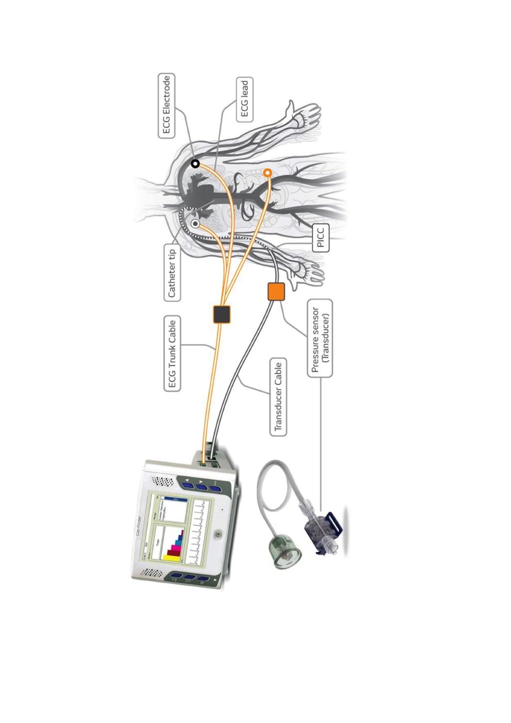
121 Catfinder Elcam
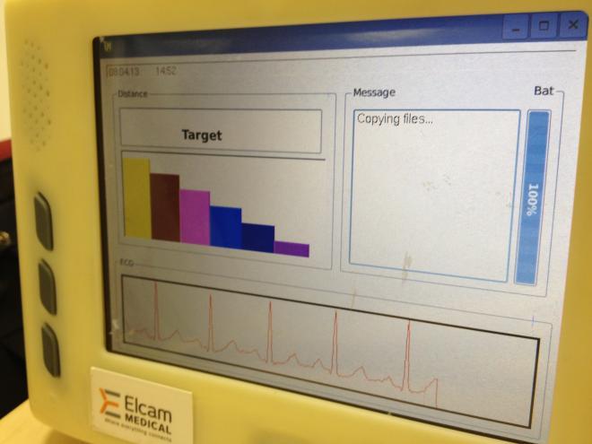
122
123 Catfinder (Elcam) A few abstracts available No published paper on peer-reviewed journals (as from Pub Med) A study just completed in our University Hospital
124 The primary endpoint of our study was to evaluate the accuracy of the CatFinder Navigational Device (CFND) as a tip location method for peripherally inserted central venous access devices in adult patients, as compared to the Intracavitary ECG method (IECG). The secondary endpoint was to evaluate CFND as a tip navigation method, able to detect the wrong direction of the catheter during insertion.

125

126
127
128 Method We studied adult patients candidate to placement of PICCs or PICC-ports. Patients with known ECG abnormalities or cardiac disease of any type were excluded. The target was to place the tip at the cavo-atrial junction. In all cases, tip location was verified by IECG. Any case of suspected wrong direction of the catheter was checked by ultrasound scan.

129
130 Conclusions (1) Applicability of CFND was 96% feasibility was 85% safety was 100% (feasibility is expected to improve by solving the technical issues above described)
131 Conclusions (2) - If compared to IECG, accuracy was 84% (considering a range of + 2cm) and 96% (+ 3cm). - Unacceptable tip positions were 3% (in all of these cases, the catheter was too short). - The accuracy in the diagnosis of a malposition in the IJV was 100%.
132 Problems with Catfinder Not cost effective Not easy to use Limited applicability It takes time Accuracy: high for tip navigation, somehow less for tip location
133 Tip navigation by modified IC-ECG
134 Using IC-ECG for tip navigation Available only using a specific dedicated ECG monitor (Delta) Based on the interpretation of the R wave (using a specific reference electrode): it is possible to detect the position of the tip inside the SVC Easy, cost-effective method for tip navigation As for tip location: It cannot differentiate between high and low SVC must be integrated with a landmark measurement method or with some other tip location method - requires a post-procedural verification of the tip unless a reliable tip location method is used during the maneuver

135

136 Control electrode
137 Pittiruti et al. : A new technique for catheter tip navigation using intracavitary ECG The new technique was used in 26 PICC placements. In all procedures, IC-ECG-based navigation signal successfully indicated whether the tip was moving towards or away from the CAJ; the catheter tip location at CAJ was confirmed by using the maximum P-wave criterion, as in traditional IC-ECG methods. There were no procedure-related complications

138 Pittiruti et al. : A new technique for catheter tip navigation using intracavitary ECG Reference electrode (upper sternum)
139 Pittiruti et al. : A new technique for catheter tip navigation using intracavitary ECG Negative R (tip hasnotentered the BCV)

140 Pittiruti et al. : A new technique for catheter tip navigation using intracavitary ECG Flat R The tip is below the reference electrode (enteringthe BCV)

141 Pittiruti et al. : A new technique for catheter tip navigation using intracavitary ECG R becomes positive (Tip hasentered the BCV)

142 Pittiruti et al. : A new technique for catheter tip navigation using intracavitary ECG R positive + peak of P (tip at the CAJ)
143 Using IC-ECG for tip navigation The ECG-based navigation method tells whether the tip has entered the BCV and the SVC, but cannot identify the CAJ. Not useful as tip location per se. Though, the same device can be used for IC-ECG tip navigation, conventional IC-ECG tip location and modified IC-ECG tip location
144 Advantages of tip navigation by Delta Easy to use Accurate No additional cost It can be used with any kind of central line It can be done with the same 3 electrodes used for ECG-based tip location Wide applicability (also in cases where ECG based tip location is not applicable)

145 DELTA Abstracts in international conferences No published study yet Very promising: easy, accurate, inexpensive, costeffective
146 Tip navigation: conclusions
147 Tip navigation for PICCs First conclusion There is no hard evidence that tip navigation is necessary during PICC insertion, though it may be useful (or reassuring for the operator). On the contrary, a proper method for tip location is always necessary.
148 Tip navigation for PICCs Second conclusion In most PICC insertions, ultrasound may be the easiest, simplest and most cost-effective method for tip navigation. It accurately detects the wrong direction to the ipsilateral IJV and the right direction into the BCV in adults; it detects the direction inside both BCV and SVC in neonates and infants. Also, it may help the operator to redirect the catheter under visual control.
149 Tip navigation for PICCs Third conclusion There is no evidence to support the use of expensive devices integrating tip location and tip navigation. Thogh, the most promising of such devices is Delta (integration of ECG-based tip location and ECG-based tip navigation). If compared to other similar devices integrating tip location with tip navigation (Sherlock 3CG, Vasonova, etc.), Delta seems to be easier, simpler, less expensive, more accurate and more costeffective.
150 TAKE HOME MESSAGE
151 Tip navigation and tip location for PICCs 1) Avoid fluoroscopy, always 2) For tip location: use conventional IC-ECG whenever possible take intoconsideration TTEand modified IC-ECG in selected cases 3) For tip navigation: use ultrasound scan of the supraclavicular area take intoconsideration ECG-navigation with Delta, if available

152 Thank you for your attention

153 GAVeCeLT th GAVeCeLT Congress - 11th PICC Day December 4th-5th-6th 2017, Florence (Italy)

154
PICC, port e trombosi cateterecorrelata: la prevenzione è legata alla tecnica di impianto!
 PICC, port e trombosi cateterecorrelata: la prevenzione è legata alla tecnica di impianto! Massimo Lamperti MD, MBA! Clinical Professor of Anesthesiology! Cleveland Clinic Lerner College of Medicine! Chief
Caroline Polley, BSN, RN, VA-BC Clinical Specialist BD
 Caroline Polley, BSN, RN, VA-BC Clinical Specialist BD Disclosures The speaker is a employee of BD. (Please consult BD product for any indications, contraindications, hazards, warnings, cautions and instructions
Peripherally Inserted Central Catheter & Midline Placement with ECG Confirmation of Tip Placement
 Title/Description: Peripherally Inserted Central Catheter & Midline Placement with ECG Confirmation of Tip Placement Department: Patient Care Services Personnel: Nursing Services Effective Date: April
Comparison Between Novel Tip Positioning Technology using ECG and Doppler and 2-D Echocardiography for the Placement of Central Catheters
 Comparison Between Novel Tip Positioning Technology using ECG and Doppler and 2-D Echocardiography for the Placement of Central Catheters Robert Wagner, MD, PhD 1, Petr Pokorny, MD 1, Jiri Cernosek, Dr.
REVIEWED/REVISED 12/12 DATE) MEDICAL STAFF (if applicable) DATE OTHER DATE Special Procedures Committee 10/12 Nursing Leadership Team 08/12
 MEDICAL STAFF (if applicable) DATE OTHER DATE Special Procedures Committee 10/12 Nursing Leadership Team 08/12") MANUAL: Clinical Page: 1 of 9 STANDARDIZED PROCEDURE I. POLICY: A. Function: To authorize the qualified Registered Nurses at St. Joseph Hospital (SJO) to insert Peripherally Inserted Central Catheters
MANUAL: Clinical Page: 1 of 9 STANDARDIZED PROCEDURE I. POLICY: A. Function: To authorize the qualified Registered Nurses at St. Joseph Hospital (SJO) to insert Peripherally Inserted Central Catheters
JMSCR Vol 05 Issue 10 Page October 2017
 www.jmscr.igmpublication.org Impact Factor 5.84 Index Copernicus Value: 71.58 ISSN (e)-2347-176x ISSN (p) 2455-0450 DOI: https://dx.doi.org/10.18535/jmscr/v5i10.79 Original Article Bedside Prediction of
Over the Wire Technique vs. Modified Seldinger Technique in Insertion of PICC
 Over the Wire Technique vs. Modified Seldinger Technique in Insertion of PICC Deniz Kasikci Department of Radiology, Jena University Hospital Friedrich-Schiller-University, Jena, Germany Disclosure Speaker
Research Article Can we predict the Position of Central Venous Catheter Tip Following Cannulation of Internal Jugular Vein?
 Cronicon OPEN ACCESS ANAESTHESIA Research Article Can we predict the Position of Central Venous Catheter Tip Following Cannulation of Internal Jugular Vein? Pradeep Marur Venkategowda 1, Surath Manimala
Depth of Central Venous Catheterization by Intracardiac ECG in Paediatric Patients
 Original Article Elmer Press Depth of Central Venous Catheterization by Intracardiac ECG in Paediatric Patients Prerana N. Shah a, b, Jithesh Appukutty a, Deepa Kane a Abstract Background: Central venous
Vascular Access Service Dott.ssa Alessandra Palo Direttore AAT Pavia - SAV Dipartimento Medicina Intensiva Fondazione IRCCS San Matteo Pavia
 Vascular Access Service Dott.ssa Alessandra Palo Direttore AAT Pavia - SAV Dipartimento Medicina Intensiva Fondazione IRCCS San Matteo Pavia Our story so far More and more patients with chronic illness
PARENTERAL NUTRITION: VASCUAR ACCESS DEVICE SELECTION
 PARENTERAL NUTRITION: VASCUAR ACCESS DEVICE SELECTION Winifred Magambo-Gasana Vascular Access Nurse Practitioner Oxford University Hospitals NHS Foundation Trust Aim An overview of the range of Vascular
You have a what, inside you?
 Costal Emergency Medicine Conference You have a what, inside you? Less than mainstream medical devices encountered in the ED. Eric Ossmann, MD, FACEP Associate Professor Duke University Medical Center
You have a what, inside you?
 Costal Emergency Medicine Conference You have a what, inside you? Less than mainstream medical devices encountered in the ED. Eric Ossmann, MD, FACEP Associate Professor Duke University Medical Center
Lines and tubes. 1 Nasogastric tubes Endotracheal tubes Central lines Permanent pacemakers Chest drains...
 Lines and tubes 1 Nasogastric tubes... 15 2 Endotracheal tubes.... 19 3 Central lines... 21 4 Permanent pacemakers.... 25 5 Chest drains... 30 This page intentionally left blank 1 Nasogastric tubes Background
Implications of Precise PICC Tip Location: The New Gold Standard in Clinical Practice?
 Implications of Precise PICC Tip Location: The New Gold Standard in Clinical Practice? Nancy Moureau, BSN, CRNI, CPUI, PICC Excellence, Inc. About the Speaker Nancy Moureau is an educator, legal consultant
Original Article. Evaluation of the Sherlock 3CG Tip Confirmation System on peripherally inserted central catheter malposition rates.
 Original Article doi:10.1111/anae.12785 Evaluation of the Sherlock 3CG Tip Confirmation System on peripherally inserted central catheter malposition rates A. J. Johnston, 1 A. Holder, 2 S. M. Bishop, 2
Peripherally inserted central catheterization is a
 DOI: 10.1097/JPN.0000000000000264 Continuing Education J Perinat Neonat Nurs Volume 31 Number 4, 326 331 Copyright C 2017 Wolters Kluwer Health, Inc. All rights reserved. Effectiveness of Intracavitary
Mary Lou Garey MSN EMT-P MedFlight of Ohio
 Mary Lou Garey MSN EMT-P MedFlight of Ohio Function Prolonged and frequent access to venous circulation Allows for patient to carry on normal life; decrease number of needle sticks Medications, parenteral
Ultrasound Guided Vascular Access. 7/25/2016
 Ultrasound Guided Vascular Access 7/25/2016 www.ezono.com 1 Objectives Indications for insertion of central and peripheral lines Complications associated with procedures Role of ultrasound in vascular
Dr. prakruthi Dept. of anaesthesiology, Rrmch, bangalore
 CENTRAL VENOUS CATHETERIZATION Dr. prakruthi Dept. of anaesthesiology, Rrmch, bangalore OBJECTIVES Introduction Indications and Contraindications Complications Technique Basic principles Specifics by Site
The use of ultrasound during and after central venous catheter insertion versus conventional chest X-ray after insertion of a central venous catheter
 ORIGINAL ARTICLE The use of ultrasound during and after central venous catheter insertion versus conventional chest X-ray after insertion of a central venous catheter M.J. Blans 1 *, H. Endeman 2, F.H.
W J C C M. World Journal of Critical Care Medicine. Focus on peripherally inserted central catheters in critically ill patients.
 W J C C M World Journal of Critical Care Medicine Submit a Manuscript: http://www.wjgnet.com/esps/ Help Desk: http://www.wjgnet.com/esps/helpdesk.aspx DOI: 10.5492/wjccm.v3.i4.80 World J Crit Care Med
Transcatheter Aortic Valve Implantation Procedure (TAVI)
") Page 1 of 5 Procedure (TAVI) Introduction Aortic stenosis (AS) is a common heart valve problem associated with heart failure and death. Surgical valve repair or replacement is recommended if AS patients
Page 1 of 5 Procedure (TAVI) Introduction Aortic stenosis (AS) is a common heart valve problem associated with heart failure and death. Surgical valve repair or replacement is recommended if AS patients
TITLE: Electrocardiograms versus X-rays for Guided Placement of Central Venous Catheter Tips: A Review of Clinical and Cost-effectiveness
 TITLE: Electrocardiograms versus X-rays for Guided Placement of Central Venous Catheter Tips: A Review of Clinical and Cost-effectiveness DATE: 18 July 2012 CONTEXT AND POLICY ISSUES Central venous catheters
Background & Indications Probe Selection
 Teresa S. Wu, MD, FACEP Director, EM Ultrasound Program & Fellowship Co-Director, Simulation Based Training Program & Fellowship Associate Program Director, EM Residency Program Maricopa Medical Center
Upper Extremity Venous Duplex Evaluation
 VASCULARTECHNOLOGY PROFESSIONAL PERFORMANCE GUIDELINES Upper Extremity Venous Duplex Evaluation This Guideline was prepared by the Professional Guidelines Subcommittee of the Society for Vascular Ultrasound
Peel-Apart Percutaneous Introducer Kits for
 Bard Access Systems Peel-Apart Percutaneous Introducer Kits for Table of Contents Contents Page Bard Implanted Ports Hickman*, Leonard*, Broviac*, Tenckhoff*, and Groshong* Catheters Introduction....................................
Port Design. Page 1. Port Placement, Removal, and Management. Selecting a Vascular Access Device. Thomas M. Vesely, MD
 Non-Dialysis Procedures Port Placement, Removal, and Management Thomas M. Vesely, MD Saint Louis, Missouri Selecting a Vascular Access Device Duration of use Number of lumens Frequency used Blood flow
Girish M Nair, Seeger Shen, Pablo B Nery, Calum J Redpath, David H Birnie
 268 Case Report Cardiac Resynchronization Therapy in a Patient with Persistent Left Superior Vena Cava Draining into the Coronary Sinus and Absent Innominate Vein: A Case Report and Review of Literature
Central Line Care and Management
 Central Line Care and Management What is a Central Line/ CVAD? (central venous access device) A vascular infusion device that terminates at or close to the heart or in one of the great vessels (aorta,
Central Venous Access Devices. Stephanie Cunningham Amy Waters
 Central Venous Access Devices Stephanie Cunningham Amy Waters 5 Must Know Facts About CVAD s 1) What are CVAD s? 2) What are CVAD s used for? 3) How are these devices put in? 4) What are the complications
Mitral Regurgitation
 UW MEDICINE PATIENT EDUCATION Mitral Regurgitation Causes, symptoms, diagnosis, and treatment This handout describes mitral regurgitation, a disease of the mitral valve. It explains how this disease is
A PATIENT S GUIDE TO THE LEFT ATRIAL APPENDAGE CLOSURE. Reducing the risk of stroke in atrial fibrillation
 A PATIENT S GUIDE TO THE LEFT ATRIAL APPENDAGE CLOSURE Reducing the risk of stroke in atrial fibrillation TABLE OF CONTENTS IMPORTANT Please Note: Information provided by Boston Scientific Corporation
Upper Extremity Venous Duplex. Michigan Sonographers Society Fall Ultrasound Symposium October 15, 2016
 Upper Extremity Venous Duplex Michigan Sonographers Society Fall Ultrasound Symposium October 15, 2016 Patricia A. (Tish) Poe, BA RVT FSVU Director of Quality Assurance Navix Diagnostix Patricia A. Poe
Shock, Monitoring Invasive Vs. Non Invasive
 Shock, Monitoring Invasive Vs. Non Invasive Paula Ferrada MD Assistant Professor Trauma, Critical Care and Emergency Surgery Virginia Commonwealth University Shock Fluid Pressors Ionotrope Intervention
A systematic review of the effectiveness of intra-cavitary. electrocardiographic guidance in improving CVAD tip placement
 A systematic review of the effectiveness of intra-cavitary electrocardiographic guidance in improving CVAD tip placement (Walker G, Rickard CM, Alexandrou E, Webster J, Chan R. Effectiveness of electrocardiographic
Kristin Wise, MD, FHM Division of General Internal Medicine and Geriatrics Hospital Medicine 2013
 Kristin Wise, MD, FHM Division of General Internal Medicine and Geriatrics Hospital Medicine 2013 Objectives for CVC Placement Understand the indications and contraindications Determine appropriate CVC
2018 HEMODIALYSIS CATHETERS CODING AND REIMBURSEMENT GUIDE
 2018 HEMODIALYSIS CATHETERS CODING AND REIMBURSEMENT GUIDE Contents Overview of Central Venous Access s for Hemodialysis 2 Procedures Using Hemodialysis s 2 Physician Reimbursement for Hemodialysis s 3
Navigating Vascular Access Issues
 Navigating Vascular Access Issues The Oley Foundation 27 th Annual Consumer/Clinician Conference Redondo Beach, CA June, 27 2012 Anita Piano, BS, RN, VA-BC Administrative Nurse, PICC Service UCLA Health
Central Venous Line Insertion
 Central Venous Line Insertion Understand the indications and risks of CVC insertion Understand and troubleshoot the seldinger technique Understand available sites and select the appropriate site for clinical
Adult Echocardiography Examination Content Outline
 Adult Echocardiography Examination Content Outline (Outline Summary) # Domain Subdomain Percentage 1 2 3 4 5 Anatomy and Physiology Pathology Clinical Care and Safety Measurement Techniques, Maneuvers,
Background: Bedside ultrasound is emerging as a useful tool in the assessment of
 Abstract: Background: Bedside ultrasound is emerging as a useful tool in the assessment of intravascular volume status by examining measurements of the inferior vena cava (IVC). Many previous studies do
Overview of CVADs. Type of device commonly used. Dwell time Flushing requirement Associated complications. lumens
 Source: Clinical Skills Management of Vascular Access Devices Pre-course handbook. Adapted with permission from NHS Lothian Employee and Education Development Team. Overview of CVADs Type of device Veins
Children s Acute Transport Service
 Children s Acute Transport Service Vascular Access Document Control Information Author Ramnarayan Author Position Consultant, CATS Document Owner Polke Document Owner Position CATS Co-ordinator Document
JOINT MEETING 2 Tricuspid club Chairpersons: G. Athanassopoulos, A. Avgeropoulou, M. Khoury, G. Stavridis
 JOINT MEETING 2 Tricuspid club Chairpersons: G. Athanassopoulos, A. Avgeropoulou, M. Khoury, G. Stavridis Similarities and differences in Tricuspid vs. Mitral Valve Anatomy and Imaging. Echo evaluation
Hydrocephalus: Electrocardiographic Localization of
 Arch. Dis. Childh., 1967, 42, 166. Hydrocephalus: Electrocardiographic Localization of the Catheter in Ventriculo-atrial Shunts G. BROCKLEHURST, J. R. W. GLEAVE, R. A. MILLAR, and AILEEN K. ADAMS From
Interesting Cases - A Case Report: Renal Cell Carcinoma With Tumor Mass In IVC And Heart. O Wenker, L Chaloupka, R Joswiak, D Thakar, C Wood, G Walsh
 ISPUB.COM The Internet Journal of Thoracic and Cardiovascular Surgery Volume 3 Number 2 Interesting Cases - A Case Report: Renal Cell Carcinoma With Tumor Mass In IVC And Heart O Wenker, L Chaloupka, R
Cigna - Prior Authorization Procedure List Cardiology
 Cigna - Prior Authorization Procedure List Cardiology Category CPT Code CPT Code Description 33206 Insertion of new or replacement of permanent pacemaker with transvenous electrode(s); atrial 33207 Insertion
Original Article Bedside prediction of peripherally inserted central catheter length: based on patient height combined with surface landmark
 Int J Clin Exp Med 2016;9(8):15928-15934 www.ijcem.com /ISSN:1940-5901/IJCEM0022838 Original Article Bedside prediction of peripherally inserted central catheter length: based on patient height combined
2D/3D in Evaluation of Atrial Septum
 2D/3D in Evaluation of Atrial Septum Roberto M Lang, MD OSTIUM SECUNDUM ASD: 2D AND 3D TNSESOPHAGEAL ECHO 1 Biplane views 90 0 3D Acquisi on Acquire 3D volume Lang RM et al. JASE 2012;25:3 46. Right atrial
About atrial fibrillation (AFib) Atrial Fibrillation (AFib) What is AFib? What s the danger? Who gets AFib?
 Atrial Fibrillation (AFib) What is AFib? What s the danger? Who gets AFib?") Understanding AFib Atrial Fibrillation (AFib) About AFib 3 How Your Heart Works 4 Types of AFib 5 Symptoms 5 Risk Factors 5 How is AFib Diagnosed? 6 Treatment 6 What to Ask Your Doctor 7 A normal heartbeat
Understanding AFib Atrial Fibrillation (AFib) About AFib 3 How Your Heart Works 4 Types of AFib 5 Symptoms 5 Risk Factors 5 How is AFib Diagnosed? 6 Treatment 6 What to Ask Your Doctor 7 A normal heartbeat
This presentation will deal with the basics of ECG description as well as the physiological basics of
 Snímka 1 Electrocardiography basics This presentation will deal with the basics of ECG description as well as the physiological basics of Snímka 2 Lecture overview 1. Cardiac conduction system functional
CATHETER MALPOSITION FOLLOWING SUPRACLAVICULAR APPROACH FOR SUBCLAVIAN VEIN CATHETERISATION
 CATHETER MALPOSITION FOLLOWING SUPRACLAVICULAR APPROACH FOR SUBCLAVIAN VEIN CATHETERISATION - Case Reports - Prem K Singh *, Zulfiquar Ali *, Girija P Rath ** and Hemanshu Prabhakar *** Abstract The supraclavicular
Case Report Sinus Venosus Atrial Septal Defect as a Cause of Palpitations and Dyspnea in an Adult: A Diagnostic Imaging Challenge
 Case Reports in Medicine Volume 2015, Article ID 128462, 4 pages http://dx.doi.org/10.1155/2015/128462 Case Report Sinus Venosus Atrial Septal Defect as a Cause of Palpitations and Dyspnea in an Adult:
Spontaneous Migration of a Central Line Catheter into the Heart. Saeed Al Hindi, MD, CABS, FRCSI* Khulood Al-Saad, MD**
 Bahrain Medical Bulletin, Vol. 29, No. 3, September 2007 Spontaneous Migration of a Central Line Catheter into the Heart Saeed Al Hindi, MD, CABS, FRCSI* Khulood Al-Saad, MD** Background: Spontaneous migration
The C.L.O.T. Tool for Identifying Strategies to Prevent PICC Catheter Occlusions
 The C.L.O.T. Tool for Identifying Strategies to Prevent PICC Catheter Occlusions page 2 TABLE OF CONTENTS Part 1: Definition and Scope of Catheter Occlusion Part 2: Predictors of Catheter Occlusion Part
The HeRO Graft. Shawn M. Gage, PA Division of Vascular Surgery Duke University Medical Center
 The HeRO Graft Shawn M. Gage, PA Division of Vascular Surgery Duke University Medical Center Faculty Disclosure I disclose the following financial relationships: CryoLife/Hemosphere, Inc. & W.L. Gore and
Appendix E: Overview of Vascular
 Appendix E: Overview of Vascular 56 Peripheral Short Catheter, less than 3 inches (7.5 cm) in length; over-the-needle catheter is most common. Inserted by percutaneous venipuncture, generally into a hand
Intro: Slide 1. Slide 2. Slide 3. Basic understanding of interventional radiology. Gain knowledge of key terms and phrases
 Slide 1 Intro: PRESENTED BY: Selena M. Moore, AAS, CCS, CPC HIMS Physician Liaison Coder This is a modified/updated presentation that was originally written by: Rosemary Waligorski, RHIT, CCS, RCC and
Hospital of the University of Pennsylvania NURSING. Insertion of Peripherally Inserted Central Catheter (PICC) and Midline Catheter (MLC)
 and Midline Catheter (MLC)") Page 1 of 11 KEYWORDS: Central Catheter Sterility REFER TO: 4B-02-09 Nursing Management of the Patient with a Central Venous Access Device HUP 1-12-24 Consent to Health Care Services SCOPE Registered Nurses
Page 1 of 11 KEYWORDS: Central Catheter Sterility REFER TO: 4B-02-09 Nursing Management of the Patient with a Central Venous Access Device HUP 1-12-24 Consent to Health Care Services SCOPE Registered Nurses
Per-Q-Cath* PICC Catheters with Excalibur Introducer* System
 Bard Access Systems Per-Q-Cath* PICC and Catheters with Excalibur Introducer* System Instructions For Use Table of Contents Table of Contents Page Contents 1 Product Description, Indications & Contraindications
Listen to Your Heart. What Everyone Needs To Know About Atrial Fibrillation & Stroke. The S-ICD System. The protection you need
 Listen to Your Heart The S-ICD System What Everyone Needs To Know About Atrial Fibrillation & Stroke The protection you need without Stroke. touching Are you your at heart risk? Increase your knowledge.
Adult Intubation Skill Sheet
 Adult Intubation 2. Opens the airway manually and inserts an oral airway *** 3. Ventilates the patient with BVM attached to oxygen at 15 lpm *** 4. Directs assistant to oxygenate the patient 5. Selects
Sterile Technique & IJ/Femoral Return Demonstration
 Sterile Technique & IJ/Femoral Return Demonstration Sterile Technique Description: This is a return demonstration checklist used to evaluate participants in the simulated hands on skills portions for certification
Advocate Christ Medical Center CVC Placement Certification Course
 Advocate Christ Medical Center CVC Placement Certification Course July 12th, 2012 Hannah Watts, MD Medical Simulation Director Modified August 10, 2017 Taajwar Khan, MD Chief Resident of Internal Medicine
Troubleshooting Technique for Hemodialysis Catheter Insertion
 Troubleshooting Technique for Hemodialysis Catheter Insertion Withoon Ungkitphaiboon Assistant Professor, Department of Surgery, Maha Chakri Sirindhorn Medical Center Srinakharinwirot University Present
INTERNAL CARDIOVERSION. Lancashire & South Cumbria Cardiac Network
 INTERNAL CARDIOVERSION Lancashire & South Cumbria Cardiac Network DC Cardioversion AF & AFL safe 1,2 efficacious 3,4 (60-94%) 5 SR - Increases exercise tolerance 6 Maintainence SR unlikely in patients
Leadless Pacing. Osama Diab Assistant Prof. of Cardiology Ain Shams University Egypt
 Leadless Pacing Osama Diab Assistant Prof. of Cardiology Ain Shams University Egypt The weakest link in Pacemaker system the lead. The more the leads the more the complications Dislodgement Fracture Insulation
Prevention of thrombosis
 Prevention of thrombosis Massimo Lamperti MD, MBA Chief of General Anaesthesia Department Anaesthesiology Institute Cleveland Clinic Abu Dhabi Clinical Professor of Anaesthesiology Cleveland Clinic Lerner
How to Approach the Patient with CRT and Recurrent Heart Failure
 How to Approach the Patient with CRT and Recurrent Heart Failure Byron K. Lee MD Associate Professor of Medicine Electrophysiology and Arrhythmia Section UCSF Update in Electrocardiography and Arrhythmias
CATHETER ABLATION CODING & REIMBURSEMENT GUIDE. Updated September 2018
 CATHETER ABLATION CODING & REIMBURSEMENT GUIDE Updated September 2018 TABLE OF CONTENTS Diagnosis Codes...3 ICD-10-CM Diagnosis Codes Coverage for Catheter Ablation Procedures....4 Medicare Other Payers
Figure 2. Normal ECG tracing. Table 1.
 Figure 2. Normal ECG tracing that navigates through the left ventricle. Following these bundle branches the impulse finally passes to the terminal points called Purkinje fibers. These Purkinje fibers are
Min Hur, Eun-Hee Kim, In-Kyung Song, Ji-Hyun Lee, Hee-Soo Kim, and Jin Tae Kim INTRODUCTION. Clinical Research
 Anesth Pain Med 2016; 11: 375-379 https://doi.org/10.17085/apm.2016.11.4.375 Clinical Research http://crossmark.crossref.org/dialog/?doi=10.17085/apm.2016.11.4.375&domain=pdf&date_stamp=2016-10-25 pissn
Document No. BMB/IFU/40 Rev No. & Date 00 & 15/11/2017 Issue No & Date 01 & 15/11/2017
 Central Venous Catheter Device Description Multi-lumen catheters incorporate separate, non-communicating vascular access lumens within a single catheter body. Minipunctur Access Sets And Trays: Used for
CARDIOINSIGHT TM NONINVASIVE 3D MAPPING SYSTEM CLINICAL EVIDENCE SUMMARY
 CARDIOINSIGHT TM NONINVASIVE 3D MAPPING SYSTEM CLINICAL EVIDENCE SUMMARY April 2017 SUPPORTING EVIDENCE RHYTHM AF VT PUBLICATIONS Driver Domains in Persistent Atrial Fibrillation (Haissaiguerre, et al)
Instructions for Use Reprocessed LASSO Circular Mapping Diagnostic Electrophysiology (EP) Catheter
 Catheter") Instructions for Use Reprocessed LASSO Circular Mapping Diagnostic Electrophysiology (EP) Catheter Caution: Federal (USA) law restricts this device to sale by or on the order of a physician. DEVICE DESCRIPTION
Instructions for Use Reprocessed LASSO Circular Mapping Diagnostic Electrophysiology (EP) Catheter Caution: Federal (USA) law restricts this device to sale by or on the order of a physician. DEVICE DESCRIPTION
Technique of ultrasound guided peripheral venous access in the emergency room
 Technique of ultrasound guided peripheral venous access in the emergency room Maria Giuseppina ANNETTA and Giancarlo SCOPPETTUOLO Catholic University, Rome, Italy Background Acutely ill pa?ents admiaed
1. CARDIOLOGY. These listings cannot be correctly interpreted without reference to the Preamble. Anes. $ Level
 1. CARDIOLOGY These listings cannot be correctly interpreted without reference to the Preamble. Anes. Referred Cases 33010 Consultation: To consist of examination, review of history, laboratory, X-ray
INTEGRATING ECHOCARDIOGRAPHY WITH CATHETER INTERVENTIONS FOR CONGENITAL HEART DISEASE. Krishna Kumar SevenHills Hospital, Mumbai, India
 INTEGRATING ECHOCARDIOGRAPHY WITH CATHETER INTERVENTIONS FOR CONGENITAL HEART DISEASE Krishna Kumar SevenHills Hospital, Mumbai, India Why talk about it? What is the big deal? Are we not stating the obvious?
Case Report Hemostasis of Left Atrial Appendage Bleed With Lariat Device
 273 Case Report Hemostasis of Left Atrial Appendage Bleed With Lariat Device Amena Hussain MD, Muhamed Saric MD, Scott Bernstein MD, Douglas Holmes MD, Larry Chinitz MD NYU Langone Medical Center, United
External Jugular Vein Catheterization Using Intra-Atrial Electrocardiogram
 Original Article DOI 10.3349/ymj.2009.50.2.222 pissn: 0513-5796, eissn: 1976-2437 Yonsei Med J 50(2):222-226, 2009 External Jugular Vein Catheterization Using Intra-Atrial Electrocardiogram Dilek Karaaslan,
Cardiac Electrical Therapies. By Omar AL-Rawajfah, PhD, RN
 Cardiac Electrical Therapies By Omar AL-Rawajfah, PhD, RN Outlines What are cardiac electrical therapies Ablation Defibrillation Cardioversion What are the nursing considerations for each type of therapy
CENTRAL VENOUS ACCESS DEVICES. BETHANY COLTON
 CENTRAL VENOUS ACCESS DEVICES. BETHANY COLTON Aims and Objectives To know what central venous access devices (CVAD) are. Types of CVADS used in haematology. To understand why we use them To know the complications
The Normal Echocardiogram
 The Normal Echocardiogram Pravin V. Patil, MD FACC Lewis Katz School of Medicine at Temple University Acknowledgments Dr. Susan Wiegers Dr. Martin Keane Temple Cardiac Sonographers Disclosures No relevant
Certification Review. Module 28. Medical Coding. Radiology
 Module 28 is the study of x-rays, using radiant energy and other imaging techniques, such as resonance imaging or ultrasound, to diagnose illnesses and diseases. Vocabulary Barium enema (BE): lower gastrointestinal
Module 1: Introduction to ECG & Normal ECG
 Module 1: Introduction to ECG & Normal ECG Importance of Correct anatomical positions Measurements & Morphologies ONLY accurate if Precise anatomical positions adhered to Standardised techniques are used
Ultrasound-guided infraclavicular axillary vein cannulation for central venous access {
 British Journal of Anaesthesia 93 (2): 188 92 DOI: 10.1093/bja/aeh187 Advance Access publication June 25, 2004 Ultrasound-guided infraclavicular axillary vein cannulation for central venous access { A.
Cardiac Resynchronisation Therapy Patient Information
 Melbourne Heart Rhythm Cardiac Resynchronisation Therapy Patient Information Normal Heart Function The heart is a pump responsible for maintaining blood supply to the body. It has four chambers. The two
A Primer on Central Venous Access: Peripherally-Inserted Central Catheters, Tunneled Catheters, and Subcutaneous Ports
 Disclosures A Primer on Central Venous Access: Peripherally-Inserted Central Catheters, Tunneled Catheters, and Subcutaneous Ports No conflicts of interest relevant to this presentation Jason W. Pinchot,
Integration of CT and fluoroscopy images in the ablative treatment of atrial fibrillation
 Clinical applications Integration of CT and fluoroscopy images in the ablative treatment of atrial fibrillation C. Kriatselis M. Tang M. Roser J-H. erds-li E. leck Department of Internal Medicine/Cardiology,
Catheter Fracture of a Totally Implantable Venous Device Due to Pinch Off Syndrome in Breast Cancer: A Case Report
 Kosin Medical Journal 2016;31:167-172. https://doi.org/10.7180/kmj.2016.31.2.167 KMJ Case Report Catheter Fracture of a Totally Implantable Venous Device Due to Pinch Off Syndrome in Breast Cancer: A Case
Sample page. POWER UP YOUR CODING with Optum360, your trusted coding partner for 32 years. Visit optum360coding.com.
 2018 Complete Guide for Interventional Radiology An in-depth guide to interventional radiology coding, billing, and reimbursement for facilities and physicians POWER UP YOUR CODING with Optum360, your
PERIOPERATIVE MANAGEMENT: CARDIAC PACEMAKERS AND DEFIBRILLATORS
 PERIOPERATIVE MANAGEMENT: CARDIAC PACEMAKERS AND DEFIBRILLATORS DR SUSAN CORCORAN CARDIOLOGIST ONCE UPON A TIME.. Single chamber pacemakers Programmed at 70/min VVI 70 UNIPOLAR SYSTEMS A Unipolar Pacing
Introduction to TEE using Heartworks Echocardiography Simulator
 Introduction to TEE using Heartworks Echocardiography Simulator Steven M. Ewer, MD Assistant Professor Division of Cardiovascular Medicine University of Wisconsin School of Medicine & Public Health Version
Double Superior Vena Cava; A Benign Cause of Widened Mediastenum and Implication on Venous Central Access
 ISPUB.COM The Internet Journal of Endovascular Medicine Volume 2 Number 1 Double Superior Vena Cava; A Benign Cause of Widened Mediastenum and Implication on Venous H Enuh, A Patel, A Chaudry, K Diaz,
Point of Care Ultrasound (PoCUS)
") Point of Care Ultrasound (PoCUS) Competency Assessment Forms AORTA Competency A Focussed Assessment of the Aorta (AAA) Guidance Please follow this guidance as closely as possible to ensure consistency
Point of Care Ultrasound (PoCUS) Competency Assessment Forms AORTA Competency A Focussed Assessment of the Aorta (AAA) Guidance Please follow this guidance as closely as possible to ensure consistency
UnitedHealthcare, UnitedHealthcare of the River Valley and Neighborhood Health Partnership Cardiology Notification and Prior Authorization Program:
 Notification and Prior Authorization Program: Electrophysiology Implant Classification Table The following chart contains the codes that require notification or prior authorization as part of UnitedHealthcare
Ultrasound Guidance Needle Techniques
 Ultrasound Guidance Needle Techniques Dr TANG Ho-ming AED/UCH USG Guidance Needle Techniques Commonly used in EM 1. Vessel cannulation-peripheral & central 2. Foreign body removal 3. Peripheral nerve/plexus
Vaxcel Implantable Ports Valved and Non-Valved. A Patient s Guide
 Vaxcel Implantable Ports Valved and Non-Valved A Patient s Guide Vaxcel Implantable Port This pamphlet provides some answers to questions you may have about your implantable port and how to care for it