If I Only Had a Brain
|
|
|
- Lionel Crawford
- 5 years ago
- Views:
Transcription


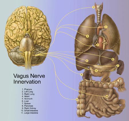
1 If I Only Had a Brain A Heart. (The Nerve!)
 Adult brain Brain")
2 Regions of the Brain Cerebral hemisphere Diencephalon Cerebellum (b) Adult brain Brain stem
 Fissure (a deep sulcus) Gyrus Sulcus Cerebral white matter Temporal lobe Cerebellum Pons Medulla oblongata Spinal")
3 Regions of the Brain: Cerebrum Precentral gyrus Frontal lobe Central sulcus Postcentral gyrus Parietal lobe Parieto-occipital sulcus (deep) Lateral sulcus Occipital lobe Cerebral cortex (gray matter) Fissure (a deep sulcus) Gyrus Sulcus Cerebral white matter Temporal lobe Cerebellum Pons Medulla oblongata Spinal cord
4 Regions of the Brain: Cerebrum Parietal lobe Left cerebral hemisphere Frontal lobe Temporal lobe Occipital lobe Cephalad Caudal Brain stem Cerebellum
5 Regions of the Cerebrum Primary motor area Premotor area Anterior association area Working memory and judgment Problem solving Language comprehension Central sulcus Primary somatic sensory area Gustatory area (taste) Speech/language (outlined by dashes) Posterior association area Broca s area (motor speech) Olfactory area Visual area Auditory area

6 Figure 7.14
 Corona radiata Fornix Internal capsule Third ventricle Pons Medulla oblongata Projection fibers Figure 7.")
7 Longitudinal fissure Lateral ventricle Basal nuclei (basal ganglia) Thalamus Superior Association fibers Commissural fibers (corpus callosum) Corona radiata Fornix Internal capsule Third ventricle Pons Medulla oblongata Projection fibers Figure 7.15
8 Third ventricle Anterior commissure Hypothalamus Optic chiasma Pituitary gland (a) Mammillary body Pons Medulla oblongata Spinal cord Cerebral hemisphere Corpus callosum Choroid plexus of third ventricle Occipital lobe of cerebral hemisphere Thalamus (encloses third ventricle) Pineal gland (part of epithalamus) Corpora quadrigemina Midbrain Cerebral aqueduct Cerebral peduncle of midbrain Fourth ventricle Choroid plexus Cerebellum Figure 7.16a
9 Cerebral hemisphere Diencephalon Cerebellum (b) Adult brain Brain stem Figure 7.12b
 Hypothalamus Pineal gland (part of epithalamus) Corpora quadrigemina Cerebral aqueduct Midbrain Cerebral peduncle of midbrain")
10 Cerebral hemisphere Corpus callosum Choroid plexus of third ventricle Occipital lobe of cerebral hemisphere Thalamus (encloses third ventricle) Hypothalamus Pineal gland (part of epithalamus) Corpora quadrigemina Cerebral aqueduct Midbrain Cerebral peduncle of midbrain Fourth ventricle
11 Radiations to cerebral cortex Visual impulses Reticular formation Ascending general sensory tracts (touch, pain, temperature) Auditory impulses Descending motor projections to spinal cord (b) Figure 7.16b
12 Thalamus Regions of the Brain: Diencephalon Surrounds the third ventricle The relay station for sensory impulses Transfers impulses to the correct part of the cortex for localization and interpretation
13 Regions of the Brain: Diencephalon Hypothalamus Under the thalamus Important autonomic nervous system center Helps regulate body temperature Controls water balance Regulates metabolism Houses the limbic center for emotions Regulates the nearby pituitary gland Produces two hormones of its own
14 Regions of the Brain: Diencephalon Epithalamus Forms the roof of the third ventricle Houses the pineal body (an endocrine gland) Includes the choroid plexus forms cerebrospinal fluid
15 Regions of the Brain: Brain Stem Attaches to the spinal cord Parts of the brain stem Midbrain Pons Medulla oblongata
16 Third ventricle Anterior commissure Hypothalamus Optic chiasma Pituitary gland (a) Mammillary body Pons Medulla oblongata Spinal cord Cerebral hemisphere Corpus callosum Choroid plexus of third ventricle Occipital lobe of cerebral hemisphere Thalamus (encloses third ventricle) Pineal gland (part of epithalamus) Corpora quadrigemina Midbrain Cerebral aqueduct Cerebral peduncle of midbrain Fourth ventricle Choroid plexus Cerebellum Figure 7.16a
17 Regions of the Brain: Brain Stem Midbrain Mostly composed of tracts of nerve fibers Has two bulging fiber tracts cerebral peduncles Has four rounded protrusions corpora quadrigemina Reflex centers for vision and hearing
18 Regions of the Brain: Brain Stem Pons The bulging center part of the brain stem Mostly composed of fiber tracts Includes nuclei involved in the control of breathing
19 Regions of the Brain: Brain Stem Medulla oblongata The lowest part of the brain stem Merges into the spinal cord Includes important fiber tracts Contains important control centers Heart rate control Blood pressure regulation Breathing Swallowing Vomiting
20 Regions of the Brain: Brain Stem Reticular Formation Diffuse mass of gray matter along the brain stem Involved in motor control of visceral organs Reticular activating system (RAS) plays a role in awake/sleep cycles and consciousness
 Auditory impulses Descending motor projections to spinal cord (b) Figure 7.")
21 Radiations to cerebral cortex Visual impulses Reticular formation Ascending general sensory tracts (touch, pain, temperature) Auditory impulses Descending motor projections to spinal cord (b) Figure 7.16b
22 Regions of the Brain: Cerebellum Two hemispheres with convoluted surfaces Provides involuntary coordination of body movements
 Pineal gland (part of epithalamus) Corpora quadrigemina Midbrain Cerebral aqueduct Cerebral peduncle of midbrain")
23 Third ventricle Anterior commissure Hypothalamus Optic chiasma Pituitary gland (a) Mammillary body Pons Medulla oblongata Spinal cord Cerebral hemisphere Corpus callosum Choroid plexus of third ventricle Occipital lobe of cerebral hemisphere Thalamus (encloses third ventricle) Pineal gland (part of epithalamus) Corpora quadrigemina Midbrain Cerebral aqueduct Cerebral peduncle of midbrain Fourth ventricle Choroid plexus Cerebellum Figure 7.16a
24 Superior vena cava Right pulmonary artery Right atrium Right pulmonary veins Fossa ovalis Right atrioventricular valve (tricuspid valve) Right ventricle Chordae tendineae Inferior vena cava Aorta Left pulmonary artery Left atrium Left pulmonary veins Pulmonary semilunar valve Left atrioventricular valve (bicuspid valve) Aortic semilunar valve Left ventricle Interventricular septum Myocardium Visceral pericardium (b) Frontal section showing interior chambers and valves. Figure 11.3b
25 The Heart: Chambers Right and left side act as separate pumps Four chambers Atria Receiving chambers Right atrium Left atrium Ventricles Discharging chambers Right ventricle Left ventricle
 Aortic semilunar valve Left ventricle Interventricular septum Myocardium")
26 Superior vena cava Right pulmonary artery Right atrium Right pulmonary veins Fossa ovalis Right atrioventricular valve (tricuspid valve) Right ventricle Chordae tendineae Inferior vena cava Aorta Left pulmonary artery Left atrium Left pulmonary veins Pulmonary semilunar valve Left atrioventricular valve (bicuspid valve) Aortic semilunar valve Left ventricle Interventricular septum Myocardium Visceral pericardium (b) Frontal section showing interior chambers and valves. Figure 11.3b
27 Left ventricle Right ventricle Muscular interventricular septum Figure 11.5
28 The Heart: Valves AV valves Anchored in place by chordae tendineae ( heart strings ) Open during heart relaxation and closed during ventricular contraction Semilunar valves Closed during heart relaxation but open during ventricular contraction Notice these valves operate opposite of one another to force a one-way path of blood through the heart
29 (a) Operation of the AV valves 1 Blood returning to the atria puts pressure against AV valves; the AV valves are forced open. 2 As the ventricles fill, AV valve flaps hang limply into ventricles. 3 Atria contract, forcing additional blood into ventricles. Ventricles 4 Ventricles contract, forcing blood against AV valve flaps. 5 AV valves close. 6 Chordae tendineae tighten, preventing valve flaps from everting into atria. AV valves open; atrial pressure greater than ventricular pressure AV valves closed; atrial pressure less than ventricular pressure Figure 11.6a, step 6

30 (b) Operation of the semilunar valves Pulmonary trunk Aorta 1 As ventricles 2 As ventricles contract and relax and intraventricular intraventricular pressure rises, pressure falls, blood is pushed up blood flows against semilunar back from arteries, valves, forcing filling the leaflets them open. of semilunar valves and forcing them to close. Semilunar valves open Semilunar valves closed Figure 11.6b, step 2

31 Capillary beds of lungs where gas exchange occurs Venae cavae Pulmonary arteries Pulmonary Circuit Pulmonary veins Aorta and branches Left atrium Left Right ventricle atrium Heart Right ventricle Systemic Circuit KEY: Oxygen-rich, CO 2 -poor blood Oxygen-poor, CO 2 -rich blood Capillary beds of all body tissues where gas exchange occurs Figure 11.4

32 Blood Flow to the Brain

33 The Circle of Willis
34 Superior vena cava Sinoatrial (SA) node (pacemaker) Left atrium Atrioventricular (AV) node Right atrium Bundle branches Purkinje fibers Atrioventricular (AV) bundle (bundle of His) Purkinje fibers Interventricular septum Figure 11.7

35 ECG Electrocariogram Sinoatrial node Atrioventricular node QRS complex R Ventricular depolarization Atrial depolarization P Ventricular repolarization T P-R Interval Q S-T Segment S Q-T Interval Time (s)
36 Cardiac Output Stroke Volume x Heart Rate Avg: 70 ml/beat x 72 beats/min = 5020 ml/ min MUST be equal from both chambers Congestive Heart Failure
37 Homeostatic Imbalances Tachycardia: abnormally fast heart rate (>100 bpm) If persistent, may lead to fibrillation Bradycardia: heart rate slower than 60 bpm May result in grossly inadequate blood circulation May be desirable result of endurance training
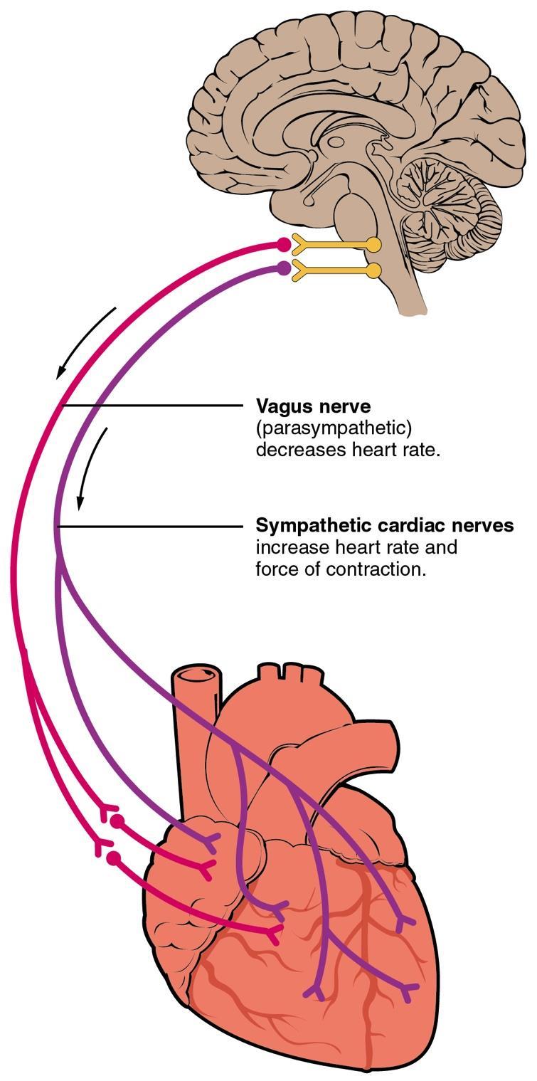
38 The Heart: Regulation of Heart Rate Increased heart rate Sympathetic nervous system Crisis Low blood pressure Hormones Epinephrine Thyroxine Exercise Decreased blood volume
39 The Heart: Regulation of Heart Rate Decreased heart rate Parasympathetic nervous system High blood pressure or blood volume Decreased venous return
40
41
42 Heart Affects the Brain Oxygen delivery CHF can lead to hypoxia
43 Brain Affects Heart Cardiovascular centers in medulla oblongata Cardioacceleratory center Cardioinhibitory center
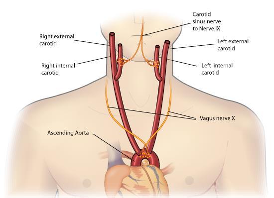
44 Nerves Carotid sinuses monitor pressure & oxygen Via Glossopharyngeal nerve (CN IX) Medulla integration of pressure & oxygen
45
46 Nerves Vagus nerve sends inhibitory impulses Sympathetic nerves send acceleratory impulses
47 To Make It All Work The heart provides the oxygenated blood to the brain The brain keeps the oxygen flowing by regulating the heart rate & strength of contraction The nerveprovide constant communication between the two organs to survive

48
The Nervous System PART B
 7 The Nervous System PART B PowerPoint Lecture Slide Presentation by Jerry L. Cook, Sam Houston University ESSENTIALS OF HUMAN ANATOMY & PHYSIOLOGY EIGHTH EDITION ELAINE N. MARIEB The Reflex Arc Reflex
The Nervous System 7PART B. PowerPoint Lecture Slide Presentation by Patty Bostwick-Taylor, Florence-Darlington Technical College
 PowerPoint Lecture Slide Presentation by Patty Bostwick-Taylor, Florence-Darlington Technical College The Nervous System 7PART B What is a reflex? What is a reflex? What is meant by the statement that
The Nervous System PART B
 7 The Nervous System PART B PowerPoint Lecture Slide Presentation by Jerry L. Cook, Sam Houston University ESSENTIALS OF HUMAN ANATOMY & PHYSIOLOGY EIGHTH EDITION ELAINE N. MARIEB Central Nervous System
The Heart. The Heart A muscular double pump. The Pulmonary and Systemic Circuits
 C H A P T E R 19 The Heart The Heart A muscular double pump circuit takes blood to and from the lungs Systemic circuit vessels transport blood to and from body tissues Atria receive blood from the pulmonary
Anatomy and Physiology (Bio 220) The Brain Chapter 14 and select portions of Chapter 16
 The Brain Chapter 14 and select portions of Chapter 16") Anatomy and Physiology (Bio 220) The Brain Chapter 14 and select portions of Chapter 16 I. Introduction A. Appearance 1. physical 2. weight 3. relative weight B. Major parts of the brain 1. cerebrum 2.
Anatomy and Physiology (Bio 220) The Brain Chapter 14 and select portions of Chapter 16 I. Introduction A. Appearance 1. physical 2. weight 3. relative weight B. Major parts of the brain 1. cerebrum 2.
10. Thick deposits of lipids on the walls of blood vessels, called, can lead to serious circulatory issues. A. aneurysm B. atherosclerosis C.
 Heart Student: 1. carry blood away from the heart. A. Arteries B. Veins C. Capillaries 2. What is the leading cause of heart attack and stroke in North America? A. alcohol B. smoking C. arteriosclerosis
Principles of Anatomy and Physiology
 Principles of Anatomy and Physiology 14 th Edition CHAPTER 14 The Brain and Cranial Nerves Introduction The purpose of the chapter is to: 1. Understand how the brain is organized, protected, and supplied
BRAIN PART I (A & B): VENTRICLES & MENINGES
: VENTRICLES & MENINGES") BRAIN PART I (A & B): VENTRICLES & MENINGES Cranial Meninges Cranial meninges are continuous with spinal meninges Dura mater: inner layer (meningeal layer) outer layer (endosteal layer) fused to periosteum
BRAIN PART I (A & B): VENTRICLES & MENINGES Cranial Meninges Cranial meninges are continuous with spinal meninges Dura mater: inner layer (meningeal layer) outer layer (endosteal layer) fused to periosteum
THE CARDIOVASCULAR SYSTEM. Part 1
 THE CARDIOVASCULAR SYSTEM Part 1 CARDIOVASCULAR SYSTEM Blood Heart Blood vessels What is the function of this system? What other systems does it affect? CARDIOVASCULAR SYSTEM Functions Transport gases,
Ch 13: Central Nervous System Part 1: The Brain p 374
 Ch 13: Central Nervous System Part 1: The Brain p 374 Discuss the organization of the brain, including the major structures and how they relate to one another! Review the meninges of the spinal cord and
Cardiovascular System
 Cardiovascular System The Heart Cardiovascular System The Heart Overview What does the heart do? By timed muscular contractions creates pressure gradients blood moves then from high pressure to low pressure
b. The groove between the two crests is called 2. The neural folds move toward each other & the fuse to create a
 Chapter 13: Brain and Cranial Nerves I. Development of the CNS A. The CNS begins as a flat plate called the B. The process proceeds as: 1. The lateral sides of the become elevated as waves called a. The
The Nervous System. Divisions of the Nervous System. Branches of the Autonomic Nervous System. Central versus Peripheral
 The Nervous System Divisions of the Nervous System Central versus Peripheral Central Brain and spinal cord Peripheral Everything else Somatic versus Autonomic Somatic Nerves serving conscious sensations
The Cardiovascular System
 Essentials of Human Anatomy & Physiology Elaine N. Marieb Seventh Edition Chapter 11 The Cardiovascular System Slides 11.1 11.19 Lecture Slides in PowerPoint by Jerry L. Cook The Cardiovascular System
BIO 210 CHAPTER 13. The Central Nervous System SUPPLEMENT 2. PowerPoint by John McGill Supplemental Notes by Beth Wyatt CEREBELLUM
 BIO 210 CHAPTER 13 The Central Nervous System SUPPLEMENT 2 PowerPoint by John McGill Supplemental Notes by Beth Wyatt CEREBELLUM Second Largest Division of the Brain Lies Below the Posterior Portion of
Lecture - Chapter 13: Central Nervous System
 Lecture - Chapter 13: Central Nervous System 1. Describe the following structures of the brain, what is the general function of each: a. Cerebrum b. Diencephalon c. Brain Stem d. Cerebellum 2. What structures
I. Anatomy of the Brain A. Cranial Meninges and Ventricles of the Brain 1. Meninges a. Dura mater 1) Endosteal/Periosteal Layer - Outer 2) Meningeal
 Endosteal/Periosteal Layer - Outer 2) Meningeal") I. Anatomy of the Brain A. Cranial Meninges and Ventricles of the Brain 1. Meninges a. Dura mater 1) Endosteal/Periosteal Layer - Outer 2) Meningeal Layer - Inner 3) Falx cerebri a) Superior sagittal sinus
I. Anatomy of the Brain A. Cranial Meninges and Ventricles of the Brain 1. Meninges a. Dura mater 1) Endosteal/Periosteal Layer - Outer 2) Meningeal Layer - Inner 3) Falx cerebri a) Superior sagittal sinus
the Cardiovascular System I
 the Cardiovascular System I By: Dr. Nabil A Khouri MD, MsC, Ph.D MEDIASTINUM 1. Superior Mediastinum 2. inferior Mediastinum Anterior mediastinum. Middle mediastinum. Posterior mediastinum Anatomy of
2. right heart = pulmonary pump takes blood to lungs to pick up oxygen and get rid of carbon dioxide
 A. location in thorax, in inferior mediastinum posterior to sternum medial to lungs superior to diaphragm anterior to vertebrae orientation - oblique apex points down and to the left 2/3 of mass on left
14 - Central Nervous System. The Brain Taft College Human Physiology
 14 - Central Nervous System The Brain Taft College Human Physiology Development of the Brain The brain begins as a simple tube, a neural tube. The tube or chamber (ventricle) is filled with cerebrospinal
Homework Week 2. PreLab 2 HW #2 Synapses (Page 1 in the HW Section)
") Homework Week 2 Due in Lab PreLab 2 HW #2 Synapses (Page 1 in the HW Section) Reminders No class next Monday Quiz 1 is @ 5:30pm on Tuesday, 1/22/13 Study guide posted under Study Aids section of website
Homework Week 2 Due in Lab PreLab 2 HW #2 Synapses (Page 1 in the HW Section) Reminders No class next Monday Quiz 1 is @ 5:30pm on Tuesday, 1/22/13 Study guide posted under Study Aids section of website
M555 Medical Neuroscience Lab 1: Gross Anatomy of Brain, Crainal Nerves and Cerebral Blood Vessels
 M555 Medical Neuroscience Lab 1: Gross Anatomy of Brain, Crainal Nerves and Cerebral Blood Vessels Anatomical Directions Terms like dorsal, ventral, and posterior provide a means of locating structures
meninges Outermost layer of the meninge dura mater arachnoid mater pia mater membranes located between bone and soft tissue of the nervous system
 membranes located between bone and soft tissue of the nervous system meninges Outermost layer of the meninge dura mater middle layer of the meninges, contains no blood vessels arachnoid mater Innermost
Brain and Cranial Nerves (Ch. 15) Human Anatomy lecture. caudal = toward the spinal cord)
 Human Anatomy lecture. caudal = toward the spinal cord)") Insight: Some cranial nerve disorders Brain and Cranial Nerves (Ch. 15) Human Anatomy lecture I. Overview (Directional terms: rostral = toward the forehead caudal = toward the spinal cord) A. 3 Major parts
Insight: Some cranial nerve disorders Brain and Cranial Nerves (Ch. 15) Human Anatomy lecture I. Overview (Directional terms: rostral = toward the forehead caudal = toward the spinal cord) A. 3 Major parts
Principles of Biomedical Systems & Devices. Lecture 8: Cardiovascular Dynamics Dr. Maria Tahamont
 Principles of Biomedical Systems & Devices Lecture 8: Cardiovascular Dynamics Dr. Maria Tahamont Review of Cardiac Anatomy Four chambers Two atria-receive blood from the vena cave and pulmonary veins Two
The Heart. Happy Friday! #takeoutyournotes #testnotgradedyet
 The Heart Happy Friday! #takeoutyournotes #testnotgradedyet Introduction Cardiovascular system distributes blood Pump (heart) Distribution areas (capillaries) Heart has 4 compartments 2 receive blood (atria)
Lab Photo Review Sheet
 9 8 0. Posterior Median Sulcus. Central Canal. Dorsal (Posterior) Horn. Ventral (Anterior) Horn. Grey Matter. White Matter. Anterior Median Fissure 8. Ventral (Anterior) Root (ramus) 9. Dorsal (Posterior)
Biology 3201 Nervous System #2- Anatomy. Components of a Nervous System
 Biology 3201 Nervous System #2- Anatomy Components of a Nervous System In any nervous system, there are 4 main components: (1) sensors: gather information from the external environment (sense organs) (2)
The Central Nervous System I. Chapter 12
 The Central Nervous System I Chapter 12 The Central Nervous System The Brain and Spinal Cord Contained within the Axial Skeleton Brain Regions and Organization Medical Scheme (4 regions) 1. Cerebral Hemispheres
Chapter 18 - Heart. I. Heart Anatomy: size of your fist; located in mediastinum (medial cavity)
") Chapter 18 - Heart I. Heart Anatomy: size of your fist; located in mediastinum (medial cavity) A. Coverings: heart enclosed in double walled sac called the pericardium 1. Fibrous pericardium: dense connective
Chapter 18 - Heart I. Heart Anatomy: size of your fist; located in mediastinum (medial cavity) A. Coverings: heart enclosed in double walled sac called the pericardium 1. Fibrous pericardium: dense connective
Human Anatomy, First Edition
 Human Anatomy, First Edition McKinley & O'Loughlin Chapter 22 : Heart 1 Functions of the Heart Center of the cardiovascular system, the heart. Connects to blood vessels that transport blood between the
The Cardiovascular System
 The Cardiovascular System The Cardiovascular System A closed system of the heart and blood vessels The heart pumps blood Blood vessels allow blood to circulate to all parts of the body The function of
Chapter 14. The Cardiovascular System
 Chapter 14 The Cardiovascular System Introduction Cardiovascular system - heart, blood and blood vessels Cardiac muscle makes up bulk of heart provides force to pump blood Function - transports blood 2
Brainstem. By Dr. Bhushan R. Kavimandan
 Brainstem By Dr. Bhushan R. Kavimandan Development Ventricles in brainstem Mesencephalon cerebral aqueduct Metencephalon 4 th ventricle Mylencephalon 4 th ventricle Corpus callosum Posterior commissure
Blood supply to the brain Blood brain barrier isolates neural tissue from general circulation
 The Brain and Cranial Nerves Objectives Name the major regions of the brain and describe their functions. Discuss the formation, circulation, and functions of the CSF. List the main components of the medulla
Cardiovascular System Notes: Physiology of the Heart
 Cardiovascular System Notes: Physiology of the Heart Interesting Heart Fact Capillaries are so small it takes ten of them to equal the thickness of a human hair. Review What are the 3 parts of the cardiovascular
Chapter 18: The Brain & Cranial Nerves. Origin of the Brain
 Chapter 18: The Brain & Cranial Nerves BIO 218 Fall 2015 Origin of the Brain The brain originates from a structure called the neural tube, which arises during a developmental stage called neurulation.
Basic Brain Structure
 The Human Brain Basic Brain Structure Composed of 100 billion cells Makes up 2% of bodies weight Contains 15% of bodies blood supply Uses 20% of bodies oxygen and glucose Brain Protection Surrounded by
Good Morning! Take out your notes and vocab 1-10! Copyright 2003 Pearson Education, Inc. publishing as Benjamin Cummings
 Good Morning! Take out your notes and vocab 1-10! Functions of the Nervous System 1. Sensory input gathering information To monitor changes occurring inside and outside the body (changes = stimuli) 2.
CEREBRUM & CEREBRAL CORTEX
 CEREBRUM & CEREBRAL CORTEX Seonghan Kim Dept. of Anatomy Inje University, College of Medicine THE BRAIN ANATOMICAL REGIONS A. Cerebrum B. Diencephalon Thalamus Hypothalamus C. Brain Stem Midbrain Pons
CHAPTER 13&14: The Central Nervous System. Anatomy of the CNS
 CHAPTER 13&14: The Central Nervous System Anatomy of the CNS in human consists of brain and spinal cord as stated earlier neurons have little support from their extracellular matrix and depend on glial
Approximately the size of your fist Location. Pericardial physiology
 Heart Anatomy Approximately the size of your fist Location Superior surface of diaphragm Left of the midline Anterior to the vertebral column, posterior to the sternum Wednesday, March 28, 2012 Muscle
The HEART. What is it???? Pericardium. Heart Facts. This muscle never stops working It works when you are asleep
 This muscle never stops working It works when you are asleep The HEART It works when you eat It really works when you exercise. What is it???? Located between the lungs in the mid thoracic region Apex
The Cardiovascular System
 PowerPoint Lecture Slide Presentation by Patty Bostwick-Taylor, Florence-Darlington Technical College The Cardiovascular System 11 PART A The Cardiovascular System A closed system of the heart and blood
Chapter 20: Cardiovascular System: The Heart
 Chapter 20: Cardiovascular System: The Heart I. Functions of the Heart A. List and describe the four functions of the heart: 1. 2. 3. 4. II. Size, Shape, and Location of the Heart A. Size and Shape 1.
Chapter 20 (2) The Heart
 The Heart") Chapter 20 (2) The Heart ----------------------------------------------------------------------------------------------------------------------------------------- Describe the component and function of
Chapter 20 (2) The Heart ----------------------------------------------------------------------------------------------------------------------------------------- Describe the component and function of
The Brain and Cranial Nerves Pg Three Main Regions of the Brain. Forebrain
 The Brain and Cranial Nerves Pg. 129 Three Main Regions of the Brain Forebrain Cerbral hemispheres Diencephalon Midbrain Brain stem Hindbrain Pons Cerebellum Medulla oblongata Interprets sensory inputs
Bellringer: The central nervous system is comprised of: What is the name of the outermost layer of the brain? a. Brain. b.
 Bellringer: The central is comprised of: a. Brain b. Spinal cord c. Sensory receptors d. Both a and b What is the name of the outermost layer of the brain? a. Pia mater b. Dura mater c. Arachnoid d. Pons
4. The two inferior chambers of the heart are known as the atria. the superior and inferior vena cava, which empty into the left atrium.
 Answer each statement true or false. If the statement is false, change the underlined word to make it true. 1. The heart is located approximately between the second and fifth ribs and posterior to the
Organization of The Nervous System PROF. MOUSAED ALFAYEZ & DR. SANAA ALSHAARAWY
 Organization of The Nervous System PROF. MOUSAED ALFAYEZ & DR. SANAA ALSHAARAWY Objectives At the end of the lecture, the students should be able to: List the parts of the nervous system. List the function
The Brain and Cranial Nerves Pg. 129
 The Brain and Cranial Nerves Pg. 129 Three Main Regions of the Brain Forebrain Cerbral hemispheres Diencephalon Midbrain Brain stem Hindbrain Pons Cerebellum Medulla oblongata Forebrain Interprets sensory
Anatomy & Physiology Central Nervous System Worksheet
 1. What are the two parts of the CNS? 2. What are the four functions of the CNS Anatomy & Physiology Central Nervous System Worksheet 3. What are the four functions of the meninges? (p430) 4. Starting
The Heart. C h a p t e r. PowerPoint Lecture Slides prepared by Jason LaPres Lone Star College - North Harris
 C h a p t e r 20 The Heart PowerPoint Lecture Slides prepared by Jason LaPres Lone Star College - North Harris Copyright 2009 Pearson Education, Inc., publishing as Pearson Benjamin Cummings Introduction
Neurology study of the nervous system. nervous & endocrine systems work together to maintain homeostasis
 Nervous System Neurology study of the nervous system nervous & endocrine systems work together to maintain homeostasis Nervous System works very fast Uses electrical signals called nerve impulses Short-lived
The Heart. Size, Form, and Location of the Heart. 1. Blunt, rounded point; most inferior part of the heart.
 12 The Heart FOCUS: The heart is composed of cardiac muscle cells, which are elongated, branching cells that appear striated. Cardiac muscle cells behave as a single electrical unit, and the highly coordinated
Organization of The Nervous System PROF. SAEED ABUEL MAKAREM
 Organization of The Nervous System PROF. SAEED ABUEL MAKAREM Objectives By the end of the lecture, you should be able to: List the parts of the nervous system. List the function of the nervous system.
Figure ) The specific chamber of the heart that is indicated by letter A is called the. Diff: 1 Page Ref: 364
 The specific chamber of the heart that is indicated by letter A is called the. Diff: 1 Page Ref: 364") Essentials of Anatomy and Physiology, 9e (Marieb) Chapter 11 The Cardiovascular System Short Answer Figure 11.1 Using Figure 11.1, identify the following: 1) The Purkinje fibers are indicated by label.
Essentials of Anatomy and Physiology, 9e (Marieb) Chapter 11 The Cardiovascular System Short Answer Figure 11.1 Using Figure 11.1, identify the following: 1) The Purkinje fibers are indicated by label.
Chapter 13 Brain and Cranial Nerves
 Chapter 13 Brain and Cranial Nerves 13-1 Brain and Cranial Nerves Brain Part of CNS contained in cranial cavity Control center for many of body s functions Much like a complex computer but more Parts of
Cardiovascular System
 Cardiovascular System Purpose Transport oxygen and nutrients Take waste products away from tissues & organs Things we learned Blood pressure: the force of blood pushing against the walls of blood vessels
The Cardiovascular System
 11 PART A The Cardiovascular System PowerPoint Lecture Slide Presentation by Jerry L. Cook, Sam Houston University ESSENTIALS OF HUMAN ANATOMY & PHYSIOLOGY EIGHTH EDITION ELAINE N. MARIEB The Cardiovascular
The Cardiovascular System: The Heart: Part A
 PowerPoint Lecture Slides prepared by Janice Meeking, Mount Royal College CHAPTER 18 The Cardiovascular System: The Heart: Part A Heart Anatomy Approximately the size of a fist Location In the mediastinum
Nervous System: Part IV The Central Nervous System The Brain
 Nervous System: Part IV The Central Nervous System The Brain Can you survive when part of your brain is destroyed? 2 Essential Knowledge 3.D.2 2. Cells communicate with each other through direct contact
11/10/2014. Muscular pump Two atria Two ventricles. In mediastinum of thoracic cavity 2/3 of heart's mass lies left of midline of sternum
 It beats over 100,000 times a day to pump over 1,800 gallons of blood per day through over 60,000 miles of blood vessels. During the average lifetime, the heart pumps nearly 3 billion times, delivering
MENTAL HOSPITAL PHONE MENU
 If you have low self-esteem, please hang up. Our operators are too busy to talk with you. MENTAL HOSPITAL PHONE MENU Hello and thank you for calling The State Mental Hospital. Please select from the following
The Cardiovascular System: The Heart
 PowerPoint Lecture Slides prepared by Meg Flemming Austin Community College C H A P T E R 12 The Cardiovascular System: The Heart Chapter 12 Learning Outcomes 12-1 12-2 Describe the anatomy of the heart,
ACTIVITY 7: NERVOUS SYSTEM HISTOLOGY, BRAIN, CRANIAL NERVES
 ACTIVITY 7: NERVOUS SYSTEM HISTOLOGY, BRAIN, CRANIAL NERVES LABORATORY OBJECTIVES: 1. Histology: Identify structures indicated on three different slides or images of nervous system tissue. These images
CIRCULATORY SYSTEM BLOOD VESSELS
 Name: Block: CIRCULATORY SYSTEM Multicellular organisms (above the level of roundworms) rely on a circulatory system to bring nutrients to, and take wastes away from, cells. In higher organisms such as
Sheep Brain Dissection
 Sheep Brain Dissection Mammalian brains have many features in common. Human brains may not be available, so sheep brains often are dissected as an aid to understanding the mammalian brain since he general
CARDIOVASCULAR SYSTEM
 CARDIOVASCULAR SYSTEM Overview Heart and Vessels 2 Major Divisions Pulmonary Circuit Systemic Circuit Closed and Continuous Loop Location Aorta Superior vena cava Right lung Pulmonary trunk Base of heart
Model 3-50B or 3-88 III VIII. Olfactory Nerve. Optic Nerve. Oculomotor Nerve. Trochlear Nerve. Trigeminal Nerve. Abducens Nerve.
 Model 3-50B or 3-88 I Olfactory Nerve II Optic Nerve Oculomotor Nerve III IV Trochlear Nerve Trigeminal Nerve V VI Abducens Nerve Glossopharyngeal Nerve IX VII Facial Nerve VIII Vestibocochlear Nerve or
Major Function of the Cardiovascular System. Transportation. Structures of the Cardiovascular System. Heart - muscular pump
 Structures of the Cardiovascular System Heart - muscular pump Blood vessels - network of tubes Blood - liquid transport vehicle brachiocephalic trunk superior vena cava right pulmonary arteries right pulmonary
The Cardiovascular System. Chapter 15. Cardiovascular System FYI. Cardiology Closed systemof the heart & blood vessels. Functions
 Chapter 15 Cardiovascular System FYI The heart pumps 7,000 liters (4000 gallons) of blood through the body each day The heart contracts 2.5 billion times in an avg. lifetime The heart & all blood vessels
The Brain. Brain. Spinal Cord. Cauda Equina
 The Brain Brain Spinal Cord Cauda Equina The Brain Ventricles- cavities in the brain filled with cerebrospinal fluid connected to the subarachnoid space- fluid filled space surrounding the brain Brain
THE CARDIOVASCULAR SYSTEM. Heart 2
 THE CARDIOVASCULAR SYSTEM Heart 2 PROPERTIES OF CARDIAC MUSCLE Cardiac muscle Striated Short Wide Branched Interconnected Skeletal muscle Striated Long Narrow Cylindrical PROPERTIES OF CARDIAC MUSCLE Intercalated
A recap of the Brain- Bio 230
 A recap of the Brain- Bio 230 This recap of the brain is to help you make sense of that 3 pound tofu blob that you carry around everyday. My hope is that if you get these basics, you can build and add
Nervous System and Brain Review. Bio 3201
 Nervous System and Brain Review Bio 3201 Dont worry about: glial cells Oligodendrocytes Satelite cells etc Nervous System - Vital to maintaining homeostasis in organisms - Comprised of : brain, spinal
Biology. Slide 1 of 37. End Show. Copyright Pearson Prentice Hall
 Biology 1 of 37 35-3 Divisions of the Nervous 2 of 37 The Nervous The human nervous system has two major divisions: central nervous system peripheral nervous system 3 of 37 The Central Nervous The Central
Lesson 14. The Nervous System. Introduction to Life Processes - SCI 102 1
 Lesson 14 The Nervous System Introduction to Life Processes - SCI 102 1 Structures and Functions of Nerve Cells The nervous system has two principal cell types: Neurons (nerve cells) Glia The functions
Dissection of the Sheep Brain
 Dissection of the Sheep Brain Laboratory Objectives After completing this lab, you should be able to: 1. Identify the main structures in the sheep brain and to compare them with those of the human brain.
Parts of the Brain. Hindbrain. Controls autonomic functions Breathing, Heartbeat, Blood pressure, Swallowing, Vomiting, etc. Upper part of hindbrain
 Parts of the Brain The human brain is made up of three main parts: 1) Hindbrain (or brainstem) Which is made up of: Myelencephalon Metencephalon 2) Midbrain Which is made up of: Mesencephalon 3) Forebrain
Biological Bases of Behavior. 3: Structure of the Nervous System
 Biological Bases of Behavior 3: Structure of the Nervous System Neuroanatomy Terms The neuraxis is an imaginary line drawn through the spinal cord up to the front of the brain Anatomical directions are
Unit 12a: The Nervous System The Brain. MDL231 Principle of Anatomy
 Unit 12a: The Nervous System The Brain MDL231 Principle of Anatomy The Brain - Overview Cerebrum T PP H midbrain Cerebellum pons m.o. Brain stem medulla oblongata (M.O.) pons midbrain (mesencephalon) Diencephalon
Chapter 13 Lecture Outline *
 Anatomy and Physiology, Seventh Edition Rod R. Seeley Idaho State University Trent D. Stephens Idaho State University Philip Tate Phoenix College Chapter 13 Lecture Outline * *See PowerPoint Image Slides
Chapter 3. Structure and Function of the Nervous System. Copyright (c) Allyn and Bacon 2004
 Allyn and Bacon 2004") Chapter 3 Structure and Function of the Nervous System 1 Basic Features of the Nervous System Neuraxis: An imaginary line drawn through the center of the length of the central nervous system, from the
Chapter 3 Structure and Function of the Nervous System 1 Basic Features of the Nervous System Neuraxis: An imaginary line drawn through the center of the length of the central nervous system, from the
stored information, making decisions, and taking action. 1. It is also the center for intellect, emotions, behavior, and memory.
 Chapter 14 - Outline I. INTRODUCTION A. The brain is the center for registering sensations, correlating them with one another and with stored information, making decisions, and taking action. 1. It is
Chapter 9. Nervous System
 Chapter 9 Nervous System Central Nervous System (CNS) vs. Peripheral Nervous System(PNS) CNS Brain Spinal cord PNS Peripheral nerves connecting CNS to the body Cranial nerves Spinal nerves Neurons transmit
Chapter 14: The Brain and Cranial Nerves. Copyright 2009, John Wiley & Sons, Inc.
 Chapter 14: The Brain and Cranial Nerves Development of the Brain Three to four-week embryo: prosencephalon, mesencephalon and rhombencephalon. Five-week embryo: telencephalon (cerebrum), diencephalon
Lab Activity 23. Cardiac Anatomy. Portland Community College BI 232
 Lab Activity 23 Cardiac Anatomy Portland Community College BI 232 Cardiac Muscle Histology Branching cells Intercalated disc: contains many gap junctions connecting the adjacent cell cytoplasm, creates
The neurvous system senses, interprets, and responds to changes in the environment. Two types of cells makes this possible:
 NERVOUS SYSTEM The neurvous system senses, interprets, and responds to changes in the environment. Two types of cells makes this possible: the neuron and the supporting cells ("glial cells"). Neuron Neurons
CV Anatomy Quiz. Dr Ella Kim Dr Pip Green
 CV Anatomy Quiz Dr Ella Kim Dr Pip Green Q1 The location of the heart is correctly described as A) lateral to the lungs. B) medial to the sternum. C) superior to the diaphragm. D) posterior to the spinal
The Brain Worksheet Sections 5-7
 The Brain Worksheet Sections 5-7 1. neuroglia 2. autonomic nervous system 3. sensory neurons 4. oligodendrocytes 5. ascending tracts 6. descending tracts 7. saltatory propagation 8. continuous propagation
- what other structures, besides the heart, does the mediastinum contain?
 Basic A & P II Dr. L. Bacha Chapter Outline (Martini & Nath 2010) An Introduction to the Cardiovascular System - read the paragraphs under this heading on page 580 The Heart is a Four Chambered Organ describe
Gross Morphology of the Brain
 Gross Morphology of the Brain Done by : Marah Marahleh & Razan Krishan *slides in bold Principal Parts of the Brain Cerebrum : largest part of the brain Diencephalon Thalamus & hypothalamus Cerebellum
The Nervous System. Functions of the Nervous System input gathering To monitor occurring inside and outside the body Changes =
 The Nervous System Functions of the Nervous System input gathering To monitor occurring inside and outside the body Changes = To process and sensory input and decide if is needed output A response to integrated
Brain ميهاربا لض اف دمح ا د The Meninges 1- Dura Mater of the Brain endosteal layer does not extend meningeal layer falx cerebri tentorium cerebelli
 .احمد د فاضل ابراهيم Lecture 15 Brain The Meninges Three protective membranes or meninges surround the brain in the skull: the dura mater, the arachnoid mater, and the pia mater 1- Dura Mater of the Brain
PSY 302: CHAPTER 3 NOTES THE BRAIN (PART II) - 9/5/17. By: Joseline
 - 9/5/17. By: Joseline") PSY 302: CHAPTER 3 NOTES THE BRAIN (PART II) - 9/5/17 By: Joseline Left 3 MAJOR FISSURES : 2HEMISPHERES Right Lateral Ventricle Central Fissure Third Ventricle Sulcus Lateral Fissure Gyros Fissure- Fissures
PSY 302: CHAPTER 3 NOTES THE BRAIN (PART II) - 9/5/17 By: Joseline Left 3 MAJOR FISSURES : 2HEMISPHERES Right Lateral Ventricle Central Fissure Third Ventricle Sulcus Lateral Fissure Gyros Fissure- Fissures
Biology 3201 Unit 1: Maintaining Dynamic Equilibrium II
 Biology 3201 Unit 1: Maintaining Dynamic Equilibrium II Ch. 12 The Nervous System (Introduction and Anatomy) The Nervous System - Introduction Cells, tissues, organs and organ systems must maintain a biological
Nervous System The Brain and Spinal Cord Unit 7b
 Nervous System The Brain and Spinal Cord Unit 7b Chetek High School Mrs. Michaelsen 9.12 Meninges A. Meninges 1. The organs of the CNS are covered by membranes a. The meninges are divided into 3 layers:
The Heart. PowerPoint Lecture Presentations prepared by Jason LaPres. Lone Star College North Harris Pearson Education, Inc.
 20 The Heart PowerPoint Lecture Presentations prepared by Jason LaPres Lone Star College North Harris An Introduction to the Cardiovascular System Learning Outcomes Describe the superficial anatomy of
Protection of the Brain. Overview of the Brain. Visual Anatomy & Physiology First Edition. Martini & Ober. Chapter 13. Lecture 20
 Visual Anatomy & Physiology First Edition Martini & Ober Chapter 13 Brain and Cranial Nerves Lecture 20 1 Overview of the Brain Functions Major Parts regulates visceral activities cerebrum (two hemispheres)