CELL INJURY AND CELL DEATH
|
|
|
- Martina Day
- 5 years ago
- Views:
Transcription
1 CELL INJURY AND CELL DEATH INTRODUCTION Cell Injury is a result of the sequence of events that occur if the limits of the adaptive capability of cells are exceeded or there is no adaptive response is possible, for specific stimuli or injurious agents. There are several of factors or occurrences that cause cell injury such as ischemia, hypoxia, chemical injury and infectious stimuli like bacteria and viruses. Different changes inside the cell s environment and organelles occur when it is injured and different mechanisms of cell injury can be observed. Depletion in ATP, mitochondrial damage, influx of Calcium, presence of free radicals and defects in the permeability of cells. In order for cells to adapt to these stresses, cells developed different cellular adaptations. These are the reversible changes in the number, size, phenotype, metabolic activity or functions of cells in response to the changes in their environment. Hypertrophy, hyperplasia, atrophy and metaplasia are some of the adaptations that are being utilized by the cells in the body as the need arises. However, if the cells weren t able to adapt to stress, it leads to cell death, to either necrosis or apoptosis. Necrosis is the type of cell death that is associated with loss of membrane integrity and leakage of cellular contents resulting to dissolution of cells. There are several types of necrosis: coagulative, liquefactive, gangrene, caseous, fat and fibrinoid necrosis. As for apoptosis, or the process of programmed cell death is a pathway of cell death that is induced by a tightly regulated suicide program in which cells destined to die activate enzymes that degrade the cells' own nuclear DNA and nuclear and cytoplasmic proteins. This case study includes photographs and micrographs of different diseases. It aims to help the students to explain descriptively the gross and histopathologic changes seen in clinical examples of cellular adaptation and cell injury. Another objective of this activity is to link the microscopic findings of specific examples of cellular adaptation and cell injury to its pathologic causes and clinical presentation. CASE 1 Clinical History: R.M., 48-year-old, male died of cerebral hemorrhage. He had a longstanding history of poorly controlled hypertension (BP = 210/140). Gross specimen: Postmortem examination of his heart showed that it weighed 835 grams (Normal = grams). The heart was cut in cross-sectional "bread loaf" slices. In this image, the rounded left ventricle is on the right and the triangular right ventricle is on the left. The wall of the left ventricle concentrically measured approximately 2.5 cm in width (normal width, ~1 cm).


2 (Photograph of the heart specimen) Normal Histology Before you look at the case, you may wish to review the histology of the normal myocardium at several increasing powers of magnification. Observe the intracytoplasmic striations and central nuclei.
.")
. An arrow indicates a papillary muscle.")
 To the right is the epicardium, with a star icon indicating the epicardial")
3 Histological Section of Patient's Heart This is a transmural histological section of the patient's heart removed at autopsy (Virtual Slide 001). For orientation, the ventricular cavity is to the left (marked with a heart icon). An arrow indicates a papillary muscle. (The endocardium is not evident at this magnification.) To the right is the epicardium, with a star icon indicating the epicardial fat. Look at this slide at higher magnification in the area marked by the box.
 component would be most likely to have been increased in the cells?")

4 Compare the patient's myocardium (on the left) with the normal myocardium (on the right) observed at the same magnification. Study Questions: 1. Describe the cytoplasmic and nuclear changes in the cells compared to normal. 2. What subcellular (organelle) component would be most likely to have been increased in the cells? Case Study Questions: 1. What pathological diagnosis (of the heart tissue) would appear on the report? 2. Explain your answer above by defining the pathological diagnosis you had made 3. Is this morphological change indicative of cell death? If not, what cellular change is this? Explain your answer 4. What specific clinical cause could explain the myocardial change in this patient? 5. What are the other etiologies that can produce similar histological changes in the heart? Additional Questions on Cellular Change The following are photographs and micrographs of different organs that had undergone cellular adaptations, identify each adaptation, identify the possible etiology and describe your answers briefly. 1. Breast

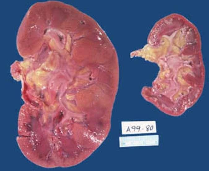
5 2. Trachea 3. Kidney
6 (ANSWER SHEET) STUDY QUESTIONS CASE STUDY QUESTIONS: ADDITIONAL QUESTIONS ON CELLULAR CHANGE PHOTO # Cell Adaptation/Change Description Etiology (END)
Mechanisms of Cell Injury
 Causes of Cell Injury 1- oxygen deprivation (anoxia) 2- physical agents 3- chemical agents 4- infections agents 5- immunologic reactions 6- genetic defects 7- nutritional imbalances Mechanisms of Cell
PREPARED BY P.DHARANI PRASAD II YEAR B.PHARM II SEM SUB:PATHOPHYSIOLOGY
 CELL INJURY UNIT I PREPARED BY P.DHARANI PRASAD II YEAR B.PHARM II SEM SUB:PATHOPHYSIOLOGY DETECTION OF CELLULAR CHANGES AFTER INJURY BY: LIGHT MICROSCOPY OR GROSS EXAMINATION DETECT CHANGES HOURS TO DAYS
number Done by Corrected by Doctor Heyam Awad
 number 4 Done by Waseem Abu Obeida Corrected by Saad Al-Hayek Doctor Heyam Awad Cell injury -in the previous lectures we talked about the causes (etiology) and the mechanism (pathogenesis) of cell injury.
Types of insult - hypoxia
 Introduction This presentation will be a guide to cell injury and cell death outline causes and pathogenesis of cell injury/death describe the morphological changes of cell injury/death Describe the process
Histopathology: Cell necrosis and cytoplasmic accumulations
 Histopathology: Cell necrosis and cytoplasmic accumulations These presentations are to help you identify basic histopathological features. They do not contain the additional factual information that you
BIOCHEMICAL INVESTIGATIONS IN THE DIAGNOSTICS OF CARDIOVASCULAR DISORDERS. As. MARUSHCHAK M.I.
 BIOCHEMICAL INVESTIGATIONS IN THE DIAGNOSTICS OF CARDIOVASCULAR DISORDERS As. MARUSHCHAK M.I. Heart attack symptoms Acute MI Measurement of cardiac enzyme levels Measure cardiac enzyme levels at regular
Chapter 1 CELL INJURY CELL DEATH CELL ADAPTATIONS. M.G.Rajanandh, Dept. of Pharmacy Practice, SRM College of Pharmacy, SRM University.
 Chapter 1 CELL INJURY CELL DEATH CELL ADAPTATIONS M.G.Rajanandh, Dept. of Pharmacy Practice, SRM College of Pharmacy, SRM University. CONCEPTS IN CELL INJURY The clinical signs and symptoms are several
Cell Adaptation, Cell Injury and Cell Death
 Cell Adaptation, Cell Injury and Cell Death Pathology:- is the study of structural and functional abnormalities that are expressed as diseases of organs and systems. Modern pathology, proposed that injury
Pathophysiology lab 2. Cellular injury and adaptation
 Pathophysiology lab 2 Cellular injury and adaptation Adaptation Cellular changes that aim to preserve cell viability and prevent cell injury. The adaptive responses include: 1. Atrophy 2. Hypertrophy 3.
shehab Moh Tarek ... ManarHajeer
 3 shehab Moh Tarek... ManarHajeer In the previous lecture we discussed the accumulation of oxygen- derived free radicals as a mechanism of cell injury, we covered their production and their pathologic
Cellular Injury, Necrosis, Apoptosis
 Cellular Injury, Necrosis, Apoptosis Cell injury results when cells are stressed and can no longer adapt Injury may progress through a reversible stage Reversible Cell Injury Reduced oxidative phosphorylation
Pathology of Hypertension
 2016-03-07 Pathology of Hypertension Honghe Zhang honghezhang@zju.edu.cn Tel:88208199 Department of Pathology ❶ Genetic predisposition ❷ Dietary factors ❸ Environmental factors ❹ Others Definition and
Cell Injury MECHANISMS OF CELL INJURY
 Cell Injury MECHANISMS OF CELL INJURY The cellular response to injurious stimuli depends on the following factors: Type of injury, Its duration, and Its severity. Thus, low doses of toxins or a brief duration
Coagulative Necrosis of Myocardium. Dr Rodney Itaki Division of Pathology
 Coagulative Necrosis of Myocardium Dr Rodney Itaki Division of Pathology Coagulative Necrosis Gross pathology: 3 day old infarct: Yellow necrosis surrounded by hyperemic borders. Arrow points to a transmural
Cellular responses to stress
 Cellular responses to stress (Adaptations, injury and death) (2 of 5) Most injurious stimuli are grouped into: Oxygen deprivation Chemical agents Infectious agents Immunologic reactions Genetic factors
CELL INJURY. Severity of Cell Injury
 GENERAL PATHOLOGY LECTURE - 3 DR. M. TARIQ JAVED Professor Department of Pathology, Faculty of Veterinary Science, University of Agriculture, Faisalabad, Pakistan. 9/11/2009 1 CELL INJURY No adaptive response
Cellular response to stress
 Cellular pathology - cell injury, death and adaptations Pathology Göran Andersson Cellular response to stress Cells differ in their capacity to tolerate changes in their microenvironment Acute, severe
SECTION 2 CELL INJURY
 Adapted myocyte Normal myocyte Reversibly-injured myocyte SECTION 2 CELL INJURY Cell death 5/4/2014 1 5/4/2014 2 Reversible Degeneration Irreversible Cellular Swelling Fatty Change Hyaline Change Amyloid
Quiz 1 Review. More Cowbell
 Quiz 1 Review More Cowbell Quiz 1 review Inflamma7on Repair Cell Injury and Adapta7on Quiz 1 review Inflamma7on Injury Acute inflammation Chronic inflammation Abscess Resolution Repair Time course Inflammation
Introduction to pathology lecture 5/ Cell injury apoptosis. Dr H Awad 2017/18
 Introduction to pathology lecture 5/ Cell injury apoptosis Dr H Awad 2017/18 Apoptosis = programmed cell death = cell suicide= individual cell death Apoptosis cell death induced by a tightly regulated
Consultant Medical Laboratory Scientist Assistant Professor of Histopathology & Cytopathology
 بسم اهلل الرحمن الرحيم By: PhD (Histopathology & Cytopathology), M.BA (Total Quality Management) Consultant Medical Laboratory Scientist Assistant Professor of Histopathology & Cytopathology Introduction
APOPTOSIS, NECROSIS AND CANCER. Dr. S. P. Pattanayak
 APOPTOSIS, NECROSIS AND CANCER Dr. S. P. Pattanayak LEARNING OBJECTIVES At the end of the lecture, students should be able to: Know the importance of cell death. Define various modes of cell death. Identify
Cellular Pathology. Histopathology Lab #2 (web) Paul Hanna Jan 2018
 Paul Hanna Jan 2018") Cellular Pathology Histopathology Lab #2 (web) Paul Hanna Jan 2018 Slide #91 Clinical History: a necropsy was performed on an aged cat the gross pathological changes included: widespread subcutaneous edema
Cellular Pathology Histopathology Lab #2 (web) Paul Hanna Jan 2018 Slide #91 Clinical History: a necropsy was performed on an aged cat the gross pathological changes included: widespread subcutaneous edema
CHAPTER ONE INTRODUCTION TO PATHOLOGY
 Dr. Nabeel Abdulwadood Rasheed 1 CHAPTER ONE INTRODUCTION TO PATHOLOGY The literal translation of the word pathology is the study (logos) of suffering (pathos). It is a discipline that bridges clinical
Mechanisms of disease
 PP Mechanisms of disease Stress and disease Homeostasis - Responsible for maintaining a constant, safe internal environment - Controlled by feedback loops o Negative feedback loop: temperature, blood glucose
Chemical and Biochemical Mechanism Of Cell Injury.
 Chemical and Biochemical Mechanism Of Cell Injury. Professor Dr. M. Tariq Javed Dept. of Pathology Faculty of Vet. Science The University Of Agriculture Faisalabad Cell Injury When the cell is exposed
Stages in the cellular response to stress & injurious stimuli
 Blok BBS 2 Departemen Patologi Anatomi Fakultas Kedokteran Universitas Sumatera Utara Medan -2011 Stages in the cellular response to stress & injurious stimuli 3/28/2011 2 1 Table 1-1.Cellular Responses
Disturbances of Circulation. Histopathology Lab #2 (Web)
") Disturbances of Circulation Histopathology Lab #2 (Web) Paul Hanna Winter 2015 Slide #96 History: pig was fine in the morning & found dead in the afternoon there was ~100 mls of clear fluid in the pericardial
Disturbances of Circulation Histopathology Lab #2 (Web) Paul Hanna Winter 2015 Slide #96 History: pig was fine in the morning & found dead in the afternoon there was ~100 mls of clear fluid in the pericardial
NECROSIS, GANGRENE. I. practical training 2 rd year Dentistry
 NECROSIS, GANGRENE. I. practical training 2 rd year Dentistry Signs of death Cardiac arrest (no pulse) Pallor mortis, paleness which happens in the 15 120 minutes after death Livor mortis, a settling of
Lecture-2 / Dr Hussain Abady Aljebori Over view of cell injury and cell death; Cell injury results when: a. cells are stressed so severely that they
 Lecture-2 / Dr Hussain Abady Aljebori Over view of cell injury and cell death; Cell injury results when: a. cells are stressed so severely that they are no longer able to adapt or b. when cells are exposed
Pathology MCQs. lipid. protein. glycogen. lipofuscin. water. Karyolysis. Cellular swelling. Involvement of a large number of cells
 Pathology MCQs 1. In hypoxic cell injury, cell swelling occurs because of increased intracellular: lipid protein glycogen lipofuscin water 2. Which of the following is a feature of apoptosis? Karyolysis
Histopathology: Glomerulonephritis and other renal pathology
 Histopathology: Glomerulonephritis and other renal pathology These presentations are to help you identify basic histopathological features. They do not contain the additional factual information that you
PATHOLOGY & PATHOPHYSIOLOGY
 PATHOLOGY & PATHOPHYSIOLOGY CHAPTER 2: CELLULAR PATHOLOGY OBJECTIVES Discuss the factors that can cause injury to cells Differentiate between the various ways that cells adapt to stressors Look at the
Hemodynamic Disorders, Thrombosis, and Shock. Richard A. McPherson, M.D.
 Hemodynamic Disorders, Thrombosis, and Shock Richard A. McPherson, M.D. Edema The accumulation of abnormal amounts of fluid in intercellular spaces of body cavities. Inflammation and release of mediators
Imaging of Coronary Artery Disease: II
 Acta Radiológica Portuguesa, Vol.XIX, nº 74, pág. 45-51, Abr.-Jun., 2007 Imaging of Coronary Artery Disease: II Jean Jeudy University of Maryland School of Medicine Department of Diagnostic Radiology Armed
[General Pathology] Introduction to Pathology
![[General Pathology] Introduction to Pathology](/thumbs/73/69585662.jpg "[General Pathology] Introduction to Pathology") Introduction to Pathology Pathology: Literally translated, pathology is the study (logos) of disease (pathos, suffering). It involves the investigation of the causes of disease and the associated changes
Introduction to Pathology Pathology: Literally translated, pathology is the study (logos) of disease (pathos, suffering). It involves the investigation of the causes of disease and the associated changes
INTRODUCTION TO HEALTH AND DISEASE BLOCK
 MBBS 1 st Yr. Lecture Dr. Annie Cheung September 25, 2002, 8:30AM 9:30 AM LT1, G/F, Academic and Administration Block Faculty of Medicine Building INTRODUCTION TO HEALTH AND DISEASE BLOCK CELL INJURY AND
Quantification of Coronary Arterial Narrowing at Necropsy in Acute Transmural Myocardial Infarction
 Quantification of Coronary Arterial Narrowing at Necropsy in Acute Transmural Myocardial Infarction Analysis and Comparison of Findings in 27 Patients and 22 Controls WILLIAM C. ROBERTS, M.D., AND ANCIL
Cellular Injury. Intracellular degeneration. By Dr. Hemn Hassan Othman PhD, Pathology Fall /20/2018 1
 Cellular Injury Intracellular degeneration By Dr. Hemn Hassan Othman PhD, Pathology Fall 2018 10/20/2018 1 Types of cell injury Cell injury is divided into: 1. Reversible cell injury 2. Irreversible cell
The basis of Disease
 General Curriculum The basis of Disease ZHOU REN 周韧 Prof., M.D., Ph.D. Institute of Pathology & Forensic Medicine Department of Pathology & Patho-physiology Zhenjiang University Judicial Evidence & Evaluation
Necrosis is death of cells and tissues in the living animal. Focal/ Multifocal necrosis- terms used for one
 Necrosis Necrosis Necrosis is death of cells and tissues in the living animal. Focal/ Multifocal necrosis- terms used for one or more, small, clearly defined areas of necrosis. Diffuse necrosis- term used
Lab title: Cell Division author: Dr. Ruth Dahlquist-Willard (modified by D. Bell)
") Corresponding Readings: Lab title: Cell Division author: Dr. Ruth Dahlquist-Willard (modified by D. Bell) Campbell Ch. 8 BIOL-100L Safety Information: We will be using laboratory glassware such as microscope
Corresponding Readings: Lab title: Cell Division author: Dr. Ruth Dahlquist-Willard (modified by D. Bell) Campbell Ch. 8 BIOL-100L Safety Information: We will be using laboratory glassware such as microscope
Cytyc Corporation - Case Presentation Archive - March 2002
 FirstCyte Ductal Lavage History: 68 Year Old Female Gail Index: Unknown Clinical History: Negative Mammogram in 1995 6 yrs. later presents with bloody nipple discharge Subsequent suspicious mammogram Suspicious
2. Obtain the following: eye guards gloves dissection tools: several blunt probes, scissors, a scalpel and forceps dissection pan sheep heart
 Week 04 Lab Heart Anatomy LEARNING OUTCOMES: Describe the gross external and internal anatomy of the heart. Identify and discuss the function of the valves of the heart. Identify the major blood vessels
Cardiac Muscle Physiology. Physiology Sheet # 8
 15 8 1 We have three types of muscles in our body: 1. Skeletal muscles. 2. Cardiac muscle. 3. Smooth muscles. The cardiovascular system consists of : Heart, cardiac vessels. The wall of the Heart has three
Myocardial Infarction
 Myocardial Infarction MI = heart attack Defined as necrosis of heart muscle resulting from ischemia. A very significant cause of death worldwide. of these deaths, 33% -50% die before they can reach the
THE ELECTROCARDIOGRAM IN ENDOMYOCARDIAL FIBROSIS
 BY From the Department of Medicine, Makerere College, and Mulago Hospital, Kampala, Uganda Received May 11, 1959 Endomyocardial fibrosis is an important cause of heart failure in this part of Africa. The
Cell injury, adaptation and death. Unite one Second Lab.
 Cell injury, adaptation and death Unite one Second Lab. The two lung abscesses seen here are examples of liquefactive necrosis in which there is a liquid center in an area of tissue injury. One abscess
Introduction to pathology
 Introduction to pathology By Dr. Mohsen Dashti Clinical Medicine & Pathology 316 1 st Lecture Lecture outilne Pathology. Disease. Cell injury. Manifestations of disease. Structural diseases. Functional
Mechanisms of Disease
 Chapter 2 Mechanisms of Disease Causes of Disease Heredity Trauma Inflammation/infection Hyperplasias/neoplasms Nutritional imbalance Impaired immunity Heredity Hereditary diseases Error in individual
DISEASE ETIOLOGY. Dynamic condition Morpho-functional alteration of one or more organ/tissue Acute or chronic Localized or systemic
 DISEASE Dynamic condition Morpho-functional alteration of one or more organ/tissue Acute or chronic Localized or systemic Etiology = studies the causes of disease Pathogenesis = starting from etiology,
Hypertrophy of cardiac muscle in the left ventricular chamber.
 The increase in the size of cells and consequently in the size of the affected organ. caused by specific hormone stimulation or by increased functional demand. ü ü Pregnancy: an adaptive response muscular
INTRODUCTION TO PATHOLOGY
 INTRODUCTION TO PATHOLOGY The literal translation of the word pathology is the study (logos) of suffering (pathos). It is a discipline that bridges clinical practice and basic sciences. Pathology is concerned
Systolic and Diastolic Currents of Injury
 Systolic and Diastolic Currents of Injury Figure 1 Action Potentials of Normal and Ischemic Tissue In Figure 1 above, the action potential of normal myocardium is represented by the solid lines and the
Diploma of Expert Practice in Histological Dissection. Examination Paper 1
 Diploma of Expert Practice in Histological Dissection Examination 2018 Paper 1 Mandatory modules short-answer questions 120 minutes 1. Attempt all questions 2. Questions may be answered in any order 3.
This is the second learning component (Learning Component 2) in our first learning module (Learning Module 1). In this component we review a very
 in our first learning module (Learning Module 1). In this component we review a very") This is the second learning component (Learning Component 2) in our first learning module (Learning Module 1). In this component we review a very basic response to injury inflammation. We ll look at examples
This is the second learning component (Learning Component 2) in our first learning module (Learning Module 1). In this component we review a very basic response to injury inflammation. We ll look at examples
Epithelium tissue system
 Epithelium tissue system Histology : is the study of the microscopic anatomy (microanatomy) of cells and tissues of plants and animals. It is commonly performed by examining cells and tissues under a light
Protocols. Harvesting and Sectioning the Ascending Aorta
 DAUGHERTY LAB Saha Cardiovascular Research Center University of Kentucky Protocols Harvesting and Sectioning the Ascending Aorta Page 1 of 7 Harvesting the ascending aorta Materials: 1. Insulin syringe
12 Lead EKG Chapter 4 Worksheet
 Match the following using the word bank. 1. A form of arteriosclerosis in which the thickening and hardening of the vessels walls are caused by an accumulation of fatty deposits in the innermost lining
Physiology sheet #2. The heart composed of 3 layers that line its lumen and cover it from out side, these layers are :
 Physiology sheet #2 * We will talk in this lecture about cardiac muscle physiology, the mechanism and the energy sources of their contraction and intracellular calcium homeostasis. # Slide 4 : The heart
MYOCARDIALINFARCTION. By: Kendra Fischer
 MYOCARDIALINFARCTION By: Kendra Fischer Outline Definition Epidemiology Clinical Aspects Treatment Effects of Exercise Exercise Testing Exercise Rx Summary and Conclusions References Break it down MYOCARDIAL
Histopathology: Hypertension and diabetes in the kidney These presentations are to help you identify basic histopathological features.
 Histopathology: Hypertension and diabetes in the kidney These presentations are to help you identify basic histopathological features. They do not contain the additional factual information that you need
Hashem Al-Dujaily. Tamer Barakat. Manar Hajeer
 1 Hashem Al-Dujaily Tamer Barakat... Manar Hajeer Introduction Pathology comes from Patho: disease/suffering and Logy: study. Therefore, Pathology is the study of disease. Pathology is the bridge between
Ischemic heart disease
 Ischemic heart disease Introduction In > 90% of cases: the cause is: reduced coronary blood flow secondary to: obstructive atherosclerotic vascular disease so most of the time it is called: coronary artery
What in the world is Histotechnology? Karen Stiffler, MA, HTL Program Director for Histotechnology
 What in the world is Histotechnology? Karen Stiffler, MA, HTL Program Director for Histotechnology The Basics of Histology Histology: the study of body tissues "histo" is from the Greek "histos" meaning
Avian Pathology. Bacterial diseases: histo slides. ECVP-ESVP Summer School 2012 Frédérique NGUYEN
 Avian Pathology Bacterial diseases: histo slides ECVP-ESVP Summer School 2012 Frédérique NGUYEN Bacterial diseases: histo slides B1. Turkey. Organs? Morphologic diagnosis? Special procedure? B2. Hen. Organ?
BIOLOGY 2290 Pathophysiology I (Health Sciences III ) Sept 2010 Instructor : Dr. Paul S. Sunga, Rm A359a, , Introduction
 Sept 2010 Instructor : Dr. Paul S. Sunga, Rm A359a, , Introduction") BIOLOGY 2290 Pathophysiology I (Health Sciences III ) Sept 2010 Instructor : Dr. Paul S. Sunga, Rm A359a, 323-5251, psunga@langara.bc.ca Introduction This course is designed to provide students with insights
BIOLOGY 2290 Pathophysiology I (Health Sciences III ) Sept 2010 Instructor : Dr. Paul S. Sunga, Rm A359a, 323-5251, psunga@langara.bc.ca Introduction This course is designed to provide students with insights
The Fundamental Unit of Life. Intext Exercise 1
 Intext Exercise 1 Who discovered cells and how? Cells were discovered in 1665 by an English Botanist, Robert Hooke. He used a primitive microscope to observe cells in a cork slice. Why is the cell called
MicroLife Review Sheet
 Name: KEY Due Date: MicroLife Review Sheet Directions: Complete the review sheet to help study for the unit test. This is only a guide of what will be on the test. Be sure to use this, notes, lab booklet,
SUPPLEMENTARY INFORMATION
 doi:10.1038/nature10188 Supplementary Figure 1. Embryonic epicardial genes are down-regulated from midgestation stages and barely detectable post-natally. Real time qrt-pcr revealed a significant down-regulation
ANATOMICAL PATHOLOGY TARIFF
 ANATOMICAL PATHOLOGY TARIFF A GUIDE TO UTILISATION. The following guidelines have been agreed by consensus of Anatomical Pathologists who are members of the Anatomical Pathologist s Group, or the National
Anatomy of the Heart
 Biology 212: Anatomy and Physiology II Anatomy of the Heart References: Saladin, KS: Anatomy and Physiology, The Unity of Form and Function 8 th (2018). Required reading before beginning this lab: Chapter
Disclosures. Parathyroid Pathology. Objectives. The normal parathyroid 11/10/2012
 Disclosures Parathyroid Pathology I have nothing to disclose Annemieke van Zante MD/PhD Assistant Professor of Clinical Pathology Associate Chief of Cytopathology Objectives 1. Review the pathologic features
Cellular Pathology Gross Pathology Laboratory 2 Cell Injury. VPM 152: General Pathology Instructor: Chelsea Martin Winter 2016
 Cellular Pathology Gross Pathology Laboratory 2 Cell Injury VPM 152: General Pathology Instructor: Chelsea Martin Winter 2016 Gross Specimens The following slides consist of images from the specimens presented
Name: SLO and Quarterly Assessment #4 Review Date: Science 8 - Mrs. Hagan
 Name: SLO and Quarterly Assessment #4 Review Date: Science 8 - Mrs. Hagan Period: 1. The data table below shows the masses and volumes of three objects (,, and ). The formula for calculating an object's
Case Scenario 1: Thyroid
 Case Scenario 1: Thyroid History and Physical Patient is an otherwise healthy 80 year old female with the complaint of a neck mass first noticed two weeks ago. The mass has increased in size and is palpable.
Early scientists who observed cells made detailed sketches of what they saw.
 Early scientists who observed cells made detailed sketches of what they saw. Early scientists who observed cells made detailed sketches of what they saw. CORK Early scientists who observed cells made detailed
DEGENERATION NECROSIS AND INFILTRATION
 DEGENERATION NECROSIS AND INFILTRATION Cellular Degenerations and Infiltrations 1. Cloudy swelling and hydropic degeneration Cloudy swelling and hydropic degeneration occur when the regulatory mechanisms
Canine Liver Eneku Wilfred Bovine Pathology
 2012-1-3 Canine Liver Eneku Wilfred Bovine Pathology Contributor: New Mexico Department of Agriculture Veterinary Diagnostic Services Signalment: 5 month old male Weimaraner dog (Canis familiaris) History:
1) Severe, crushing substernal chest pain 2) radiate to the neck, jaw, epigastrium, or left arm. 3- rapid and weak pulse 4- nausea (posterior MI).
 Severe, crushing substernal chest pain 2) radiate to the neck, jaw, epigastrium, or left arm. 3- rapid and weak pulse 4- nausea (posterior MI).") 1) Severe, crushing substernal chest pain 2) radiate to the neck, jaw, epigastrium, or left arm. 3- rapid and weak pulse 4- nausea (posterior MI). 5- cardiogenic shock (massive MIs >40% of the left ventricle)
1) Severe, crushing substernal chest pain 2) radiate to the neck, jaw, epigastrium, or left arm. 3- rapid and weak pulse 4- nausea (posterior MI). 5- cardiogenic shock (massive MIs >40% of the left ventricle)
Dr. Heba Kalbouneh. Dr. Heba Kalbouneh. Dr. Heba Kalbouneh
 Dr. Heba Kalbouneh Dr. Heba Kalbouneh Dr. Heba Kalbouneh Tissue: is a group of cells that serve the same function, they are surrounded by extra cellular matrix. The 4 basic types of tissue: 1. epithelial
 Question 1: Who discovered cells and how? Cells were discovered in 1665 by an English Botanist, Robert Hooke. He used a primitive microscope to observe cells in a cork slice. Question 2: Why is the cell
Lesson 1. Cell Theory - Statements - Exceptions. Categorizing Cells - Prokaryotes vs Eukaryotes
 Lesson 1 Cell Theory - Statements - Exceptions Categorizing Cells - Prokaryotes vs Eukaryotes The Cell Theory The discovery of cells and their structure is linked to the development of the magnifying lenses,
MITOSIS IN ONION ROOTLET CELLS
 Lesson 6: CELL CYCLE, MITOSIS Name: Group: MITOSIS IN ONION ROOTLET CELLS Permanent slide: onion rootlet stained with acetorcein The particular mitotic phases are visible in the onion rootlet cells. Chromosomes
The basis of Disease
 General Curriculum The basis of Disease ZHOU REN 周韧 Prof., M.D., Ph.D. Institute of Pathology & Forensic Medicine Department of Pathology & Patho-physiology Zhenjiang University Judicial Evidence & Evaluation
Class IX Chapter 5 The Fundamental Unit of Life Science
 1 Class IX Chapter 5 The Fundamental Unit of Life Science Question 1: Who discovered cells and how? Cells were discovered in 1665 by an English Botanist, Robert Hooke. He used a primitive microscope to
I. ADAPTATION TO ENVIRONMENTAL STRESS. A. Hypertrophy:
 د.جواهر محي الدين Lec:2&3 Cellular Reaction to Injury I. ADAPTATION TO ENVIRONMENTAL STRESS II. HYPOXIC CELL INJURY III. FREE RADICAL INJURY IV. CHEMICAL CELL INJURY V. NECROSIS VI. APOPTOSIS VII. REVERSIBLE
CNS pathology Third year medical students. Dr Heyam Awad 2018 Lecture 12: CNS tumours 2/3
 CNS pathology Third year medical students Dr Heyam Awad 2018 Lecture 12: CNS tumours 2/3 Pilocytic astrocytoma Relatively benign ( WHO grade 1) Occurs in children and young adults Mostly: in the cerebellum
Department of medical physiology 7 th week and 8 th week
 Department of medical physiology 7 th week and 8 th week Semester: winter Study program: Dental medicine Lecture: RNDr. Soňa Grešová, PhD. Department of medical physiology Faculty of Medicine PJŠU Cardiovascular
Advanced Multi-Layer Speckle Strain Permits Transmural Myocardial Function Analysis in Health and Disease:
 Advanced Multi-Layer Speckle Strain Permits Transmural Myocardial Function Analysis in Health and Disease: Clinical Case Examples Jeffrey C. Hill, BS, RDCS Echocardiography Laboratory, University of Massachusetts
General Pathology Theory Syllabus for II B.D.S.
 General Pathology Theory Syllabus for II B.D.S. Sr. No. Topic (Must Know) (Desirable to know) 1.Introduction to Pathology - Different sections in pathology - The Cell in health - Normal cell structure
The Adrenal Glands. I. Normal adrenal gland A. Gross & microscopic B. Hormone synthesis, regulation & measurement. II.
 The Adrenal Glands Thomas Jacobs, M.D. Diane Hamele-Bena, M.D. I. Normal adrenal gland A. Gross & microscopic B. Hormone synthesis, regulation & measurement II. Hypoadrenalism III. Hyperadrenalism; Adrenal
Arterioles of Kidney and Pancreas in Cases
 Arterioles of Kidney and Pancreas in Cases of Cardiac Hypertrophy of Undetermined Causation By HAROLD A. FERRIS, JR., M.D. The arterioles in the kidneys and pancreas in 50 consecutive necropsy cases of
c Ischemia (30 min) Reperfusion (8 w) Supplementary Figure bp 300 bp Ischemia (30 min) Reperfusion (4 h) Dox 20 mg/kg i.p.
 Reperfusion (8 w) Supplementary Figure bp 300 bp Ischemia (30 min) Reperfusion (4 h) Dox 20 mg/kg i.p.") a Marker Ripk3 +/ 5 bp 3 bp b Ischemia (3 min) Reperfusion (4 h) d 2 mg/kg i.p. 1 w 5 w Sacrifice for IF size A subset for echocardiography and morphological analysis c Ischemia (3 min) Reperfusion (8
a Marker Ripk3 +/ 5 bp 3 bp b Ischemia (3 min) Reperfusion (4 h) d 2 mg/kg i.p. 1 w 5 w Sacrifice for IF size A subset for echocardiography and morphological analysis c Ischemia (3 min) Reperfusion (8
Pathophysiology of Cardiovascular System. Dr. Hemn Hassan Othman, PhD
 Pathophysiology of Cardiovascular System Dr. Hemn Hassan Othman, PhD hemn.othman@univsul.edu.iq What is the circulatory system? The circulatory system carries blood and dissolved substances to and from
Dr/ Sherein Saeid AbdElgayed, ph.d
 هللامسب Dr/ Sherein Saeid AbdElgayed, ph.d Professor of Veterinary Pathology, Cairo University, Giza, Egypt. Chairman of the Editorial Board of Arab Journal of Science & Research Publishing (AJSRP) http://www.ajsrp.com
Myocardial Ischemia in Infants
 THE ANNALS OF THORACIC SURGERY Journal of The Society of Thoracic Surgeons and the Southern Thoracic Surgical Association VOLUME 8 NUMBER 5 NOVEMBER 1969. * Myocardial Ischemia in Infants Its Role in Three
Muscle Tissue. General concepts. Classification of muscle. I. Functional classification is based on the type of neural control.
 Muscle Tissue LEARNING OBJECTIVES 1. Identify the three types of muscle tissue at the light microscopic level. 2. List and compare the structural and functional features of each of the three muscle fiber