Alicia Armour, MA, BS, RDCS
|
|
|
- Trevor Goodwin
- 5 years ago
- Views:
Transcription

1 Alicia Armour, MA, BS, RDCS
2 No disclosures
3 Review 2D Speckle Strain (briefly) Discuss some various patient populations & disease pathways where Strain can be helpful Discuss how to acquire images for Strain analysis
4 Deformation dealing with shape and volume change Distance between some particles changes Angle between particle lines may change Longitudinal Strain: shortening or lengthening; e: change in length per length e=(l -l o )/l o = Δl/l o (dimensionless) l and l o are the final and original lengths of a linear object Shortening is a negative extension (e<0)

5 2D Speckle Tracking (our focus today) Myocardial deformation (fractional change in length of a myocardial segment) Unitless, expressed as a percentage Positive (lengthening) or negative (shortening) values Current and Evolving Echocardiographic Techniques for the Quantitative Evaluation of Cardiac Mechanics: ASE/EAE Consensus Statement on Methodology and Indications Mor-Avi et al., JASE 2011; 24:
6 What images are required What spectral Doppler information is required How they need to be optimized How to analyze
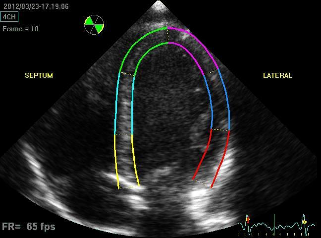
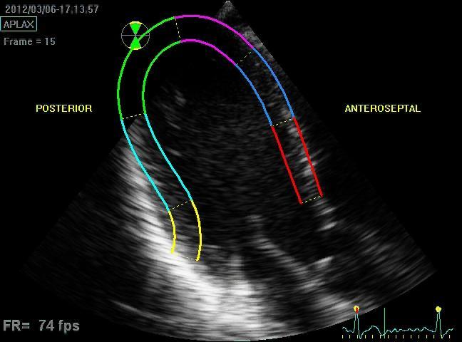
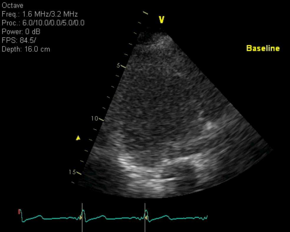
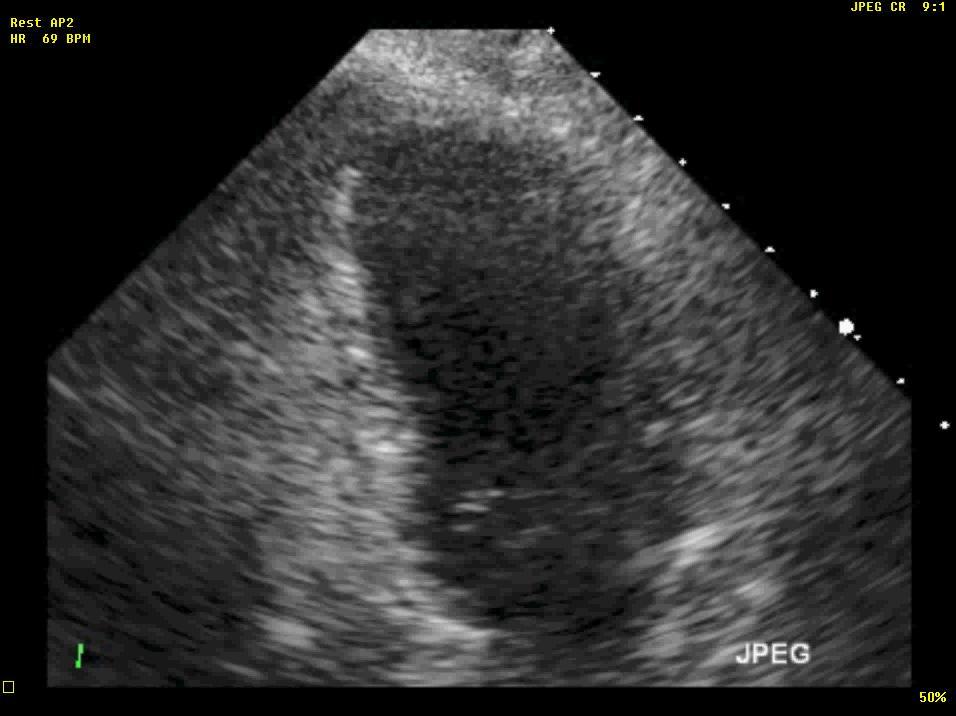
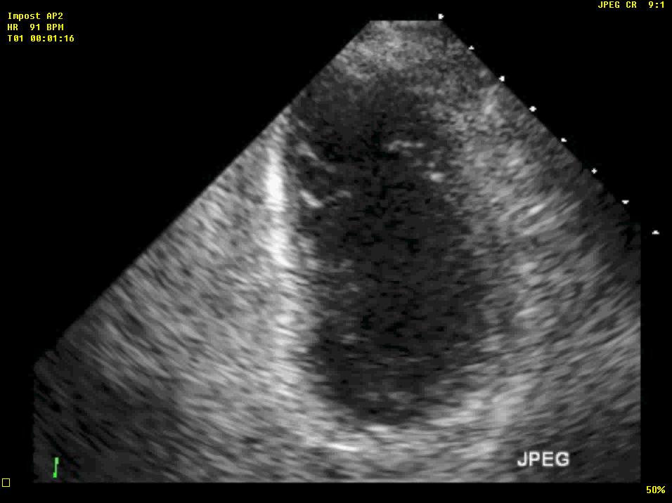
7 2D Images Required: SAX from the apex, mid-level (paps) and mitral valve (for radial strain as applicable) Apical 4 chamber Apical 2 chamber Apical Long Doppler images Required (for AoV closure timing): CW through AoV PW of LVOT


8 Not all patients have studio quality images, and it still tracks!!!
9 Most programs require a frame rate of 60-90Hz How to optimize that image: LV focus Less depth Narrow sector arc Images need speckle to track, so focus on endocardial border definition
10 Need images to be acquired within 5 beats for consistency Breath hold/same time in respiration Acquire 3 beat loops Some systems need 100ms before & after each cardiac cycle for analysis Acquiring 3 beat loops gives you more options for analysis afterwards & can overcome pre/post cycle timing issues
11
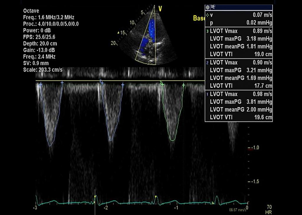
12 Most systems require aortic valve (AoV) timingopening & closing CW through AoV PW the LVOT Some systems require R to R timing as well (any spectral Doppler image will work). If you want to include mitral timing-pw MV inflow

13 Measure on the valve clicks (for CW or PW as applicable)
14 Can override spectral Doppler timing in most systems Can use 2D image but Our lab has found more variance in timing & less accuracy with using the 2D images If you use persistence or smoothing you 2D image may also provide inaccurate data
15 Find the best Apical 4, 2 & long axis images Find your spectral Doppler image for tracing Set AoV timing information Use analysis system image by image Some systems require the following sequence: Apical Long, Apical 4ch, Apical 2Ch
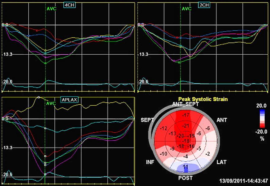
16


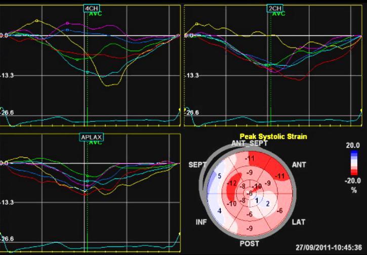
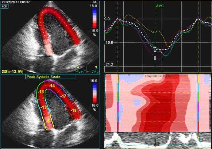
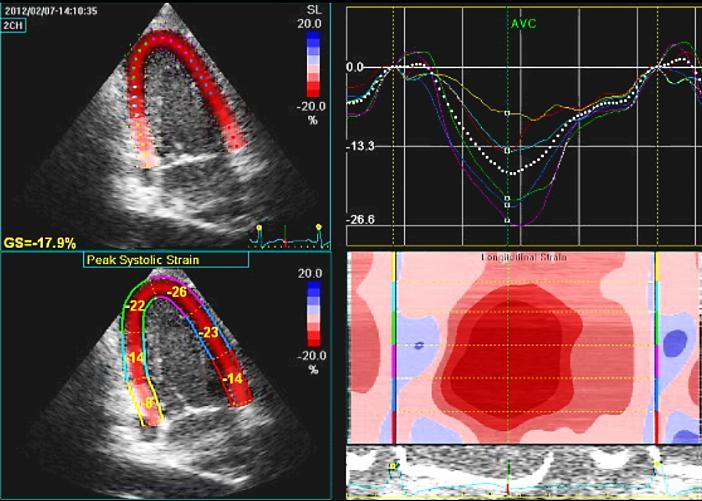
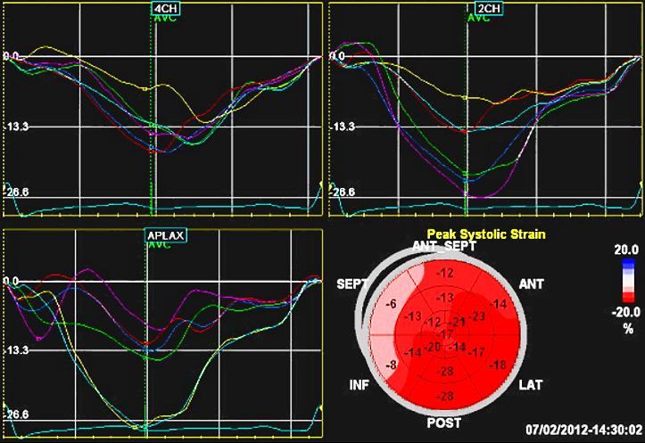
17 Average Global Peak Longitudinal Strain: -6.7% (abnormal)
18 Ensure the tracking! Is the tracking moving with the endocardium? Change the way you look at the image-watch the longitudinal movement Are there any ballistic movements through the tracking? Watch the base!
19
20
21 Check the waveforms! Do they make sense? What do they mean? What are the other things shown in the analysis?


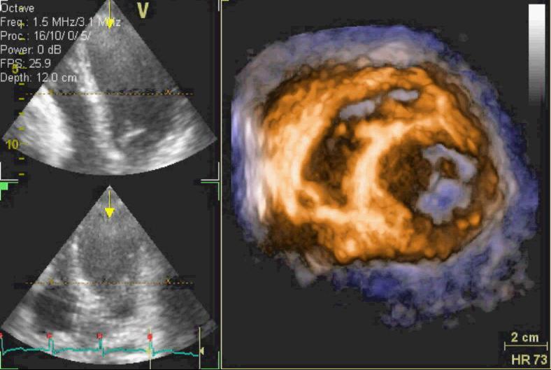
22 What do the waveforms show? What are they similar to?

23 AVC Waveforms trace the regional strain throughout the cardiac cycle EKG
24


25 GS = -13.9% Regional systolic strains


26 Peak Systolic Strain for each region Global Peak Strain for each view (an average) Overall Global L. Peak Strain (another average)

27 Red = speckles moving toward each other longitudinally Deep red = more negative strain Blue = speckles moving away from each other longitudinally White = little to no movement

28 AVC Colors represent the movement of the speckles throughout the cardiac cycle
29 Most systems produce a Bullseye The numbers displayed are the peak longitudinal strain before AoV closure All peak strains for each view & the Global Strain are averages (of averages )

30 Average Global Longitudinal Peak Strain (GLPS_Avg): -16.7% Peak Systolic Strain for each region prior to AVC
31 Images required: Apical Long, 4ch, 2ch Frame Rate of 60-90Hz LV focused view (less depth) with narrow sector arc 3 beat loops with a breath hold (same point in respiration each time) Heart Rate within 5 beats CW of AoV or PW of LVOT
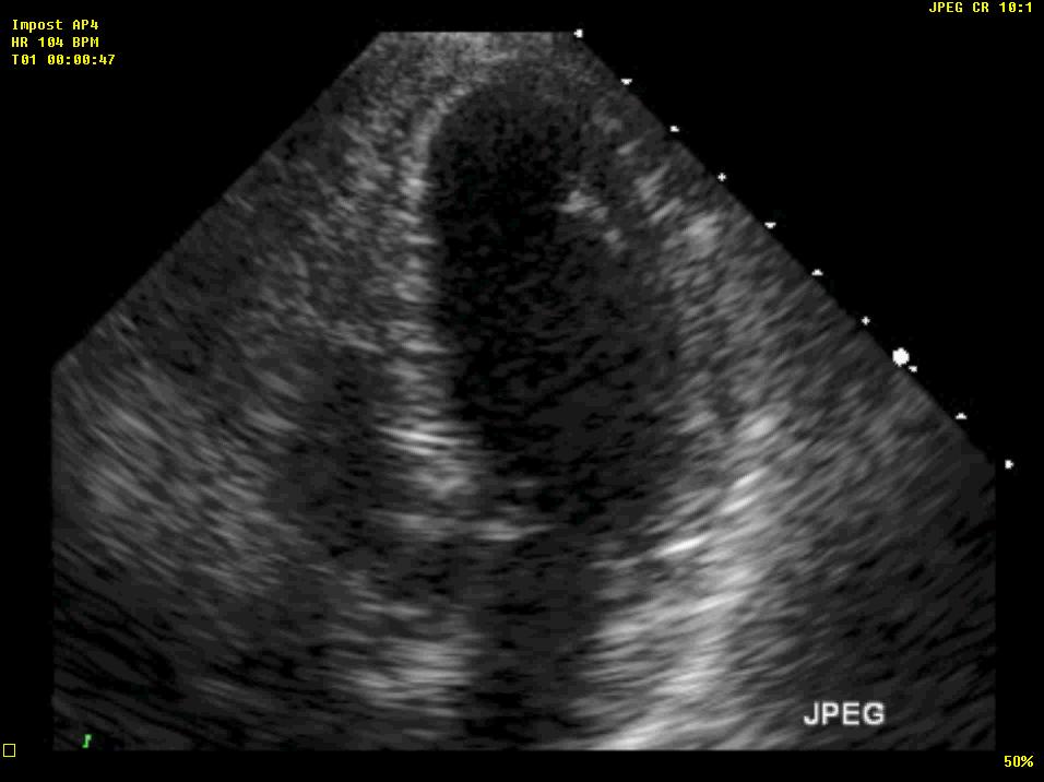
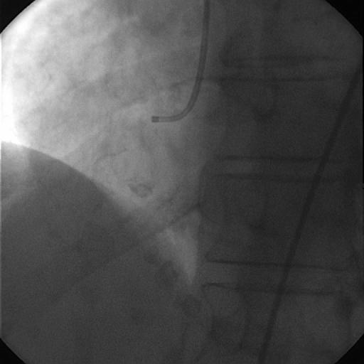
32 Measure AoV timing Follow software for sequence & setting points
33 Advantages: Analysis can be performed in any direction within the imaging plane Depending on spatial resolution epicardial, midwall & endocardial function may be analyzed as well Semiautomatic LV segment definition shows well in most patients making it possible to analyze in most patients successfully Current and Evolving Echocardiographic Techniques for the Quantitative Evaluation of Cardiac Mechanics: ASE/EAE Consensus Statement on Methodology and Indications Mor-Avi et al., JASE 2011; 24:
34 Disadvantages: Image quality dependent Acoustic shadowing or reverberations may cause underestimation of true deformation Difference among vendors Tracking algorithms use spatial smoothing & prior knowledge of normal LV function (erroneous regional dysfunction) Relies on assumption that morphologic details can be tracked in consecutive frames, which may not be true when out of plane motion occurs (requiring high temporal resolution) Current and Evolving Echocardiographic Techniques for the Quantitative Evaluation of Cardiac Mechanics: ASE/EAE Consensus Statement on Methodology and Indications Mor-Avi et al., JASE 2011; 24:
 Dyssynchrony Activation-delay induced")
35 Myocardial Mechanics Cardiac Resynchronization Therapy (CRT) Dyssynchrony Activation-delay induced cardiomyopathy
 used for Stroke Volume & Cardiac Output Depending on how your lab works: Time to Peak")
36 Overall goal: to get the walls to all contract at the same time Still need required Doppler information (MV inflow, PW of LVOT) used for Stroke Volume & Cardiac Output Depending on how your lab works: Time to Peak Waveforms
37 We use the waveforms, specifically observing regional patterns. Overall GLPS is also used in conjunction with Doppler information & 3D LVEF
38
39 61 year old female Heart Failure (non-ischemic cardiomyopathy), NYHA class II-III symptoms BiV/ICD in 2006 (lateral lead placement) AVR & MV repair 2002 Diabetes, hyperlipidemia, hypothyroidism, COPD, obstructive sleep apnea (OSA), chronic kidney disease (CKD)
40

41 Global Longitudinal Peak Strain Average -7.8%

42 Average VTI 19 cm
43 Tried various settings for sensed AV delay (SAV) and LV offset/pre-activation using VTI as improvement parameter End settings: SAV delay 120 ms VTI 25 cm (from 19 cm at baseline) LV pre-excitation -30 ms VTI 30.5 cm (from 19 cm at baseline) GLPS post-optimization -10.3% vs. -7.8% at baseline


44 LV-30 Average VTI 31 cm (final setting) Baseline Average VTI 19 cm


45 Baseline Bulls eye Final Settings Bulls eye Average GLPS -7.8% Average GLPS -10.3%
46 Post MI, is myocardium still viable? What other echo parameters can detect rejection? Are there any other useful echo tools for serial evaluation of cancer treatment patients that detect cardiotoxicity early? Besides LVEF & LV dimensions, how can I tell an athlete s heart from HCM or a hypertrophied heart? Regional changes in HCM patients-can this be tracked by echo as well as MRI? Can strain help detect LV dysfunction early with valvular disease?
47 Strain analysis increases sensitivity in detecting subclinical cardiac involvement in diseases such as: amyloidosis, diabetes and hypertensive heart disease. It will also show change in LV function after cancer treatment. Current and Evolving Echocardiographic Techniques for the Quantitative Evaluation of Cardiac Mechanics: ASE/EAE Consensus Statement on Methodology and Indications Mor-Avi et al., JASE 2011; 24:
48 Cancer treatment patients Coronary Artery Disease Valvular Disease
49 Cardiotoxicity after anthracycline (ANT) therapy can be as high as 18-26%; upwards of 36%. Heart failure can occur up to 20 years after successful treatment. 1 Several clinical trials have shown that while ejection fraction (EF) and fractional shortening remain normal, early signs of cardiotoxicity can be seen in Doppler and Strain parameters Editorial Comment: Identification of Anthracycline Cardiotoxicity: Left Ventricular Ejection Fraction Is Not Enough, Eidem, B W, MD, JASE Strain Rate Imaging Detects Early Cardiac Effects of Pegylated Liposomal Doxorubicin as Adjuvant Therapy in Elderly Patients with Breast Cancer, Jurcut et al., JASE 2008;21:
50 One research study in adolescents showed changes at 4 & 8 months in Longitudinal Peak Systolic Strain (LPSS) (ANT group averaged 18.1% vs. Control at 20.5%.) Impaired/decreased areas were observed for basal and mid averages. 3. Changes in Left Ventricular Longitudinal Strain with Anthracycline Chemotherapy in Adolescents Precede Subsequent Decreased Left Ventricular Ejection Fraction, Poterucha, Joseph et al., JASE 2012; 25:733-40
51 From Changes in LV Longitudinal Strain with Anthracycline, Poterucha et al., JASE 2012; 25:733-40
52 LPSS detected changes earlier than changes in LVEF (visual and calculated). Doppler parameters also indicated changes in the ANT group earlier than LVEF changes. 2D Speckle-Tracking LV LPSS can be useful in serial clinical monitoring for ANT cardiotoxicity. From Changes in Left Ventricular Longitudinal Strain with Anthracycline Chemotherapy in Adolescents Precede Subsequent Decreased Left Ventricular Ejection Fraction, Poterucha et al., JASE 2012; 25:733-40

53
54

55
56 Subendocardium most vulnerable area LV longitudinal mechanics at rest may be attenuated in patients with CAD Recent research shows: Correlation between Doppler-derived strain rate and coronary stenosis (85% specificity & 64% sensitivity). STE LS can be indicative of CAD extent From Assessment of Myocardial Mechanics Using Speckle Tracking Echocardiography: Fundamentals and Clinical Applications, Geyer, H. et al., JASE 2010;23:351-69

57 Stress Echo: strain changes can be indicative of ischemic myocardium & provide diagnostic information Resting cardiac strain can help define transmural extent of myocardial infarction (MI) and presence of viable myocardium. From Assessment of Myocardial Mechanics Using Speckle Tracking Echocardiography: Fundamentals and Clinical Applications, Geyer, H. et al., JASE 2010;23:351-69
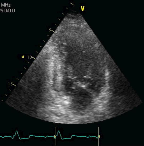
58 71 y.o. female Chest pain radiating to neck No prior history of CAD Sent to CDU for Stress Echo

59 Stress Echo 71 y.o. female (Target HR 126, Max HR 149bpm)

60 Stress Echo

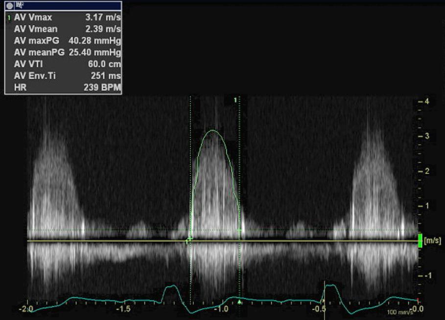
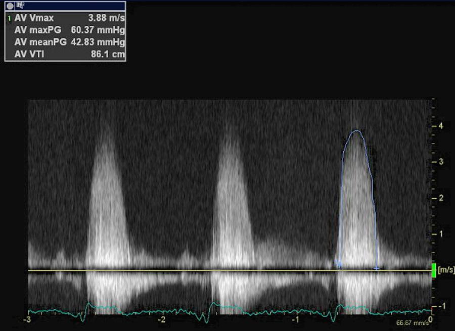
61

62 LV Strain Study 71 y.o. female
63 LV Strain Study Report 71 y.o. female
64 Coronary Angiography 71 y.o. female
65 LV remodeling can occur with or without symptoms even in the setting of severe valvular disease Strain-Tracking Echocardiography (STE) detects remodeling/cardiac dysfunction improving the yield of 2D echocardiography in patients with valvular diseases. Assessment of Myocardial Mechanics Using Speckle Tracking Echocardiography: Fundamentals and Clinical Applications, Geyer, H. et al., JASE 2010;23:351-69
66 LVH (with preserved LVEF) due to increased afterload Speckle-tracking longitudinal strain is impaired (or reduced ) with severe AS; while radial & circumferential strains remain relatively preserved. Post aortic valve replacement (AVR), all 3 forms of strain show improvement. Assessment of Myocardial Mechanics Using Speckle Tracking Echocardiography: Fundamentals and Clinical Applications, Geyer, H. et al., JASE 2010;23:351-69
67 68yo male with history of non-ischemic cardiomyopathy & valvular disease Seen initially to evaluate valvular disease & dyssynchrony
68


69
70 Not a significant improvement in LV function but patient reports feeling better. Small improvements in Global LS as well as regional strains. Increase in mean AoV gradient (from 25mmHg to 42mmHg)

71
72 Aortic Insufficiency (AI): Increase in LV end-diastolic volume & preload Remodeling to compensate which can mask onset of LV dysfunction DTI-derived strain (& strain rate): longitudinal & radial peak systolic strain correlate with LV ESV & EDV STE global longitudinal strain (LS) is reduced/impaired in patients with AI & bicuspid aortic valves. STE global radial strain shows changes immediately post AVR, but after 6mo follow up shows improvement. Assessment of Myocardial Mechanics Using Speckle Tracking Echocardiography: Fundamentals and Clinical Applications, Geyer, H. et al., JASE 2010;23:351-69
73 Mitral Regurgitation DTI-derived strain: longitudinal & radial strain reduced/impaired with severe MR & directly related to LV stroke volume (SV), diameter & contractility. STE-derived LS reported impaired with severe MR earlier than circumferential & radial strain. LV twist mechanics are reported to remain preserved. Assessment of Myocardial Mechanics Using Speckle Tracking Echocardiography: Fundamentals and Clinical Applications, Geyer, H. et al., JASE 2010;23:351-69
74 Cardiomyopathies (in general) Hypertrophic Cardiomyopathies (HCM) Athlete s Heart Sarcoidosis, Lupus Amyloidosis Myocardial Infarction And the list can go on
75
Implementing New Technology
 Implementing New Technology PP16 Imaging Conference Bicol Hospital, Legaspi City, Philippines July 2016 David Adams, ACS, RCS, RDCS, FASE Duke University Medical Center Echo Evolution By Dr. Zoghbi Handheld/POC
2/2/2011. Strain and Strain Rate Imaging How, Why and When? Movement vs Deformation. Doppler Myocardial Velocities. Movement. Deformation.
 Strain and Strain Rate Imaging How, Why and When? João L. Cavalcante, MD Advanced Cardiac Imaging Fellow Cleveland Clinic Foundation Disclosures: No conflicts of interest Movement vs Deformation Movement
LV FUNCTION ASSESSMENT: WHAT IS BEYOND EJECTION FRACTION
 LV FUNCTION ASSESSMENT: WHAT IS BEYOND EJECTION FRACTION Jamilah S AlRahimi Assistant Professor, KSU-HS Consultant Noninvasive Cardiology KFCC, MNGHA-WR Introduction LV function assessment in Heart Failure:
Strain and Strain Rate Imaging How, Why and When?
 Strain and Strain Rate Imaging How, Why and When? João L. Cavalcante, MD Advanced Cardiac Imaging Fellow Cleveland Clinic Foundation Disclosures: No conflicts of interest Movement vs Deformation Movement
Advanced Multi-Layer Speckle Strain Permits Transmural Myocardial Function Analysis in Health and Disease:
 Advanced Multi-Layer Speckle Strain Permits Transmural Myocardial Function Analysis in Health and Disease: Clinical Case Examples Jeffrey C. Hill, BS, RDCS Echocardiography Laboratory, University of Massachusetts
How To Perform Strain Imaging; Step By Step Approach. Maryam Bo Khamseen Echotechnoligist II EACVI, ARDMS, RCS King Abdulaziz Cardiac Center- Riyadh
 How To Perform Strain Imaging; Step By Step Approach Maryam Bo Khamseen Echotechnoligist II EACVI, ARDMS, RCS King Abdulaziz Cardiac Center- Riyadh Outlines: Introduction Describe the basic of myocardium
Tissue Doppler and Strain Imaging. Steven J. Lester MD, FRCP(C), FACC, FASE
, FACC, FASE") Tissue Doppler and Strain Imaging Steven J. Lester MD, FRCP(C), FACC, FASE Relevant Financial Relationship(s) None Off Label Usage None a. Turn the wall filters on and turn down the receiver gain. b. Turn
Tissue Doppler and Strain Imaging Steven J. Lester MD, FRCP(C), FACC, FASE Relevant Financial Relationship(s) None Off Label Usage None a. Turn the wall filters on and turn down the receiver gain. b. Turn
Nancy Goldman Cutler, MD Beaumont Children s Hospital Royal Oak, Mi
 Nancy Goldman Cutler, MD Beaumont Children s Hospital Royal Oak, Mi Identify increased LV wall thickness (WT) Understand increased WT in athletes Understand hypertrophic cardiomyopathy (HCM) Enhance understanding
Myocardial Strain Imaging in Cardiac Diseases and Cardiomyopathies.
 Myocardial Strain Imaging in Cardiac Diseases and Cardiomyopathies. Session: Cardiomyopathy Tarun Pandey MD, FRCR. Associate Professor University of Arkansas for Medical Sciences Disclosures No relevant
Tissue Doppler and Strain Imaging
 Tissue Doppler and Strain Imaging Steven J. Lester MD, FRCP(C), FACC, FASE Relevant Financial Relationship(s) None Off Label Usage None 1 Objective way with which to quantify the minor amplitude and temporal
OPTIMIZING ECHO ACQUISTION FOR STRAIN AND DIASTOLOGY
 OPTIMIZING ECHO ACQUISTION FOR STRAIN AND DIASTOLOGY October 8, 2017 Deborah Agler, ACS, RDCS, FASE Coordinator of Education and Training Cleveland Clinic General Principles Diastology Clinical Data Heart
Tissue Doppler and Strain Imaging
 Tissue Doppler and Strain Imaging Steven J. Lester MD, FRCP(C), FACC, FASE Relevant Financial Relationship(s) None Off Label Usage None 1 Objective way with which to quantify the minor amplitude and temporal
Three-dimensional Wall Motion Tracking:
 Three-dimensional Wall Motion Tracking: A Novel Echocardiographic Method for the Assessment of Ventricular Volumes, Strain and Dyssynchrony Jeffrey C. Hill, BS, RDCS, FASE Jennifer L. Kane, RCS Gerard
VECTORS OF CONTRACTION
 1/3/216 Strain, Strain Rate, and Torsion: Myocardial Mechanics Simplified and Applied VECTORS OF CONTRACTION John Gorcsan, MD University of Pittsburgh, Pittsburgh, PA Shortening Thickening Twisting No
Tissue Doppler Imaging in Congenital Heart Disease
 Tissue Doppler Imaging in Congenital Heart Disease L. Youngmin Eun, M.D. Department of Pediatrics, Division of Pediatric Cardiology, Kwandong University College of Medicine The potential advantage of ultrasound
Cardiac Chamber Quantification by Echocardiography
 Cardiac Chamber Quantification by Echocardiography Maryam Bokhamseen, RCS, RCDS, EACVI Echotechnologist ǁ, Non invasive Cardiac Laboratory King Abdulaziz Cardiac Center. Outline: Introduction. Background
Altered left ventricular geometry and torsional mechanics in high altitude-induced pulmonary hypertension:
 Altered left ventricular geometry and torsional mechanics in high altitude-induced pulmonary hypertension: a 3-D echocardiographic study B.W. De Boeck,* S. Kiencke, C. Dehnert, K. Auinger, # M. Maggiorini,
Strain/Untwisting/Diastolic Suction
 What Is Diastole and How to Assess It? Strain/Untwisting/Diastolic Suction James D. Thomas, M.D., F.A.C.C. Cardiovascular Imaging Center Department of Cardiology Cleveland Clinic Foundation Cleveland,
Global left ventricular circumferential strain is a marker for both systolic and diastolic myocardial function
 Global left ventricular circumferential strain is a marker for both systolic and diastolic myocardial function Toshinari Onishi 1, Samir K. Saha 2, Daniel Ludwig 1, Erik B. Schelbert 1, David Schwartzman
Load and Function - Valvular Heart Disease. Tom Marwick, Cardiovascular Imaging Cleveland Clinic
 Load and Function - Valvular Heart Disease Tom Marwick, Cardiovascular Imaging Cleveland Clinic Indications for surgery in common valve lesions Risks Operative mortality Failed repair - to MVR Operative
Strain Imaging: Myocardial Mechanics Simplified and Applied
 9/28/217 Strain Imaging: Myocardial Mechanics Simplified and Applied John Gorcsan III, MD Professor of Medicine Director of Clinical Research Division of Cardiology VECTORS OF CONTRACTION Shortening Thickening
Aortic Stenosis: Spectrum of Disease, Low Flow/Low Gradient and Variants
 Aortic Stenosis: Spectrum of Disease, Low Flow/Low Gradient and Variants Martin G. Keane, MD, FASE Professor of Medicine Lewis Katz School of Medicine at Temple University Basic root structure Parasternal
Evaluation of Left Ventricular Function and Hypertrophy Gerard P. Aurigemma MD
 Evaluation of Left Ventricular Function and Hypertrophy Gerard P. Aurigemma MD Board Review Course 2017 43 year old health assistant Severe resistant HTN LT BSA 2 Height 64 1 Here is the M mode echocardiogram
Echocardiographic Assessment of the Left Ventricle
 Echocardiographic Assessment of the Left Ventricle Theodora Zaglavara, MD, PhD, BSCI/BSCCT Department of Cardiovascular Imaging INTERBALKAN EUROPEAN MEDICAL CENTER 2015 The quantification of cardiac chamber
LV geometric and functional changes in VHD: How to assess? Mi-Seung Shin M.D., Ph.D. Gachon University Gil Hospital
 LV geometric and functional changes in VHD: How to assess? Mi-Seung Shin M.D., Ph.D. Gachon University Gil Hospital LV inflow across MV LV LV outflow across AV LV LV geometric changes Pressure overload
Quantification of Cardiac Chamber Size
 2017 KSE 2017-11-25 Quantification of Cardiac Chamber Size Division of Cardiology Keimyung University Dongsan Medical Center In-Cheol Kim M.D., Ph.D. LV size and function Internal linear dimensions PLX
Assessment of LV systolic function
 Tutorial 5 - Assessment of LV systolic function Assessment of LV systolic function A knowledge of the LV systolic function is crucial in the undertanding of and management of unstable hemodynamics or a
Back to Basics: Common Errors In Quantitation In Everyday Practice
 Back to Basics: Common Errors In Quantitation In Everyday Practice Deborah Agler, ACS, RDCS, FASE October 9, 2017 ASE: Echo Florida Rebecca T. Hahn, MD Director of Interventional Echocardiography Professor
Velocity, strain and strain rate: Doppler and Non-Doppler methods. Thoraxcentre, Erasmus MC,Rotterdam
 Velocity, strain and strain rate: Doppler and Non-Doppler methods J Roelandt J. Roelandt Thoraxcentre, Erasmus MC,Rotterdam Basics of tissue Doppler imaging Instantaneous annular velocity profiles IVCT
Value of echocardiography in chronic dyspnea
 Value of echocardiography in chronic dyspnea Jahrestagung Schweizerische Gesellschaft für /Schweizerische Gesellschaft für Pneumologie B. Kaufmann 16.06.2016 Chronic dyspnea Shortness of breath lasting
HYPERTROPHY: Behind the curtain. V. Yotova St. Radboud Medical University Center, Nijmegen
 HYPERTROPHY: Behind the curtain V. Yotova St. Radboud Medical University Center, Nijmegen Disclosure of interest: none Relative wall thickness (cm) M 0.22 0.42 0.43 0.47 0.48 0.52 0.53 F 0.24 0.42 0.43
10/7/2013. Systolic Function How to Measure, How Accurate is Echo, Role of Contrast. Thanks to our Course Director: Neil J.
 Systolic Function How to Measure, How Accurate is Echo, Role of Contrast Neil J. Weissman, MD MedStar Health Research Institute & Professor of Medicine Georgetown University Washington, D.C. No Disclosures
How to assess ischaemic MR?
 ESC 2012 How to assess ischaemic MR? Luc A. Pierard, MD, PhD, FESC, FACC Professor of Medicine Head, Department of Cardiology University Hospital Sart Tilman, Liège ESC 2012 No conflict of interest Luc
Adel Hasanin Ahmed 1 LV MORPHOLOGY
 Adel Hasanin Ahmed 1 LV MORPHOLOGY The left ventricular wall comprises three layers- middle circumferential layer and superficial and deep longitudinal layers: 1. Subepicardial longitudinal layer (25%
DISCLOSURE. Myocardial Mechanics. Relevant Financial Relationship(s) Off Label Usage
 Off Label Usage") 7th Annual Team Echocardiography: The Heart of Cardiovascular Medicine Tissue Doppler, Strain, Speckle: What? How? Christopher J Kramer RDCS Aurora Medical Group Advanced Cardiovascular Services, Aurora
7th Annual Team Echocardiography: The Heart of Cardiovascular Medicine Tissue Doppler, Strain, Speckle: What? How? Christopher J Kramer RDCS Aurora Medical Group Advanced Cardiovascular Services, Aurora
Mechanisms of False Positive Exercise Electrocardiography: Is False Positive Test Truly False?
 Mechanisms of False Positive Exercise Electrocardiography: Is False Positive Test Truly False? Masaki Izumo a, Kengo Suzuki b, Hidekazu Kikuchi b, Seisyo Kou b, Keisuke Kida b, Yu Eguchi b, Nobuyuki Azuma
Dr. Dermot Phelan MB BCh BAO PhD European Society of Cardiology 2012
 Relative Apical Sparing of Longitudinal Strain Using 2- Dimensional Speckle-Tracking Echocardiography is Both Sensitive and Specific for the Diagnosis of Cardiac Amyloidosis. Dr. Dermot Phelan MB BCh BAO
Chamber Quantitation Guidelines: What is New?
 Chamber Quantitation Guidelines: What is New? Roberto M Lang, MD J AM Soc Echocardiogr 2005; 18:1440-1463 1 Approximately 10,000 citations iase in itune Cardiac Chamber Quantification: What is New? Database
Echocardiographic Evaluation of the Cardiomyopathies. Stephanie Coulter, MD, FACC, FASE April, 2016
 Echocardiographic Evaluation of the Cardiomyopathies Stephanie Coulter, MD, FACC, FASE April, 2016 Cardiomyopathies (CMP) primary disease intrinsic to cardiac muscle Dilated CMP Hypertrophic CMP Infiltrative
좌심실수축기능평가 Cardiac Function
 Basic Echo Review Course 좌심실수축기능평가 Cardiac Function Seonghoon Choi Cardiology Hallym university LV systolic function Systolic function 좌심실수축기능 - 심근의수축으로심실에서혈액을대동맥으로박출하는기능 실제임상에서 LV function 의의미 1Diagnosis
RIGHT VENTRICULAR SIZE AND FUNCTION
 RIGHT VENTRICULAR SIZE AND FUNCTION Edwin S. Tucay, MD, FPCC, FPCC, FPSE Philippine Society of Echocardiography Quezon City, Philippines Echo Mission, BRTTH, Legaspi City, July 1-2, 2016 NO DISCLOSURE
Measuring cardiac tissue motion and strain
 Ultrasound Measuring cardiac tissue motion and strain Automated Cardiac Motion Quantification A.I. (acmq A.I. ) David Prater, MS, Clinical Scientist, Philips Jane Vogel, MD, Senior Product Manager, Philips
Coronary artery disease (CAD) risk factors
 risk factors") Background Coronary artery disease (CAD) risk factors CAD Risk factors Hypertension Insulin resistance /diabetes Dyslipidemia Smoking /Obesity Male gender/ Old age Atherosclerosis Arterial stiffness precedes
Background Coronary artery disease (CAD) risk factors CAD Risk factors Hypertension Insulin resistance /diabetes Dyslipidemia Smoking /Obesity Male gender/ Old age Atherosclerosis Arterial stiffness precedes
Aortic valve Stenosis: Insights in the evaluation of LV function. Erwan DONAL Cardiologie CHU Rennes
 Aortic valve Stenosis: Insights in the evaluation of LV function Erwan DONAL Cardiologie CHU Rennes erwan.donal@chu-rennes.fr Preload Afterload Myocardial Fiber Shortening Circumferential Longitudinal
Vevo 2100 System Cardio Measurements. Dieter Fuchs, PhD FUJIFILM VisualSonics, Inc.
 Vevo 2100 System Cardio Measurements Dieter Fuchs, PhD FUJIFILM VisualSonics, Inc. dfuchs@visualsonics.com Instructions This document is a guideline on how to assess cardiac function in rodents imaged
Cardiology for the Practitioner Advanced Cardiac Imaging: Worth the pretty pictures?
 Keenan Research Centre Li Ka Shing Knowledge Institute Cardiology for the Practitioner Advanced Cardiac Imaging: Worth the pretty pictures? Howard Leong-Poi, MD, FRCPC Associate Professor of Medicine St.
Top 10 Facts in Contrast Echocardiography. Pamela R. Burgess, BS, RDCS, RDMS, RVT, FASE
 Top 10 Facts in Contrast Echocardiography Pamela R. Burgess, BS, RDCS, RDMS, RVT, FASE Presenter Disclosure The following relationship exist related to this presentation: Pamela R. Burgess, BS, RDCS, RDMS,
Novel echocardiographic modalities: 3D echo, speckle tracking and strain rate imaging. Potential roles in sports cardiology. Stefano Caselli, MD, PhD
 Novel echocardiographic modalities: 3D echo, speckle tracking and strain rate imaging. Potential roles in sports cardiology. Stefano Caselli, MD, PhD Ospedale San Pietro Fatebenefratelli Rome, Italy Differential
3D-stress echocardiography Bernard Cosyns, MD, PhD
 3D-stress echocardiography Bernard Cosyns, MD, PhD No Disclosure The Pro-Technology bias Sicari et al. Cardiovascular Ultrasound 2006, 4:11 Overview 2D stress echocardiography: main limitations 3D echocardiography:
Diastology Disclosures: None. Dias2011:1
 Diastology 2011 James D. Thomas, M.D., F.A.C.C. Cardiovascular Imaging Center Department of Cardiology Cleveland Clinic Foundation Cleveland, Ohio, USA Disclosures: None Dias2011:1 Is EVERYBODY a member!?!
Stage of Valvular AS. Outline 10/14/16. Low-flow and Other Challenges to the Assessment of Aortic Stenosis. Severe AS
 Low-flow and Other Challenges to the Assessment of Aortic Stenosis Nithima Ratanasit, MD, FACC, FASE Siriraj Hospital, Mahidol University Bangkok, Thailand Outline Types of low-flow aortic stenosis Assessment
Diagnostic approach to heart disease
 Diagnostic approach to heart disease Initial work up History Physical exam Chest radiographs ECG Special studies Echocardiography Cardiac catheterization Echocardiography principles Technique of producing
Hemodynamic Assessment. Assessment of Systolic Function Doppler Hemodynamics
 Hemodynamic Assessment Matt M. Umland, RDCS, FASE Aurora Medical Group Milwaukee, WI Assessment of Systolic Function Doppler Hemodynamics Stroke Volume Cardiac Output Cardiac Index Tei Index/Index of myocardial
Heart Failure. Cardiac Anatomy. Functions of the Heart. Cardiac Cycle/Hemodynamics. Determinants of Cardiac Output. Cardiac Output
 Cardiac Anatomy Heart Failure Professor Qing ZHANG Department of Cardiology, West China Hospital www.blaufuss.org Cardiac Cycle/Hemodynamics Functions of the Heart Essential functions of the heart to cover
Dobutamine Stress testing In Low Flow, Low EF, Low Gradient Aortic Stenosis Case Studies
 Dobutamine Stress testing In Low Flow, Low EF, Low Gradient Aortic Stenosis Case Studies Mitral Regurgitation The New ASE Guidelines: Role of 2D/3D and CMR William A. Zoghbi MD, FASE, MACC Professor and
Echo assessment of the failing heart
 Echo assessment of the failing heart Mark K. Friedberg, MD The Labatt Family Heart Center The Hospital for Sick Children Toronto, Ontario, Canada Cardiac function- definitions Cardiovascular function:
22 nd Annual Conference of the Saudi Heart Association Riyadh, Saudi Arabia
 22 nd Annual Conference of the Saudi Heart Association Riyadh, Saudi Arabia New Echocardiographic Modalities to Evaluate Ventricular Function in Congenital Heart Disease: Tissue Doppler & Strain Rate Imaging
Quantitation of right ventricular dimensions and function
 SCCS Basics of cardiac assessment Quantitation of right ventricular dimensions and function Tomasz Kukulski, MD PhD Dept of Cardiology, Congenital Heart Disease and Electrotherapy Silesian Medical University
Prosthetic valve dysfunction: stenosis or regurgitation
 Prosthetic valve dysfunction: stenosis or regurgitation Jean G. Dumesnil MD, FRCP(C), FACC, FASE(Hon) Quebec Heart and Lung Institute, Québec, Québec No disclosures Possible Causes of High Gradients in
Appendix II: ECHOCARDIOGRAPHY ANALYSIS
 Appendix II: ECHOCARDIOGRAPHY ANALYSIS Two-Dimensional (2D) imaging was performed using the Vivid 7 Advantage cardiovascular ultrasound system (GE Medical Systems, Milwaukee) with a frame rate of 400 frames
Left atrial function. Aliakbar Arvandi MD
 In the clinic Left atrial function Abstract The left atrium (LA) is a left posterior cardiac chamber which is located adjacent to the esophagus. It is separated from the right atrium by the inter-atrial
Utility of Echocardiography
 Hypertrophic Cardiomyopathy and Beyond- Echo Hawaii 2018 Lawrence Rudski MD FRCPC FACC FASE Professor of Medicine Director, Division of Cardiology and Azrieli Heart Center Jewish General Hospital, McGill
Managing Hypertrophic Cardiomyopathy with Imaging. Gisela C. Mueller University of Michigan Department of Radiology
 Managing Hypertrophic Cardiomyopathy with Imaging Gisela C. Mueller University of Michigan Department of Radiology Disclosures Gadolinium contrast material for cardiac MRI Acronyms Afib CAD Atrial fibrillation
Evaluation of Left Ventricular Diastolic Dysfunction by Doppler and 2D Speckle-tracking Imaging in Patients with Primary Pulmonary Hypertension
 ESC Congress 2011.No 85975 Evaluation of Left Ventricular Diastolic Dysfunction by Doppler and 2D Speckle-tracking Imaging in Patients with Primary Pulmonary Hypertension Second Department of Internal
Effect of loading and geometry on functional parameters
 Effect of loading and geometry on functional parameters Piet Claus Cardiovascular Imaging and Dynamics Department of Cardiovascular Diseases Leuven University, Leuven, Belgium 5 th European Echocardiography
Velocity Vector Imaging as a new approach for cardiac magnetic resonance: Comparison with echocardiography
 Velocity Vector Imaging as a new approach for cardiac magnetic resonance: Comparison with echocardiography Toshinari Onishi 1, Samir K. Saha 2, Daniel Ludwig 1, Erik B. Schelbert 1, David Schwartzman 1,
Review of Cardiac Imaging Modalities in the Renal Patient. George Youssef
 Review of Cardiac Imaging Modalities in the Renal Patient George Youssef ECHO Left ventricular hypertrophy (LVH) assessment Diastolic dysfunction Stress ECHO Cardiac CT angiography Echocardiography - positives
Restrictive Cardiomyopathy
 ESC Congress 2011, Paris Imaging Unusual Causes of Cardiomyopathy Restrictive Cardiomyopathy Kazuaki Tanabe, MD, PhD Professor of Medicine Chair, Division of Cardiology Izumo, Japan I Have No Disclosures
Athlete s Heart vs. Cardiomyopathy
 Athlete s Heart vs. Cardiomyopathy Linda D. Gillam, MD, MPH, FASE Chair, Department of Cardiovascular Medicine Medical Director, Cardiovascular Service Line Former Team Cardiologist to the New York Jets
Acute impairment of basal left ventricular rotation but not twist and untwist are involved in the pathogenesis of acute hypertensive pulmonary oedema
 Acute impairment of basal left ventricular rotation but not twist and untwist are involved in the pathogenesis of acute hypertensive pulmonary oedema A.D. Margulescu 1,2, R.C. Sisu 1,2, M. Florescu 2,
ASE Guidelines on Aortic Regurgitation What Do I Measure? Case Studies
 ASE Guidelines on Aortic Regurgitation What Do I Measure? Case Studies Mitral Regurgitation The New ASE Guidelines: Role of 2D/3D and CMR William A. Zoghbi MD, FASE, MACC Professor and Chairman, Department
Ejection across stenotic aortic valve requires a systolic pressure gradient between the LV and aorta. This places a pressure load on the LV.
 Valvular Heart Disease Etiology General Principles Cellular and molecular mechanism of valve damage Structural pathology Functional pathology - stenosis/regurgitation Loading conditions - pressure/volume
Assessing Function by Echocardiography in VHD Asymptomatic Severe Organic MR. Dr. Julien Magne, PhD Sart Tilman Liège, BELGIUM
 Assessing Function by Echocardiography in VHD Asymptomatic Severe Organic MR Dr. Julien Magne, PhD Sart Tilman Liège, BELGIUM Conflict of Interest Disclosure None Why to assess LV function in asymptomatic
Echocardiographic Cardiovascular Risk Stratification: Beyond Ejection Fraction
 Echocardiographic Cardiovascular Risk Stratification: Beyond Ejection Fraction October 4, 2014 James S. Lee, M.D., F.A.C.C. Associates in Cardiology, P.A. Silver Spring, M.D. Disclosures Financial none
Feasibility and limitations of 2D speckle tracking echocardiography
 ORIGINAL ARTICLE 204 A prospective study in daily clinical practice Feasibility and limitations of 2D speckle tracking echocardiography Lina Melzer, Anja Faeh-Gunz, Barbara Naegeli, Burkhardt Seifert*,
Right Ventricular Strain in Normal Healthy Adult Filipinos: A Retrospective, Cross- Sectional Pilot Study
 Right Ventricular Strain in Normal Healthy Adult Filipinos: A Retrospective, Cross- Sectional Pilot Study By Julius Caesar D. de Vera, MD Jonnah Fatima B. Pelat, MD Introduction Right ventricle contributes
B-Mode measurements protocols:
 Application Note How to Perform the Most Commonly Used Measurements from the Cardiac Measurements Package associated with Calculations of Cardiac Function using the Vevo Lab Objective The Vevo LAB offline
1. LV function and remodeling. 2. Contribution of myocardial ischemia due to CAD, and
 1 The clinical syndrome of heart failure in adults is commonly associated with the etiologies of ischemic and non-ischemic dilated cardiomyopathy, hypertrophic cardiomyopathy, hypertensive heart disease,
Mechanisms of heart failure with normal EF Arterial stiffness and ventricular-arterial coupling. What is the pathophysiology at presentation?
 Mechanisms of heart failure with normal EF Arterial stiffness and ventricular-arterial coupling What is the pathophysiology at presentation? Ventricular-arterial coupling elastance Central arterial pressure
Adult Echocardiography Examination Content Outline
 Adult Echocardiography Examination Content Outline (Outline Summary) # Domain Subdomain Percentage 1 2 3 4 5 Anatomy and Physiology Pathology Clinical Care and Safety Measurement Techniques, Maneuvers,
Ejection across stenotic aortic valve requires a systolic pressure gradient between the LV and aorta. This places a pressure load on the LV.
 Valvular Heart Disease General Principles Etiology Cellular and molecular mechanism of valve damage Structural pathology Functional pathology - stenosis/regurgitation Loading conditions - pressure/volume
Comprehensive Echo Assessment of Aortic Stenosis
 Comprehensive Echo Assessment of Aortic Stenosis Smonporn Boonyaratavej, MD, MSc King Chulalongkorn Memorial Hospital Bangkok, Thailand Management of Valvular AS Medical and interventional approaches to
How does the heart pump? From sarcomere to ejection volume
 How does the heart pump? From sarcomere to ejection volume Piet Claus Cardiovascular Imaging and Dynamics Department of Cardiovascular Diseases University Leuven, Leuven, Belgium Course on deformation
Cardiac MRI: Cardiomyopathy
 Cardiac MRI: Cardiomyopathy Laura E. Heyneman, MD I do not have any relevant financial relationships with any commercial interests Cardiac MRI: Cardiomyopathy Laura E. Heyneman, MD Duke University Medical
Diastolic Function: What the Sonographer Needs to Know. Echocardiographic Assessment of Diastolic Function: Basic Concepts 2/8/2012
 Diastolic Function: What the Sonographer Needs to Know Pat Bailey, RDCS, FASE Technical Director Beaumont Health System Echocardiographic Assessment of Diastolic Function: Basic Concepts Practical Hints
Martin G. Keane, MD, FASE Temple University School of Medicine
 Martin G. Keane, MD, FASE Temple University School of Medicine Measurement of end-diastolic LV internal diameter (LVIDd) made by properly-oriented M-Mode techniques in the Parasternal Long Axis View (PLAX):
How NOT to miss Hypertrophic Cardiomyopathy? Adaya Weissler-Snir, MD University Health Network, University of Toronto
 How NOT to miss Hypertrophic Cardiomyopathy? Adaya Weissler-Snir, MD University Health Network, University of Toronto Introduction Hypertrophic cardiomyopathy is the most common genetic cardiomyopathy,
Conflict of interest: none declared
 The value of left ventricular global longitudinal strain assessed by three-dimensional strain imaging in the early detection of anthracycline-mediated cardiotoxicity C. Mornoş, A. Ionac, D. Cozma, S. Pescariu,
MYOCARDIAL DEFORMATION IMAGING ON EXERCISE IN CHRONIC PRIMARY MITRAL REGURGITATION
 MYOCARDIAL DEFORMATION IMAGING ON EXERCISE IN CHRONIC PRIMARY MITRAL REGURGITATION A thesis submitted to the University of Manchester for the degree of Doctor of Medicine in the Faculty of Medical and
ECHO HAWAII. Role of Stress Echo in Valvular Heart Disease. Not only ischemia! Cardiomyopathy. Prosthetic Valve. Diastolic Dysfunction
 Role of Stress Echo in Valvular Heart Disease ECHO HAWAII January 15 19, 2018 Kenya Kusunose, MD, PhD, FASE Tokushima University Hospital Japan Not only ischemia! Cardiomyopathy Prosthetic Valve Diastolic
Assessment of cardiac function with 3D echocardiography. Đánh giá chức năng tim bằng siêu âm tim 3D
 Assessment of cardiac function with 3D echocardiography Đánh giá chức năng tim bằng siêu âm tim 3D TS. BS. Nguyễn Thị Thu Hoài Viện Tim Mạch Quốc Gia Việt Nam TỪ SIÊU ÂM M-mode ĐẾN SIÊU ÂM 3D TỪ SIÊU ÂM
Νεότερα ςτην Υπερηχοκαρδιογραφία. Βαςίλειοσ Καμπερίδησ Clinical research fellow in Cardiology
 Νεότερα ςτην Υπερηχοκαρδιογραφία Βαςίλειοσ Καμπερίδησ Clinical research fellow in Cardiology Disclosures ESC training grant EACVI research grant HCS training grant ELIKAR research grant Evolution of Echocardiography
Basic Assessment of Left Ventricular Systolic Function
 WINFOCUS BASIC ECHO (WBE) Basic Assessment of Left Ventricular Systolic Function Ritesh Dhar, MD Director, Echocardiography Lab and Staff Cardiologist Intermountain Medical Center Murray, Utah Outline
The importance of left atrium in LV diastolic function
 II Baltic Heart Failure Meeting and Congress of Latvian Society of Cardiology The importance of left atrium in LV diastolic function Dr. Artem Kalinin Eastern Clinical University Hospital Riga 30.09.2010.
Fetal cardiac function: what to use and does it make a difference?
 17 th International Conference on Prenatal Diagnosis and Therapy Lisbon, June 2013 Fetal cardiac function: what to use and does it make a difference? Fàtima Crispi Department of Maternal-Fetal Medicine,
Basic Approach to the Echocardiographic Evaluation of Ventricular Diastolic Function
 Basic Approach to the Echocardiographic Evaluation of Ventricular Diastolic Function J A F E R A L I, M D U N I V E R S I T Y H O S P I T A L S C A S E M E D I C A L C E N T E R S T A F F C A R D I O T
DECLARATION OF CONFLICT OF INTEREST. None
 DECLARATION OF CONFLICT OF INTEREST None Hot Topics in Echocardiography: The position of the EAE EAE / ASE recommendation about Echo Assessment of Cardiac Mechanics Jens-Uwe Voigt Dpt. of Cardiovascular
Disclosures. Stress Echocardiography 2010 Appropriate Use & Further Applications. Stress Echo Clinical Utility 9/8/10
 2010 & Further Applications Disclosures None Geoffrey A. Rose, MD FACC FASE Director, Cardiac Ultrasound Laboratory Sanger Heart & Vascular Institute Clinical Utility and Event-free Survival Cortigiani,
What s New in Cardiac MRI
 What s New in Cardiac MRI Katie M. Hawthorne, MD Director, Cardiac MRI Main Line Health Philadelphia Cardiovascular Summit November 18, 2017 Cardiac MRI: Disclosure 2 Disclosures No financial disclosures
Congenital. Unicuspid Bicuspid Quadricuspid
 David Letterman s Top 10 Aortic Stenosis The victim can be anyone: Echo is the question and the answer!!!! Hilton Head Island Echocardiography Conference 2012 Timothy E. Paterick, MD, JD, MBA Christopher