Danil Hammoudi.MD 1/12/2009
|
|
|
- Dwight Jones
- 5 years ago
- Views:
Transcription
 in the body.")
 that carries oxygen poor")


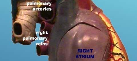
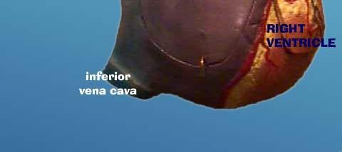
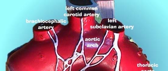
1 Danil Hammoudi.MD Aorta the biggest and longest artery (a blood vessel carrying blood away from the heart) in the body. It carries oxygen rich blood from the left ventricle of the heart to the body.inferior vena cava a large vein (a blood vessel carrying blood to the heart) that carries oxygen poor blood to the right atrium from the lower half of the body. Left atrium the left upper chamber of the heart. It receives oxygen rich blood from the lungs via the pulmonary vein. Left ventricle the left lower chamber of the heart. It pumps the blood through the aortic valve into the aorta. Mitral valve the valve between the leftatrium and the left ventricle. It prevents the back flow flowofbloodof from the ventricle to the atrium. Pulmonary artery the blood vessel that carries oxygen poor blood from the right ventricle of the heart to the lungs. Pulmonary valve the flaps between the right ventricle and the pulmonary artery. When the ventricle contracts, the valve opens, causing blood to rush into the pulmonary artery. When the ventricle relaxes, the valves close, preventing the back flow of blood from the pulmonary artery to the right atrium. Pulmonary vein the blood vessel that carries oxygen rich blood from the lungs to the left atrium of the heart. Right atrium the right upper chamber of the heart. It receives oxygen poor blood from the body through the inferior vena cava a and the superior vena cava. a Right ventricle the right lower chamber of the heart. It pumps the blood into the pulmonary artery. Septum the muscular wall that separates the left and right sides of the heart. Superior vena cava a large vein that carries oxygen poor blood to the right atrium from the upper parts of the body. Tricuspid valve the flaps between the right atrium and the right ventricle. It is composed of three leaflike parts and prevents the back flow of blood from the ventricle to the atrium. 1
 1.")

 4.")
 5.")
 6.")


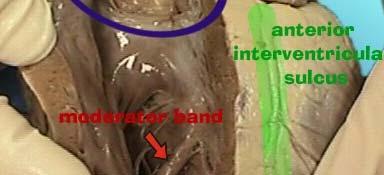
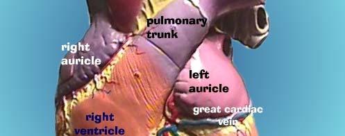
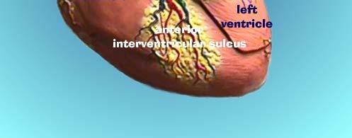
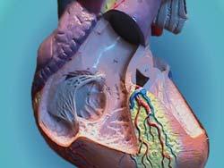
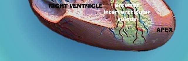
2 External Heart (Anterior View) 1. Right Auricle 2. Right Ventricle 3. Brachiocephalic Artery (Oxygenated) 4. Aortic Arch (Oxygenated) 5. Pulmonary Artery (Deoxygenated) 6. Left Auricle 7. Interventricular Sulcus 8. Left Ventricle The wooden probe is in the Aorta, forceps are in the brachiocephalic artery, & a metal probe is in the pulmonary artery. Anterior or ventral surface 2
 1.")
 2.")


 7.")


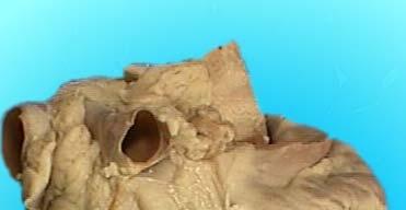
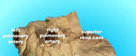
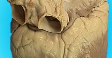
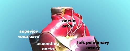
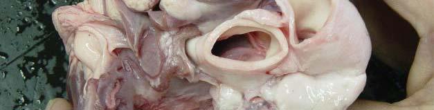
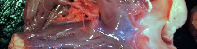
3 External Heart (Posterior View) 1. Brachiocephalic Artery (Oxygenated) 2. Aortic Arch (Oxygenated) 3. Openings for Pulmonary Veins (Oxygenated) 4. Opening for Inferior Vena Cava (Deoxygenated) 5. Left Ventricle 6. Opening for Superior Vena Cava (Deoxygenated) 7. Right Auricle 8. Right Ventricle anterior or ventral surface posterior or dorsal surface. The right atrium has a partial cut through it. The vena cava & pulmonary veins are not easily located. 3


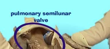
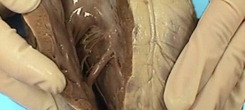
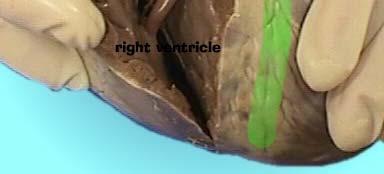
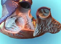
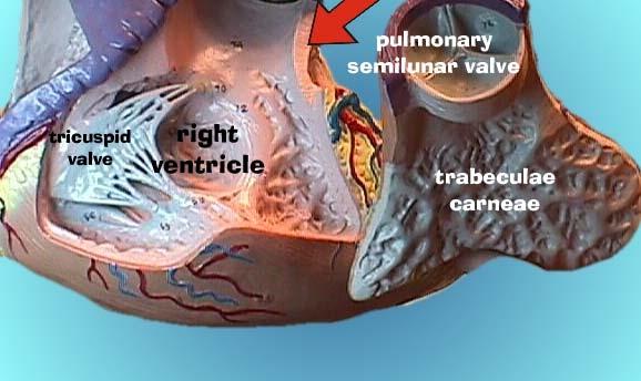
4 posterior or dorsal surface. A small pair of forceps is in the superior vena cava, large forceps in pulmonary vein & a probe goes into the inferior vena cava. Right Side Heart Opened (Anterior View) 1. Right Ventricle 2. Papillary Muscle 3. Moderator Band 4. Pulmonary Semilunar Valve 5. Pulmonary Artery 6. Left Ventricle 4


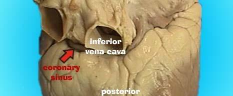
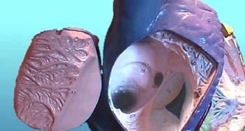
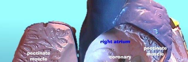
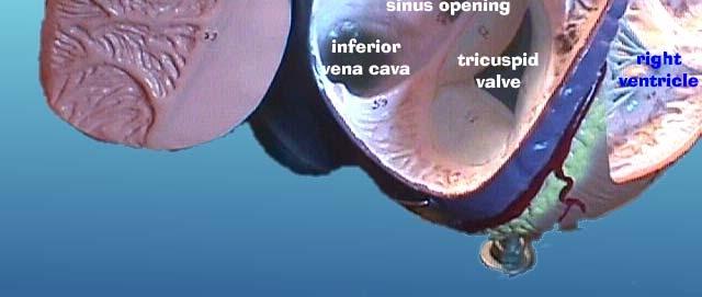
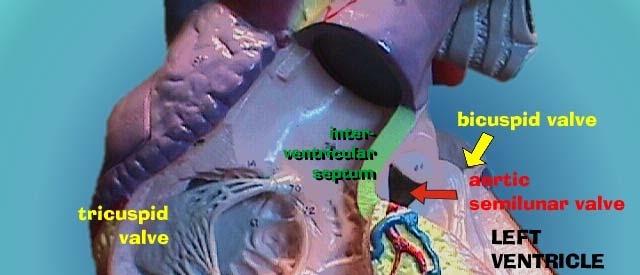
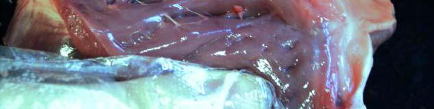
5 Right Side Heart Opened (Posterior View) 1. Right Atrium 2. Right Ventricle 3. Myocardium (Note Thinness) 4. Opening of Coronary Sinus 5. Inferior Vena Cava 6. Superior Vena Cava 7. Pectinate Muscle 8. Tricuspid Valve 9. Papillary Muscle 10. Moderator Band Left Side Heart Opened (Lateral View) 1. Pulmonary Artery 2. Brachiocephalic Artery 3. Aorta 4. Aortic Seminlunar Valve 5. Opening for Coronary Artery 6. Trabeculae Carnae of Left Ventricle 7. Ventricular Myocardium (Note Thickness) 5
 1.")


6 Left Side Heart Opened (Posterior View) 1. Aortic Arch 2. Left Atrium 3. Mitral Valve 4. Chordae Tendineae 5. Left Ventricle 6. Myocardium (Note Thickness) internal view. The left atrium & ventricle are on your left. A relatively large section of the right ventricle is also visible on the right. 6


7 internal view. The left atrium & left ventricle are on your right. The aorta & its semilunar valves are also visible. 7


8 8

9 TO PRACTICE 9
10 HEART MODELS 10
11 11

12 12
13 13

14 14
15 HEART DISSECTION 15


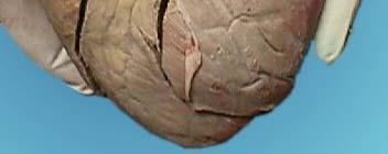
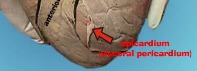
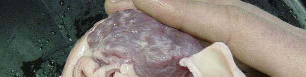
16 Intact Heart This shows the pig heart from the front, with the portion on the right ihtof the picture being the left side of the heart and vice versa. The aorta is clearly visible at the top, with an atrium on eitherside side, whilethe ventricles are in the bottom left. 16


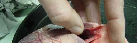
17 The first incision is along the right ventricle. The right ventricle can be identified by squeezing the heart, since the myocardium on the right side is much less rigid than that of the left ventricle. This allows us to see the tricuspidvalve andthe right ventricular outflow tract which includes the pulmonary valve. Longitudinal Cut The right ventricle has been cut open from the bottom towards the top. In this picture, the myocardium is being held back. My finger is stuck underneath one leaflet of the tricuspid valve, which leads to the pulmonary valve. 17


18 The Tricuspid Valve up close The tricuspid valve allows blood to flow from the right atrium into the right ventricle. Pulmonary Valve When the heart is contracting, the pulmonary valve is open because the blood pushes the cusps out of the way. After contracting, the ventricles begin to relax and the pulmonary valve closes and prevents backflow (called regurgitation) of blood into the ventricle. 18


 valve The mitral valve")

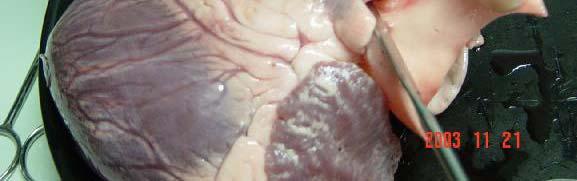
19 The Left Ventricle This longitudinal incision extends from the bottom to the top of the left ventricle, then continues up into the atrium to allow us to view the entire left heart. The Mitral (bicuspid) valve The mitral valve prevents blood from flowing back into the left atrium The mitral valve is positioned between the atrium (at top) and ventricle (at bottom). 19


20 Left Ventricular Outflow Blood flows into the ventricles by passing through h the mitral valve, but can you see where it flows out? This is a bit of a trick question because the outflow tract is hidden behind the mitral valves 20
Heart Dissection. 5. Locate the tip of the heart or the apex. Only the left ventricle extends all the way to the apex.
 Heart Dissection Page 1 of 6 Background: The heart is a four-chambered, hollow organ composed primarily of cardiac muscle tissue. It is located in the center of the chest in between the lungs. It is the
CJ Shuster A&P2 Lab Addenum Beef Heart Dissection 1. Heart Dissection. (taken from Johnson, Weipz and Savage Lab Book)
") CJ Shuster A&P2 Lab Addenum Beef Heart Dissection 1 Heart Dissection. (taken from Johnson, Weipz and Savage Lab Book) Introduction When you have finished examining the model, you are ready to begin your
CJ Shuster A&P2 Lab Addenum Beef Heart Dissection 1 Heart Dissection. (taken from Johnson, Weipz and Savage Lab Book) Introduction When you have finished examining the model, you are ready to begin your
LAB: Sheep or Pig Heart Dissection
 Biology 12 Name: Circulatory System Per: Date: Observation: External Anatomy LAB: Sheep or Pig Heart Dissection 1. Line a dissecting tray with paper towel for easy clean up as the heart is fatty and will
THE CARDIOVASCULAR SYSTEM. Part 1
 THE CARDIOVASCULAR SYSTEM Part 1 CARDIOVASCULAR SYSTEM Blood Heart Blood vessels What is the function of this system? What other systems does it affect? CARDIOVASCULAR SYSTEM Functions Transport gases,
The Heart. Happy Friday! #takeoutyournotes #testnotgradedyet
 The Heart Happy Friday! #takeoutyournotes #testnotgradedyet Introduction Cardiovascular system distributes blood Pump (heart) Distribution areas (capillaries) Heart has 4 compartments 2 receive blood (atria)
2. Obtain the following: eye guards gloves dissection tools: several blunt probes, scissors, a scalpel and forceps dissection pan sheep heart
 Week 04 Lab Heart Anatomy LEARNING OUTCOMES: Describe the gross external and internal anatomy of the heart. Identify and discuss the function of the valves of the heart. Identify the major blood vessels
An Illustrated 1. Dissection Guide. To The... Mammalian. rr= Heart. Right ventricle+---, by David H. Hall
 An Illustrated 1. Dissection Guide. To The... Mammalian rr= Heart ventricle+---, by David H. Hall The Mam.malian Heart Because mammals are warm blooded (endothermic) and generally very active animals,
THE HEART OBJECTIVES: LOCATION OF THE HEART IN THE THORACIC CAVITY CARDIOVASCULAR SYSTEM
 BIOLOGY II CARDIOVASCULAR SYSTEM ACTIVITY #3 NAME DATE HOUR THE HEART OBJECTIVES: Describe the anatomy of the heart and identify and give the functions of all parts. (pp. 356 363) Trace the flow of blood
ACTIVITY 9: BLOOD AND HEART BLOOD
 ACTIVITY 9: BLOOD AND HEART OBJECTIVES: 1) How to get ready: Read Chapters 21 & 22, McKinley et al., Human Anatomy, 4e. All text references are for this textbook. Read dissection instructions BEFORE YOU
Read Chapters 21 & 22, McKinley et al
 ACTIVITY 9: BLOOD AND HEART OBJECTIVES: 1) How to get ready: Read Chapters 21 & 22, McKinley et al., Human Anatomy, 5e. All text references are for this textbook. Read dissection instructions BEFORE YOU
Read Me. covering the Heart Anatomy. Labs. textbook. use. car: you
 Heart Anatomy Lab Pre-Lab Exercises Read Me These exercises should be done before coming to lab, after watching the videos covering the Heart Anatomy Labs. Answer the questions in this guide using the
THE HEART. Unit 3: Transportation and Respiration
 THE HEART Unit 3: Transportation and Respiration The Circulatory System Also called the Cardiovascular System Circulates blood in the body Transports nutrients, oxygen, carbon dioxide, hormones, and blood
Lab Activity 23. Cardiac Anatomy. Portland Community College BI 232
 Lab Activity 23 Cardiac Anatomy Portland Community College BI 232 Cardiac Muscle Histology Branching cells Intercalated disc: contains many gap junctions connecting the adjacent cell cytoplasm, creates
This lab activity is aligned with Visible Body s A&P app. Learn more at visiblebody.com/professors
 1 This lab activity is aligned with Visible Body s A&P app. Learn more at visiblebody.com/professors 2 PRE-LAB EXERCISES: A. Watch the video 29.1 Heart Overview and make the following observations: 1.
Chapter 14. The Cardiovascular System
 Chapter 14 The Cardiovascular System Introduction Cardiovascular system - heart, blood and blood vessels Cardiac muscle makes up bulk of heart provides force to pump blood Function - transports blood 2
LAB 12-1 HEART DISSECTION GROSS ANATOMY OF THE HEART
 LAB 12-1 HEART DISSECTION GROSS ANATOMY OF THE HEART Because mammals are warm-blooded and generally very active animals, they require high metabolic rates. One major requirement of a high metabolism is
DISSECTION OF A SHEEP HEART
 DISSECTION OF A SHEEP HEART I. INTRODUCTION A. You will soon appreciate the point made previously the heart models just don t teach us what a real heart is like! Dissecting a sheep heart will give you
Chapter 20 (1) The Heart
 The Heart") Chapter 20 (1) The Heart Learning Objectives Describe the location and structure of the heart Describe the path of a drop of blood from the superior vena cava or inferior vena cava through the heart out
Chapter 20 (1) The Heart Learning Objectives Describe the location and structure of the heart Describe the path of a drop of blood from the superior vena cava or inferior vena cava through the heart out
Human Anatomy and Physiology Chapter 19 Worksheet 1- The Heart
 Human Anatomy and Physiology Chapter 19 Worksheet 1- The Heart Name Date Period 1. The "double pump" function of the heart includes the right side, which serves as the circuit pump, while the left side
The Heart. The Heart A muscular double pump. The Pulmonary and Systemic Circuits
 C H A P T E R 19 The Heart The Heart A muscular double pump circuit takes blood to and from the lungs Systemic circuit vessels transport blood to and from body tissues Atria receive blood from the pulmonary
Ch 19: Cardiovascular System - The Heart -
 Ch 19: Cardiovascular System - The Heart - Give a detailed description of the superficial and internal anatomy of the heart, including the pericardium, the myocardium, and the cardiac muscle. Trace the
2. right heart = pulmonary pump takes blood to lungs to pick up oxygen and get rid of carbon dioxide
 A. location in thorax, in inferior mediastinum posterior to sternum medial to lungs superior to diaphragm anterior to vertebrae orientation - oblique apex points down and to the left 2/3 of mass on left
Chapter 14. Circulatory System Images. VT-122 Anatomy & Physiology II
 Chapter 14 Circulatory System Images VT-122 Anatomy & Physiology II The mediastinum Dog heart Dog heart Cat heart Dog heart ultrasound Can see pericardium as distinct bright line Pericardial effusion Fluid
Ch.15 Cardiovascular System Pgs {15-12} {15-13}
 Ch.15 Cardiovascular System Pgs {15-12} {15-13} E. Skeleton of the Heart 1. The skeleton of the heart is composed of rings of dense connective tissue and other masses of connective tissue in the interventricular
LECTURE 5. Anatomy of the heart
 LECTURE 5. Anatomy of the heart Main components of the CVS: Heart Blood circulatory system arterial compartment haemomicrocirculatory (=microvascular) compartment venous compartment Lymphatic circulatory
the Cardiovascular System I
 the Cardiovascular System I By: Dr. Nabil A Khouri MD, MsC, Ph.D MEDIASTINUM 1. Superior Mediastinum 2. inferior Mediastinum Anterior mediastinum. Middle mediastinum. Posterior mediastinum Anatomy of
The Heart. Made up of 3 different tissue: cardiac muscle tissue, nerve tissue, and connective tissue.
 The Heart The Heart Made up of 3 different tissue: cardiac muscle tissue, nerve tissue, and connective tissue. Your heart pumps with a regular beat (Heart Rate) Your heart rate can change depending on
#4 Cardiovascular I The Heart
 Page1 #4 Cardiovascular I The Heart Objectives: Identify a list of human heart structures using a virtual human dissection Dissect a sheep heart to identify external and internal structures Identify a
THE HEART. A. The Pericardium - a double sac of serous membrane surrounding the heart
 THE HEART I. Size and Location: A. Fist-size weighing less than a pound (250 to 350 grams). B. Located in the mediastinum between the 2 nd rib and the 5 th intercostal space. 1. Tipped to the left, resting
Anatomy of the Heart
 Biology 212: Anatomy and Physiology II Anatomy of the Heart References: Saladin, KS: Anatomy and Physiology, The Unity of Form and Function 8 th (2018). Required reading before beginning this lab: Chapter
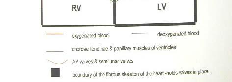
(2) (1) (3) (4) BLOOD PATHWAY ASSESSMENT RUBRIC
 (1) (3) (4) BLOOD PATHWAY ASSESSMENT RUBRIC") BLOODPATHWAYASSESSMENT(4) BLOOD%PATHWAY%ASSESSMENT%(3)% BLOODPATHWAYASSESSMENT(3) (4) (3) (2) (1) Using a completely blank diagram of the heart, all valves, chambers, great vessels, and direction of blood
BLOODPATHWAYASSESSMENT(4) BLOOD%PATHWAY%ASSESSMENT%(3)% BLOODPATHWAYASSESSMENT(3) (4) (3) (2) (1) Using a completely blank diagram of the heart, all valves, chambers, great vessels, and direction of blood
Anatomy of the Heart. Figure 20 2c
 Anatomy of the Heart Figure 20 2c Pericardium & Myocardium Remember, the heart sits in it s own cavity, known as the mediastinum. The heart is surrounded by the Pericardium, a double lining of the pericardial
Cardiovascular System. Heart Anatomy
 Cardiovascular System Heart Anatomy 1 The Heart Location & general description: Atria vs. ventricles Pulmonary vs. systemic circulation Coverings Walls The heart is found in the mediastinum, the medial
CIRCULATORY SYSTEM BLOOD VESSELS
 Name: Block: CIRCULATORY SYSTEM Multicellular organisms (above the level of roundworms) rely on a circulatory system to bring nutrients to, and take wastes away from, cells. In higher organisms such as
DISSECTING A PIG S HEART
 DISSECTING A PIG S HEART LAB 59 OBSERVATION STUDENT BOOK Chapter 6, page 185 Goal Locate and observe structures of a mammal s heart. Observation criteria Identify the structures of the heart indicated
Blood and Heart. Student Learning Objectives:
 Blood and Heart Student Learning Objectives: Identify the major components of the blood. Identify the primary structures associated with the heart Follow the blood through the path of the circulation.
The Cardiovascular System (Heart)
") The Cardiovascular System The Cardiovascular System (Heart) A closed system of the heart and blood vessels The heart pumps blood Blood vessels allow blood to circulate to all parts of the body The function
The Cardiovascular System The Cardiovascular System (Heart) A closed system of the heart and blood vessels The heart pumps blood Blood vessels allow blood to circulate to all parts of the body The function
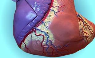
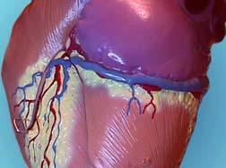
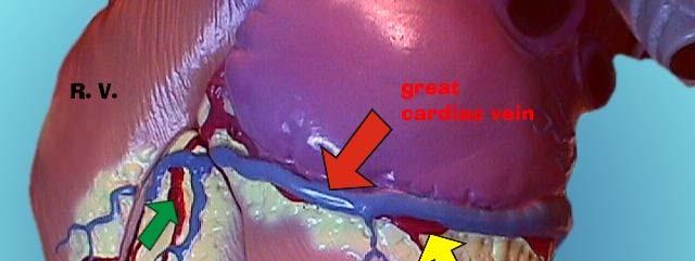
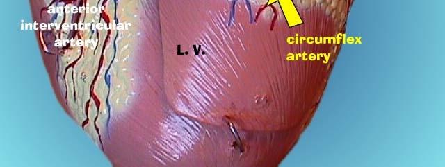
AP2 Lab 3 Coronary Vessels, Valves, Sounds, and Dissection
 AP2 Lab 3 Coronary Vessels, Valves, Sounds, and Dissection Project 1 - BLOOD Supply to the Myocardium (Figs. 18.5 &18.10) The myocardium is not nourished by the blood while it is being pumped through the
Heart Anatomy. 7/5/02 Stephen G Davenport 1
 Heart Anatomy Copyright 1999, Stephen G. Davenport, No part of this publication may be reproduced, stored in a retrieval system, or transmitted, in any form without prior written permission. 7/5/02 Stephen
The Cardiovascular System. Chapter 15. Cardiovascular System FYI. Cardiology Closed systemof the heart & blood vessels. Functions
 Chapter 15 Cardiovascular System FYI The heart pumps 7,000 liters (4000 gallons) of blood through the body each day The heart contracts 2.5 billion times in an avg. lifetime The heart & all blood vessels
Biology Unit 3 The Human Heart P
 Biology 2201 Unit 3 The Human Heart P 314-321 Structure and Function of the Human Heart Structure of the Human Heart Has four Chambers (2 Atria and 2 Ventricles) Made of Cardiac Muscle Found in Chest Cavity
The HEART. What is it???? Pericardium. Heart Facts. This muscle never stops working It works when you are asleep
 This muscle never stops working It works when you are asleep The HEART It works when you eat It really works when you exercise. What is it???? Located between the lungs in the mid thoracic region Apex
Cardiovascular System Note-Taking Guide
 FUNctions: Name: 3-27-14 Cardiovascular System Note-Taking Guide Heart: Pumps and delivers through the body Blood: Vessels: Delivers and to the body Carries waste and Maintains homeostasis - Carries blood
Lab 16. The Cardiovascular System Heart and Blood Vessels. Laboratory Objectives
 Lab 16 The Cardiovascular System Heart and Blood Vessels Laboratory Objectives Describe the anatomical structures of the heart to include the pericardium, chambers, valves, and major vessels. Describe
Human Anatomy, First Edition
 Human Anatomy, First Edition McKinley & O'Loughlin Chapter 22 : Heart 1 Functions of the Heart Center of the cardiovascular system, the heart. Connects to blood vessels that transport blood between the
Introduction to Anatomy. Dr. Maher Hadidi. Bayan Yanes. April/9 th /2013
 Introduction to Anatomy Dr. Maher Hadidi Bayan Yanes 27 April/9 th /2013 KEY POINTS: 1) Right side of the heart 2) Papillary muscles 3) Left side of the heart 4) Comparison between right and left sides
Circulatory Systems. All cells need to take in nutrients and expel metabolic wastes.
 Circulatory Systems All cells need to take in nutrients and expel metabolic wastes. Single celled organisms: nutrients from the environment can diffuse (or be actively transported) directly in to the cell
MODULE 2: CARDIOVASCULAR SYSTEM ANTOMY An Introduction to the Anatomy of the Heart and Blood vessels
 MODULE 2: CARDIOVASCULAR SYSTEM ANTOMY An Introduction to the Anatomy of the Heart and Blood vessels The cardiovascular system includes a pump (the heart) and the vessels that carry blood from the heart
Anatomy lab -1- Imp note: papillary muscle Trabeculae Carneae chordae tendineae
 Anatomy lab -1- Imp note: the arrangement of this sheet is different than the lab recording, it has been arranged in a certain way to make it easier to study. When you open the left ventricle you can see
The Cardiovascular System Part I: Heart Outline of class lecture After studying part I of this chapter you should be able to:
 The Cardiovascular System Part I: Heart Outline of class lecture After studying part I of this chapter you should be able to: 1. Describe the functions of the heart 2. Describe the location of the heart,
The Cardiovascular System
 11 PART A The Cardiovascular System PowerPoint Lecture Slide Presentation by Jerry L. Cook, Sam Houston University ESSENTIALS OF HUMAN ANATOMY & PHYSIOLOGY EIGHTH EDITION ELAINE N. MARIEB The Cardiovascular
Cardiovascular System Notes: Physiology of the Heart
 Cardiovascular System Notes: Physiology of the Heart Interesting Heart Fact Capillaries are so small it takes ten of them to equal the thickness of a human hair. Review What are the 3 parts of the cardiovascular
Chapter 18 - Heart. I. Heart Anatomy: size of your fist; located in mediastinum (medial cavity)
") Chapter 18 - Heart I. Heart Anatomy: size of your fist; located in mediastinum (medial cavity) A. Coverings: heart enclosed in double walled sac called the pericardium 1. Fibrous pericardium: dense connective
Chapter 18 - Heart I. Heart Anatomy: size of your fist; located in mediastinum (medial cavity) A. Coverings: heart enclosed in double walled sac called the pericardium 1. Fibrous pericardium: dense connective
CIRCULATION & GAS EXCHANGE
 AP BIOLOGY ACTIVITY2.13 Text:Campbell,v.8,chapter42 NAME DATE HOUR CIRCULATION & GAS EXCHANGE 1. In general, what is the function of transport systems? 2. What method/structure do most invertebrates use
CV Anatomy Quiz. Dr Ella Kim Dr Pip Green
 CV Anatomy Quiz Dr Ella Kim Dr Pip Green Q1 The location of the heart is correctly described as A) lateral to the lungs. B) medial to the sternum. C) superior to the diaphragm. D) posterior to the spinal
ACTIVITY: The Heart Cycle
 ACTIVITY: The Heart Cycle In this activity, you will follow the flow of blood through the heart. Your heart has two jobs to do, and its two sides have separate responsibilities: The left side pumps oxygen-enriched
The Heart. Size, Form, and Location of the Heart. 1. Blunt, rounded point; most inferior part of the heart.
 12 The Heart FOCUS: The heart is composed of cardiac muscle cells, which are elongated, branching cells that appear striated. Cardiac muscle cells behave as a single electrical unit, and the highly coordinated
2.01 Remember the structures of the circulatory system
 2.01 Remember the structures of the circulatory system Essential questions What are the structures of blood? What are the structures of the circulatory system? circulatory system 2 Structures of the circulatory
HUMAN HEART. Learn the following structures on the heart models.
 HUMAN HEART Learn the following structures on the heart models. The human heart has four chambers that consist of the right atrium, left atrium, right ventricle, and left ventricle. The atria are smaller
Cardiovascular System Module 3: Heart Anatomy *
 OpenStax-CNX module: m49683 1 Cardiovascular System Module 3: Heart Anatomy * Donna Browne Based on Heart Anatomy by OpenStax This work is produced by OpenStax-CNX and licensed under the Creative Commons
Figure ) The specific chamber of the heart that is indicated by letter A is called the. Diff: 1 Page Ref: 364
 The specific chamber of the heart that is indicated by letter A is called the. Diff: 1 Page Ref: 364") Essentials of Anatomy and Physiology, 9e (Marieb) Chapter 11 The Cardiovascular System Short Answer Figure 11.1 Using Figure 11.1, identify the following: 1) The Purkinje fibers are indicated by label.
Essentials of Anatomy and Physiology, 9e (Marieb) Chapter 11 The Cardiovascular System Short Answer Figure 11.1 Using Figure 11.1, identify the following: 1) The Purkinje fibers are indicated by label.
THE HEART. Structure & Function
 THE HEART Structure & Function SARAH JOHANSON BIOTECH ENGINEERING, 2015 Function of the Heart: The heart is muscular organ that sits centrally in the thorax region of the body, but is skewed and twisted
Lab 6: Blood. BIO104 Laboratory Handouts 147. Unit 12: Blood and Lymphatics. 1. Blood Characteristics Volume Functions Composition -
 147 Lab 6: Blood Unit 12: Blood and Lymphatics Ex. 12-1: Formed Elements (Cells) of Blood, p. 313-316 1. Blood Characteristics Volume Functions Composition - 2. Leukocytes (WBCs) a. WBC count normal b.
THE HEART. Structure & Function
 THE HEART Structure & Function SARAH JOHANSON BIOTECH ENGINEERING, 2015 Function of the Heart: The heart is muscular organ that sits centrally in the thorax region of the body, but is skewed and twisted
10. Thick deposits of lipids on the walls of blood vessels, called, can lead to serious circulatory issues. A. aneurysm B. atherosclerosis C.
 Heart Student: 1. carry blood away from the heart. A. Arteries B. Veins C. Capillaries 2. What is the leading cause of heart attack and stroke in North America? A. alcohol B. smoking C. arteriosclerosis
Health Science 20 Circulatory System Notes
 Health Science 20 Circulatory System Notes Functions of the Circulatory System The circulatory system functions mainly as the body s transport system. It transports: o Oxygen o Nutrients o Cell waste o
Aim: Transport- Why is it so important to multicellular organisms?
 Aim: Transport- Why is it so important to multicellular organisms? I.Transportthe absorption and circulation that allows substances to pass into or out of cells and move throughout the organism. A. absorptionsubstances
Cardiovascular System- Heart. Miss Wheeler Unit 8
 Cardiovascular System- Heart Miss Wheeler Unit 8 Overview CARDIOVASCULAR SYSTEM heart vessels Made up of heart, blood vessels, and blood Functions Heart- pump blood Vessels- (veins, arteries, capillaries)
human anatomy 2016 lecture thirteen Dr meethak ali ahmed neurosurgeon
 Heart The heart is a hollow muscular organ that is somewhat pyramid shaped and lies within the pericardium in the mediastinum. It is connected at its base to the great blood vessels but otherwise lies
Large Arteries of Heart
 Cardiovascular System (Part A-2) Module 5 -Chapter 8 Overview Arteries Capillaries Veins Heart Anatomy Conduction System Blood pressure Fetal circulation Susie Turner, M.D. 1/5/13 Large Arteries of Heart
AP2 Lab 1 - Blood & Heart
 AP2 Lab 1 - Blood & Heart Project 1 - Formed Elements Identification & Recognition See fig. 17.10 and Table 17.2. Instructor may also provide other images. Note: See Fig. 17.11 All formed elements are
Chapter 27 The Heart and Blood Vessels
 Chapter 27 The Heart and Blood Vessels Most animals have a closed blood system. The blood flows continuously in vessels back to the heart. In an open system the blood is pumped into open ended tubes and
Cardiovascular system Physiology Sheet (1)
") Cardiovascular system Physiology Sheet (1) In any patient the most important thing is the cardiorespiratory system, if any patient comes to you with a car accident for example the first thing that you
Cardiovascular system Physiology Sheet (1) In any patient the most important thing is the cardiorespiratory system, if any patient comes to you with a car accident for example the first thing that you
Heart and Lung Dissection
 Heart and Lung Dissection Name(s) Before you begin any work or dissection of your specimen, please try to identify the following. You will need to illustrate what you see, so make sure to note size, texture
Lab Photo Review Sheet
 9 8 0. Posterior Median Sulcus. Central Canal. Dorsal (Posterior) Horn. Ventral (Anterior) Horn. Grey Matter. White Matter. Anterior Median Fissure 8. Ventral (Anterior) Root (ramus) 9. Dorsal (Posterior)
d) the pulmonary vein b) the superior vena cava e) the inferior vena cava c) the pulmonary artery The large vessels of a sheep s heart
 the pulmonary vein b) the superior vena cava e) the inferior vena cava c) the pulmonary artery The large vessels of a sheep s heart") CHAPTER 8 Investigation 8.A: Identifying Structures of the Circulatory System Question: What features of a mammalian heart can you identify in a real or virtual heart? What route does blood take through
CHAPTER 8 Investigation 8.A: Identifying Structures of the Circulatory System Question: What features of a mammalian heart can you identify in a real or virtual heart? What route does blood take through
INTRODUCTORY REMARKS:
 INTRODUCTORY REMARKS: The circulatory system provides a way for the blood to be transported throughout the body. This provides nutrients to the cells and allows wastes to be removed. Open vs. Closed Circulatory
CIRCULATORY SYSTEM TASK CARDS Worksheet
 CIRCULATORY SYSTEM TASK CARDS Worksheet Name: Date: Instructions: Put the answers to each task card in the numbered boxes on the chart. 1 a) left semilunar valve / aortic valve b) blood would backflow
37 1 The Circulatory System
 H T H E E A R T 37 1 The Circulatory System The circulatory system and respiratory system work together to supply cells with the nutrients and oxygen they need to stay alive. a) The respiratory system:
Cardiovascular System
 Cardiovascular System Purpose Transport oxygen and nutrients Take waste products away from tissues & organs Things we learned Blood pressure: the force of blood pushing against the walls of blood vessels
Middle mediastinum---- heart & pericardium. Dep. of Human Anatomy Zhou Hongying
 Middle mediastinum---- heart & pericardium Dep. of Human Anatomy Zhou Hongying eaglezhyxzy@163.com Subdivisions of the mediastinum Contents of Middle mediastinum Heart Pericardium: a serous sac enclosing
Scrub In: Red blood cells are called: Which component of blood is necessary for the initiation of the blood clotting process:
 Scrub In: Red blood cells are called: a. erythrocytes b. leukocytes c. melanocytes d. thrombocytes Which component of blood is necessary for the initiation of the blood clotting process: a. erythrocytes
Heart. Heart 2-Tunica media: middle layer (media ='middle') muscle fibers (smooth or cardiac).
 muscle fibers (smooth or cardiac).") t. innermost lumenal General Circulatory system heart and blood vessels walls have 3 layers (inside to outside) 1-Tunica interna: aka tunica intima layer--lumenal layer epithelium--endothelium simple squamous
t. innermost lumenal General Circulatory system heart and blood vessels walls have 3 layers (inside to outside) 1-Tunica interna: aka tunica intima layer--lumenal layer epithelium--endothelium simple squamous
The Cardiovascular System
 The Cardiovascular System The Manila Times College of Subic Prepared by: Stevens B. Badar, RN, MANc THE HEART Anatomy of the Heart Location and Size approx. the size of a person s fist, hollow and cone-shaped,
Lesson 10 Circulatory System (Nelson p.88-93)
") Name: Date: Lesson 10 Circulatory System (Nelson p.88-93) Learning Goals: A. I can explain the primary functions of the circulatory system in animals. B. I can identify and explain all the parts of the
Name: Date: Lesson 10 Circulatory System (Nelson p.88-93) Learning Goals: A. I can explain the primary functions of the circulatory system in animals. B. I can identify and explain all the parts of the
Your heart is a muscular pump about the size of your fist, located
 How Your Heart Works Your heart is a muscular pump about the size of your fist, located slightly to the left and behind your breastbone. Its function is to pump blood throughout your body. As your heart
The Mammalian Circulatory System
 The Mammalian Heart The Mammalian Circulatory System Recall: What are the 3 cycles of the mammalian circulatory system? What are their functions? What are the three main vessel types in the mammalian circulatory
11/10/2014. Muscular pump Two atria Two ventricles. In mediastinum of thoracic cavity 2/3 of heart's mass lies left of midline of sternum
 It beats over 100,000 times a day to pump over 1,800 gallons of blood per day through over 60,000 miles of blood vessels. During the average lifetime, the heart pumps nearly 3 billion times, delivering
The Cardiovascular System
 Essentials of Human Anatomy & Physiology Elaine N. Marieb Seventh Edition Chapter 11 The Cardiovascular System Slides 11.1 11.19 Lecture Slides in PowerPoint by Jerry L. Cook The Cardiovascular System
Transport in Animals (IGCSE Biology Syllabus )
") Transport in Animals (IGCSE Biology Syllabus 2016-2018) Blood o Red blood cells: heamoglobin and oxygen transport o White blood cells: phagocyte phagocytosis (engulf pathogen, vesicles fuse with vacuole,
Transport in Animals (IGCSE Biology Syllabus 2016-2018) Blood o Red blood cells: heamoglobin and oxygen transport o White blood cells: phagocyte phagocytosis (engulf pathogen, vesicles fuse with vacuole,
Cardiovascular System. I. Structures of the heart A. : Pericardium sack that surrounds the heart
 Cardiovascular System I. Structures of the heart A. : Pericardium sack that surrounds the heart 1. : Pericardial Cavity serous fluid filled space between the heart and the pericardium B. Heart Wall 1.
- what other structures, besides the heart, does the mediastinum contain?
 Basic A & P II Dr. L. Bacha Chapter Outline (Martini & Nath 2010) An Introduction to the Cardiovascular System - read the paragraphs under this heading on page 580 The Heart is a Four Chambered Organ describe
INVESTIGATING THE STRUCTURE OF THE HEART
 Luke Micallef name Heart Health: A Beginner s guide to cardiovascular disease Section INVESTIGATING THE STRUCTURE OF THE HEART Guide to Week 1 home practical Note: You will also need to download a copy
2. What makes up the most of your blood? least of your blood? 1. Look like red discs, have a pale center, no nucleus, similar in size
 I Can Statements I can identify the major components of blood and where they are formed. Identify the four components of blood in the diagram below. 1. Label each section of the pie chart with the correct
The Circulatory System
 The Circulatory System Science Matters Chapter 8 Introduction Living things need a transport system to carry things around the body. In humans its called The Circulatory system. The parts of the system
STRUCTURES OF THE CARDIOVASCULAR SYSTEM
 STRUCTURES OF THE CARDIOVASCULAR SYSTEM CARDIOVASCULAR SYSTEM Also called the circulatory system Consists of the heart, arteries, veins, and capillaries Main function is to pump/circulate oxygenated blood
The blood returns from the body and enters right atrium using the vena cava. It passes through the tricuspid valve to the right ventricle.
 The blood returns from the body and enters right atrium using the vena cava. It passes through the tricuspid valve to the right ventricle. From this camber, it passes through the pulmonary semilunar valve
Two semilunar valves. Two atrioventricular valves. Valves of the heart. Left atrioventricular or bicuspid valve Mitral valve
 The Heart 3 Valves of the heart Two atrioventricular valves Two semilunar valves Right atrioventricular or tricuspid valve Left atrioventricular or bicuspid valve Mitral valve Aortic valve Pulmonary valve
Circulation. Circulation = is a process used for the transport of oxygen, carbon! dioxide, nutrients and wastes through-out the body
 Circulation Circulation = is a process used for the transport of oxygen, carbon! dioxide, nutrients and wastes through-out the body Heart = muscular organ about the size of your fist which pumps blood.