Echocardiography Conference
|
|
|
- Marjorie Patterson
- 5 years ago
- Views:
Transcription
1 Echocardiography Conference David Stultz, MD Cardiology Fellow, PGY-6 September 20, 2005
2
3
4 Atrial Septal Aneurysm Bulging of Fossa Ovalis Associated commonly with Atrial septal defect or small perforations Left to Right or Right to Left shunt
5 Diagnostic criteria for ASA Diameter of base of atrial septal anuerysm >=15mm Bulging of atrial septum >=15mm Motion of the septum during respiration >=15mm Zanchetta
6 Associated abnormalities Cardiac Ascending aorta aneurysm Elongated thoracic aorta Pericardial effusion Constrictive pericarditis Tricuspid regurgitation Redundant Eustacian Valve Pulmonary Pulmonary embolism Right pneumanectomy COPD ARDS OSA Zanchetta
7 SPARC Study Prevelence of ASA is 2.9% in general population 7.9% in patients with cerebral ischemic events 56% of pts with ASA have a Patent Foramen Ovale (with Right-Left shunt) Paradoxic embolism is major mechanism of vascular events Zanchetta
8 Chest Guidelines 1% prevalence of ASA in autopsy studies 3-4% prevalence in TEE studies of nonstroke patients High incidence of PFO with ASA (70-83%) Anecdotal reports of thrombus in aneurysm Deeb

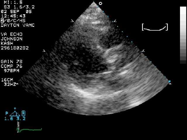
9 FIGURE Apical four-chamber view recorded in a patient with an atrial septal aneurysm. Note the marked bulging of the atrial septum into the cavity of the left atrium (arrow). The right-hand panel is recorded after injection of saline contrast medium. Note the contrast medium has filled the right ventricular cavity, and there are numerous individual microbubbles seen in the cavity of both the left atrium and left ventricle, consistent with a right-to-left shunt through fenestrations in the atrial septal aneurysm. Abbreviations are as per previous figures. Braunwald
10 Feigenbaum
11 Feigenbaum
12 Feigenbaum
13 Feigenbaum
14 Chest Guidelines Aspirin useful for primary prevention of CVA in patients with atrial septal aneurysm and/or PFO In patients with CVA and documented embolic source (DVT) + ASA/PFO with R- L shunt, coumadin recommended long term Deeb

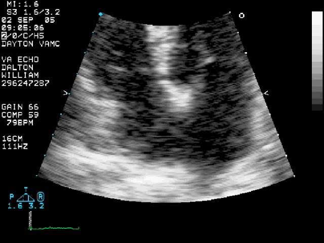
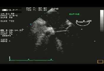
15 Next case Apical 4 Chamber
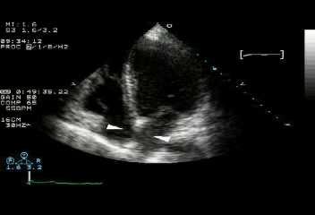
16 Parasternal Long Axis

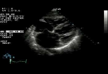
17 Basal Short Axis

18 Subcostal view

19 Parasternal Long M-Mode
20 Diagnostic Features of Tamponade RA diastolic collapse Occurs when intrapericardial pressure >=4 Common and early finding High sensitivity, low specificity Best seen on apical and subcostal view RV diastolic collapse Occurs when intrapericardial pressure >=6-8 Occurs after RA compression Lower sensitivity, but high specificity and negative predictive values Best seen on parasternal long, short axis, and subcostal view Roldan
21 Doppler inflow patterns Mitral During inspiration, decline of 30-50% of E and A velocities Tricuspid During inspiration, increase of E wave (>35-80%) and A wave (>25-50%) Normal variation is 10-25% Roldan

22 Mitral Valve Inflow Doppler

23 Tricuspid Valve Inflow Doppler
24 Next Case
25
26 Pulmonary Hypertension RV Pressure overload Flattened or curved septum during late systole and early diastole (D-shaped LV) RV Volume overload D-shape LV seen during mid-diastole; during systole normal contour is seen
27
28 Next Case
29

30 M-Mode of Mitral Valve

31 Mitral inflow doppler (Pressure Half time)

32 Aortic Regurgitation doppler (Pressure Half time)
33 References Deeb N. Salem, MD, FCCP, Chair; Denise Hartnett Daudelin, RN, MPH; Herbert J. Levine, MD; Stephen G. Pauker, MD; Mark H. Eckman, MD and Joshua Riff, BSc. Antithrombotic Therapy in Valvular Heart Disease. Chest. 2001;119:207S-219S. Zanchetta M, Rigatelli G, Pedon L, Zennaro M, Maiolino P, Onorato E. Role of intracardiac echocardiography in atrial septal abnormalities. J Interv Cardiol Feb;16(1):63-77.
Adel Hasanin Ahmed 1
 Adel Hasanin Ahmed 1 PERICARDIAL DISEASE The pericardial effusion ends anteriorly to the descending aorta and is best visualised in the PLAX. PSAX is actually very useful sometimes for looking at posterior
Echocardiography as a diagnostic and management tool in medical emergencies
 Echocardiography as a diagnostic and management tool in medical emergencies Frank van der Heusen MD Department of Anesthesia and perioperative Care UCSF Medical Center Objective of this presentation Indications
Fig.1 Normal appearance of RV in SAX:
 Tutorial 7 - Assessment of the right heart Assessment of the Right heart The right heart assessment clinically and echocardiographically is not a very important part of mainstream cardiology. In the ICU,
Adult Echocardiography Examination Content Outline
 Adult Echocardiography Examination Content Outline (Outline Summary) # Domain Subdomain Percentage 1 2 3 4 5 Anatomy and Physiology Pathology Clinical Care and Safety Measurement Techniques, Maneuvers,
Certificate in Clinician Performed Ultrasound (CCPU) Syllabus. Rapid Cardiac Echo (RCE)
 Syllabus. Rapid Cardiac Echo (RCE)") Certificate in Clinician Performed Ultrasound (CCPU) Syllabus Rapid Cardiac Echo (RCE) Purpose: Rapid Cardiac Echocardiography (RCE) This unit is designed to cover the theoretical and practical curriculum
Certificate in Clinician Performed Ultrasound (CCPU) Syllabus Rapid Cardiac Echo (RCE) Purpose: Rapid Cardiac Echocardiography (RCE) This unit is designed to cover the theoretical and practical curriculum
Atrial Septal Defects
 Supplementary ACHD Echo Acquisition Protocol for Atrial Septal Defects The following protocol for echo in adult patients with atrial septal defects (ASDs) is a guide for performing a comprehensive assessment
Appendix II: ECHOCARDIOGRAPHY ANALYSIS
 Appendix II: ECHOCARDIOGRAPHY ANALYSIS Two-Dimensional (2D) imaging was performed using the Vivid 7 Advantage cardiovascular ultrasound system (GE Medical Systems, Milwaukee) with a frame rate of 400 frames
Pediatric Echocardiography Examination Content Outline
 Pediatric Echocardiography Examination Content Outline (Outline Summary) # Domain Subdomain Percentage 1 Anatomy and Physiology Normal Anatomy and Physiology 10% 2 Abnormal Pathology and Pathophysiology
Uncommon Doppler Echocardiographic Findings of Severe Pulmonic Insufficiency
 Uncommon Doppler Echocardiographic Findings of Severe Pulmonic Insufficiency Rahul R. Jhaveri, MD, Muhamed Saric, MD, PhD, FASE, and Itzhak Kronzon, MD, FASE, New York, New York Background: Two-dimensional
PROSTHETIC VALVE BOARD REVIEW
 PROSTHETIC VALVE BOARD REVIEW The correct answer D This two chamber view shows a porcine mitral prosthesis with the typical appearance of the struts although the leaflets are not well seen. The valve
MITRAL STENOSIS. Joanne Cusack
 MITRAL STENOSIS Joanne Cusack BSE Breakdown Recognition of rheumatic mitral stenosis Qualitative description of valve and sub-valve calcification and fibrosis Measurement of orifice area by planimetry
PART II ECHOCARDIOGRAPHY LABORATORY OPERATIONS ADULT TRANSTHORACIC ECHOCARDIOGRAPHY TESTING
 PART II ECHOCARDIOGRAPHY LABORATORY OPERATIONS ADULT TRANSTHORACIC ECHOCARDIOGRAPHY TESTING STANDARD - Primary Instrumentation 1.1 Cardiac Ultrasound Systems SECTION 1 Instrumentation Ultrasound instruments
Index. K Knobology, TTE artifact, image resolution, ultrasound, 14
 A Acute aortic regurgitation (AR), 124 128 Acute aortic syndrome (AAS) classic aortic dissection diagnosis, 251 263 evolutive patterns, 253 255 pathology, 250 251 classifications, 247 248 incomplete aortic
Pericardial Diseases. Smonporn Boonyaratavej, MD. Division of Cardiology, Department of Medicine Chulalongkorn University
 Pericardial Diseases Smonporn Boonyaratavej, MD Division of Cardiology, Department of Medicine Chulalongkorn University Cardiac Center, King Chulalongkorn Memorial Hospital 21 AUGUST 2016 Pericardial
Cardiac ultrasound protocols
 Cardiac ultrasound protocols IDEXX Telemedicine Consultants Two-dimensional and M-mode imaging planes Right parasternal long axis four chamber Obtained from the right side Displays the relative proportions
PERICARDIAL DIAESE. Kaijun Cui Associated professor Sichuan University
 PERICARDIAL DIAESE Kaijun Cui Associated professor Sichuan University CLASSIFICATION acute pericarditis pericardial effusion cardiac tamponade constrictive pericarditis congenitally absent pericardium
British Society of Echocardiography
 British Society of Echocardiography Affiliated to the British Cardiac Society A Minimum Dataset for a Standard Adult Transthoracic Echocardiogram From the British Society of Echocardiography Education
2D/3D in Evaluation of Atrial Septum
 2D/3D in Evaluation of Atrial Septum Roberto M Lang, MD OSTIUM SECUNDUM ASD: 2D AND 3D TNSESOPHAGEAL ECHO 1 Biplane views 90 0 3D Acquisi on Acquire 3D volume Lang RM et al. JASE 2012;25:3 46. Right atrial
Patent Foramen Ovale: Diagnosis and Treatment
 Patent Foramen Ovale: Diagnosis and Treatment Anthony DeMaria Judy and Jack White Chair in Cardiology University of California, San Diego At one time or another a Grantee, Sponsored Speaker or Ad-hoc Consultant
The Doppler Examination. Katie Twomley, MD Wake Forest Baptist Health - Lexington
 The Doppler Examination Katie Twomley, MD Wake Forest Baptist Health - Lexington OUTLINE Principles/Physics Use in valvular assessment Aortic stenosis (continuity equation) Aortic regurgitation (pressure
Constrictive Pericarditis Pitfalls in MR Diagnosis Cylen Javidan-Nejad Associate Professor Mallinckrodt Institute of Radiology Washington University
 Constrictive Pericarditis Pitfalls in MR Diagnosis Cylen Javidan-Nejad Associate Professor Mallinckrodt Institute of Radiology Washington University in St. Louis Goal o To review the imaging criteria of
HISTORY. Question: What category of heart disease is suggested by this history? CHIEF COMPLAINT: Heart murmur present since early infancy.
 HISTORY 18-year-old man. CHIEF COMPLAINT: Heart murmur present since early infancy. PRESENT ILLNESS: Although normal at birth, a heart murmur was heard at the six week check-up and has persisted since
Pulmonary Hypertension. Echocardiography: Pearls & Pitfalls
 Pulmonary Hypertension Echocardiography: Pearls & Pitfalls Αθανάσιος Γ. Κουτσάκης Ειδικευόμενος Καρδιολογίας Α Καρδιολογική Κλινική ΑΠΘ Σεμινάρια Ομάδων Εργασίας Ελληνικής Καρδιολογικής Εταιρείας Ιωάννινα,
Copyright 2017 American College of Emergency Physicians. All rights reserved.
 POLICY Approved April 2017 Guidelines for the Use of Transesophageal Echocardiography (TEE) in the ED for Cardiac Arrest Approved by the ACEP Board of Directors April 2017 1. Introduction The American
ΚΑΡΔΙΟΛΟΓΟΣ EUROPEAN ACCREDITATION IN TRANSTHORACIC AND TRANSESOPHAGEAL ECHOCARDIOGRAPHY
 1 ΚΑΡΔΙΟΛΟΓΟΣ EUROPEAN ACCREDITATION IN TRANSTHORACIC AND TRANSESOPHAGEAL ECHOCARDIOGRAPHY 2 Constrictive pericarditis (CP) is characterized by impaired ventricular filling due to a stiffened or noncompliant
Case # 1. Page: 8. DUKE: Adams
 Case # 1 Page: 8 1. The cardiac output in this patient is reduced because of: O a) tamponade physiology O b) restrictive physiology O c) coronary artery disease O d) left bundle branch block Page: 8 1.
Data Collected: June 17, Reported: June 30, Survey Dates 05/24/ /07/2010
 Job Task Analysis for ARDMS Pediatric Echocardiography Data Collected: June 17, 2010 Reported: Analysis Summary For: Pediatric Echocardiography Exam Survey Dates 05/24/2010-06/07/2010 Invited Respondents
ECHOCARDIOGRAPHIC APPROACH TO CONGENITAL HEART DISEASE: THE UNOPERATED ADULT
 ECHOCARDIOGRAPHIC APPROACH TO CONGENITAL HEART DISEASE: THE UNOPERATED ADULT Karen Stout, MD, FACC Divisions of Cardiology University of Washington Medical Center Seattle Children s Hospital NO DISCLOSURES
Cardiac Ausculation in the Elderly
 Cardiac Ausculation in the Elderly 박성하 신촌세브란스병원심장혈관병원심장내과 Anatomy Surface projection of the Heart and Great Vessels Evaluating pulsation Superior vena cava Rt. pulmonary artery Right atrium Right ventricle
Disclosures. Cardiac Ultrasound. Introductory Case. 80 y/o male Syncope at home Emesis x 3 in ambulance Looks sick. No pain.
 Disclosures Cardiac Ultrasound Justin A Davis, MD MPH RDMS Subchief for Emergency Ultrasound Kaiser Permanente East Bay Medical Center I have nothing to disclose. Introductory Case HR 118 BP 65/43 RR 27
Intro to Bedside Ultrasound. Cardiac Ultrasound
 Intro to Bedside Ultrasound Cardiac Ultrasound TEACHERS University of California-Irvine School of Medicine Nathan Molina nathan.d.molina@gmail.com Trevor Plescia taplescia90@gmail.com Jack Silva jpsilva42@gmail.com
Breakout Session: Transesophageal Echocardiography
 Breakout Session: Transesophageal Echocardiography Doris Ockert, MD Andrew Schroeder, MD University of Wisconsin School of Medicine and Public Health Jutta Novalija, MD, PhD Medical College of Wisconsin
Pulmonary arteriovenous fistula
 International Journal of Medical Imaging 2014; 2(2): 34-38 Published online April 10, 2014 (http://www.sciencepublishinggroup.com/j/ijmi) doi: 10.11648/j.ijmi.20140202.16 Pulmonary arteriovenous fistula
Atrial Septal Defect Closure. Stephen Brecker Director, Cardiac Catheterisation Labs
 Stephen Brecker Director, Cardiac Catheterisation Labs ADVANCED ANGIOPLASTY Incorporating The Left Main 5 Plus Course Conflicts of Interest The following companies have supported educational courses held
ISUOG Basic Training. Obtaining & Interpreting Heart Views Correctly Alfred Abuhamad, USA. Basic training. Editable text here
 ISUOG Basic Training Obtaining & Interpreting Heart Views Correctly Alfred Abuhamad, USA Learning Objectives 6, 7 & 8 At the end of the lecture you will be able to: describe how to assess cardiac situs
POST GRADUATE DIPLOMA IN CLINICAL CARDIOLOGY (PGDCC) Term-End Examination June, 2012 MCC- 002: FUNDAMENTALS OF CARDIOVASCULAR SYSTEM - II
 Term-End Examination June, 2012 MCC- 002: FUNDAMENTALS OF CARDIOVASCULAR SYSTEM - II") No. of Printed Pages :12 MCC-002 POST GRADUATE DIPLOMA IN CLINICAL CARDIOLOGY (PGDCC) Term-End Examination 00330 June, 2012 MCC- 002: FUNDAMENTALS OF CARDIOVASCULAR SYSTEM - II Time : 2 hours Maximum Marks
No. of Printed Pages :12 MCC-002 POST GRADUATE DIPLOMA IN CLINICAL CARDIOLOGY (PGDCC) Term-End Examination 00330 June, 2012 MCC- 002: FUNDAMENTALS OF CARDIOVASCULAR SYSTEM - II Time : 2 hours Maximum Marks
Diagnostic approach to heart disease
 Diagnostic approach to heart disease Initial work up History Physical exam Chest radiographs ECG Special studies Echocardiography Cardiac catheterization Echocardiography principles Technique of producing
Uptofate Study Summary
 CONGENITAL HEART DISEASE Uptofate Study Summary Acyanotic Atrial septal defect Ventricular septal defect Patent foramen ovale Patent ductus arteriosus Aortic coartation Pulmonary stenosis Cyanotic Tetralogy
Little is known about the degree and time course of
 Differential Changes in Regional Right Ventricular Function Before and After a Bilateral Lung Transplantation: An Ultrasonic Strain and Strain Rate Study Virginija Dambrauskaite, MD, Lieven Herbots, MD,
ASCeXAM / ReASCE. Practice Board Exam Questions Monday Morning
 ASCeXAM / ReASCE Practice Board Exam Questions Monday Morning Ultrasound Physics Artifacts Doppler Physics Imaging, Knobology, and Artifacts Echocardiographic Evaluation of the RV Tricuspid and Pulmonary
2/4/2011. Nathan Kerner, M.D.
 Nathan Kerner, M.D. Definition Elevated pressures - cut off usually >40 mmhg pulmonary artery systolic pressure (PASP) Usually associated with elevated pulmonary vascular resistance (PVR) measured in dynessec/cm
ECHOCARDIOGRAPHY DATA REPORT FORM
 Patient ID Patient Study ID AVM - - Date of form completion / / 20 Initials of person completing the form mm dd yyyy Study period Preoperative Postoperative Operative 6-month f/u 1-year f/u 2-year f/u
PRACTICAL ECHOCARDIOGRAPHY IN THE ADULT with Doppler and color-doppler flow imaging
 PRACTICAL ECHOCARDIOGRAPHY IN THE ADULT with Doppler and color-doppler flow imaging PRACTICAL ECHOCARDIOGRAPHY IN THE ADULT with Doppler and color-doppler flow imaging by J.P.M. HAMER Thoraxcentre, Department
Hemodynamic Assessment. Assessment of Systolic Function Doppler Hemodynamics
 Hemodynamic Assessment Matt M. Umland, RDCS, FASE Aurora Medical Group Milwaukee, WI Assessment of Systolic Function Doppler Hemodynamics Stroke Volume Cardiac Output Cardiac Index Tei Index/Index of myocardial
Department of Cardiac, Thoracic and Vascular Sciences University of Padua Cardiac Tamponade. Echocardiography in Diagnosis and Management
 Department of Cardiac, Thoracic and Vascular Sciences University of Padua Cardiac Tamponade. Echocardiography in Diagnosis and Management Luigi P. Badano, MD, FESC, FACC Declaration of interest **Dr. Badano
CMS Limitations Guide - Radiology Services
 CMS Limitations Guide - Radiology Services Starting October 1, 2015, CMS will update their existing medical necessity limitations on tests and procedures to correspond to ICD-10 codes. This limitations
Normal TTE/TEE Examinations
 Normal TTE/TEE Examinations Geoffrey A. Rose, MD FACC FASE Sanger Heart & Vascular Institute Before you begin imaging... Obtain the patient s Height Weight BP PLAX View PLAX View Is apex @ 9-10 o clock?
Rotation: Echocardiography: Transthoracic Echocardiography (TTE)
") Rotation: Echocardiography: Transthoracic Echocardiography (TTE) Rotation Format and Responsibilities: Fellows rotate in the echocardiography laboratory in each clinical year. Rotations during the first
Rotation: Echocardiography: Transthoracic Echocardiography (TTE) Rotation Format and Responsibilities: Fellows rotate in the echocardiography laboratory in each clinical year. Rotations during the first
The Fontan circulation. Folkert Meijboom
 The Fontan circulation Folkert Meijboom What to expect? Why a Fontan-circulation Indications How does it work Types of Fontan circulation Historical overview Role of echocardiography What to expect? Why
Radiology of the respiratory/cardiac diseases (part 2)
") Cardiology Cycle - Lecture 6 436 Teams Radiology of the respiratory/cardiac diseases (part 2) Objectives Done By Team Leaders: Khalid Alshehri Hanin Bashaikh Team Members: Leena Alwakeel Aroob Alhuthail
Cardiology Cycle - Lecture 6 436 Teams Radiology of the respiratory/cardiac diseases (part 2) Objectives Done By Team Leaders: Khalid Alshehri Hanin Bashaikh Team Members: Leena Alwakeel Aroob Alhuthail
Giovanni Di Salvo MD, PhD, FESC Second University of Naples Monaldi Hospital
 Giovanni Di Salvo MD, PhD, FESC Second University of Naples Monaldi Hospital VSD is one of the most common congenital cardiac abnormalities in the newborn. It can occur as an isolated finding or in combination
Echocardiography in Systemic Embolization. January 29, 2007 Joe M. Moody, Jr, MD UTHSCSA and STVHCS
 Echocardiography in Systemic Embolization January 29, 2007 Joe M. Moody, Jr, MD UTHSCSA and STVHCS Neurologic Events and Cardiac Source of Embolus For patients who present with evidence of abrupt arterial
Making the Black Box of the Heart More Transparent!
 Making the Black Box of the Heart More Transparent! Elena Peña MD Assistant Professor of Radiology University of Ottawa Cardiothoracic Radiologist The Ottawa Hospital 79th CAR Annual Scientific Meeting,
Choose the grading of diastolic function in 82 yo woman
 Question #1 Choose the grading of diastolic function in 82 yo woman E= 80 cm/s A= 70 cm/s LAVI < 34 ml/m 2 1= Grade 1 2= Grade 2 3= Grade 3 4= Normal 5= Indeterminate 2018 MFMER 3712003-1 Choose the grading
ECHOCARDIOGRAPHY SERVICE OBJECTIVES FOR ECHOCARDIOGRAPHY IN THE McGILL CARDIOLOGY TRAINING PROGRAM
 ECHOCARDIOGRAPHY SERVICE OBJECTIVES FOR ECHOCARDIOGRAPHY IN THE McGILL CARDIOLOGY TRAINING PROGRAM As stipulated by Royal College training requirements, residents undergo a minimum of 6 months of training
Comprehensive Hemodynamics By Doppler Echocardiography. The Echocardiographic Swan-Ganz Catheter.
 Comprehensive Hemodynamics By Doppler Echocardiography. The Echocardiographic Swan-Ganz Catheter. Itzhak Kronzon, MD, FASE, FACC, FESC, FAHA, FACP, FCCP North Shore HS, LIJ/Lenox Hill Hospital, New York
List of Videos. Video 1.1
 Video 1.1 Video 1.2 Video 1.3 Video 1.4 Video 1.5 Video 1.6 Video 1.7 Video 1.8 The parasternal long-axis view of the left ventricle shows the left ventricular inflow and outflow tract. The left atrium
P = 4V 2. IVC Dimensions 10/20/2014. Comprehensive Hemodynamic Evaluation by Doppler Echocardiography. The Simplified Bernoulli Equation
 Comprehensive Hemodynamic Evaluation by Doppler Echocardiography Itzhak Kronzon, MD North Shore LIJ/ Lenox Hill Hospital New York, NY Disclosure: Philips Healthcare St. Jude Medical The Simplified Bernoulli
cardiac imaging planes planning basic cardiac & aortic views for MR
 cardiac imaging planes planning basic cardiac & aortic views for MR Dianna M. E. Bardo, M. D. Assistant Professor of Radiology & Cardiovascular Medicine Director of Cardiac Imaging cardiac imaging planes
Echocardiographic Guidance During Placement of the Buttoned Double-Disk Device for Atrial Septa1 Defect Closure
 Echocardiographic Guidance During Placement of the Buttoned Double-Disk Device for Atrial Septa1 Defect Closure L. LUANN MINICH, M.D., and A. REBECCA SNIDER, M.D. Department of Pediatrics, C.S. Mott Children
Congenital Heart Defects
 Normal Heart Congenital Heart Defects 1. Patent Ductus Arteriosus The ductus arteriosus connects the main pulmonary artery to the aorta. In utero, it allows the blood leaving the right ventricle to bypass
The Cardiovascular System Part I: Heart Outline of class lecture After studying part I of this chapter you should be able to:
 The Cardiovascular System Part I: Heart Outline of class lecture After studying part I of this chapter you should be able to: 1. Describe the functions of the heart 2. Describe the location of the heart,
(Ann Thorac Surg 2008;85:845 53)
") I Made Adi Parmana The utility of intraoperative TEE has become increasingly more evident as anesthesiologists, cardiologists, and surgeons continue to appreciate its potential application as an invaluable
I Made Adi Parmana The utility of intraoperative TEE has become increasingly more evident as anesthesiologists, cardiologists, and surgeons continue to appreciate its potential application as an invaluable
CMR for Congenital Heart Disease
 CMR for Congenital Heart Disease * Second-line tool after TTE * Strengths of CMR : tissue characterisation, comprehensive access and coverage, relatively accurate measurements of biventricular function/
Antithrombotic Summit Basel 2012 Basel, 26. April Peter T. Buser Klinik Kardiologie Unviersitätsspital Basel
 Antithrombotic Summit Basel 2012 Basel, 26. April 2012 Peter T. Buser Klinik Kardiologie Unviersitätsspital Basel Background stroke = third-leading cause of death among adults 1/5 of stroke survivors require
Echocardiography for the Electrophysiologist: Day-to-day practice. Emmanuel Fares, MD
 Echocardiography for the Electrophysiologist: Day-to-day practice Emmanuel Fares, MD EP and pacing service, Department of Cardiovascular Medicine, Cairo University Agenda Role of echo in arrhythmia management:
Changes in the Venous Pulse Waveform in Pericardial Effusion Revealed by Doppler. Benoy N Shah MD(Res) MRCP FESC & Dhrubo J Rakhit PhD FRCP FACC
 MRCP FESC & Dhrubo J Rakhit PhD FRCP FACC") Page 1 of 5 Title of image and video article Changes in the Venous Pulse Waveform in Pericardial Effusion Revealed by Doppler Echocardiography of the Superior Vena Cava Authors Benoy N Shah MD(Res) MRCP
Page 1 of 5 Title of image and video article Changes in the Venous Pulse Waveform in Pericardial Effusion Revealed by Doppler Echocardiography of the Superior Vena Cava Authors Benoy N Shah MD(Res) MRCP
Echocardiography in Adult Congenital Heart Disease
 Echocardiography in Adult Congenital Heart Disease Michael Vogel Kinderherz-Praxis München CHD missed in childhood Subsequent lesions after repaired CHD Follow-up of cyanotic heart disease CHD missed in
Echocardiographic Evaluation of the Cardiomyopathies. Stephanie Coulter, MD, FACC, FASE April, 2016
 Echocardiographic Evaluation of the Cardiomyopathies Stephanie Coulter, MD, FACC, FASE April, 2016 Cardiomyopathies (CMP) primary disease intrinsic to cardiac muscle Dilated CMP Hypertrophic CMP Infiltrative
A New Radiopaque Surgical Suture* Juro WADA, M.D. and Masahiro ENDO, M.D.
 A New Radiopaque Surgical Suture* Juro WADA, M.D. and Masahiro ENDO, M.D. SUMMARY We have developed a new X-ray visible suture. It is a polyester suture containing platinum wires. The radiopaque suture
Coronary Anomalies & Hemodynamic Identification
 Coronary Anomalies & Hemodynamic Identification David Stultz, MD Cardiology Fellow, PGY 6 May 2, 2006 Anomaly #1 Anomaly #2 Anomaly #3 Figure 18-27 Anomalous origin of the left circumflex artery.
Echocardiography in the Adult with Congenital Heart Disease
 1 1 Echocardiography in the Adult with Congenital Heart Disease Julie A. Kovach Indications for Echocardiography in the Evaluation of the Adult with Congenital Heart Disease........ 279 Indications and
Echo Doppler Assessment of Right and Left Ventricular Hemodynamics.
 Echo Doppler Assessment of Right and Left Ventricular Hemodynamics. Itzhak Kronzon, MD, FASE, FACC, FESC, FAHA, FACP, FCCP Northwell, Lenox Hill Hospital, New York Professor of Cardiology Hofstra University
Doppler-echocardiographic findings in a patient with persisting right ventricular sinusoids
 Zurich Open Repository and Archive University of Zurich Main Library Strickhofstrasse 39 CH-8057 Zurich www.zora.uzh.ch Year: 1990 Doppler-echocardiographic findings in a patient with persisting right
Evaluation of the Right Ventricle in Candidates for Right Ventricular Assist Device Implantation.
 Evaluation of the Right Ventricle in Candidates for Right Ventricular Assist Device Implantation. Evaluation of RVAD Function. Ioannis A Paraskevaidis Attikon University Hospital Historical Perspective
M-Mode Echocardiography Is it still Alive? Itzhak Kronzon, MD,FASE. Sampling Rate M-Mode: 1800 / sec 2D: 30 / sec
 M-Mode Echocardiography Is it still Alive? Itzhak Kronzon, MD,FASE Honoraria: Philips Classical M-mode Echocardiography M-Mode offers better time and image resolution. Sampling Rate M-Mode: 1800 / sec
EAE RECOMMENDATIONS FOR TRANSESOPHAGEAL ECHO. Cardiac Sources of Embolism. Luigi P. Badano, MD, FESC
 EAE RECOMMENDATIONS FOR TRANSESOPHAGEAL ECHO. Cardiac Sources of Embolism Luigi P. Badano, MD, FESC Background Stroke is the 3 cause of death in several industrial countries; Embolism accounts for 15-30%
Normal Pericardial Physiology
 Normal Pericardial Physiology Normal pericardium contains 20-30 ml of lymphoid fluid lubricating function that facilitates normal myocardial rotation and translation during each cardiac cycle in that the
How to assess an adult with a Ventricular Septal Defect. When it should be closed and how?
 TTE AND TEE VDS ASSESSMENT. WHAT S S THE SIZE, WHERE ARE THE MARGINS? How to assess an adult with a Ventricular Septal Defect. When it should be closed and how? Dr Gianfranco Butera, MD, PhD Dr Gianfranco
Evaluation of Left Ventricular Diastolic Dysfunction by Doppler and 2D Speckle-tracking Imaging in Patients with Primary Pulmonary Hypertension
 ESC Congress 2011.No 85975 Evaluation of Left Ventricular Diastolic Dysfunction by Doppler and 2D Speckle-tracking Imaging in Patients with Primary Pulmonary Hypertension Second Department of Internal
Echo Emergencies. Outline. Michael H. Picard, MD Massachusetts General Hospital Harvard Medical School No disclosures
 Echo Emergencies Michael H. Picard, MD Massachusetts General Hospital Harvard Medical School No disclosures Outline Common emergency / on call scenarios Tamponade Pulmonary embolism/rv strain Cardiogenic
The impacts of pericardial effusion on the heart function of infants and young children with respiratory syncytial virus infection
 The impacts of pericardial effusion on the heart function of infants and young children with respiratory syncytial virus infection Author(s): Muslim M. Al Saadi, Abdullah S. Al Jarallah Vol. 13, No. 1
Outline. Echocardiographic Assessment of Pericardial Effusion/Tamponade: The Essentials
 Echocardiographic Assessment of Pericardial Effusion/Tamponade: The Essentials John R Schairer DO FACC Henry Ford Heart and Vascular Institute No Disclosures Outline Normal Anatomy and Physiology Pathophysiology
TAMPONADE CARDIAQUE. Dr Cédrick Zaouter TUSAR 15 décembre 2015
 TAMPONADE CARDIAQUE Dr Cédrick Zaouter TUSAR 15 décembre 2015 OUTLINE History Incidence Definition Pathophysiology Aetiologies Investigations - Echocardiography Treatment of cardiac tamponade Pericardial
POST GRADUATE DIPLOMA IN CLINICAL CARDIOLOGY (PGDCC) MCC-002 : FUNDAMENTALS OF CARDIOVASCULAR SYSTEM-II
 MCC-002 : FUNDAMENTALS OF CARDIOVASCULAR SYSTEM-II") 13 MCC-002 Total No. of Questions : 90] [Total No. of Printed Pages : 16 POST GRADUATE DIPLOMA IN CLINICAL CARDIOLOGY (PGDCC) Term-End Examination June, 2014 MCC-002 : FUNDAMENTALS OF CARDIOVASCULAR SYSTEM-II
13 MCC-002 Total No. of Questions : 90] [Total No. of Printed Pages : 16 POST GRADUATE DIPLOMA IN CLINICAL CARDIOLOGY (PGDCC) Term-End Examination June, 2014 MCC-002 : FUNDAMENTALS OF CARDIOVASCULAR SYSTEM-II
DISCLOSURE. Echocardiography in Systemic Diseases: Questions. Relevant Financial Relationship(s) None. Off Label Usage None 5/7/2018
 None. Off Label Usage None 5/7/2018") Echocardiography in Systemic Diseases: Questions Sunil Mankad, MD, FACC, FCCP, FASE Associate Professor of Medicine Mayo Clinic College of Medicine Director, Transesophageal Echocardiography Associate
Echocardiography in Systemic Diseases: Questions Sunil Mankad, MD, FACC, FCCP, FASE Associate Professor of Medicine Mayo Clinic College of Medicine Director, Transesophageal Echocardiography Associate
Right Heart Evaluation ASE Guidelines Review. Chris Mann RDCS, RCS, FASE Faculty, Echocardiography Pitt Community College Greenville, NC
 Right Heart Evaluation ASE Guidelines Review Chris Mann RDCS, RCS, FASE Faculty, Echocardiography Pitt Community College Greenville, NC Objectives Briefly review right atrial and right ventricular anatomy
Transcatheter Closure of Atrial Septal Defect Preserves Right Ventricular Function
 Cronicon OPEN ACCESS Transcatheter Closure of Atrial Septal Defect Preserves Right Ventricular Function CARDIOLOGY Research Article Osama Sanad Arafa 1, Tarek HelmyAboelazm 1, Shaimaa Ahmed Mostafa 1 *,
Cardiac Catheterization Cases Primary Cardiac Diagnoses Facility 12 month period from to PRIMARY DIAGNOSES (one per patient)
") PRIMARY DIAGNOSES (one per patient) Septal Defects ASD (Atrial Septal Defect) PFO (Patent Foramen Ovale) ASD, Secundum ASD, Sinus venosus ASD, Coronary sinus ASD, Common atrium (single atrium) VSD (Ventricular
PRIMARY DIAGNOSES (one per patient) Septal Defects ASD (Atrial Septal Defect) PFO (Patent Foramen Ovale) ASD, Secundum ASD, Sinus venosus ASD, Coronary sinus ASD, Common atrium (single atrium) VSD (Ventricular
좌심실수축기능평가 Cardiac Function
 Basic Echo Review Course 좌심실수축기능평가 Cardiac Function Seonghoon Choi Cardiology Hallym university LV systolic function Systolic function 좌심실수축기능 - 심근의수축으로심실에서혈액을대동맥으로박출하는기능 실제임상에서 LV function 의의미 1Diagnosis
ECHOCARDIOGRAPHY. Patient Care. Goals and Objectives PF EF MF LF Aspirational
 Patient Care Be able to: Perform and interpret basic TTE and X cardiac Doppler examinations Perform and interpret a comprehensive X TTE and cardiac Doppler examination Perform and interpret a comprehensive
the Cardiovascular System I
 the Cardiovascular System I By: Dr. Nabil A Khouri MD, MsC, Ph.D MEDIASTINUM 1. Superior Mediastinum 2. inferior Mediastinum Anterior mediastinum. Middle mediastinum. Posterior mediastinum Anatomy of
Echocardiography can noninvasively
 Bedside echocardiography in the assessment of the critically ill Yanick Beaulieu, MD, FRCPC Advances in ultrasound technology continue to enhance its diagnostic applications in daily medical practice.
HISTORY. Question: How do you interpret the patient s history? CHIEF COMPLAINT: Dyspnea of two days duration. PRESENT ILLNESS: 45-year-old man.
 HISTORY 45-year-old man. CHIEF COMPLAINT: Dyspnea of two days duration. PRESENT ILLNESS: His dyspnea began suddenly and has been associated with orthopnea, but no chest pain. For two months he has felt
8/31/2016. Mitraclip in Matthew Johnson, MD
 Mitraclip in 2016 Matthew Johnson, MD 1 Abnormal Valve Function Valve Stenosis Obstruction to valve flow during that phase of the cardiac cycle when the valve is normally open. Hemodynamic hallmark - pressure
Primary (idiopathic) pulmonary hypertension
 pulmonary hypertension") Echocardiographic Features of Primary Pulmonary Hypertension Eduardo Bossone, MD, PhD, Thanh H. Duong-Wagner, MD, Giuseppe Paciocco, MD, Hakan Oral, MD, Mark Ricciardi, MD, David S. Bach, MD, Melvyn Rubenfire,
Echocardiographic Features of Primary Pulmonary Hypertension Eduardo Bossone, MD, PhD, Thanh H. Duong-Wagner, MD, Giuseppe Paciocco, MD, Hakan Oral, MD, Mark Ricciardi, MD, David S. Bach, MD, Melvyn Rubenfire,
Echo in Pulmonary HTN
 Echo in Pulmonary HTN Steven A. Goldstein MD FACC FASE Professor of Medicine Georgetown University Medical Center MedStar Heart Institute Washington Hospital Center Monday, October 10, 2017 Pulmonary Artery
ACCIAHA Guidelines for the Clinical
 AIIA Medical/Scientific Statement Special Report 2323 ACCIAHA Guidelines for the Clinical Application of Echocardiography A Report of the American College of Cardiology/ American Heart Association Task
HISTORY. Question: What category of heart disease is suggested by the fact that a murmur was heard at birth?
 HISTORY 23-year-old man. CHIEF COMPLAINT: Decreasing exercise tolerance of several years duration. PRESENT ILLNESS: The patient is the product of an uncomplicated term pregnancy. A heart murmur was discovered
Emergency department bedside echocardiography diagnosis of massive pulmonary embolism with direct visualization of thrombus in the pulmonary artery
 Crit Ultrasound J (2011) 3:155 160 DOI 10.1007/s13089-011-0081-4 CASE REPORT Emergency department bedside echocardiography diagnosis of massive pulmonary embolism with direct visualization of thrombus