Looking Outside the Box: Incidental Extracardiac Finding in Echo
|
|
|
- Sophie Jackson
- 5 years ago
- Views:
Transcription
1 Looking Outside the Box: Incidental Extracardiac Finding in Echo Dr. Aijaz Shah Head of Division, Adult Echocardiography Laboratory Prince Sultan Cardiac Centre Riyadh
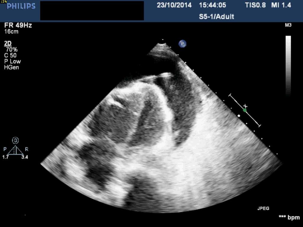
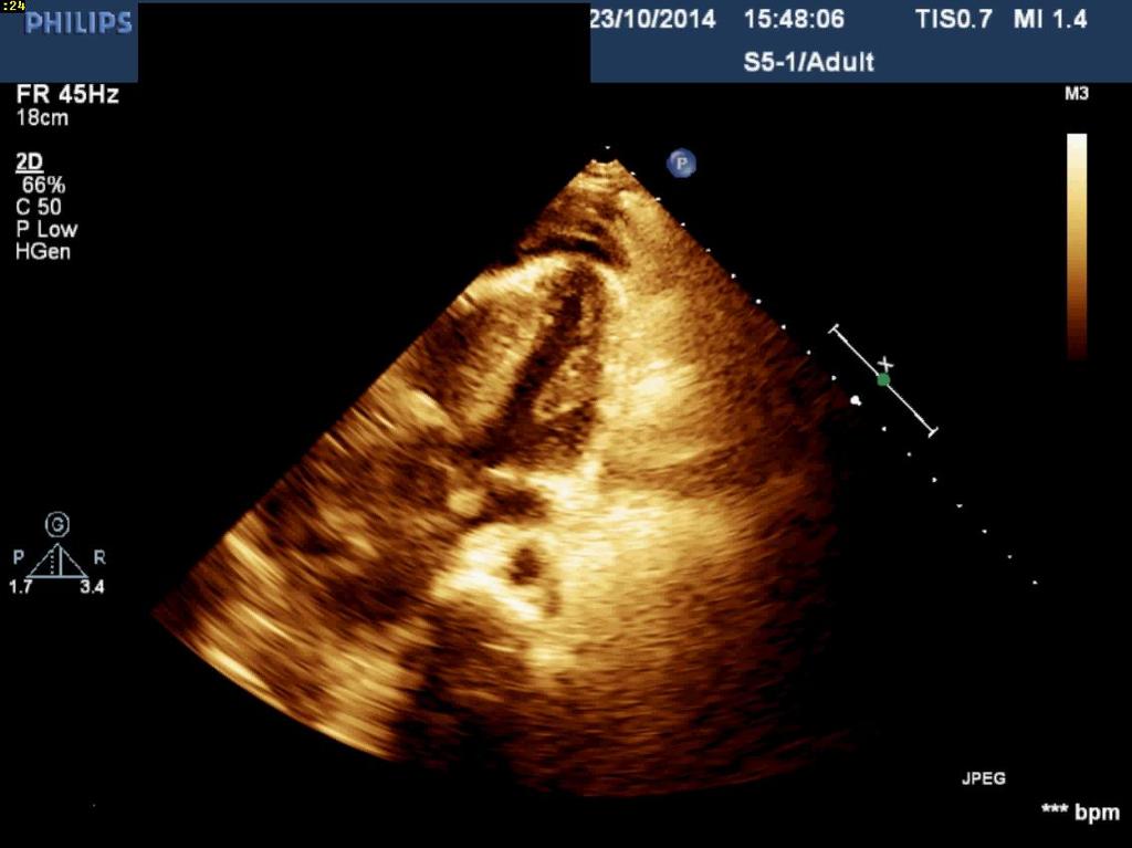
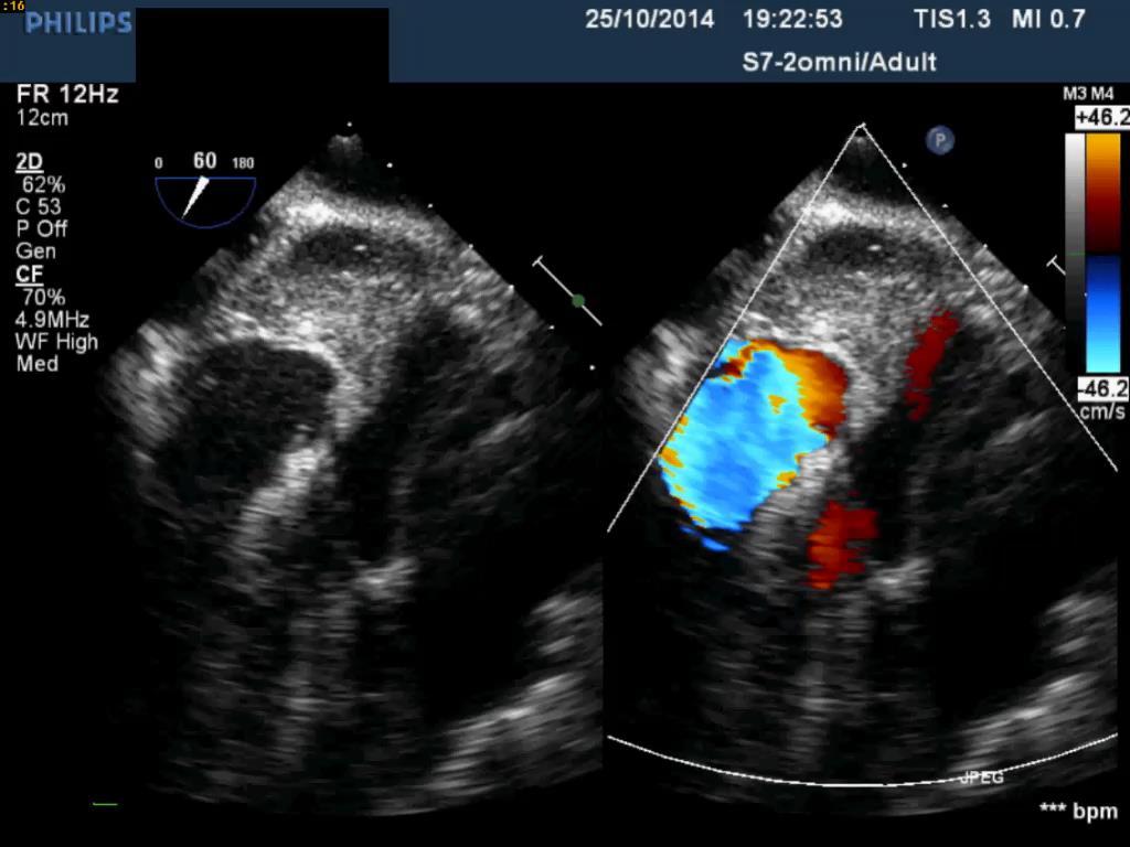
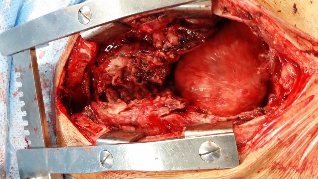
2 Case 1 17 year old boy presented to the ER: Left sided chest pain aggravated with breathing Low back pain Shortness of breath Fatigue Had felt febrile on and off since the past few months. General examination: Thin, pale boy, mildly tachypneic, afebrile Tachycardic with HR of 125 Blood pressure 93/60 mmhg
3

4
5
6 Echo-guided Pericardiocentesis

7 Two days later in the ICU Patient is more distressed Tachypnic 25/min Tachycardic HR 135bpm BP 92/50 mmhg
8


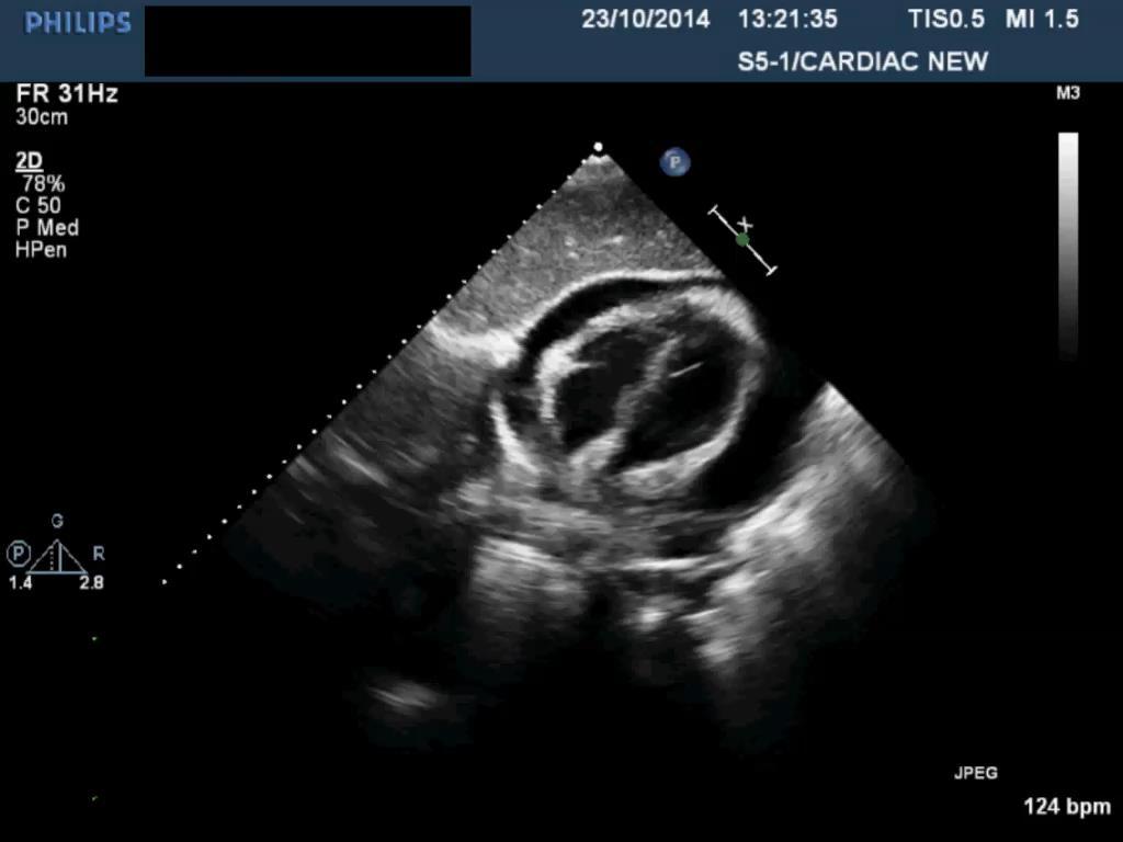
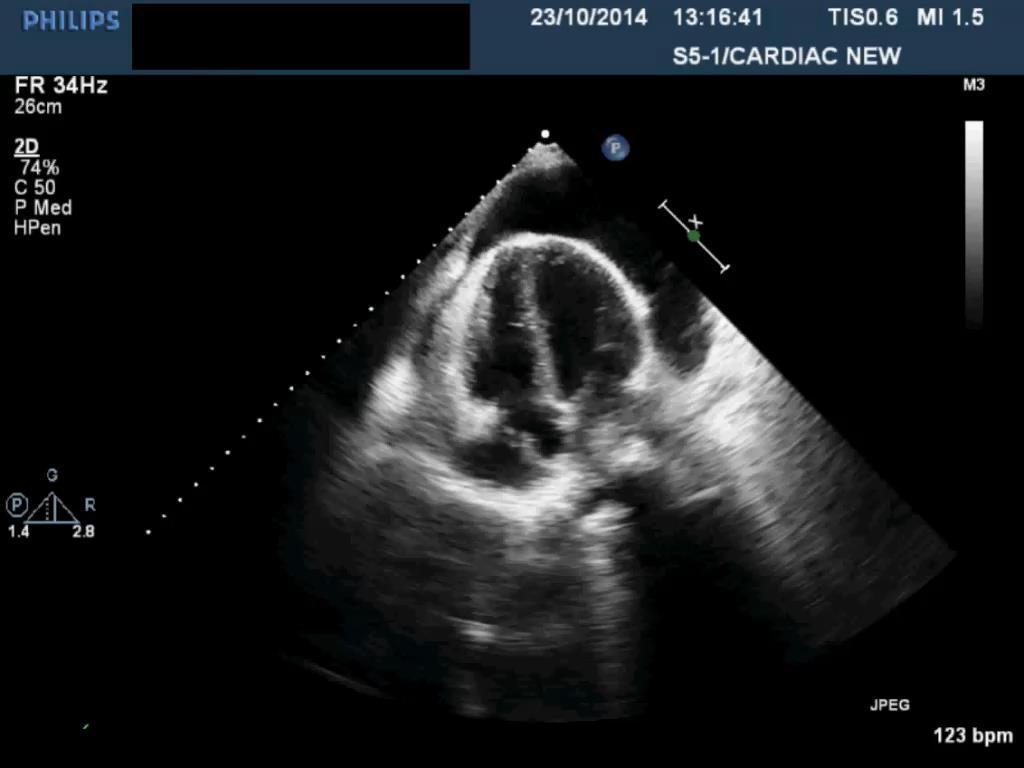
9 Echo Report: Large pericardial effusion Large hematoma compressing the RV
10

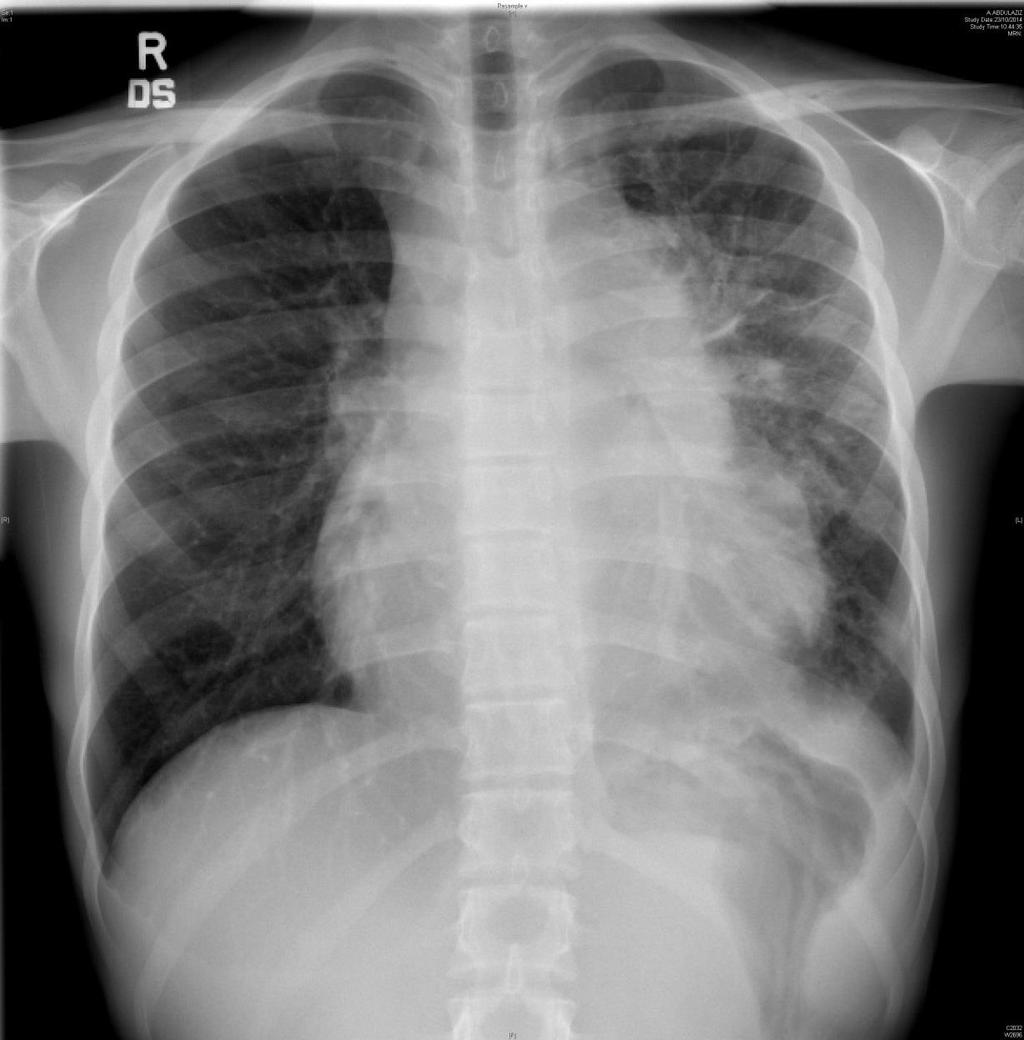
11 Missed clue: Wide superior mediastinum on chest X ray Diffuse Large B-cell Lymphoma stage IV Started on chemotherapy. Regression of tumor size after few weeks.

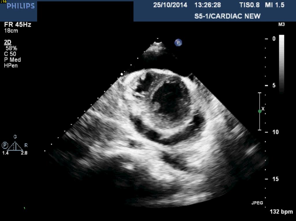
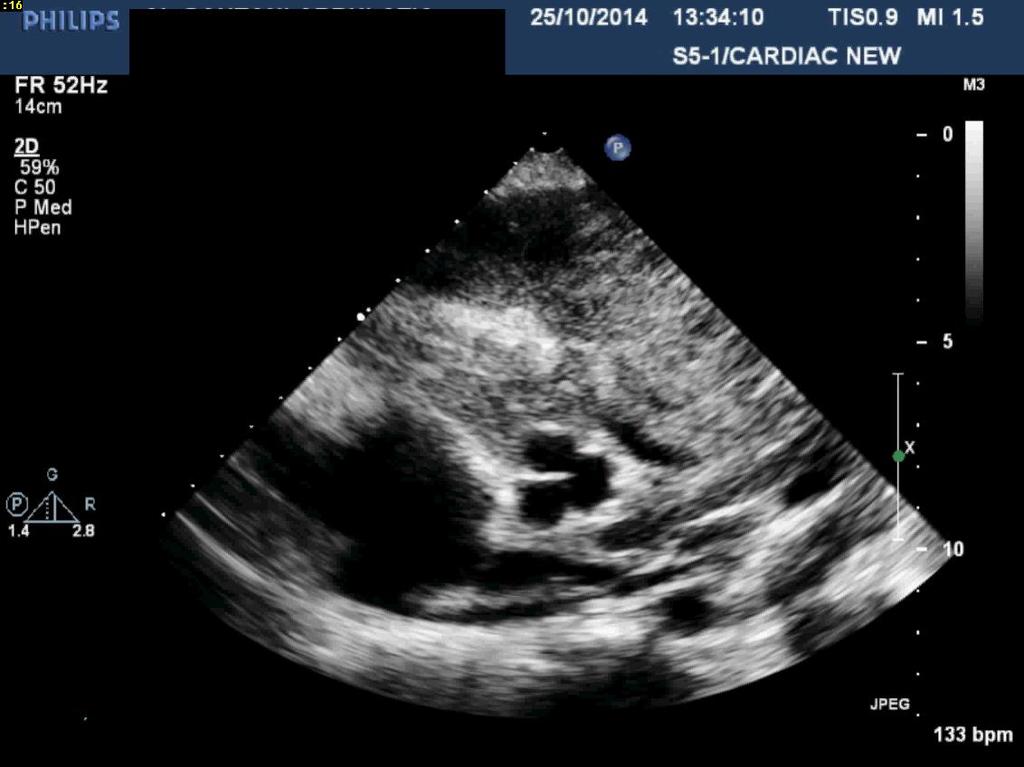
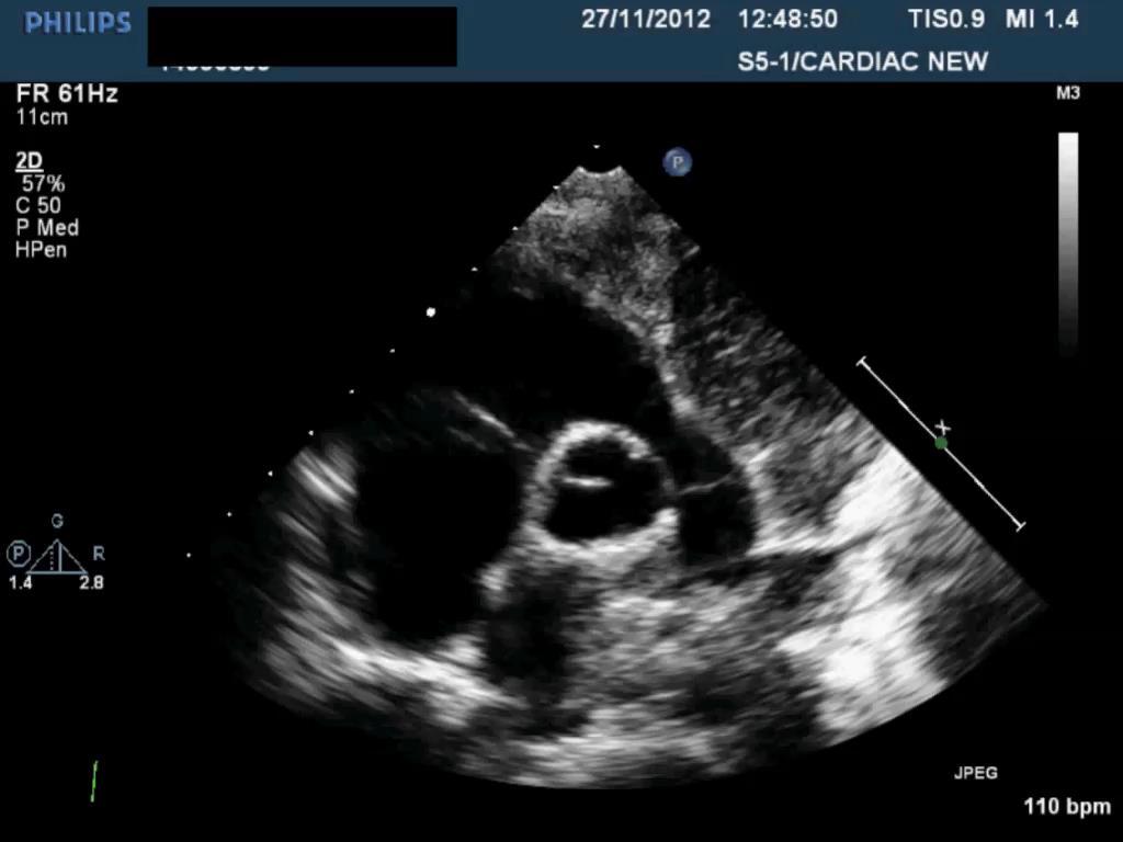
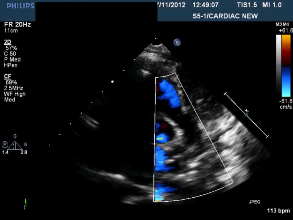
12 15 year old girl diagnosed as Hodgkin s lymphoma (nodular sclerosing) Planned for chemotherapy Echocardiography was requested Report: positive findings Small pericardial effusion Small PDA No mention of ECF in report Case 2
13


14 The patient had successful treatment Currently in remission


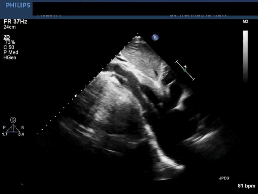
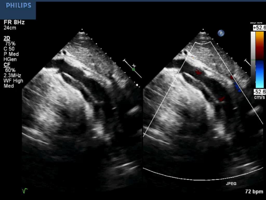
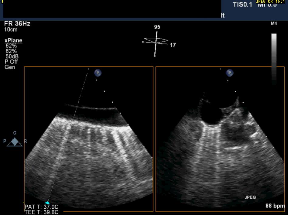
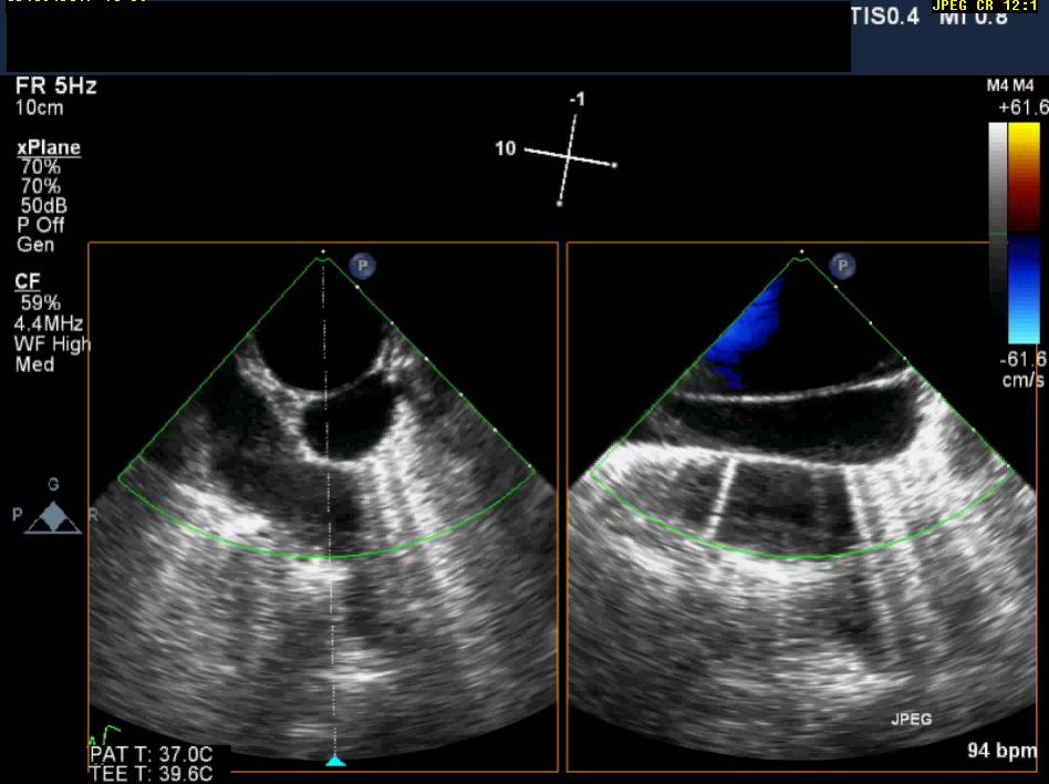
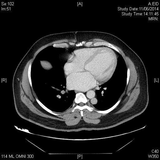
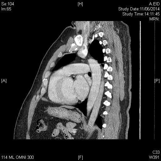
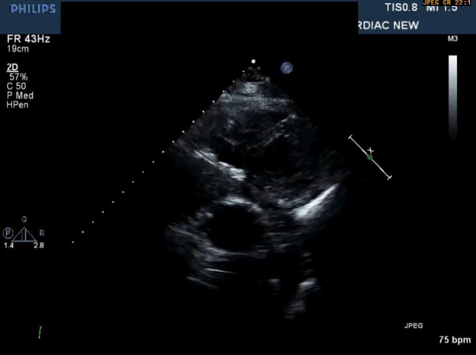
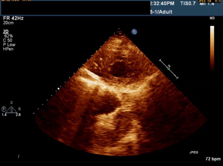
15 Case 3 43 male known case of metastatic colon cancer Admitted for chemotherapy Echocardiography requested prior to start of chemotherapy No significant cardiac pathology noted Subcostal images showed liver abnormality Report read:? Liver masses-suggest CT abdomen
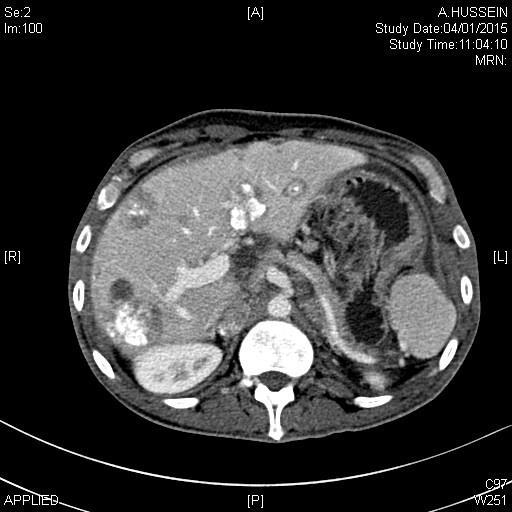
16


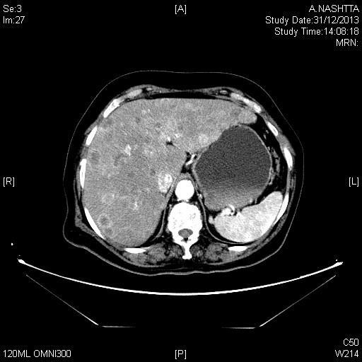
17 Case 4 78 year old lady Known case of metastatic breast cancer on chemotherapy Patient has moderate aortic stenosis Subcostal views showed abnormality Report read:?liver masses? mets
18

19 47 year old male diabetic and hypertensive Focal segmental glomerulonephritis Chronic kidney disease Hyperlipidemia Raised liver enzymes and. Liver biopsy showed non-specific fatty infiltration of liver MRI liver reported normal liver morphology and signal intensity and no focal hepatic lesions Case 5
")
20 Echocardiography was requested because of chest pain No significant cardiac pathology Subcostal views showed ECF Report read: Abnormal liver texture need U/S Ultrasound abdomen: Diffuse hepatic steatosis (fatty liver)
21 Case 6 53 Hypertensive male not on any medication Admitted as a case of CVA CT brain showed intracerebral hemorrhage. Echocardiogram requested as initial work up Echo report: Severe LV hypertrophy Ejection fraction =45-50% Mild to moderate mitral regurgitation
22 Extracardiac findings


23 Incidental ECF Abdominal Aorta Dissection
24 TTE report: There is dissection flap across entire seen portion of the abdominal aorta, there is flap seen in the descending aorta just distal to the subclavian artery. No flap seen in the ascending aorta CT report after TTE: Extensive dissection involving the thoracoabdominal aorta starting just beyond the left subclavian artery extending downward all the way down to the left renal artery


25 Case 7 78 male HTN, DM and known case of ischemic heart disease Post PCI to LAD TTE: EF 25% moderately severe mitral regurgitation PA systolic pressure mmhg TEE requested to assess the severity of mitral regurgitation. TEE reported severe MR and additional ECF: Severe atheromatous disease of descending aorta Aortic dissection.

26 Dissection of Descending Aorta


27 Case 8 56 male complains of chest pain and shortness of breath Diagnosed as mitral valve prolapse with severe mitral regurgitation and impaired LV function EF=40% TEE was requested for assessment of mitral valve

28 Descending Aorta Imaging
29
30 TEE Report Extracardiac finding Aortic dissection descending aorta, no entry point seen, no flap in ascending aorta. No evident thrombus CT scan is needed for full assessment CT Report There is congenital anomaly consisting of hemiazygous continuation of left sided IVC Left IVC coursing superiorly lateral to the descending aorta up to the junction between left subclavian and left internal jugular vein


31 Left hemiazygous continuation of left IVC


32 Pulmonary Artery Thrombus IVC Thrombus
33 Case 9
34 Extracardiac Findings in Echo, Where do we Stand? Echocardiography is the most widely used imaging modality for assessing cardiovascular anatomy and physiology. > 20 million echocardiographic studies are performed annually in USA. There is dilemma on how to report the incidental extracardiac findings. The standardized echo report (as per ASE recommendations) does not include reporting of incidental extracardiac findings. There are no training guidelines for diagnosing and reporting extracardiac findings. Few data regarding extracardiac findings exist
35 Definition of ECF Definition is not established. Most would agree about: Pleural effusion Ascites Liver abnormality Lung or mediastinal mass Abdominal aorta abnormalities Hiatus hernia Many argue that descending aortic abnormality in TEE is not extracardiac Ascending aorta abnormalities are not considered ECF Experienced and skilled physicians can identify extracardiac findings especially if they are trained in non cardiac pathology If one is not trained to see and abnormality, the detection sensitivity will be low.
36 What is the Incidence of ECF by Echocardiography? Myocardial perfusion imaging: 1-4% Cardiac CT and CMR: 10-60% in different studies Echocardiography: Not much data is available Khosa et.al: 7.5% AlKhouli et.al: 4.4 %
37 Prevalence of Non-Cardiac Pathology on Clinical Transthoracic Echocardiography. Khosa et.al 2008 Subcostal images from 1008 TTE studies reported by experienced sonographers (level 2 or level 3) were analyzed by 2 radiologists separately. Classification of ECF: Benign: Do not alter patient management or require follow up assessment Simple hepatic cysts Hemangioma Cholelithiasis. Intermediate: Might require some form of follow-up assessment Pleural effusions Ascites Cholecystitis Worrisome: Potentially have major impact on patient management Metastasis Venous thrombosis.
 Patients, n=")

 Intermediate: 52 (67%)")
 Previously")
38 Benign: 20 (26%) Previously known: 13 (65%) Patients, n= 922 TTEs, n= 1008 Inpatient TTEs, n=443 Outpatients, n=565 ECFs, n=77(7.5%) Intermediate: 52 (67%) Previously known: 35 (67%) Worrisome: 5 (7%) Previously known: 3 (60%)
39 Category Inpatient ( n = 47 [61%]) Outpatient ( n = 30 [39%]) Benign ( n = 20 [26%]) Hepatic cyst (7) Hepatic cyst (8) Hemangioma (3) Uncomplicated cholelithiasis (1) Renal collecting system fullness (1) Indeterminate ( n = 52 [67%]) Ascites (16) Pleural effusion (14) Cholecystitis (4) Choledocolithiasis (1) Indeterminate hepatic cyst (3) Ascites (5) Pleural effusion (5) Cholecystitis (1) Dilated gallbladder with polyp (1) Indeterminate hepatic cyst (1) Calcified adrenal gland (1) Worrisome ( n = 5 [7%]) IVC filling defect (1) Liver metastasis (1) IVC stenosis (1) Liver involvement by sarcoid (1) Portal vein thrombus (1)
40 Conclusion: The incidence of ECF on TTE imaging is much smaller than other modalities: Cardiac CT: 23% CMR: 43% The incidence of potentially management altering findings was only 3.8%. Majority of them were previously known None of those not previously known had an impact on patient outcomes after downstream analysis. Analysis of additional views of a subset of 300 patients showed additional ECF in 6 (2%) patients: 4 pleural effusions and two hernias. All were non management altering.
41 ECF on Routine Echocardiographic Examinations. Alkhouli et.al ,067 echocardiographic studies both TTE and TEE were screened ECFs were defined as findings in : Liver Lung and pleura Abdomen Mediastinum Vena cava Pulmonary artery Descending to aorta A comprehensive list of keywords (that experienced echoacardiographers use to describe ECFs) was developed and used to search all the echocardiographic reports in the database. The list of keywords captured ECFs in 98% of patients
42 Sensitivity analysis of 350 TTE images by 2 experienced readers Reader 1 Reader 2 Overall Total studies reviewed Agreed with positive ECF Agreed with negative ECF Disagreed with positive ECF Disagreed with negative ECF /55 (75%) /295 (95%) /55 (25%) /295 (5%)
43 Total incidence of ECFs was 4.4% ECF TTE ( n = 39,269) Pleural effusion 1,032 (2.6%) 45 (2.5%) Lung or mediastinal mass 4 (<0.1%) 1 (<0.1%) Ascites 129 (0.3%) 3 (0.2%) Liver abnormalities 41 (0.1%) 0 (0%) Hernia 13 (<0.1%) 1 (<0.1%) DA dilatation 141 (0.4%) 0 (0%) DA thrombus or ulcer 3 (<0.1%) 34 (2%) DA severe atheroma 4 (<0.1%) 107 (6%) Mild to moderate DA atheroma 19 (<0.1%) 206 (11.5%) IVC thrombosis 10 (<0.1%) 2 (0.1%) Pulmonary embolism 2 (0.1%) 0 (0%) Total 1398(3.5%) 399(22.2%) TEE ( n = 1,798)
44 Clinical follow up review was done up to 17.8 months ECF Incidence TTE Inpatient Followup available DA dilatation % 117 (83%) Ascites % 107 (81%) Liver abnormality 105 (74%) 114 (86%) Follow-up studies (31%) 20 0 (0%) 41 98% 23 (56%) 36 (88%) 12 3 (25%) Hernia 13 93% 12 (92%) 8 (62%) 2 2 (100%) Echocardiography led to new diagnosis requiring further management IVC mass/ thrombus % 11 (92%) 12 (100%) 7 7 (100%) Mediastinal mass Pulmonary embolus 6 83% 6 (100%) 6 (100%) 6 3 (50%) 2 100% 2 (100%) 2 (100%) 2 2 (100%)
45 Conclusions: In this large database ECFs were found in 4.4% of studies Majority of ECFs are likely low-risk (pleural effusion, ascites, hernia) Reporting ECFs by echo led to changes in management in 32% of patients with available follow up data There is variable practice patterns of experienced readers with regards to identification of ECFs Cardiologists usually report ECFs as general descriptions rather than specific diagnosis
46 What Should we do? The incidence of important management altering ECFs by TTE and TEE is very low Cardiology and radiology co-reading will not be efficient or cost effective. However some ECFs do lead to management changes. Focused training of sonographers and physician echoacardiographers may be needed on non cardiac pathology especially in liver and mediastinal pathology.
47 What to do? Echocardiography societies should develop training guidelines and make recommendations for reporting of ECFs Protocol should be developed for describing important ECFs intte and TEE Appropriate action for each category of risk should be specified. If you don t see it doesn t mean that it is not there
Detailed Order Request Checklists for Cardiology
 Next Generation Solutions Detailed Order Request Checklists for Cardiology 8600 West Bryn Mawr Avenue South Tower Suite 800 Chicago, IL 60631 www.aimspecialtyhealth.com Appropriate.Safe.Affordable 2018
Adult Echocardiography Examination Content Outline
 Adult Echocardiography Examination Content Outline (Outline Summary) # Domain Subdomain Percentage 1 2 3 4 5 Anatomy and Physiology Pathology Clinical Care and Safety Measurement Techniques, Maneuvers,
Diseases of the Aorta
 Diseases of the Aorta ASE Review 2018 Susan E Wiegers, MD, FASE, FACC Professor of Medicine My great friend Dr. Roberto Lang Disclosure None related to this presentation 1 Objectives Aneurysm Dissection
ADVANCED CARDIOVASCULAR IMAGING. Medical Knowledge. Goals and Objectives PF EF MF LF Aspirational
 Medical Knowledge Goals and Objectives PF EF MF LF Aspirational Know the basic principles of magnetic resonance imaging (MRI) including the role of the magnetic fields and gradient coil systems, generation
Rotation: Echocardiography: Transthoracic Echocardiography (TTE)
") Rotation: Echocardiography: Transthoracic Echocardiography (TTE) Rotation Format and Responsibilities: Fellows rotate in the echocardiography laboratory in each clinical year. Rotations during the first
Rotation: Echocardiography: Transthoracic Echocardiography (TTE) Rotation Format and Responsibilities: Fellows rotate in the echocardiography laboratory in each clinical year. Rotations during the first
Presenter: Steven Brust, HCS-D, HCS-H Product Manager, Home Health Coding Center
 Presenter: Steven Brust, HCS-D, HCS-H Product Manager, Home Health Coding Center Pinpoint & properly assign the appropriate heart failure codes Left- vs. Right-sided Left ventricular failure (LVF) may
Cardiac MRI in ACHD What We. ACHD Patients
 Cardiac MRI in ACHD What We Have Learned to Apply to ACHD Patients Faris Al Mousily, MBChB, FAAC, FACC Consultant, Pediatric Cardiology, KFSH&RC/Jeddah Adjunct Faculty, Division of Pediatric Cardiology
ERDHEIM-CHESTER DISEASE LUNG & HEART ISSUES
 ERDHEIM-CHESTER DISEASE LUNG & HEART ISSUES GIULIO CAVALLI, M.D. INTERNAL MEDICINE AND CLINICAL IMMUNOLOGY IRCCS SAN RAFFAELE HOSPITAL VITA-SALUTE SAN RAFFAELE UNIVERSITY MILAN, ITALY cavalli.giulio@hsr.it
Cardiac Masses. Cardiac Masses: Considerations. Dennis A. Tighe, MD, FASE. University of Massachusetts Medical School Worcester, MA 4/16/2018
 Cardiac Masses Dennis A. Tighe, MD, FASE University of Massachusetts Medical School Worcester, MA Cardiac Masses: Considerations Definition of the mass Nature Location Benign or malignant Presentation
Index. Note: Page numbers of article titles are in boldface type.
 Index Note: Page numbers of article titles are in boldface type. A Acute coronary syndrome(s), anticoagulant therapy in, 706, 707 antiplatelet therapy in, 702 ß-blockers in, 703 cardiac biomarkers in,
Echocardiography as a diagnostic and management tool in medical emergencies
 Echocardiography as a diagnostic and management tool in medical emergencies Frank van der Heusen MD Department of Anesthesia and perioperative Care UCSF Medical Center Objective of this presentation Indications
Top 10 Facts in Contrast Echocardiography. Pamela R. Burgess, BS, RDCS, RDMS, RVT, FASE
 Top 10 Facts in Contrast Echocardiography Pamela R. Burgess, BS, RDCS, RDMS, RVT, FASE Presenter Disclosure The following relationship exist related to this presentation: Pamela R. Burgess, BS, RDCS, RDMS,
Severity of AS Degree of AV calcification (? Bicuspid AV), annulus size, & aortic root
, annulus size, & aortic root") The role of Cardiac Imaging modalities in evaluation & selection of patients for Trans-catheter Aortic Valve Implantation Dr.Saeed AL Ahmari Consultant Cardiologist Prince Sultan Cardaic Center, Riyadh
The role of Cardiac Imaging modalities in evaluation & selection of patients for Trans-catheter Aortic Valve Implantation Dr.Saeed AL Ahmari Consultant Cardiologist Prince Sultan Cardaic Center, Riyadh
Pericardial Diseases. Smonporn Boonyaratavej, MD. Division of Cardiology, Department of Medicine Chulalongkorn University
 Pericardial Diseases Smonporn Boonyaratavej, MD Division of Cardiology, Department of Medicine Chulalongkorn University Cardiac Center, King Chulalongkorn Memorial Hospital 21 AUGUST 2016 Pericardial
Radiology of the respiratory/cardiac diseases (part 2)
") Cardiology Cycle - Lecture 6 436 Teams Radiology of the respiratory/cardiac diseases (part 2) Objectives Done By Team Leaders: Khalid Alshehri Hanin Bashaikh Team Members: Leena Alwakeel Aroob Alhuthail
Cardiology Cycle - Lecture 6 436 Teams Radiology of the respiratory/cardiac diseases (part 2) Objectives Done By Team Leaders: Khalid Alshehri Hanin Bashaikh Team Members: Leena Alwakeel Aroob Alhuthail
Cardiac Masses. Dennis A. Tighe, MD, FASE. University of Massachusetts Medical School Worcester, MA
 Cardiac Masses Dennis A. Tighe, MD, FASE University of Massachusetts Medical School Worcester, MA Cardiac Masses: Considerations Definition of the mass Nature Location Benign or malignant Presentation
Certificate in Clinician Performed Ultrasound (CCPU) Syllabus. Rapid Cardiac Echo (RCE)
 Syllabus. Rapid Cardiac Echo (RCE)") Certificate in Clinician Performed Ultrasound (CCPU) Syllabus Rapid Cardiac Echo (RCE) Purpose: Rapid Cardiac Echocardiography (RCE) This unit is designed to cover the theoretical and practical curriculum
Certificate in Clinician Performed Ultrasound (CCPU) Syllabus Rapid Cardiac Echo (RCE) Purpose: Rapid Cardiac Echocardiography (RCE) This unit is designed to cover the theoretical and practical curriculum
Pulmonary Embolism. Thoracic radiologist Helena Lauri
 Pulmonary Embolism Thoracic radiologist Helena Lauri 8.5.2017 Statistics 1-2 out of 1000 adults annually are diagnosed with deep vein thrombosis (DVT) and/or pulmonary embolism (PE) About half of patients
Echocardiographic Cardiovascular Risk Stratification: Beyond Ejection Fraction
 Echocardiographic Cardiovascular Risk Stratification: Beyond Ejection Fraction October 4, 2014 James S. Lee, M.D., F.A.C.C. Associates in Cardiology, P.A. Silver Spring, M.D. Disclosures Financial none
Cardiac Radiography. Jared D. Christensen, M.D.
 Cardiac Radiography Jared D. Christensen, M.D. Cardiac radiography Jared D. Christensen, M.D. Overview Basic Concepts Technique Normal anatomy Cases Technique 3 Standard Views Posterior-Anterior (PA) Anterior-Posterior
Interesting Cases - A Case Report: Renal Cell Carcinoma With Tumor Mass In IVC And Heart. O Wenker, L Chaloupka, R Joswiak, D Thakar, C Wood, G Walsh
 ISPUB.COM The Internet Journal of Thoracic and Cardiovascular Surgery Volume 3 Number 2 Interesting Cases - A Case Report: Renal Cell Carcinoma With Tumor Mass In IVC And Heart O Wenker, L Chaloupka, R
Pericardial Disease: Case Examples. Echo Fiesta 2017
 Pericardial Disease: Case Examples Echo Fiesta 2017 2014 2014 MFMER MFMER 3346252-1 slide-1 Objectives Have a systematic approach to evaluation of constriction 2014 MFMER 3346252-2 CASE 1 2013 MFMER 3248567-3
2/4/2011. Nathan Kerner, M.D.
 Nathan Kerner, M.D. Definition Elevated pressures - cut off usually >40 mmhg pulmonary artery systolic pressure (PASP) Usually associated with elevated pulmonary vascular resistance (PVR) measured in dynessec/cm
Acute Aortic Syndromes
 Acute Aortic Syndromes Carole J. Dennie, MD Acute Thoracic Aortic Syndromes Background Non-Traumatic Acute Thoracic Aortic Syndromes Carole Dennie MD FRCPC Associate Professor of Radiology and Cardiology
2019 Qualified Clinical Data Registry (QCDR) Performance Measures
 Performance Measures") 2019 Qualified Clinical Data Registry (QCDR) Performance Measures Description: This document contains the 18 performance measures approved by CMS for inclusion in the 2019 Qualified Clinical Data Registry
2019 Qualified Clinical Data Registry (QCDR) Performance Measures Description: This document contains the 18 performance measures approved by CMS for inclusion in the 2019 Qualified Clinical Data Registry
Follow-up of Aortic Dissection: How, How Often, Which Consequences Euro Echo 2011
 Follow-up of Aortic Dissection: How, How Often, Which Consequences Euro Echo 2011 Susan E. Wiegers, MD, FASE Director of Clinical Echocardiography Hospital of the University of Pennsylvania Disclosure
ASE 2011 Appropriate Use Criteria for Echocardiography
 ASE 2011 Appropriate Use Criteria for Echocardiography Table 1. TTE for General Evaluation of Cardiac Structure and Function 1 2 Suspected Cardiac Etiology General With TTE Symptoms or conditions potentially
CMS Limitations Guide - Radiology Services
 CMS Limitations Guide - Radiology Services Starting October 1, 2015, CMS will update their existing medical necessity limitations on tests and procedures to correspond to ICD-10 codes. This limitations
ACUTE AORTIC SYNDROMES
 ACUTE AORTIC SYNDROMES AGNETA FLINCK MD, PhD Dept. of Thoracic Radiology Sahlgrenska University Hospital ACUTE AORTIC SYNDROMES Aortic dissection Intramural hematoma (IMH) 5-20% Penetrating atherosclerotic
ARIC HEART FAILURE HOSPITAL RECORD ABSTRACTION FORM. General Instructions: ID NUMBER: FORM NAME: H F A DATE: 10/13/2017 VERSION: CONTACT YEAR NUMBER:
 ARIC HEART FAILURE HOSPITAL RECORD ABSTRACTION FORM General Instructions: The Heart Failure Hospital Record Abstraction Form is completed for all heart failure-eligible cohort hospitalizations. Refer to
Vascular Imaging in the Pediatric Abdomen. Jonathan Swanson, MD
 Vascular Imaging in the Pediatric Abdomen Jonathan Swanson, MD Goals and Objectives To understand the imaging approach, appearance, and clinical manifestations of the common pediatric abdominal vascular
Double Superior Vena Cava; A Benign Cause of Widened Mediastenum and Implication on Venous Central Access
 ISPUB.COM The Internet Journal of Endovascular Medicine Volume 2 Number 1 Double Superior Vena Cava; A Benign Cause of Widened Mediastenum and Implication on Venous H Enuh, A Patel, A Chaudry, K Diaz,
7. Echocardiography Appropriate Use Criteria (by Indication)
") Criteria for Echocardiography 1133 7. Echocardiography Criteria (by ) Table 1. TTE for General Evaluation of Cardiac Structure and Function Suspected Cardiac Etiology General With TTE 1. Symptoms or conditions
Criteria for Echocardiography 1133 7. Echocardiography Criteria (by ) Table 1. TTE for General Evaluation of Cardiac Structure and Function Suspected Cardiac Etiology General With TTE 1. Symptoms or conditions
cardiac imaging planes planning basic cardiac & aortic views for MR
 cardiac imaging planes planning basic cardiac & aortic views for MR Dianna M. E. Bardo, M. D. Assistant Professor of Radiology & Cardiovascular Medicine Director of Cardiac Imaging cardiac imaging planes
Cardiac Mass and Mass-like Structures
 KSE 2017 Basic Echo Review Course (4) Nov 26, 2017 Cardiac Mass and Mass-like Structures Sun Hwa Lee, MD, PhD Chonbuk National University Hospital & Medical School Introduction Although cardiac tumors
Review of Cardiac Imaging Modalities in the Renal Patient. George Youssef
 Review of Cardiac Imaging Modalities in the Renal Patient George Youssef ECHO Left ventricular hypertrophy (LVH) assessment Diastolic dysfunction Stress ECHO Cardiac CT angiography Echocardiography - positives
Radiology of hepatobiliary diseases
 GI cycle - Lecture 14 436 Teams Radiology of hepatobiliary diseases Objectives 1. To Interpret plan x-ray radiograph of abdomen with common pathologies. 2. To know the common pathologies presentation.
Heart and Lungs. LUNG Coronal section demonstrates relationship of pulmonary parenchyma to heart and chest wall.
 Heart and Lungs Normal Sonographic Anatomy THORAX Axial and coronal sections demonstrate integrity of thorax, fetal breathing movements, and overall size and shape. LUNG Coronal section demonstrates relationship
Job Task Analysis for ARDMS Abdomen Data Collected: June 30, 2011
 Job Task Analysis for ARDMS Abdomen Data Collected: June 30, 2011 Reported: Analysis Summary for: Abdomen Examination Survey Dates 06/13/2011-06/26/2011 Invited Respondents 6,000 Surveys with Demographics
AORTIC ANEURYSM. howmed.net
 AORTIC ANEURYSM howmed.net ANATOMY It is important to understand the anatomy of the aorta Need to know the extent of the aneurysm Need to know the vessels involved This helps with Medical or Surgical management
Aortic Valve Practice Guidelines: What Has Changed and What You Need to Know
 Aortic Valve Practice Guidelines: What Has Changed and What You Need to Know James F. Burke, MD Program Director Cardiovascular Disease Fellowship Lankenau Medical Center Disclosure Dr. Burke has no conflicts
Pediatric Echocardiography Examination Content Outline
 Pediatric Echocardiography Examination Content Outline (Outline Summary) # Domain Subdomain Percentage 1 Anatomy and Physiology Normal Anatomy and Physiology 10% 2 Abnormal Pathology and Pathophysiology
COMPREHENSIVE EVALUATION OF FETAL HEART R. GOWDAMARAJAN MD
 COMPREHENSIVE EVALUATION OF FETAL HEART R. GOWDAMARAJAN MD Disclosure No Relevant Financial Relationships with Commercial Interests Fetal Echo: How to do it? Timing of Study -optimally between 22-24 weeks
Index. K Knobology, TTE artifact, image resolution, ultrasound, 14
 A Acute aortic regurgitation (AR), 124 128 Acute aortic syndrome (AAS) classic aortic dissection diagnosis, 251 263 evolutive patterns, 253 255 pathology, 250 251 classifications, 247 248 incomplete aortic
Choose the grading of diastolic function in 82 yo woman
 Question #1 Choose the grading of diastolic function in 82 yo woman E= 80 cm/s A= 70 cm/s LAVI < 34 ml/m 2 1= Grade 1 2= Grade 2 3= Grade 3 4= Normal 5= Indeterminate 2018 MFMER 3712003-1 Choose the grading
JFICMI Basic Critical Care Echocardiography (BCCE)
") JFICMI Basic Critical Care Echocardiography (BCCE) 2017 Introduction The International expert statement on training standards for critical care ultrasonography position paper published in Intensive Care
JFICMI Basic Critical Care Echocardiography (BCCE) 2017 Introduction The International expert statement on training standards for critical care ultrasonography position paper published in Intensive Care
ASCeXAM / ReASCE. Practice Board Exam Questions. Tuesday Morning
 ASCeXAM / ReASCE Practice Board Exam Questions Tuesday Morning Congenital Heart Disease in Adults Congenital Heart Disease Cases Diastolic Function Pericardial Disease Congenital Heart Disease Cases Michael
You Won t Believe What I Saw on. Disclosures. Goals. Dimensions 2013 October 18 th Michael Pfeiffer, MD. No Financial Disclosures
 You Won t Believe What I Saw on that ECHO! Dimensions 2013 October 18 th Michael Pfeiffer, MD Disclosures No Financial Disclosures Goals Review unusual and unique echocardiographic images. Briefly present
New ASE Guidelines: What you must know
 New ASE Guidelines: What you must know Federico M Asch MD, FASE, FACC Chair, ASE Guidelines and Standards Committee Medstar Washington Hospital Center Medstar Health Research Institute Georgetown University
HAND held echocardiography a dangerous toy?
 HAND held echocardiography a dangerous toy? Eva Gerdts Professor of Medicine University of Bergen Visiting Professor of Medicine Federico II University, Napoli Diagnostic accuracy Vscan vs high end echocardiograph
Congestive Heart Failure or Heart Failure
 Congestive Heart Failure or Heart Failure Dr Hitesh Patel Ascot Cardiology Group Heart Failure Workshop April, 2014 Question One What is the difference between congestive heart failure and heart failure?
Chapter 5 Section 1.1
 Radiology Chapter 5 Section 1.1 Issue Date: March 7, 1986 Authority: 32 CFR 199.4(a), (b)(2)(x), (c)(2)(viii), (e)(14) and 32 CFR 199.6(d)(2) Copyright: CPT only 2006 American Medical Association (or such
Chapter 5 Section 1.1. Diagnostic Radiology (Diagnostic Imaging)
") Radiology Chapter 5 Section 1.1 Issue Date: March 7, 1986 Authority: 32 CFR 199.4(a), (b)(2)(x), (c)(2)(viii), (e)(14) and 32 CFR 199.6(d)(2) 1.0 CPT 1 PROCEDURE CODES 70010-72292, 73000-76499, 77071-77084,
Radiology Chapter 5 Section 1.1 Issue Date: March 7, 1986 Authority: 32 CFR 199.4(a), (b)(2)(x), (c)(2)(viii), (e)(14) and 32 CFR 199.6(d)(2) 1.0 CPT 1 PROCEDURE CODES 70010-72292, 73000-76499, 77071-77084,
Endovascular treatment of acquired arteriovenous fistula with severe hemodynamic effects: a case report
 Endovascular treatment of acquired arteriovenous fistula with severe hemodynamic effects: a case report The Leipzig Interventional Course, January 24 27, 2017 El Samman K., Šedivý P., Šnajdrová A., Přindišová
Case 47 Clinical Presentation
 93 Case 47 C Clinical Presentation 45-year-old man presents with chest pain and new onset of a murmur. Echocardiography shows severe aortic insufficiency. 94 RadCases Cardiac Imaging Imaging Findings C
Clinical Indications for Echocardiography
 Clinical Indications for Echocardiography Echocardiography is widely utilised and potential applications are increasing with advances in technology. The aim of this document is two-fold: 1) To define clinical
Conflict Disclosures. Vermont Cardiac Network. Outline. Series Learning Objectives 4/27/2016. Scott E. Friedman April 28, 2016
 Conflict Disclosures Vermont Cardiac Network The Speaker has reported no significant financial relationship with any companies whose product may be germane to the content of their presentations or who
Susan P. D Anna MSN, APRN BC February 14, 2019
 Is there Equal Opportunity in Heart Failure?? Susan P. D Anna MSN, APRN BC February 14, 2019 Disclosures: I have no financial disclosures. I am not an expert on this topic, but see a lot of women with
Pericardial Diseases/Tamponade Illustrative Cases
 Pericardial Diseases/Tamponade Illustrative Cases Jae K. Oh, MD Echo Hawaii 2017 2012 MFMER 3200268v3(2010)-1 Case #1 47 year old man Chest pain Not exertional Normal Examination 2016 MFMER slide-2 1 47
DR. DO NGUYEN TIN CHILDREN HOSPITAL 1
 DR. DO NGUYEN TIN CHILDREN HOSPITAL 1 BACKGROUND Congenital extrahepatic porto-systemic shunt (CEPS), known as Abernethy malformation, is a rare malformation. Intestinal and/or splenic venous blood bypasses
New Cardiovascular Devices and Interventions: Non-Contrast MRI for TAVR Abhishek Chaturvedi Assistant Professor. Cardiothoracic Radiology
 New Cardiovascular Devices and Interventions: Non-Contrast MRI for TAVR Abhishek Chaturvedi Assistant Professor Cardiothoracic Radiology Disclosure I have no disclosure pertinent to this presentation.
Multimodality Imaging of the Thoracic Aorta
 Multimodality Imaging of the Thoracic Aorta Steven Goldstein MD, FACC Director Noninvasive Cardiology MedStar Heart and Vascular Institute Washington Hospital Center Saturday, October 8, 2016 DISCLOSURE
DISCLOSURE. Echocardiography in Systemic Diseases: Questions. Relevant Financial Relationship(s) None. Off Label Usage None 5/7/2018
 None. Off Label Usage None 5/7/2018") Echocardiography in Systemic Diseases: Questions Sunil Mankad, MD, FACC, FCCP, FASE Associate Professor of Medicine Mayo Clinic College of Medicine Director, Transesophageal Echocardiography Associate
Echocardiography in Systemic Diseases: Questions Sunil Mankad, MD, FACC, FCCP, FASE Associate Professor of Medicine Mayo Clinic College of Medicine Director, Transesophageal Echocardiography Associate
Normal Sonographic Anatomy
 hapter 2:The Liver DUNSTAN ABRAHAM Normal Sonographic Anatomy Homogeneous, echogenic texture (Figure 2-1) Measures approximately 15 cm in length and 10 12.5 cm anterior to posterior; measurement taken
Valvular Heart Disease: Assessment and Timing of Intervention. Graham Cole Consultant Cardiologist Imperial College Healthcare NHS Trust
 Valvular Heart Disease: Assessment and Timing of Intervention Graham Cole Consultant Cardiologist Imperial College Healthcare NHS Trust Disclosures: Speaker fee: Bayer Acknowledgements: Matt Shun-Shin
Image Library Case Listing:
 Image Library Case Listing: 1. Giant left atrial myxoma with mitral valve damage 2. Type A aortic dissection 3. Primum ASD 4. Aortic Transection from motor vehicle accident 5. Snake thrombus in right atrium
Assignable revenue codes: Explanation of services:
 computed tomography Chest/Cardiac Assignable revenue codes: Explanation of services: 0350 CT Scan General Classification 0351 CT Scan Head Scan 0352 CT Scan Body Scan 0359 CT Scan Other CT Scans Known
Adult Congenital Heart Disease: What All Echocardiographers Should Know Sharon L. Roble, MD, FACC Echo Hawaii 2016
 1 Adult Congenital Heart Disease: What All Echocardiographers Should Know Sharon L. Roble, MD, FACC Echo Hawaii 2016 DISCLOSURES I have no disclosures relevant to today s talk 2 Why should all echocardiographers
Echo Emergencies. Outline. Michael H. Picard, MD Massachusetts General Hospital Harvard Medical School No disclosures
 Echo Emergencies Michael H. Picard, MD Massachusetts General Hospital Harvard Medical School No disclosures Outline Common emergency / on call scenarios Tamponade Pulmonary embolism/rv strain Cardiogenic
Breakout Session: Transesophageal Echocardiography
 Breakout Session: Transesophageal Echocardiography Doris Ockert, MD Andrew Schroeder, MD University of Wisconsin School of Medicine and Public Health Jutta Novalija, MD, PhD Medical College of Wisconsin
Ve V rmont rmon Card Car iac d Netw Ne ork tw Scott E. Friedman April 28, 2016
 Vermont Cardiac Network Scott E. Friedman April 28, 2016 Conflict Disclosures Th S k h d i ifi fi i l l i hi ih The Speaker has reported no significant financial relationship with any companies whose product
March yr. old male, newspaper writer, with worsening dyspnea /orthopnea past few months
 Case 1 March 2016 59 yr. old male, newspaper writer, with worsening dyspnea /orthopnea past few months PMH diabetes, celiac disease Reports chest discomfort, positional coughing and pedal edema last 10
Cigna - Prior Authorization Procedure List: Radiology & Cardiology
 Cigna - Prior Authorization Procedure List: Radiology & Cardiology Category CPT Code CPT Code Description 93451 Right heart catheterization 93452 Left heart catheterization 93453 Combined right and left
An aneurysm is a localized abnormal dilation of a blood vessel or the heart Types: 1-"true" aneurysm it involves all three layers of the arterial
 An aneurysm is a localized abnormal dilation of a blood vessel or the heart Types: 1-"true" aneurysm it involves all three layers of the arterial wall (intima, media, and adventitia) or the attenuated
PART II ECHOCARDIOGRAPHY LABORATORY OPERATIONS ADULT TRANSTHORACIC ECHOCARDIOGRAPHY TESTING
 PART II ECHOCARDIOGRAPHY LABORATORY OPERATIONS ADULT TRANSTHORACIC ECHOCARDIOGRAPHY TESTING STANDARD - Primary Instrumentation 1.1 Cardiac Ultrasound Systems SECTION 1 Instrumentation Ultrasound instruments
Cardiovascular manifestations of HIV
 Cardiovascular manifestations of HIV Prabhakar Rajiah, MBBS, MD, FRCR Associate Professor of Radiology Associate Director, Cardiac CT and MRI University of Texas Southwestern Medical Center, Dallas, USA
Dobutamine Stress testing In Low Flow, Low EF, Low Gradient Aortic Stenosis Case Studies
 Dobutamine Stress testing In Low Flow, Low EF, Low Gradient Aortic Stenosis Case Studies Mitral Regurgitation The New ASE Guidelines: Role of 2D/3D and CMR William A. Zoghbi MD, FASE, MACC Professor and
ECHOCARDIOGRAPHY. Patient Care. Goals and Objectives PF EF MF LF Aspirational
 Patient Care Be able to: Perform and interpret basic TTE and X cardiac Doppler examinations Perform and interpret a comprehensive X TTE and cardiac Doppler examination Perform and interpret a comprehensive
TAVR: Echo Measurements Pre, Post And Intra Procedure
 2017 ASE Florida, Orlando, FL October 10, 2017 8:00 8:25 AM 25 min TAVR: Echo Measurements Pre, Post And Intra Procedure Muhamed Sarić MD, PhD, MPA Director of Noninvasive Cardiology Echo Lab Associate
pulmonary valve on, 107 pulmonary valve vegetations on, 113
 INDEX Adriamycin-induced cardiomyopathy, 176 Amyloidosis, 160-161 echocardiographic abnormalities in, 160 intra-mural tumors similar to, 294 myocardial involvement in, 160-161 two-dimensional echocardiography
Multimodality Imaging of Anomalous Left Coronary Artery from the Pulmonary
 1 IMAGES IN CARDIOVASCULAR ULTRASOUND 2 3 4 Multimodality Imaging of Anomalous Left Coronary Artery from the Pulmonary Artery 5 6 7 Byung Gyu Kim, MD 1, Sung Woo Cho, MD 1, Dae Hyun Hwang, MD 2 and Jong
Abdomen Sonography Examination Content Outline
 Abdomen Sonography Examination Content Outline (Outline Summary) # Domain Subdomain Percentage 1 2 3 Anatomy, Perfusion, and Function Pathology, Vascular Abnormalities, Trauma, and Postoperative Anatomy
Cardiac Mass in a 15-Year-Old Boy
 Cardiac Mass in a 15-Year-Old Boy Echocardiographic Case Report Hortensia Vuçini Department of Cardiology and Cardiac Surgery UHC Mother Theresa Tirana, Albania October 20, 2007 Case Presentation 15 year-old
When Should I Order a Stress Test or an Echocardiogram
 When Should I Order a Stress Test or an Echocardiogram Updates in Cardiology 2015 March 7, 2015 Donald L. Lappé, MD, FAHA, FACC Chairman, Cardiovascular Department Medical Director, Intermountain Cardiovascular
PULMONARY VENOLOBAR SYNDROME. Dr.C.Anandhi DNB Resident, Southern Railway Headquarters Hospital.
 PULMONARY VENOLOBAR SYNDROME Dr.C.Anandhi DNB Resident, Southern Railway Headquarters Hospital. Presenting complaint: 10 yrs old girl with recurrent episodes of lower respiratory tract infection from infancy.
SONOGRAPHER & NURSE LED VALVE CLINICS
 SONOGRAPHER & NURSE LED VALVE CLINICS Frequency of visits and alerts AORTIC STENOSIS V max > 4.0 m/s or EOA < 1.0 cm 2 V max 3.5 4.0 m/s + Ca+ V max 3.0 4.0 m/s or EOA 1.0-1.5 cm 2 V max 2.5 3.0 m/s every
Heart Failure. Cardiac Anatomy. Functions of the Heart. Cardiac Cycle/Hemodynamics. Determinants of Cardiac Output. Cardiac Output
 Cardiac Anatomy Heart Failure Professor Qing ZHANG Department of Cardiology, West China Hospital www.blaufuss.org Cardiac Cycle/Hemodynamics Functions of the Heart Essential functions of the heart to cover
How to Assess and Treat Obstructive Lesions
 How to Assess and Treat Obstructive Lesions Erwin Oechslin, MD, FESC, FRCPC, Director, Congenital Cardiac Centre for Adults Peter Munk Cardiac Centre University Health Network/Toronto General Hospital
AORTIC DISSECTIONS Current Management. TOMAS D. MARTIN, MD, LAT Professor, TCV Surgery Director UF Health Aortic Disease Center University of Florida
 AORTIC DISSECTIONS Current Management TOMAS D. MARTIN, MD, LAT Professor, TCV Surgery Director UF Health Aortic Disease Center University of Florida DISCLOSURES Terumo Medtronic Cook Edwards Cryolife AORTIC
Pregnancy and Heart Disease Sharon L. Roble, MD Echo Hawaii 2016
 1 Pregnancy and Heart Disease Sharon L. Roble, MD Echo Hawaii 2016 DISCLOSURES I have no disclosures relevant to today s talk 2 Cardiovascular Effects of Pregnancy Anatomic Ventricular muscle mass increases
Imaging abdominal vascular emergencies. V.Stoynova
 Imaging abdominal vascular emergencies V.Stoynova Abdominal vessels V. Stoynova 2 Acute liver bleeding trauma anticoagulant therapy liver disease : HCC, adenoma, meta, FNH, Hemangioma Diagnosis :CT angiography
The Chest X-ray for Cardiologists
 Mayo Clinic & British Cardiovascular Society at the Royal College of Physicians, London : 21-23-October 2013 Cases-Controversies-Updates 2013 The Chest X-ray for Cardiologists Michael Rubens Royal Brompton
Policy #: 222 Latest Review Date: March 2009
 Name of Policy: MRI Phase-Contrast Flow Measurement Policy #: 222 Latest Review Date: March 2009 Category: Radiology Policy Grade: Active Policy but no longer scheduled for regular literature reviews and
Cardiac Imaging Tests
 Cardiac Imaging Tests http://www.medpagetoday.com/upload/2010/11/15/23347.jpg Standard imaging tests include echocardiography, chest x-ray, CT, MRI, and various radionuclide techniques. Standard CT and
Appendix 5. EFSUMB Newsletter. Gastroenterological Ultrasound
 EFSUMB Newsletter 87 Examinations should encompass the full range of pathological conditions listed below A log book listing the types of examinations undertaken should be kept Training should usually
Cardiac tamponade and Pericardiocentesis Made Easy
 Cardiac tamponade and Pericardiocentesis Made Easy www.cardiconcept.com Etiology of pericardial diseases. Non Infectious cause Infectious cause European Heart Journal (2015) 36, 2921 2964 Recommendations
2014 CPT Radiology Codes Requiring Review
 CT Head 1 70480 CT orbit, sella or posterior fossa; w/o contrast 1 CT Head 1 70481 CT orbit, sella or posterior fossa; with CT orbit, sella or posterior fossa; w/o contrast CT Head 1 70482 followed by
CT of Acute Thoracic Aortic Syndromes Stuart S. Sagel, M.D.
 CT of Acute Thoracic Aortic Syndromes Stuart S. Sagel, M.D. Thoracic Aortic Aneurysms Atherosclerotic Dissection Penetrating ulcer Mycotic Inflammatory (vasculitis) Traumatic Aortic Imaging Options Catheter
Congenital Heart Disease Cases
 Congenital Heart Disease Cases Sabrina Phillips, MD FACC FASE Mayo Clinic Congenital Heart Disease Center 2013 MFMER slide-1 No Disclosures 2013 MFMER slide-2 1 CASE 1 2013 MFMER slide-3 63 year old Woman
Case Presentation. By Eman El Sharkawy Ass. Professor of cardiology Alexandria University
 Case Presentation By Eman El Sharkawy Ass. Professor of cardiology Alexandria University 6m old baby girl Past history : - At the age of 2m attack of fever, diarrhea, mouth ulcers, difficult breast feeding