Accuracy of transvaginal ultrasound and magnetic resonance imaging in diagnosis and extension of pelvic endometriosis
|
|
|
- Della Robbins
- 5 years ago
- Views:
Transcription
1 Accuracy of transvaginal ultrasound and magnetic resonance imaging in diagnosis and extension of pelvic endometriosis A.Salem, Kh. Fakhfakh, S. Mehiri, Y. Ben Brahim, F. Ben Amara, H. Rajhi, R. Hamza, N.MNIF Radiology Department, Charles Nicole Hospital, Tunis Tunisia
2 Introduction Endometriosis: presence of endometrial glands and stroma outside the uterus. Cause of pelvic pain, dysmenorrhoea, dyspareunia, dyschesia and urinary symptoms Endometriosis is associated with infertility The aim of this study is to assess the accuracy of transvaginal ultrasound and MRI in the diagnosis and the extension of pelvic endometriosis and to illustrate the main pelvic locations
3 Materials, Methods and Results six patients aged between 27 and 42 years complain of dysmenorrhrea and dyspaneuria; two patients complain of pain during evacuation and micturation. All patients underwent clinical examination, transvaginal ultrasound and pelvic MRI. MRI technique: 4mm-thick sections and 1mm gap Field of view 224x512pixels Sagittal and transverse SET2 Transverse SET1 with and without fat suppression Transverse SET1 with fat suppression after intravenous injection of gadolinium
4 patients age symptoms transvaginal ultrasound MRI IVU N 1 42years Left pelvic pain Left endometrioma Left endometrioma Uterosacral ligaments, vagina N 2 27years dysmenorrhoea Left endometrioma Left endometrioma N 3 37years dysmenorrhoea dyspareunia Non specific heterogenous ovarian cysts, thickening of posterior vaginal wall Non hemorragic ovarian cysts, mass of the Torus uterosacral ligaments, posterior Vaginal wall, rectum N 4 29years chronic pelvic pain Right endometrioma Bilateral endometriomas Right ureteral endometriosis Uterosacral ligaments N 5 39years Chronic pelvic pain Bilateral endometriomas Torus nodule Sigmoid implant endometriomas Uterosacral ligaments Vagina, torus, rectum N 6 35years Infertility Bilateral endometriomas Bilateral endometriomas


5 Patient N 1 Sagittal SE T2: thickening of posterior vaginal wall containing hyperintense T2 foci Sagittal SE T2: thickening of uterosacral ligament hypointense T2 transverse SE T2: left ovarian endometrioma hypointense T2 transverse SE T1 with FS: left ovarian endometrioma hyperintense T1FS


6 Patient N 2 transverse SE T2: left ovarian endometrioma hypointense T2 transverse SE T1: left ovarian endometrioma hyperintense T1


7 Patient N 3 Transvaginal sonography:multiple hetregenous cysts Transvaginal sonography: thickening of posterior vaginal wall containing little cysts


8 Patient N 3 Sagittal SE T2: hypointense mass in the torus uterinus and posterior vaginal wall containing hyperintense T2 foci and extending to the anterior wall of rectum transverse SE T2: non hemorragic left ovarian cysts and adhesion with posterior wall of uterus transverse SE T1: no ovarian hemorragic signal T1 transverse SE T1: thickening of posterior vaginal wall containing hyperintense T1 foci (hemorragic)

 containing")


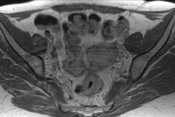
9 Patient N 4 Sagittal and coronal SE T2: bilateral endometriomas hypointense T2 (star), hypointense mass of the torus and posterior vaginal wall (arrow) containing little cysts hyperintense T2 sagittal SE T1: hyperintense T1 endometriomas, hypointense mass of the torus and posterior wall of vagina containing hyperintense foci sagittal SE T1 FS Gado: ovarian endometriomas hyperintense T1, mass of the torus with enhancement after injection of gadolinium transverse SE T1 FS: bilateral endometriomas hyperintense T1


10 Patient N 4 Pelvic sonography: right ovarian heterogenous cyst Intravenous urography: right pelvic ureteral stenosis due to ureteral endometriosis


11 Patient N 5 Transvaginal sonography: thickening of the torus and the right uterosacral ligament and hypoechoic implant of endometriosis in the anterior wall of rectum Rectal sonography: hypoechoic mass of the anterior wall of rectum


12 Patient N 5 Sagittal SE T2: hypointense mass in the torus uterinus extending to the anterior wall of rectum Transverse SE T2: hypointense mass in the torus uterinus, thichning of right uterosacral ligament and mass of the anterior wall of rectum Transverse SE T1 gado: mass in the torus uterinus, thichning of right uterosacral ligament and mass of the anterior wall of rectum


13 Patient N 6 Sagittal and coronal SE T2: hypointense T2 bilateral ovarian endometriomas Sagittal SE T1 and transverse SE T1FS: hyperintense T2 bilateral ovarian endometriomas
14 Discussion: Pelvic endpmetriosis include ovarian endometriomas, superficial peritoneal lesions and deep infiltrating endometriosis (DIE). DIE can be anterieor involving the bladder or posterior involving the uterosacral ligaments, the vagina and the intestine especially the rectum and the sigmoid colon

15 Anatomical distribution of pelvic endometriosis Colorectal endometriosis Péritoneal endometriosis Torus endometriosis Ovarian endometrioma Recto vaginal septum bladder implant
16 Physical examination Posterior pelvic endometriosis is diagnosed by physical examination in 60% reported by Chapron et al (2002). Extent of involvment of DIE remains difficult to determine by physical examination and requires further exploration as transvaginal sonography, rectal sonography and pelvic MRI.
17 Transvaginal sonography First investigation in pelvic disorders: explore the whole pelvic cavity; well tolerated by patients It is sufficient to diagnose ovarian endometriomas as in all our cases ( Mais et al, 1993; Guerriero et al, 1996) The most accurate investigation for the diagnosis of bladder endometriosis (Fedele et al,1997; Balleyguier et al, 2002) Accuracy in the diagnosis of endometriosis involving uterosacral ligaments and the colorectum ranges from 77 to 97% (Bazot and al, 2003)
18 Transvaginal sonography(2) Limits: Requires a great experience from the operator Virginity Impossibility of determining the distance between rectal lesion and the anal margins and the depth of rectal wall involvment Rectal endoscopic sonography can accurrately diagnose posterior pelvic lesions but miss anterior pelvic lesions, implant in the pouch of Douglas and endometriomas Rectal implant in patient n 5 was detected with both transvaginal and endorectal sonography in the second look after MRI.
19 pelvic MRI (1) Main limitation of sonographic techniques: focus on limited anatomic area of the pelvis MRI: evaluation of overall pelvic extension: anterior and posterior deep pelvic endometriosis Involvment of the torus and the uterosacral ligaments is the most frequent location of deep endometriosis seen in Four of our patients: Mass or thickening of the torus uterinus: insertion of the uterosacral ligaments on the posterior wall of uterus: hypointense T1 and T2 with hemorragic or cystic foci. Nodule or thickening of uterosacral ligaments with regular or irregular margins
20 pelvic MRI (2) Vagina: mass or thickening of posterior vaginal wall with or without foci Anterior wall of rectum or sigmoid colon: Disappearance of the fat tissue lying between the uterus and the rectum Disappearance of the hypointense signal T2 of the anterior wall of the rectum Mass extending on the anterior wall of the rectum MRI precises degree of extension and distance between the lower limit of the fibrotic mass and the rectal-anal junction Pouch of Douglas: partial or complete obliteration with or without fluid collection
21 pelvic MRI (3): Rectovaginal septum: nodule or mass passing through the lower border of the posterior lip of the cervix Bladder involvment: nodule or mass of the vesico-uterine pouch Obliteration of the hypointense signal T2 of the bladder wall Protrusion of the nodule in the lumen with invasion of the mucosal layer Intestinal tract involvment: intraperitoneal locations: sigmoid colon, rectum, adhesions to the posterior wall of the uterine body Frozen pelvis: extension of endometriosis to multiple adjacent pelvic structures
22 Conclusion physical examination is fundamental for establishing the initial diagnostic suspicion of deep infiltrating endometriosisanddefinesexaminationsthatwillbe requested Transvaginal sonography is the first investigation in this pelvic disorder: sufficient in ovarian endometriomas and in anterior pelvic involvment but less accurate in the posterior pelvic endometriosis and depends on the operator experience MRI has the advantages of aiding in the detection of all deep pelvic sites of endometriosis and providing accurate depiction of extension of the disease for the surgeon to warn him in extensive cases that dissection can be particularly difficult.
ENDOMETRIOSIS AS A COMMON CAUSE OF PELVIC PAIN
 ENDOMETRIOSIS AS A COMMON CAUSE OF PELVIC PAIN M.Basta Nikolić, S. Stojanović, O. Nikolić, T. Mrđanin, D. Donat, V. Žigić Center for Radiology, Clinical Center of Vojvodina Novi Sad Chronic pelvic pain
Deep pelvic endometriosis: MR imaging with laparoscopic and histologic correlation
 Deep pelvic endometriosis: MR imaging with laparoscopic and histologic correlation Poster No.: C-0372 Congress: ECR 2012 Type: Scientific Exhibit Authors: S. Gispert; Barcelona/ES DOI: 10.1594/ecr2012/C-0372
Deep Pelvic Endometriosis: MR Imaging for Diagnosis and Prediction of Extension of Disease 1
 Marc Bazot, MD Emile Darai, MD, PhD Roula Hourani, MD Isabelle Thomassin, MD Annie Cortez, MD Serge Uzan, MD Jean-Noël Buy, MD Index terms: Bladder, diseases, 83.3192 Bladder, MR, 83.121411, 83.121412,
Endometriosis - MRI findings with anatomic-pathologic correlation
 Endometriosis - MRI findings with anatomic-pathologic correlation Poster No.: C-2551 Congress: ECR 2015 Type: Educational Exhibit Authors: E. Matos, A. T. Almeida, A. Sanches; Vila Nova de Gaia/PT Keywords:
Endometriosis: Endometriosis. Overview 2/24/19. Systematic approach to scanning for deep infiltrating endometriosis
 Endometriosis Endometriosis: Superficial endometriosis Ovarian endometrioma Deep infiltrating endometriosis (DIE) TVS is an accurate and reliable diagnostic tool for diagnosing DIE Diagnostic performance
ADENOMYOSIS CHRONIC PELVIC PAIN IN WOMEN IMAGING CHRONIC PELVIC PAIN IN WOMEN CHRONIC PELVIC PAIN IN WOMEN ADENOMYOSIS: PATHOLOGY ADENOMYOSIS
 CHRONIC PELVIC PAIN IN WOMEN IMAGING CHRONIC PELVIC PAIN IN WOMEN MOSTAFA ATRI, MD Dipl. Epid. UNIVERSITY OF TORONTO Non-menstrual pain of 6 months Prevalence 15%: 18-50 years of age 10-40% of gynecology
 INTRAUTERINE DEVICE = IUD INTRAUTERINE DEVICE = IUD CONGENITAL DISORDERS Pyometra = pyometrea is a uterine infection, it is accumulation of purulent material in the uterine cavity. Ultrasound is usually
Journal of Medical Imaging and Radiation Oncology
 Journal of Medical Imaging and Radiation Oncology 61 (2017) 767 773 MEDICAL IMAGING PICTORIAL ESSAY MRI findings in deep infiltrating endometriosis: A pictorial essay Anitha L Thalluri, 1 Steven Knox 1,2,3
The many faces of Endometriosis
 The many faces of Endometriosis Beryl Benacerraf M.D Harvard Medical School What is Endometriosis? Endometriosis is defined as the presence of normal endometrial tissue occurring outside of the endometrial
Usual and unusual endometriosis locations. an MRI based approach
 Usual and unusual endometriosis locations. an MRI based approach Poster No.: C-1950 Congress: ECR 2014 Type: Educational Exhibit Authors: E. E. Martin, M. I. BOLAÑO VEGA, D. L. PINEDA, S. JARUFFE, E. P.
Case 9539 Endometriosis in the canal of Nuck
 Case 9539 Endometriosis in the canal of Nuck Monteiro V, Cunha TM Section: Genital (Female) Imaging Published: 2011, Sep. 27 Patient: 26 year(s), female Authors' Institution V Monteiro 1 TM Cunha 2 1 Unidade
CNGOF Guidelines for the Management of Endometriosis
 CNGOF Guidelines for the Management of Endometriosis Anatomoclinical forms of endometriosis Definitions Endometriosis is defined as the presence of endometrial tissue containing both glands and stroma
Imaging of intestinal involvement in endometriosis
 Diagnostic and Interventional Imaging (2013) 94, 281 291 ICONOGRAPHIC REVIEW / Genito-urinary imaging Imaging of intestinal involvement in endometriosis A. Massein a,, E. Petit a,b, M.A. Darchen b, J.
Investigations and management of severe endometriosis
 Investigations and management of severe endometriosis Dr Jim Tsaltas Head of Gynaecological Endoscopy and Endometriosis Surgery Monash Health Monash University Dept of O&G Melbourne IVF Freemasons Hospital
Posterior Deep Endometriosis. What is the best approach? Posterior Deep Endometriosis. Should we perform a routine excision of the vagina??
 Posterior Deep Endometriosis What is the best approach? Dept Gyn Obst Polyclinique Hotel Dieu CHU Clermont Ferrand France Posterior Deep Endometriosis Organs involved - Peritoneum - Uterine cervix -Rectum
MRI of Endometriosis with Pre and Post- Operative Correlation
 MRI of Endometriosis with Pre and Post- Operative Correlation Shannon P. Sheedy, Candice A. Bookwalter, Wendaline M. VanBuren Mayo Clinic Rochester, Department of Radiology SCBT-MR - Nashville, TN September
Moneli Golara Consultant Obstetrician and Gynaecologist Barnet Hospital Royal Free NHS Trust
 Moneli Golara Consultant Obstetrician and Gynaecologist Barnet Hospital Royal Free NHS Trust Endometriosis one of the most common conditions requiring treatment Growth of endometrial like tissue outside
FDG-PET value in deep endometriosis
 Gynecol Surg (2011) 8:305 309 DOI 10.1007/s10397-010-0652-6 ORIGINAL ARTICLE FDG-PET value in deep endometriosis A. Setubal & S. Maia & C. Lowenthal & Z. Sidiropoulou Received: 3 December 2010 / Accepted:
Magnetic resonance imaging characteristics of deep endometriosis
 Human Reproduction vol.14 no.4 pp.1080 1086, 1999 Magnetic resonance imaging characteristics of deep endometriosis Karen Kinkel 1,2,4, Charles Chapron 3, Corinne Balleyguier 1, Xavier Fritel 3, Jean-Bernard
Endometriosis د. نجمه محمود كلية الطب جامعة بغداد فرع النسائية والتوليد
 Endometriosis د. نجمه محمود كلية الطب جامعة بغداد فرع النسائية والتوليد Objectives:- To know what is endometriosis The sites where it occur To explain its itiology & pathogenesis To know the clinical features
Posterior Deep Endometriosis. What is the best approach? Dept Gyn Obst CHU Clermont Ferrand France
 Posterior Deep Endometriosis What is the best approach? Dept Gyn Obst CHU Clermont Ferrand France Posterior Deep Endometriosis Organs involved - Peritoneum - Uterine cervix - Rectum - Vagina Should we
JMSCR Vol 05 Issue 06 Page June 2017
 www.jmscr.igmpublication.org Impact Factor 5.84 Index Copernicus Value: 83.27 ISSN (e)-2347-176x ISSN (p) 2455-0450 DOI: https://dx.doi.org/10.18535/jmscr/v5i6.29 MRI in Clinically Suspected Uterine and
Pelvic Angiogram - Male
 Pelvic Angiogram - Male Common iliac artery Internal iliac artery Lateral sacral artery Iliolumbar artery Posterior trunk of internal iliac artery Superior gluteal artery Internal pudendal artery External
Endometriosis Redux: A review of the usual and unusual manifestations with pathological correlation
 Endometriosis Redux: A review of the usual and unusual manifestations with pathological correlation Karen Tran-Harding MD; Rashmi T. Nair MD; Adrian Dawkins MD; Andres Ayoob MD; James Lee MD; Scott Stevens
MR Findings of Extrauterine Mu llerian Adenosarcoma Associated with Deep Pelvic Endometriosis 1
 MR Findings of Extrauterine Mullerian Adenosarcoma Associated with Deep Pelvic Endometriosis 1 Dae Kun Oh, M.D., Chan Kyo Kim, M.D., Byung Kwan Park, M.D., Ji Young Kim, M.D. 2 Extrauterine mu llerian
Towards early diagnosis, what would be the best strategy. Leng Jinhua Peking Union Medical College Hospital China 2015-May, Paris
 Towards early diagnosis, what would be the best strategy Leng Jinhua Peking Union Medical College Hospital China 2015-May, Paris 1 Conflicts of interest! No conflict 2 Barriers to early diagnosis! Lack
Multidisciplinary approach of endometriosis: do's and dont's
 Multidisciplinary approach of endometriosis: do's and dont's Poster No.: C-2024 Congress: ECR 2013 Type: Educational Exhibit Authors: A. Guerra, A. Setubal, F. Osorio, F. Faustino, D. Vilarinho, V. Mascarenhas,
Deep infiltrating endometriosis MR imaging with surgical correlation
 Review rticle Deep infiltrating endometriosis MR imaging with surgical correlation Xue Tang 1, Rennan Ling 1, Jingshan Gong 1, Dongdong Mei 1, Yan Luo 1, Minge Li 2, Jianmin Xu 1, Liguo Ma 2 1 Department
A Case Report Hydronephrosis and Hydrodureter due to Ureteral Deep Infiltrating Endometriosis mimic Ureteral Stricture Suryamanggala SI 1, Satria ML 2
 A Case Report Hydronephrosis and Hydrodureter due to Ureteral Deep Infiltrating Endometriosis mimic Ureteral Stricture Suryamanggala SI 1, Satria ML 2 1 Departement of Obstetric and Gynecology Faculty
Deep pelvic endometriosis: MR evaluation
 Deep pelvic endometriosis: MR evaluation Poster No.: C-2074 Congress: ECR 2012 Type: Educational Exhibit Authors: R. J. Méndez, J. Barrera Ortega; Madrid/ES Keywords: Abdomen, Genital / Reproductive system
Value of MRI in Characterizing Adnexal Masses
 The Journal of Obstetrics and Gynecology of India (July August 2015) 65(4):259 266 DOI 10.1007/s13224-015-0730-9 PHOTO ESSAY Value of MRI in Characterizing Adnexal Masses Alpana Karnik 1 Raina Anil Tembey
ENDOMETRIOSIS When and how to implement treatment
 ENDOMETRIOSIS When and how to implement treatment Francisco Carmona Hospital Clínic ENDOMETRIOSIS TREATMENT It depends on the severity of symptoms the patient's desire for pregnancy the extent of disease
Surgery of symptomatic DIE is required
 Laparoscopic treatment of deeply infiltrating endometriosis i ESRHE 27/11/2009 Leuven M Nisolle, J Dequesne, C Innocenti, JM Foidart University of Liège,Belgium Deep infiltrating endometriosis Rectovaginal
Female pelvic MRI for infertility: Radiological findings in a cohort of patients referred by a fertility specialist.
 Female pelvic MRI for infertility: Radiological findings in a cohort of patients referred by a fertility specialist. Poster No.: C-0684 Congress: ECR 2016 Type: Scientific Exhibit Authors: S. Saha, S.
We are IntechOpen, the world s leading publisher of Open Access books Built by scientists, for scientists. International authors and editors
 We are IntechOpen, the world s leading publisher of Open Access books Built by scientists, for scientists 3,500 108,500 1.7 M Open access books available International authors and editors Downloads Our
Definition Endometriosis is the presence of functioning endometrial tissue outside the cavity of the uterus.
 Dept. of Obstetrics t and Gynecology Faculty of Medicine University of Sumatera Utara Endometriosis Definition Endometriosis is the presence of functioning endometrial tissue outside the cavity of the
Carcinoma della cervice uterina
 Carcinoma della cervice uterina 16/01/2018 Carpi Dott. Matteo Generali U.O. Ginecologia Epidemiology Second most common malignancy in women worlwide CASI: 493.293 DECESSI: 273.505 In Italy, 3000-3400 new
Disclosure. Acknowledgement. What is the Best Workup for Rectal Cancer Staging: US/MRI/PET? Rectal cancer imaging. None
 What is the Best Workup for Rectal Cancer Staging: US/MRI/PET? Zhen Jane Wang, MD Assistant Professor in Residence UC SF Department of Radiology Disclosure None Acknowledgement Hueylan Chern, MD, Department
Endometriosis. What you need to know. 139 Dumaresq Street Campbelltown Phone Fax
 Endometriosis What you need to know 139 Dumaresq Street Campbelltown Phone 4628 5292 Fax 4628 0349 www.nureva.com.au September 2015 What is Endometriosis? Endometriosis is a condition whereby the lining
Pelvic Pain: Overlooked
 EDUCATION EXHIBIT 3 Pelvic Pain: Overlooked and Underdiagnosed Gynecologic Conditions 1 CME FEATURE See accompanying test at http:// www.rsna.org /education /rg_cme.html LEARNING OBJECTIVES FOR TEST 1
A Practical Approach to Adnexal Masses
 A Practical Approach to Adnexal Masses Darcy J. Wolfman, MD Section Chief of Genitourinary Imaging American Institute for Radiologic Pathology Clinical Associate Johns Hopkins Community Radiology Division
Agreement between the preoperative findings and the operative diagnosis in patients with deep endometriosis
 Agreement between the preoperative findings and the operative diagnosis in patients with deep endometriosis Marcio Bezerra Barcellos, Bernardo Lasmar & Ricardo Lasmar Archives of Gynecology and Obstetrics
Laparoscopic approach to severe endometriosis
 Center for minimal access Surgery in Gynecology Department of Gynaecology and Obstetrics Hospital Sachsenhausen Frankfurt Academic Teaching hospital University of Frankfurt Laparoscopic approach to severe
ENDOMETRIOSIS, What Radiologists Should Know
 ENDOMETRIOSIS, What Radiologists Should Know Poster No.: C-0815 Congress: ECR 2013 Type: Educational Exhibit Authors: E. Alvarez Moreno, M. Jimenez De La Peña, L. Herraiz Hidalgo, 1 1 3 4 2 5 T. Quintana
Role of imaging in the preoperative assessment of pelvic and extrapelvic endometriosis: a pictorial essay
 Role of imaging in the preoperative assessment of pelvic and extrapelvic endometriosis: a pictorial essay Poster No.: C-1064 Congress: ECR 2015 Type: Authors: Keywords: DOI: Scientific Exhibit M. Peri,
Accuracy of magnetic resonance imaging and rectal endoscopic sonography for the prediction of location of deep pelvic endometriosis
 Human Reproduction pp. 1 7, 2007 Hum. Reprod. Advance Access published February 15, 2007 doi:10.1093/humrep/dem008 Accuracy of magnetic resonance imaging and rectal endoscopic sonography for the prediction
Endometrioma With Calcification Simulating a Dermoid on Sonography
 Case Report Endometrioma With Calcification Simulating a Dermoid on Sonography Kiran A. Jain, MD Several investigators have explored the sonographic diagnostic criteria of endometriomas. Endometriomas
Radiological assessment of infertility: A pictorial review
 Radiological assessment of infertility: A pictorial review Poster No.: C-1681 Congress: ECR 2015 Type: Educational Exhibit Authors: J. P. Walsh, N. Healy, M. O'sullivan, S. Harte, M. T. Knox; Dublin/ IE
Sonovaginography is a new technique for assessing rectovaginal endometriosis
 FERTILITY AND STERILITY VOL. 79, NO. 4, APRIL 2003 Copyright 2003 American Society for Reproductive Medicine Published by Elsevier Science Inc. Printed on acid-free paper in U.S.A. Sonovaginography is
Lab Monitor Images Dissection of the Abdominal Vasculature + Lower Digestive System
 Lab Monitor Images Dissection of the Abdominal Vasculature + Lower Digestive System Stomach & Duodenum Frontal (AP) View Nasogastric tube 2 1 3 4 Stomach Pylorus Duodenum 1 Duodenum 2 Duodenum 3 Duodenum
Endometriosis and Deep endometriosis
 Endometriosis and Deep endometriosis 16/01/2018 Dott Matteo Generali AUSL Modena Carpi U.O. Ostetricia e Ginecologia Definizione L endometriosi è una patologia ginecologica, benigna, cronica, ormonodipendente
EDUCATIONAL OBJECTIVE: Readers will order appropriate imaging if patients present with symptoms that suggest endometriosis
 IMAGING IN PRACTICE CME CREDIT EDUCATIONAL OBJECTIVE: Readers will order appropriate imaging if patients present with symptoms that suggest endometriosis GIUSEPPE LO MONTE, MD Department of Morphology,
Gynecological Surgery
 Eisenberg et al. Gynecological Surgery (2017) 14:19 DOI 10.1186/s10397-017-1022-4 Gynecological Surgery ORIGINAL ARTICLE Open Access Applying a statistical method in transvaginal ultrasound training: lessons
Malignant Transformation of Endometriosis: Magnetic Resonance Imaging Aspects
 Malignant Transformation of Endometriosis: Magnetic Resonance Imaging Aspects Poster No.: C-0084 Congress: ECR 2014 Type: Scientific Exhibit Authors: E. A. Yukhno, I. Trofimenko, G. Trufanov; St. Petersburg/RU
Malignant Transformation of Endometriosis: Magnetic Resonance Imaging Aspects
 Malignant Transformation of Endometriosis: Magnetic Resonance Imaging Aspects Poster No.: C-0084 Congress: ECR 2014 Type: Scientific Exhibit Authors: E. A. Yukhno, I. Trofimenko, G. Trufanov; St. Petersburg/RU
Diagnostic value of transvaginal tenderness-guided ultrasonography for the prediction of location of deep endometriosis
 Human Reproduction Vol.23, No.11 pp. 2452 2457, 2008 Advance Access publication on July 29, 2008 doi:10.1093/humrep/den293 Diagnostic value of transvaginal tenderness-guided ultrasonography for the prediction
TheFormationofaScoringSystemtoDiagnoseEndometriosis. The Formation of a Scoring System to Diagnose Endometriosis
 Global Journal of Medical Research: E Gynecology and Obstetrics Volume 18 Issue 1 Version 1.0 Type: Double Blind Peer Reviewed International Research Journal Publisher: Global Journals Online ISSN: 49-4618
Genitourinary Imaging Pictorial Essay
 rown et al. MRI of the Female Pelvis Genitourinary Imaging Pictorial Essay Downloaded from www.ajronline.org by 37.44.202.41 on 12/17/17 from IP address 37.44.202.41. Copyright RRS. For personal use only;
Difference Between PCOS and Endometriosis
 Difference Between PCOS and Endometriosis www.differencebetween.com Key Difference PCOS vs Endometriosis Ovaries play an important role in the reproduction and the maintenance of the female body. They
Freedom of Information
 ND ref. FOI/16/309 Freedom of Information Thank you for your 19/10/16 request for the following information: Under the Freedom of Information Act, please could you fill out the following Freedom of Information
Program Schedule. 3 rd Annual Collaborative Symposium: Update in Minimally Invasive Gynecologic Surgery
 Program Schedule 3 rd Annual Collaborative Symposium: Update in Minimally Invasive Gynecologic Surgery Thursday, February 5, 2015 6:45 a.m. Registration and Breakfast 7:25 a.m. Welcome / Announcements
Endometriosis. essentials for general practice. pull out & keep update. Part one. as seen in
 as seen in pull out & keep update 23 Endometriosis essentials for general practice Part one This Update is the first in a two-part series on It covers the epidemiology, pathogenesis, symptoms, signs and
Tom K Holland 1*, Alfred Cutner 2, Ertan Saridogan 2, Dimitrios Mavrelos 1, Kate Pateman 2 and Davor Jurkovic 2
 Holland et al. BMC Women's Health 2013, 13:43 RESEARCH ARTICLE Open Access Ultrasound mapping of pelvic endometriosis: does the location and number of lesions affect the diagnostic accuracy? a multicentre
Pitfall in differentiation of hemorrhagic vs. fatty lesions in female pelvis using fat saturated...
 IOSR Journal of Dental and Medical Sciences (IOSR-JDMS) e-issn: 2279-0853, p-issn: 2279-0861.Volume 14, Issue 3 Ver. V (Mar. 2015), PP 86-90 www.iosrjournals.org Pitfall in Differentiation of Hemorrhagic
Adenomyosis by myometrial Invasion of endometriosis: Comparison with typical adenomyosis
 Adenomyosis by myometrial Invasion of endometriosis: Comparison with typical adenomyosis Poster No.: C-1294 Congress: ECR 2010 Type: Scientific Exhibit Topic: Genitourinary Authors: S. Moon, H. K. Lim,
Endometriosis. *Chocolate cyst in the ovary
 Endometriosis What is endometriosis? Endometriosis is a common condition in young women. It's chronic, painful, and it often progressively gets worse over the time. *Chocolate cyst in the ovary Normally,
Endometrial line thickness in different conditions.
 Endometrial line thickness in different conditions 1 Endometrial thickens in response to Rising estrogen levels during the menstrual cycle and then shedding endometrial at the times of menses 2 The thickens
Management of Gynae Problems in Primary Care David Griffiths FRCOG The Great Western Hospital Swindon. A brief overview
 Management of Gynae Problems in Primary Care David Griffiths FRCOG The Great Western Hospital Swindon A brief overview Pelvic Pain Challenge to the physician In UK 1 Million sufferers 20% of all gynae
Clinical Case Reports: Open Access
 Clinical Case Reports: Open Access Mini Review Vol 1 Iss 2 Surgical Management of Endometriosis- A Mini Review Kanika Chopra *, Debasis Dutta and Kanika Jain Department of Minimally Invasive Gynaecology,
Peritoneum: Def. : It is a thin serous membrane that lines the walls of the abdominal and pelvic cavities and clothes the viscera.
 Peritoneum: Def. : It is a thin serous membrane that lines the walls of the abdominal and pelvic cavities and clothes the viscera. Layers of the peritoneum: 1. Outer Layer ( Parietal Peritoneum) : lines
Surgical treatment of deep endometriosis and risk of recurrence
 Journal of Minimally Invasive Gynecology (2005) 12, 508-513 Surgical treatment of deep endometriosis and risk of recurrence Michele Vignali, MD, Stefano Bianchi, MD, Massimo Candiani, MD, Giovanna Spadaccini,
High-field (3T) magnetic resonance defecography with functional assessment of the evacuation phase: A pictorial essay
 magnetic resonance defecography with functional assessment of the evacuation phase: A pictorial essay") High-field (3T) magnetic resonance defecography with functional assessment of the evacuation phase: A pictorial essay Poster No.: C-430 Congress: ECR 2009 Type: Educational Exhibit Topic: Abdominal and
High-field (3T) magnetic resonance defecography with functional assessment of the evacuation phase: A pictorial essay Poster No.: C-430 Congress: ECR 2009 Type: Educational Exhibit Topic: Abdominal and
Essentials of Clinical MR, 2 nd edition. 73. Urinary Bladder and Male Pelvis
 73. Urinary Bladder and Male Pelvis Urinary bladder carcinoma is best locally staged with MRI. It is important however to note that a thickened wall (> 5 mm) is a non-specific finding seen in an underfilled
2/24/19. Myometrial evaluation. Size Echotexture. Homogeneous Heterogeneous. Adenomyosis Fibroids. Adenomyosis. MUSA guidelines
 Content Adenomyosis and MUSA guidelines for myometrial disorders Adenomyosis MUSA guidelines Dr Lufee Wong FRANZCOG, MPH, DDU Recommended reporting guidelines Fibroids Adenomyosis Myometrial evaluation
Gynecology Dr. Sallama Lecture 3 Genital Prolapse
 Gynecology Dr. Sallama Lecture 3 Genital Prolapse Genital(utero-vaginal )prolapse is extremely common, with an estimated 11% of women undergoing at least one operation for this condition. Definition: A
MR Imaging of the Adnexal Masses: A Review
 Page54 Review of Literature NJR 2011;1(1):54 60; Available online at www.nranepal.org MR Imaging of the Adnexal Masses: A Review I Ahmad 1, S Kirmani 1, M Rashid 2, K Ahmad 3 1 Department of Radiodiagnosis,
yechniques,!nd Instrumentation
 yechniques,!nd Instrumentation l FERTILITY AND STERILITY Copyright 1996 American Society for Reproductive Medicine Vol. 6, No.1, January 1996 Printed on acid-free paper in U. S. A Laparoscopically assisted
Top Tips for Gynaecological Ultrasound. Catherine Kirkpatrick Consultant Sonographer Dublin Oct 2018
 Top Tips for Gynaecological Ultrasound Catherine Kirkpatrick Consultant Sonographer Dublin Oct 2018 We can all scan a pelvis so what can we do to improve? Uterus, endometrium and ovaries, got it covered!
Characteristic images of deeply in ltrating rectosigmoid endometriosis on transvaginal and transrectal ultrasonography
 Human Reproduction Vol.18, No.6 pp. 1328±1333, 2003 DOI: 10.1093/humrep/deg243 Characteristic images of deeply in ltrating rectosigmoid endometriosis on transvaginal and transrectal ultrasonography Kaori
3D Dynamic Ultrasound In Obstructed Defecation
 3D Dynamic Ultrasound In Obstructed Defecation By Ramy Salahudin Abdelkader Assist. Lecturer of General Surgery Cairo University Introduction Pelvic floor is complex system, with passive and active components
LAPAROSCOPIC REPAIR OF PELVIC FLOOR
 LAPAROSCOPIC REPAIR OF PELVIC FLOOR Dr. R. K. Mishra Elements comprising the Pelvis Bones Ilium, ischium and pubis fusion Ligaments Muscles Obturator internis muscle Arcus tendineus levator ani or white
Prevention of Surgical Injuries in Gynecology
 in Gynecology John K. Chan, M.D. Division of Gynecologic Oncology Overview Review anatomy, etiology, intraoperative, postoperative management, prevention of injuries to: 1. Urinary tract 2. Gastrointestinal
Deep endometriosis surgery
 JDD Lyon 24-25/11/2016 Deep endometriosis surgery Philippe R. Koninckx *,*** Anastasia Ussia **,*** *Prof em KU leuven Belgium, Univ Oxford UK, Univ Sacro Cuore, Italy, Honorary Consultant UK, Hon Prof
Dysmenorrhoea Gynaecology د.شيماءعبداالميرالجميلي. Aetiology of secondary dysmenorrhea
 30-11-2014 Gynaecology Dysmenorrhoea د.شيماءعبداالميرالجميلي Dysmenorrhoea is defined as painful menstruation. It is experienced by 45 95 per cent of women of reproductive age.primary Spasmodic Dysmenorrhea
Comparative study of high resolusion ultrasonography and magnetic resonance imaging in diagnosing traumatic knee injuries & pathologies
 Original article: Comparative study of high resolusion ultrasonography and magnetic resonance imaging in diagnosing traumatic knee injuries & pathologies Dr. Rakesh Gujjar*, Dr. R. P. Bansal, Dr. Sandeep
International Journal of Gynecology and Obstetrics
 International Journal of Gynecology and Obstetrics 118 (2012) 42 46 Contents lists available at SciVerse ScienceDirect International Journal of Gynecology and Obstetrics journal homepage: www.elsevier.com/locate/ijgo
Endometriosis Information Leaflet
 Endometriosis Information Leaflet What is Endometriosis? Endometriosis is a condition where tissue similar to the lining of the womb (endometrium) is found outside the womb. About 1 out of 10 women of
FDG-PET Findings in an Ovarian Endometrioma: A Case Report
 FDG-PET Findings in an Ovarian Endometrioma: A Case Report Jia-Huei Lin 1, Victor Chit-kheng Kok 2, Jian-Chiou Su 3 1 Department of Nuclear medicine, Kuang Tien General Hospital, Sha-Lu, Taichung, Taiwan
Focused Assessment with Sonography in Trauma (FAST) UC Irvine School of Medicine
 UC Irvine School of Medicine") Focused Assessment with Sonography in Trauma (FAST) UC Irvine School of Medicine Purpose of FAST exam Quickly evaluate patient s status in emergency situations Blunt or penetrating trauma Visualize fluid
Focused Assessment with Sonography in Trauma (FAST) UC Irvine School of Medicine Purpose of FAST exam Quickly evaluate patient s status in emergency situations Blunt or penetrating trauma Visualize fluid
Outline - MRI - CT - US. - Combinations of imaging modalities for treatment planning
 Imaging Outline - MRI - CT - US - Combinations of imaging modalities for treatment planning Imaging Part 1: MRI MRI for cervical cancer high soft tissue contrast multiplanar imaging MRI anatomy: the normal
4 Mousa Al-abbadi. Ola Al-juneidi. Abdul-rahman Ibrahim
 4 Mousa Al-abbadi Ola Al-juneidi Abdul-rahman Ibrahim Cervical Cancer We previously talked about human papilloma virus (HPV). There are almost 140 serotypes of HPV so far. Certain serotypes (14 of them)
C. CHAPRON*, M. VIEIRA*, N. CHOPIN*, C. BALLEYGUIER, H. BARAKAT*, I. DUMONTIER, G. ROSEAU, A. FAUCONNIER*, H. FOULOT* and B.
 Ultrasound Obstet Gynecol 2004; 24: 175 179 Published online 14 July 2004 in Wiley InterScience (www.interscience.wiley.com). DOI: 10.1002/uog.1107 Accuracy of rectal endoscopic ultrasonography and magnetic
Category Term Definition Comments 1 Major Categories 1a
 Working Lexicon Categories, Terms & Definitions Category Term Definition Comments 1 Major Categories 1a Physiologic Category (consistent with normal ovarian physiology) Follicle Simple 3 cm in premenopausal
By:Dr:ISHRAQ MOHAMMED
 By:Dr:ISHRAQ MOHAMMED Protrusion of an organ or structure beyond its normal confines. Prolapses are classified according to their location and the organs contained within them. 1-Anterior vaginal wall
Semiotics in Radiology
 Adelino Santos Health Technology College Coimbra, Portugal Collaboration of António Agudo Student of Radiology College of Health Technology Coimbra, Portugal What are the most important points to evaluate
SOUTH AFRICAN GUIDELINE FOR TREATMENT OF ENDOMETRIOSIS
 SOUTH AFRICAN GUIDELINE FOR TREATMENT OF ENDOMETRIOSIS SASREG PUBLICATION Recommended treatment protocols for the South African patient population based on the European Society of Human Reproduction and
Endometriosis an Enigma- Review Article
 Volume 2 Issue 1 2018 Page 212 to 217 Editorial Gynaecology and Perinatology ISSN: 2576-8301 Endometriosis an Enigma- Review Article Dr. Sreelatha S 1 *, Dr. Shruthi A 2, Dr. Vandana Ambastha 2, Dr. Asha
Case 1. Gynaecology Case Presentation. Objectives. Disclosures 22/10/ year old female Clinical history: Assess right ovarian cyst
 Gynaecology Case Presentation Organ Imaging 2016 University of Toronto Sarah Johnson 39 year old female Clinical history: Assess right ovarian cyst Clinically diagnosed endometriosis Started fertility
Diagnostic accuracy and potential limitations of transvaginal sonography for bladder endometriosis
 Ultrasound Obstet Gynecol 2009; 34: 595 600 Published online 14 October 2009 in Wiley InterScience (www.interscience.wiley.com). DOI: 10.1002/uog.7356 Diagnostic accuracy and potential limitations of transvaginal
ENDOMETRIOSIS WITH LYMPHATIC SPREAD P. Narmadha 1, P. Viswanathan 2, Rehana Tippoo 3, U. Manohar 4, Lavanya Kumari 5
 ENDOMETRIOSIS WITH LYMPHATIC SPREAD P. Narmadha 1, P. Viswanathan 2, Rehana Tippoo 3, U. Manohar 4, Lavanya Kumari 5 HOW TO CITE THIS ARTICLE: P. Narmadha, P. Viswanathan, Rehana Tippoo, U. Manohar, Lavanya