CYTOLOGY OF EUS- GUIDED FNA OF THE PANCREAS AND THE UPPER GI TRACT
|
|
|
- Candice Hawkins
- 5 years ago
- Views:
Transcription
1 CYTOLOGY OF EUS- GUIDED FNA OF THE PANCREAS AND THE UPPER GI TRACT Barbara A. Centeno, M.D. Vice-Chair, Clinical Services Assistant Chief of Pathology Director of Cytopathology Department of Anatomic Pathology/Moffitt Cancer Center Professor

2 Endoscopic Ultrasound Guided FNA Courtesy of Dr. William Brugge, Massachusetts General Hospital
3 Pre-Analytic Work-up Clinical history Age Pancreatoblastoma in infants Solid-pseudopapillary tumor in younger age group Mucinous cystic neoplasm in middle-aged women Gender Solid pseudo-papillary tumor and mucinous cystic neoplasm in females Previous history of carcinoma Risk factors for ductal adenocarcinoma Familial syndromes Lead to PDAC in young patients (23 yo)
4 Imaging Appearance Solid Mixed Solid and Cystic Cystic Benign/Non -Neoplastic Acute pancreatitis Chronic pancreatitis Autoimmune pancreatitis Ectopic spleen Neoplastic Ductal adenocarcinoma Neuroendocrine tumors Acinar cell carcinoma Pancreatoblastoma Lymphoma, plasmacytoma Metastasis Benign/Non- Neoplastic Groove pancreatitis Neoplastic Solidpseudopapillary neoplasm Neuroendocrine tumors Ductal adenocarcinoma Acinar cell carcinoma Metastases and secondary tumors Benign/Non- Neoplastic Mature teratoma Epidermoid cyst in ectopic spleen Lymphoepithelial cyst Pseudocyst Squamoid cyst of pancreatic ducts Neoplastic Intraductal papillary mucinous neoplasms Mucinous cystic neoplasms Cystic neuroendocrine tumors Serous cystic neoplasm Acinar cell cystadenocarcinoma Algorithmic approach of pancreatic masses based on imaging features
5 Pre-Analytic Work-up cont. Radiological findings Some lesions may present with characteristic findings ie: starburst pattern in serous cystadenoma sausage shaped pancreas in autoimmune pancreatitis Evidence of invasion beyond the pancreas and vascular invasion are features of adenocarcinoma
6 Pre-Analytic Work-Up cont. What kind of technique was used to guide the aspiration Transabdominal approaches more likely to have hepatocytes or mesothelial cells Endoscopic ultrasound will have significant gastrointestinal epithelium Location determines whether it is duodenal or gastric contaminant Correlate clinical and imaging findings with cytological and ancillary findings

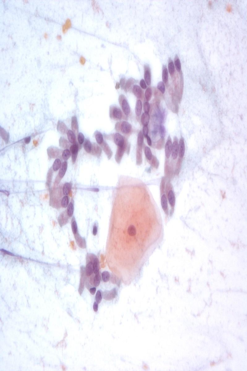
7 Esophagus Squamous Epithelium
8 Gastric Epithelium

9 Gastric Epithelium Large flat sheets Cytoplasm of foveolar cells is columnar, with mucin Nuclei round and evenly spaced, uniform in size

10 Gastric Epithelium Sometimes show small grooves, inclusions and small nucleoli Associated with mucin


11 Gastric Epithelium Stripped Nuclei Mucin and stripped nuclei Degenerative grooves Inclusions

12 Gastric Parietal Cells and Chief Cells Pitfall: May be mistaken for acinar cells or hepatocytes


13 Gastric Foveolar Epithelium Cup shaped mucin

14 Duodenum Typically large sheets Columnar enterocytes with a microvillus brush border with interspersed goblet cells

15 Duodenal Epithelium Nuclei round and uniform Background mucin is typically thin,
16 Enterocytes have a microvillous brush border
17 Primary Neoplasms Of The Pancreas Ductal phenotype (ductal neoplasms) Ductal Adenocarcinoma and variants Cystic neoplasms Serous cystic neoplasms (centroacinar cells) Mucinous cystic neoplasms Intraductal papillary mucinous neoplasms Acinar cell phenotype Acinar cell carcinomas Acinar cell cystadenoma Acinar cell cystadenocarcinoma Islet cell phenotype Pancreatic neuroendocrine tumors Unknown histogenesis Pancreatoblastoma Solid pseudo-papillary neoplasms
18 Solid Tumors Morphology Ductal Pattern Ductal adenocarcinoma Solid Cellular Pattern Neuroendocrine Tumor Acinar cell carcinoma Solid pseudopapillary tumor Pancreatoblastoma


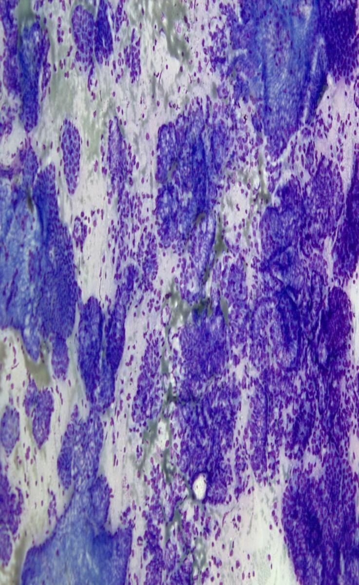
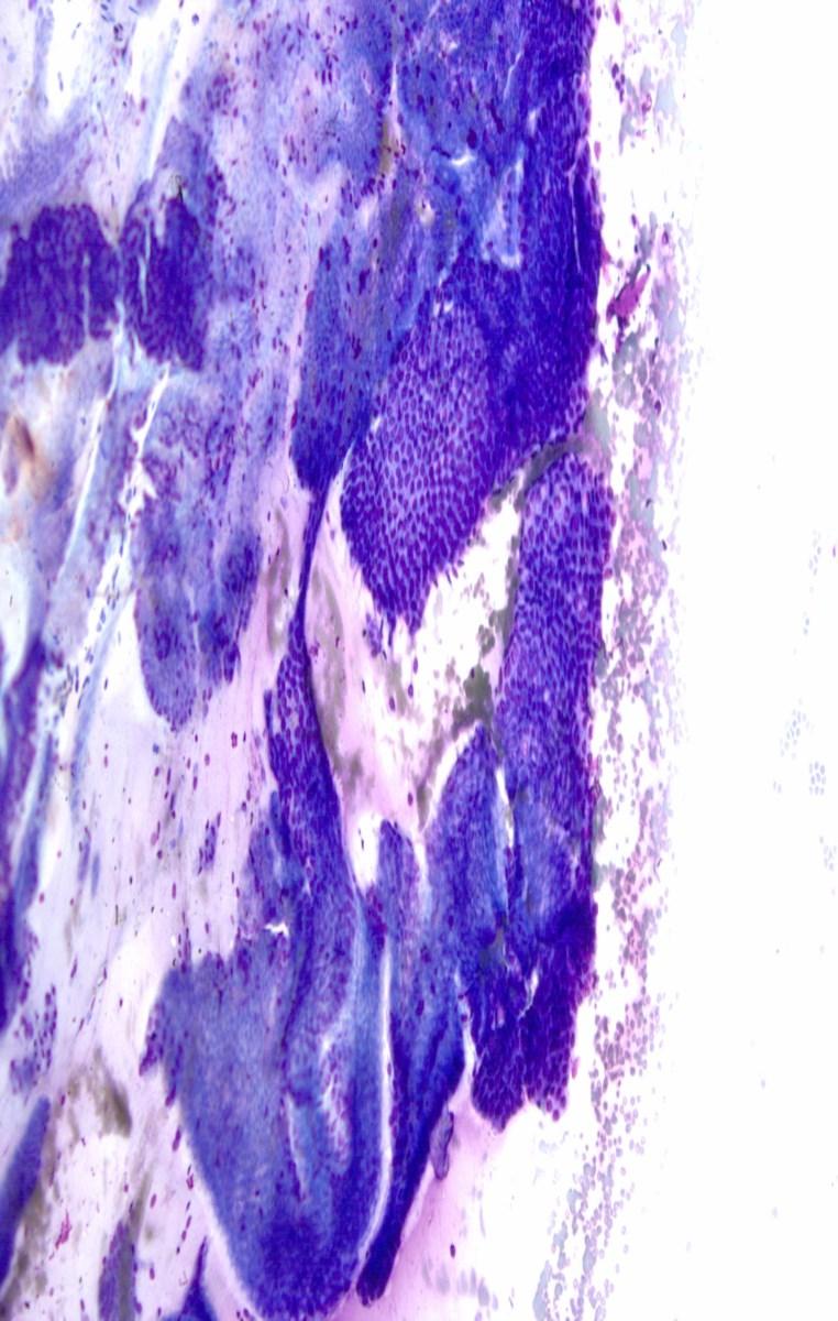
19 Ductal Pattern=Ductal Adenocarcinoma Ductal Pattern with Desmoplasia Ductal Pattern Low power view

 Solid Pseudopapillary Tumor (SPPN)")
20 Solid, Cellular Pattern Proliferation of neoplastic cells without stromal desmoplasia NonDuctal Neoplasms Pancreatic Neuroendocrine Tumor (PanNET) Acinar Cell Carcinoma (ACC) Solid Pseudopapillary Tumor (SPPN) Pancreatoblastoma (PB)
21 Diagnostic Cytopathology. April 2014 vol. 42 (4)
22 Management of Pancreatic Masses Benign Medical management, minimal intervention or observation Neoplastic Surgery or observation Premalignant Observation LGD, MGD Resection HGD/Carcinoma Malignant Surgery, neoadjuvant therapy, adjuvant therapy
23 Issues with Cytology Reporting of Pancreatobiliary Specimens Nonstandardized reporting lead to confusion for the clinicians treating the patient with a pancreatic mass or lesion Lack of epithelial cells used as criteria for nondiagnostic IPMN and MCN not uniformly handled, thick mucin still indicative of underlying neoplasm, even without neoplastic cells Pseudocysts will lack epithelial cells Serous cystadenoma may lack neoplastic cells Cases signed out as c/w cyst contents What cytological criteria should be used to interpret a lesion as IPMN or MCN? Atypical mucinous epithelium classified as atypical, rather than neoplastic, or as suspicious for carcinoma
24 Issues with Cytology Reporting of Pancreatobiliary Specimens cont. Was high grade dysplasia in IPMN atypical, suspicious or malignant? Serous cystadenoma classified as negative or atypical Were neuroendocrine tumor and solid pseudopapillary neoplasms suspicious, positive, or other?
25 Purpose of Standardization Unify reporting of disease categories among pathologists Reduce and improve inter and intraobserver variability Provide clinically relevant information for patient management To reflect the current understanding of the biology of disease entities
26 Categories I. Nondiagnostic II. Negative (for malignancy) III. Atypical IV. Neoplastic: benign or other V. Suspicious (for malignancy) VI. Positive (for malignancy) or malignant * only for laboratory systems where the information system requires it.
27 NONDIAGNOSTIC Category I
28 Nondiagnostic Definition: A non-diagnostic cytology specimen is one that provides no useful information or diagnostic information about the sampled mass. Discordant imaging and cytology findings Cyst fluids that yield insufficient material for ancillary studies Any cellular atypia precludes a non-diagnostic category Caveats: Nondiagnostic does not equal unsatisfactory Unsatisfactory indicates that a specimen cannot be evaluated, Acellular cyst fluid without other specific features is still representative of the cyst aspirated
29 Nondiagnostic Cytological Criteria Preparation or obscuring artifact Gastrointestinal contaminant Benign acinar and ductal epithelium derived from a solid or cystic mass lesion Acellular aspirates of a pancreatic mass or pancreatic brushing Acellular aspirate of a cyst without evidence of mucinous etiology Lack of thick, background mucin and/or oncotic cells Lack of elevated CEA Lack of KRAS or GNAS mutations
30 Incidental Cysts in the Pancreas Incidental cysts of the pancreas are common More likely to be neoplastic rather than benign 30% of all incidental masses were IPMN in one series 55% were MCN or IPMN (preinvasive precursors), only 4% were pseudocyst Pathologist is reviewing the cyst fluid to assess for the presence of pre-invasive precursors lesions Pseudocyst diagnosis of exclusion

31 Cyst Contents
32 NEGATIVE Category II
33 Negative Definition: Adequate cellular and/or extracellular material to evaluate and or define a nonneoplastic lesion identified on imaging Includes the presence of normal pancreatic parenchyma in the appropriate clinical setting Vague fullness of the pancreas No distinct pancreatic mass
34 Differential Diagnosis Benign pancreas Acute Pancreatitis Chronic Pancreatitis Autoimmune pancreatitis Splenule/Ectopic spleen/accessory spleen Pseudocyst Lymphoepithelial cyst
35 ATYPICAL Category III
36 Atypical Definition: Cells with cytoplasmic, nuclear, or architectural features not consistent with normal or reactive cellular components of the pancreas or bile ducts, and insufficient features to classify them as a neoplasm or suspicious for a high grade malignancy The findings do not explain the lesion identified on imaging Follow-up evaluation is warranted
37 Atypical Cytological Criteria Cytological specimen contains cellular or extracellular tissue that displays morphological features that cannot comfortably be classified as reactive or benign, but which are also insufficient to classify as definitively neoplastic or as suspicious for malignancy. Examples: Biliary brush specimens with mucinous epithelium and other atypical findings Atypical mucinous epithelium in a pancreatic aspirate PanIN Gastric conatminant IPMN Cellular component is suggestive of a PanNET or SPN but the sample is of insufficient quantity or quality for definitive diagnosis
38 NEOPLASTIC Category IV
39 Neoplastic: Benign Definition: The cytological specimen is sufficiently cellular and representative, with or without the context of clinical, imaging and ancillary studies to be diagnostic of a benign neoplasm. Neoplasms included in tis category: Serous cystadenoma Schwannoma Cystic teratoma
40 Neoplastic: Other Definition: Defines a neoplasm that is either premalignant, or a low grade malignant neoplasm. Neoplasms included in this category: Neoplasm that is preinvasive cancer IPMN or MCN with LGD, MGD, HGD IOPN Solid cellular neoplasm Pancreatic neuroendocrine tumor Solid pseudopapillary neoplasm Extra-adrenal paraganglioma Gastrointestinal Stromal tumor
41 Rationale Established to provide a category for neoplasms that were either not clearly benign, such as serous cystadenoma, nor clearly aggressive, and high grade in their behavior, such as ductal adenocarcinoma. Standardize cytological nomenclature and terminology to correlate with WHO 2010 classification and terminology. The words tumor and neoplasm connote a neoplasm, but not a malignancy Patients with neoplasms in this category may have the option of being managed conservatively PanNET may be observed IPMN with low risk features may be observed. The categories of atypical and suspicious connote an indeterminate interpretation. Does not define these a benign or malignant
42 Mucinous Cysts Nonneoplastic and neoplastic mucinous cysts Nonneoplastic mucinous cyst (mucinous duct lesion) Foregut cysts Nonneoplastic cysts lack mutations Cytological and cyst fluid features may overlap Mucin only Elevated CEA Lack KRAS and GNAS Mucinous cyst, not otherwise specified Characteristic mucin with oncotic cells, no epithelium for evaluation Mucinous cyst with low grade epithelial atypia (low grade and moderate grade dysplasia) (IPMN or MCN) Mucinous cyst with high grade epithelial atypia (high grade dysplasia and adenocarcinoma)
43 Criteria for Mucinous Cyst Fluid viscous, clear or white Cytology shows mucinous background as described, +/- neoplastic epithelium Ancillary studies CEA elevated Mutational analyses KRAS mutated in IPMN and MCN GNAS mutations in IPMN RNF43 mutations in IPMN and MCN
 Comment: No neoplastic epithelium is present for evaluation of dysplasia.")
44 Neoplastic: Other Imaging: Large, multiloculated cyst in the tail of the pancreas with evident connect to the pancreatic ductal system Report: Adequacy: Satisfactory Interpretation: Thick background mucin, histiocytes, and oncotic cells, consistent with IPMN Category: Neoplastic: Other (other) Comment: No neoplastic epithelium is present for evaluation of dysplasia. The cytological findings correlated with the imaging findings support the above interpretation.

45 Mucinous cyst, NOS Background
46 Management of IPMN 2012 Consensus Guidelines Consensus conference of the International Association of Pancreatology has proposed guidelines for the management of patients with pancreatic cysts suspected to be IPMNs These guidelines use a combination of clinical history, gender, imaging characteristics, cytology and cyst fluid analysis to determine therapeutic and surveillance strategies. Resection without tissue confirmation: Main duct IPMN > 10 mm Obstructive jaundice a/w cyst in HOP Enhancing solid component
47 Management of IPMN 2012 Consensus Guidelines Worrisome features Patient evaluated by EUS These include: Cyst >= 3 cm Thickened, enhancing cyst walls Main duct 5-9 mm Nonenhancing mural nodule Abrupt change in caliber of pancreatic duct with distal pancreatic atrophy EUS worrisome features Definite mural nodule Main duct features suspicious for involvement Cytology: suspicious or positive Strongly consider surgery if > 3 cm Consider surgery if 2-3 cm
48 What is suspicious or positive cytology? Cells that indicate the presence of high grade dysplasia or invasive carcinoma Role of cytology: Screening for high risk cytological features Terms: High grade dysplasia present or absent High grade epithelial atypia: incorporates high grade dysplasia and invasive carcinoma absence of high-grade atypia with the absence of high risk imaging features of dilated MPD and mural nodule gives a NPV of > 90%


49 Low-Grade Dysplasia Abundant columnar mucin containing cytoplasm Basally located nuclei Columnar cytoplasm with mucin Basally located nuclei


50 Moderate dysplasia Pseudostratification


51 High-Grade Dysplasia Papillary tufts, nuclei extend to luminal border Mitoses


 DOI: 10.1002/cncy.21344, wileyonlinelibrary.")
52 Original Article Cytological Criteria of High-Grade Epithelial Atypia in the Cyst Fluid of Pancreatic Intraductal Papillary Mucinous Neoplasms Martha B. Pitman, MD 1 ; Barbara A. Centeno, MD 2 ; Ebubekir S. Daglilar, MD 3 ; William R. Brugge, MD 3 ; and Mari Mino-Kenudson, MD 1 Cancer cytopathology.. Published online August 12, 2013 in Wiley Online Library (wileyonlinelibrary.com) DOI: /cncy.21344, wileyonlinelibrary.com Duodenum: 12 u LGEA: cells 12u 40 Cancer Cytopathology January 2014 HGEA: cells <12 u
")
53 High-grade epithelial atypia Includes high grade dysplasia and invasive carcinoma Criteria High N/C Nuclear membrane irregularities Abnormal chromatin Hypo- or hyperchromasia Background necrosis Cancer (Cancer Cytopathol) 2013;121:
54 SUSPICIOUS Category V
55 Suspicious Some, but not all of the criteria of a specific malignant neoplasm are present, mainly for pancreatic adenocarcinoma diagnosis. The features are qualitatively or quantitatively insufficient for a conclusive diagnosis. Confirmatory ancillary testing or substantial clinical and radiological findings must be present and discussed during a treatment planning conference, or similar correlation conference.
56 POSITIVE Category VI
57 Positive/Malignant Unequivocal display of malignant cytological change Diagnoses: Adenocarcinoma and its variant Neuroendocrine carcinoma, small and large cell type Pancreatoblastoma Acinar cell carcinoma Lymphoma Metastases Sarcomas in this region, secondarily involving the pancreas
58 Ancillary Testing Solid Masses Flow cytometry Work-up for probable lymphomas Immunohistochemistry Reactive Glands vs. Adenocarcinoma S100P, IMP3, mesothelin, DPC4, VHL Differential diagnosis of solid cellular neoplasms PanNET, SPN, ACC, PB» PanNET: CK +, neuroendocrine markers+» SPN: b-catenin +, vimentin+, loss of e-cadherin» ACC: chymotrypsin, trypsin, and bcl 10+ Metastases IHC panel depending on the differential diagnosis
59 Ancillary Testing Cystic Masses Biochemical Viscosity Elevated in MCN/IPMN/IOPN Ostwald viscometer, or finger pull CEA: >192ng/ml False positives from duplication cysts, mesothelial inclusion cysts, LECP, etc Amylase: >1000s for pseudocyst Elevated level does not mean it is a pseudocyst, falsely elevated in IPMN, MCN and other neoplasms
60 Ancillary Testing Cystic Masses Molecular (for pancreatic cysts) RAS GNAS RNF43 supports a neoplastic mucinous cyst, does not distinguish between IPMN or MCN Supports IPMN No correlation with grade Mucinous neoplastic etiology Does not distinguish between IPMN and MCN
61 Ancillary Testing Cystic Masses Molecular (for pancreatic cysts) 3P deletions 3p25, VHL gene, supports serous cystadenoma Other 3p deletions also found in serous cystadenoma CTNNB1 Mutations support solid pseudopapillary neoplasm TP53, CDKN2A, SMAD4/DPC4 Loss suports a high risk cyst High grade dysplasia or risk to progression
62
63 EUS-FNA OF THE UPPER GI TRACT Utilized for masses in the submucosal and deep layers of the GI tract 80-90% sensitivity Layers: Mucosal and muscularis mucosa NETs Submucosal lipoma
64 EUS-FNA OF THE UPPER GI TRACT Muscularis propria GIST, schwannoma, leiomyoma Serosa/adventitia Metastatic disease
Standardized Terminology in Pancreatobiliary Cytology: The Papanicolaou Society Guidelines
 Standardized Terminology in Pancreatobiliary Cytology: The Papanicolaou Society Guidelines Barbara Ann Centeno. M.D. Vice-Chair, Clinical Services, Anatomic Pathology Assistant Chief, Pathology Service
Pancreatico-biliary cytology: a practical approach to diagnosis. Corina Cotoi
 Pancreatico-biliary cytology: a practical approach to diagnosis Corina Cotoi Pancreatico-biliary lesions Solid: Ductal adenocarcinoma Cholangiocarcinoma Acinar cell carcinoma Neuroendocrine tumour / carcinoma
Pancreatitis: A Potential Pitfall in Endoscopic Ultrasound Guided Pancreatic FNA
 Pancreatitis: A Potential Pitfall in Endoscopic Ultrasound Guided Pancreatic FNA Jack Yang, MD Department of Pathology, Medical University of South Carolina Objectives Understand the indication of EUS
Patient History. A 58 year old man presents with a 16 mm cyst in the pancreatic tail. The cyst is unilocular with a thick wall and no mural nodule.
 Case 1 Martha Bishop Pitman, MD Director of Cytopathology Massachusetts General Hospital Associate Professor of Pathology Harvard Medical School Boston, MA Patient History A 58 year old man presents with
Case 1. Case 1: EUS Report 5/1/2017. Interesting Cases of Pancreatic Masses
 Interesting Cases of Pancreatic Masses Martha Bishop Pitman, MD Professor of Pathology Harvard Medical School Director of Cytopathology Massachusetts General Hospital Boston, MA MASSACHUSETTS GENERAL PHYSICIANS
Evaluation and Management of Cystic Lesions of the Pancreas: When to Resect, When to Follow and When to Forget
 Evaluation and Management of Cystic Lesions of the Pancreas: When to Resect, When to Follow and When to Forget Randall Brand, MD Professor of Medicine Division of Gastroenterology, Hepatology and Nutrition
Standardization of Nomenclature
 Standardized Terminology and Nomenclature for Pancreaticobiliary Cytopathology from the Papanicolaou Society of Cytopathology Lester J. Layfield, M.D. Professor and Chair University of Missouri Pathology
Pancreatic Cystic Lesions 원자력병원
 Pancreatic Cystic Lesions 원자력병원 박선 후 Lines of cellular differentiation Ductal Acinar Undetermined Ductal adenocarcinoma Serous/ mucinous tumor Intraductal papillary mucinous neoplasm Acinar cell carcinoma
Outline 11/2/2017. Pancreatic EUS-FNA general aspects. Cytomorphologic features of solid neoplasms/lesions of the pancreas
 ENDOSCOPIC ULTRASOUND GUIDED-FINE NEEDLE ASPIRATION CYTOLOGY OF PANCREAS Khalid Amin M.D. Assistant Professor Department of Laboratory Medicine and Pathology University of Minnesota Outline Pancreatic
Select problems in cystic pancreatic lesions
 Disclosure Select problems in cystic pancreatic lesions Five Prime Therapeutics shareholder Adicet Bio shareholder Bristol-Meyer Squibb advisory board grace.kim@ucsf.edu Pancreatic cystic lesions Intraductal
40th European Congress of Cytology Liverpool, UK, 2-5 th October 2016
 40th European Congress of Cytology Liverpool, UK, 2-5 th October 2016 EUS FNA of abdominal organs: An approach to reporting and triage for ancillary testing Date and time: Sunday 2 nd October 2016 15.00-16.30
Conflicts of Interest
 Standardized Terminology and Nomenclature for Pancreaticobiliary Cytopathology from the Papanicolaou Society of Cytopathology Lester J. Layfield, M.D. Professor and Chair University of Missouri Pathology
An Approach to Pancreatic Cysts. Introduction
 An Approach to Pancreatic Cysts Nalini M. Guda, MD Aurora St. Luke s Medical Center, Milwaukee Clinical Adjunct Professor of Medicine, University of Wisconsin School of Medicine and Public Health Introduction
Video Microscopy Tutorial 19
 Video Microscopy Tutorial 19 EUS FNA of Pancreatic Cysts Martha Pitman, MD There are no disclosures necessary. EUS-FNA of Pancreatic Cysts Martha Bishop Pitman, M.D. Massachusetts General Hospital Harvard
Management A Guideline Based Approach to the Incidental Pancreatic Cysts. Common Cystic Pancreatic Neoplasms.
 Management 2016 A Guideline Based Approach to the Incidental Pancreatic Cysts ISMRM 2016 Masoom Haider, MD, FRCP(C) Professor of Radiology, University of Toronto Clinician Scientist, Ontario Institute
Advances in Pancreatic. Cytology. Martha B. Pitman, MD. Disclosure of Relevant Financial Relationships
 Advances in Pancreatic Cytology Martha B. Pitman, MD Disclosure of Relevant Financial Relationships USCAP requires that all planners (Education Committee) in a position to influence or control the content
Pancreatic Cytopathology: A pragmatic approach. By Dr Miguel Perez-Machado MD. PhD. MRCPath Royal Free Hospital
 Pancreatic Cytopathology: A pragmatic approach. By Dr Miguel Perez-Machado MD. PhD. MRCPath Royal Free Hospital Indications of Cytology Sampling To document malignancy in patients with malignant appearing
Objectives. Pancreatic Cysts. Benefits and Limitations of the Cytologic Assessment of Cystic Pancreatic Lesions and Masses
 Benefits and Limitations of the Cytologic Assessment of Cystic Pancreatic Lesions and Masses Michelle D. Reid, MD, MSc Associate 1/24/15 Professor Director of Cytology - EUHM Michelle D Reid MD, MS Emory
Appendix 4: WHO Classification of Tumours of the pancreas 17
 S3.01 The WHO histological tumour type must be recorded. CS3.01a The histological type of the tumour should be recorded based on the current WHO classification 17 (refer to Appendices 4-7). Appendix 4:
Biliary Tract Neoplasia: A Cyto-histologic Review. Michelle Reid, MD, MSc Professor of Pathology Director of Cytopathology Emory University Hospital
 Biliary Tract Neoplasia: A Cyto-histologic Review Michelle Reid, MD, MSc Professor of Pathology Director of Cytopathology Emory University Hospital Bile Duct Brushings (BDB) BDBs are the initial diagnostic
The role of endoscopy in the diagnosis and treatment of cystic pancreatic neoplasms
 The role of endoscopy in the diagnosis and treatment of cystic pancreatic neoplasms CYSTIC LESIONS AND FLUID COLLECTIONS OF THE PANCREAS Their pathology ranges from pseudocysts and pancreatic necrosis
Pancreatic Cysts. Darius C. Desai, MD FACS St. Luke s University Health Network
 Pancreatic Cysts Darius C. Desai, MD FACS St. Luke s University Health Network None Disclosures Incidence Widespread use of cross sectional imaging Seen in over 2% of patients having abdominal imaging
Suspicious Cytologic Diagnostic Category in Endoscopic Ultrasound-Guided FNA of the Pancreas: Follow-Up and Outcomes
 Suspicious Cytologic Diagnostic Category in Endoscopic Ultrasound-Guided FNA of the Pancreas: Follow-Up and Outcomes Evan A. Alston, MD 1 ; Sejong Bae, PhD 2 ; and Isam A. Eltoum, MD, MBA 1 BACKGROUND:
Neoplasias Quisticas del Páncreas
 SEAP -Aproximación Práctica a la Patología Gastrointestinal- Madrid, 26 de mayo, 2006 Neoplasias Quisticas del Páncreas Gregory Y. Lauwers, M.D. Director, Service Massachusetts General Hospital Harvard
DIAGNOSTIC CHALLENGES Pancreas FNAB. Dr. M. Weir Oct 2017
 DIAGNOSTIC CHALLENGES Pancreas FNAB Dr. M. Weir Oct 2017 CONFLICT OF INTEREST DISCLOSURE I have not had in the past 3 years, a financial interest, arrangement or affiliation with one or more organizations
ROSE in EUS guided FNA of Pancreatic Lesions
 ROSE in EUS guided FNA of Pancreatic Lesions Guy s Hospital, London, 16 April 2018 Laxmi Batav Imperial College NHS Trust Imperial College NHS Trust Cytology Workload Cervical Cytology 57,500 (decreases
Evaluation of AGA and Fukuoka Guidelines for EUS and surgical resection of incidental pancreatic cysts
 Evaluation of AGA and Fukuoka Guidelines for EUS and surgical resection of incidental pancreatic cysts Authors Alexander Lee 1, Vivek Kadiyala 2,LindaS.Lee 3 Institutions 1 Texas Digestive Disease Consultants,
Pancreatic Cytopathology: The Solid Neoplasms
 Pancreatic Cytopathology: The Solid Neoplasms Syed Z. Ali, M.D. Professor of Pathology and Radiology Director of Cytopathology The Johns Hopkins Hospital Baltimore, Maryland Pancreatic Cytopathology: Past,
ACG Clinical Guideline: Diagnosis and Management of Pancreatic Cysts
 ACG Clinical Guideline: Diagnosis and Management of Pancreatic Cysts Grace H. Elta, MD, FACG 1, Brintha K. Enestvedt, MD, MBA 2, Bryan G. Sauer, MD, MSc, FACG (GRADE Methodologist) 3 and Anne Marie Lennon,
Outline. Intraductal Papillary Mucinous Neoplasm (IPMN) Guideline Review 4/6/2017. Case Example Background Classification Histology Guidelines
 Guideline Review 4/6/2017. Case Example Background Classification Histology Guidelines") Intraductal Papillary Mucinous Neoplasm (IPMN) Guideline Review The Nurse Practitioner Association New York State Capital Region Teaching Day Matthew Warndorf MD Case Example Background Classification
Intraductal Papillary Mucinous Neoplasm (IPMN) Guideline Review The Nurse Practitioner Association New York State Capital Region Teaching Day Matthew Warndorf MD Case Example Background Classification
Pancreatic Cystic Neoplasms: Guidelines and beyond
 Pancreatic Cystic Neoplasms: Guidelines and beyond Kenneth J. Chang, MD, FACG, FASGE Executive Director, Comprehensive Digestive Disease Center Professor and Chief, Gastroenterology Vincent & Anna Kong
Endoscopic Ultrasound-guided FNA Cytology of the Pancreas
 Endoscopic Ultrasound-guided FNA Cytology of the Pancreas 67 th Annual California Society of Pathologists 2014 Laura Tabatabai, MD Professor of Pathology University of California, San Francisco Overview
Biliary tract tumors
 Short Course 2010 Annual Fall Meeting of the Korean Society for Pathologists Biliary tract tumors Joon Hyuk Choi, M.D., Ph.D. Professor, Department of Pathology, Yeungnam Univ. College of Medicine, Daegu,
Matthew McCollough, M.D. April 9, 2009 University of Louisville
 Matthew McCollough, M.D. April 9, 2009 University of Louisville List the differential diagnosis for pancreatic cysts Review the epidemiology Illustrate the types of cysts through case discussions Discuss
EUS-guided FNAB. Differential Diagnosis 5/1/2017. EUS-FNA of Solid and Cystic Lesions:
 EUS-FNA of Solid and Cystic Lesions: Part 1: Solid Masses Martha Bishop Pitman, M.D. Director, Cytopathology Massachusetts General Hospital Professor of Pathology Harvard Medical School Boston, MA EUS-guided
Pancreatic Cysts. Pancreatic Cysts. Multidisciplinary and Multimodal Approach To the Pre-Operative Diagnosis of Pancreatic Cysts
 Multidisciplinary and Multimodal Approach To the Pre-Operative Diagnosis of Pancreatic Cysts Martha Bishop Pitman, M.D. Director of Cytopathology Massachusetts General Hospital Professor of Pathology Harvard
ENDOSCOPIC ULTRA SOUND GUIDED FNA OF GI TRACT AND PANCREAS
 ENDOSCOPIC ULTRA SOUND GUIDED FNA OF GI TRACT AND PANCREAS Prof. Fernando Schmitt Medical Faculty of Porto University, Porto, Portugal IPATIMUP General Secretary of the International Academy of Cytology
Salivary Gland Cytology: A Clinical Approach to Diagnosis and Management of Atypical and Suspicious Lesions
 Salivary Gland Cytology: A Clinical Approach to Diagnosis and Management of Atypical and Suspicious Lesions W.C. Faquin, M.D., Ph.D. Massachusetts General Hospital Harvard Medical School, USA Marc Pusztaszeri,
Team approach is essential incorporating: radiology, gastroenterology, surgery and pathology Successful performance is operator dependent
 Committee V Post Brushing and FNA Testing Managment Options Daniel Kurtycz, M.D. Chair Committee V Committee V - Post brushing and FNA management options Team approach is essential incorporating: radiology,
Objectives. Salivary Gland FNA: The Milan System. Role of Salivary Gland FNA 04/26/2018
 Salivary Gland FNA: The Milan System Dr. Jennifer Brainard Section Head Cytopathology Cleveland Clinic Objectives Introduce the Milan System for reporting salivary gland cytopathology Define cytologic
The Pancreas. Basic Anatomy. Endocrine pancreas. Exocrine pancreas. Pancreas vasculature. Islets of Langerhans. Acinar cells Ductal System
 SGNA: Back to Basics Rogelio G. Silva, MD Assistant Clinical Professor of Medicine University of Illinois at Chicago Department of Medicine Division of Gastroenterology Advocate Christ Medical Center GI
Guidelines for Pancreaticobiliary Cytology from the Papanicolaou Society of Cytopathology: A Review
 Guidelines for Pancreaticobiliary Cytology from the Papanicolaou Society of Cytopathology: A Review Martha B. Pitman, MD 1 and Lester J. Layfield, MD 2 The newest installment on state-of-the-art standards
EUS-FNA OF PANCREATIC EXOCRINE TUMORS COMPARISON OF EXPERIENCES WITH PATHOLOGICAL DIAGNOSIS
 EUS-FNA OF PANCREATIC EXOCRINE TUMORS COMPARISON OF EXPERIENCES WITH PATHOLOGICAL DIAGNOSIS Vincenzo Canzonieri, M.D. CRO - Aviano National Cancer Institute Dept of Pathology EUS European Cyto-Pathologist
Pancreatobiliary Frozen Section Nightmares
 Pancreatobiliary Frozen Section Nightmares Aatur D. Singhi, MD PhD Assistant Professor University of Pittsburgh Medical Center Department of Pathology singhiad@upmc.edu Objectives Briefly give an overview
The Role of Molecular Analysis in the Diagnosis and Surveillance of Pancreatic Cystic Neoplasms
 JOP. J Pancreas (Online) 20 Mar 20; (2):-9. RESEARCH ARTICLE The Role of Molecular Analysis in the Diagnosis and Surveillance of Pancreatic Cystic Neoplasms Megan Winner, Amrita Sethi 2, John M Poneros
State of the Art Symposium
 State of the Art Symposium Syed Z. Ali, MD There is no disclosure necessary Martha B. Pitman, MD There is no disclosure necessary Lester Layfield, MD There is no disclosure necessary Joseph Herman, MD
Oncocytic-Appearing Salivary Gland Tumors. Oncocytic, Cystic, Mucinous, and High Grade Salivary Gland Tumors SALIVARY GLAND FNA: PART II
 William C. Faquin, MD, PhD Professor of Pathology Harvard Medical School Director of Head and Neck Pathology Massachusetts Eye and Ear Massachusetts General Hospital SALIVARY GLAND FNA: PART II Oncocytic,
New Diagnoses Need New Approaches: A Glimpse into the Near Future of Gynecologic Pathology
 New Diagnoses Need New Approaches: A Glimpse into the Near Future of Gynecologic Pathology United States and Canadian Academy of Pathology 102 nd Annual Meeting Baltimore, Maryland Christina S. Kong, M.D.
5/21/2018. An Update on Pancreas Neoplasms. Arief Suriawinata, M.D. Lines of Differentiation in Pancreatic Neoplasms
 An Update on Pancreas Neoplasms Arief Suriawinata, M.D. Professor of Pathology and Laboratory Medicine Geisel School of Medicine at Dartmouth Department of Pathology and Laboratory Medicine Dartmouth-Hitchcock
Pancreatic intraepithelial
 Pancreatic intraepithelial neoplasia (PanIN) Markéta Hermanová St. Anne s University Hospital Brno Faculty of Medicine, Masaryk University Precursor lesions of invasive pancreatic cancer Pancreatic intraepithelial
EUS-FNAB of GI Tract: Specimen Handling and Triage
 EUS-FNAB of GI Tract: Specimen Handling and Triage Amy Ly, M.D. Director, Fine Needle Aspiration Service Massachusetts General Hospital Assistant Professor of Pathology Harvard Medical School Boston, MA
We herein describe new concepts in the diagnosis of the
 The Evolving Role of Pathology in New Developments, Classification, Terminology, and Diagnosis of Pancreatobiliary Neoplasms Michelle D. Reid, MD; Melinda M. Lewis, MD; Field F. Willingham, MD, MPH; N.
Update on Thyroid FNA The Bethesda System. Shikha Bose M.D. Associate Professor Cedars Sinai Medical Center
 Update on Thyroid FNA The Bethesda System Shikha Bose M.D. Associate Professor Cedars Sinai Medical Center Thyroid Nodules Frequent occurrence Palpable: 4-7% of adults Ultrasound: 10-31% Majority benign
Anatomy of the biliary tract
 Harvard-MIT Division of Health Sciences and Technology HST.121: Gastroenterology, Fall 2005 Instructors: Dr. Jonathan Glickman Anatomy of the biliary tract Figure removed due to copyright reasons. Biliary
Kenneth D. Chi, MD Medical Director GI Lab Advocate Lutheran General Hospital
 Kenneth D. Chi, MD Medical Director GI Lab Advocate Lutheran General Hospital Advances in Digestive Health for the Primary Care Physician Symposium May 2, 2015 None Case Presentation Types of Pancreatic
Cystic pancreatic lesions A proposal for a network approach. Chris Briggs Consultant HPB Surgeon Peninsula HPB Unit Derriford Hospital, Plymouth
 Cystic pancreatic lesions A proposal for a network approach Chris Briggs Consultant HPB Surgeon Peninsula HPB Unit Derriford Hospital, Plymouth Aims Brief overview of cystic pancreatic lesions International
Atypia in Diagnostic Cytopathology: Strategies to Reduce Overuse A CQI initiative
 in Diagnostic Cytopathology: Strategies to Reduce Overuse A CQI initiative Dr. Mariamma Joseph and Susan McRae Objectives Review cytology literature on standardization of as a diagnostic term Address the
"Atypical": Criteria and
 "Atypical": Criteria and Controversies Esther Rossi MD PhD MIAC Division of Anatomic Pathology and Cytology Catholic University of Sacred Heart Rome, Italy CASE HISTORY In 2015, 45 y/o woman underwent
Predictors of Malignancy in Thyroid Fine-Needle Aspirates Cyst Fluid Only Cases
 Predictors of Malignancy in Thyroid Fine-Needle Aspirates Cyst Fluid Only Cases Can Potential Clues of Malignancy Be Identified? Mohammad Jaragh, MD 1 ; V. Bessie Carydis, MMedSci (Cytol) 1 ; Christina
ROSE (Rapid on site evaluation) in EUS-FNA in solid and cystic pancreatic lesions: possibilities and limits
 in EUS-FNA in solid and cystic pancreatic lesions: possibilities and limits") ROSE (Rapid on site evaluation) in EUS-FNA in solid and cystic pancreatic lesions: possibilities and limits Charitini Salla, MD Head of Cytopathology Department, Hygeia Hospital, Athens, Greece There is
ROSE (Rapid on site evaluation) in EUS-FNA in solid and cystic pancreatic lesions: possibilities and limits Charitini Salla, MD Head of Cytopathology Department, Hygeia Hospital, Athens, Greece There is
Intraductal Papillary Mucinous Neoplasms: We Still Have a Way to Go! Francesco M. Serafini, MD, FACS
 Intraductal Papillary Mucinous Neoplasms: We Still Have a Way to Go! Francesco M. Serafini, MD, FACS Brooklyn VAMC September 21 st GI Grand Rounds - What is it? - Clinical entity that has emerged from
Thyroid follicular neoplasms in cytology. Ulrika Klopčič Institute of Oncology, Department of Cytopathology, Ljubljana, Slovenia
 Thyroid follicular neoplasms in cytology Ulrika Klopčič Institute of Oncology, Department of Cytopathology, Ljubljana, Slovenia Lecture overview importance of FNAB in assessing thyroid lesions follicular
Biliary cytolgy and pancreatic endoscopic ultrasound-guided FNA. Leena Krogerus Helsinki, FINLAND
 Biliary cytolgy and pancreatic endoscopic ultrasound-guided FNA Leena Krogerus Helsinki, FINLAND Reasons for biliary cytology PSC- is a pre-neoplastc condition in youg individulas, the cure of which is
5/2/2018. Low Grade Dysplasia of GI Tract. High Grade Dysplasia of GI Tract. Dysplasia in Gastrointestinal Tract: Practical Pearls and Issues
 Dysplasia in Gastrointestinal Tract: Practical Pearls and Issues Arief Suriawinata, M.D. Professor of Pathology and Laboratory Medicine Geisel School of Medicine at Dartmouth Department of Pathology and
Chronic pancreatitis mimicking intraductal papillary mucinous neoplasm of the pancreas; Report of tow cases
 Jichi Medical University Journal Chronic pancreatitis mimicking intraductal papillary mucinous neoplasm of the pancreas; Report of tow cases Noritoshi Mizuta, Hiroshi Noda, Nao Kakizawa, Nobuyuki Toyama,
ASSESSMENT QUESTIONS: THE BASICS
 ASSESSMENT QUESTIONS: THE BASICS Diane Davis Davey, MD Univ. of Central Florida Chair, Continuing Education Oversight Committee Disclosures I am President of the American Board of Pathology for 2012. None
EUS-guided FNAB. Differential Diagnosis 3/14/2018. EUS-FNA of Solid and Cystic Lesions: Part 1: Solid Masses
 EUS-FNA of Solid and Cystic Lesions: Part 1: Solid Masses Martha Bishop Pitman, M.D. Director, Cytopathology Massachusetts General Hospital Professor of Pathology Harvard Medical School Boston, MA EUS-guided
Salivary Gland Cytology
 Salivary Gland Cytology Diagnostic challenges and potential pitfalls Tarik M. Elsheikh, MD Professor and Medical Director Anatomic Pathology Cleveland Clinic FNA Salivary Gland Lesions Indications Distinguish
The Pathologist s Role in the Diagnosis and Management of Neoplasia in Barrett s Oesophagus Cian Muldoon, St. James s Hospital, Dublin
 The Pathologist s Role in the Diagnosis and Management of Neoplasia in Barrett s Oesophagus Cian Muldoon, St. James s Hospital, Dublin 24.06.15 Norman Barrett Smiles [A brief digression - Chair becoming
FNA of Thyroid. Toward a Uniform Terminology With Management Guidelines. NCI NCI Thyroid FNA State of the Science Conference
 FNA of Thyroid NCI NCI Thyroid FNA State of the Science Conference Toward a Uniform Terminology With Management Guidelines Thyroid Thyroid FNA Cytomorphology NCI Thyroid FNA State of the Science Conference
Papillary Lesions of the Breast A Practical Approach to Diagnosis. (Arch Pathol Lab Med. 2016;140: ; doi: /arpa.
 Papillary Lesions of the Breast A Practical Approach to Diagnosis (Arch Pathol Lab Med. 2016;140:1052 1059; doi: 10.5858/arpa.2016-0219-RA) Papillary lesions of the breast Span the spectrum of benign,
Objectives. Atypical Glandular Cells. Atypical Endocervical Cells. Reactive Endocervical Cells
 2013 California Society of Pathologists 66 th Annual Meeting San Francisco, CA Atypical Glandular Cells to Early Invasive Adenocarcinoma: Cervical Cytology and Histology Christina S. Kong, MD Associate
CT 101 :Pancreas and Spleen
 CT 101 :Pancreas and Spleen Shikha Khullar,, MD, MPH Division of Radiology University of South Alabama The Pancreas Normal Pancreas 3 Phase Pancreatic CT Non contrast Arterial phase : 30-35 35 second
Workshop 9. Cytopathology of Solid and Cystic Neoplasms of the Pancreas: Everything You Need To Know For Daily Practice.
 Workshop 9 Cytopathology of Solid and Cystic Neoplasms of the Pancreas: Everything You Need To Know For Daily Practice Michelle Reid, MD Momin Siddiqui, MD,FIAC There are no disclosures necessary. Cytopathology
Diagnostic performance of endoscopic ultrasound-guided fine-needle aspiration in pancreatic lesions
 European Review for Medical and Pharmacological Sciences 2018; 22: 1397-1401 Diagnostic performance of endoscopic in pancreatic lesions Q.-M. WU 1, Y.-N. GUO 1, Y.-Q. XU 1 Digestive Department of Beijing
X-ray Corner. Imaging of The Pancreas. Pantongrag-Brown L
 X-ray Corner 125 Imaging of The Pancreas Modern imaging modalities commonly used in pancreas include ultrasound (US), CT, and MRI. Pancreas is a retroperitoneal organ which makes it difficult to visualize
Mody. AIS vs. Invasive Adenocarcinoma of the Cervix
 Common Problems in Gynecologic Pathology Michael T. Deavers, M.D. Houston Methodist Hospital, Houston, Texas Common Problems in Gynecologic Pathology Adenocarcinoma in-situ (AIS) of the Cervix vs. Invasive
INTRODUCTION TO PATHOLOGICAL TECHNIQUES. 1. Types of routine biopsy procedures 2. Special exams (IHC, FISH)
") INTRODUCTION TO PATHOLOGICAL TECHNIQUES 1. Types of routine biopsy procedures 2. Special exams (IHC, FISH) Biopsy-Indications Diffuse/multifocal lesions (neoplastic, inflammatory, etc) Etiology of the
INTRODUCTION TO PATHOLOGICAL TECHNIQUES 1. Types of routine biopsy procedures 2. Special exams (IHC, FISH) Biopsy-Indications Diffuse/multifocal lesions (neoplastic, inflammatory, etc) Etiology of the
Medical Policy An independent licensee of the Blue Cross Blue Shield Association
 PathFinderTG Molecular Testing Page 1 of 20 Medical Policy An independent licensee of the Blue Cross Blue Shield Association Title: PathFinderTG Molecular Testing Professional Institutional Original Effective
Biliary cytolgy and pancreatic endoscopic ultrasound-guided. Helsinki, FINLAND
 Biliary cytolgy and pancreatic endoscopic ultrasound-guided d FNA Leena Krogerus Helsinki, FINLAND Reasons for biliary cytology Obstructions PSC, a premalignant condition, the cure of which is transplantation
Index 179. Genital tract contaminants, 17, 20, 22, 150 papilloma virus-infected cells, 47 squamous cells, sources of, 7
 Index Accuracy of urinary cytology, 166 Acute inflammatory cells, 38 catheter sample, 39 herpes simplex infections, 44 carcinomas, 104, 105 non-viral inclusions, 52, 53 voided urine, 17 Adenocarcinoma
Molecular Testing for the Management of Pancreatic Cysts or Barrett Esophagus
 Molecular Testing for the Management of Pancreatic Cysts or Barrett Esophagus Policy Number: 2.04.52 Last Review: 8/2018 Origination: 5/2008 Next Review: 8/2019 Policy Blue Cross and Blue Shield of Kansas
PANCREATIC CYTOPATHOLOGY: PRACTICAL POINTS TO AVOID COMMON PITFALLS
 PANCREATIC CYTOPATHOLOGY: PRACTICAL POINTS TO AVOID COMMON PITFALLS Barbara A. Centeno, M.D. Senior Member and Director of Cytopathology/H. Lee Moffitt Cancer Center and Research Institute Professor/ Departments
TBSRTC 1- Probabilistic approach and Relationship to Clinical Algorithms
 The Benefits of a Uniform Reporting System for Thyroid Cytopathology BETHESDA REPORTING SYSTEM Prof. Fernando Schmitt Department of Pathology and Oncology, Medical Faculty of Porto University Head of Molecular
Dr Claire Smith, Consultant Radiologist St James University Hospital Leeds
 Dr Claire Smith, Consultant Radiologist St James University Hospital Leeds Imaging in jaundice and 2ww pathway Image protocol Staging Limitations Pancreatic cancer 1.2.4 Refer people using a suspected
Intro to Gallbladder & Pancreas Pathology
 Cholecystitis acute chronic Gallbladder tumors Adenomyoma (benign) Adenocarcinoma Pancreatitis acute chronic Pancreatic tumors Intro to Gallbladder & Pancreas Pathology Helen Remotti M.D. Gallstones (Cholelithiasis)
Salivary Glands 3/7/2017
 Salivary Glands 3/7/2017 Goals and objectives Focus on the entities unique to H&N Common board type facts Information for your future practice Salivary Glands Salivary Glands Major gland. Paratid. Submandibular.
Hepatobiliary and Pancreatic Malignancies
 Hepatobiliary and Pancreatic Malignancies Gareth Eeson MD MSc FRCSC Surgical Oncologist and General Surgeon Kelowna General Hospital Interior Health Consultant, Surgical Oncology BC Cancer Agency Centre
Cytopathology Study Day 16 April RCPath - BAC. Digital cytology: EUS FNA pancreas and head and neck
 Cytopathology Study Day 16 April 2017 Guy s Hospital London RCPath - BAC Digital cytology: EUS FNA pancreas and head and neck R. Dina MD, FIAC, FRCPath Consultant Cyto/Histopathologist Hon Sen Lecturer
Thyroid Nodules: Understanding FNA Cytology (The Bethesda System for Reporting of Thyroid Cytopathology) Shamlal Mangray, MB, BS
 Shamlal Mangray, MB, BS") Thyroid Nodules: Understanding FNA Cytology (The Bethesda System for Reporting of Thyroid Cytopathology) Shamlal Mangray, MB, BS Attending Pathologist Rhode Island Hospital, Providence, RI DISCLOSURE:
Thyroid Nodules: Understanding FNA Cytology (The Bethesda System for Reporting of Thyroid Cytopathology) Shamlal Mangray, MB, BS Attending Pathologist Rhode Island Hospital, Providence, RI DISCLOSURE:
Cystic Pancreatic Lesions: Approach to Diagnosis
 Cystic Pancreatic Lesions: Approach to Diagnosis Poster No.: R-0130 Congress: RANZCR-AOCR 2012 Type: Educational Exhibit Authors: A. AGARWAL, R. M. Mendelson; Perth/AU Keywords: Cysts, Biopsy, Endoscopy,
Gynecologic Cytopathology: Glandular lesions
 Gynecologic Cytopathology: Glandular lesions Lin Wai Fung (MSc, MPH, CMIAC) 17/4/2014 Glandular lesions of the uterus Endocervix Endometrium Normal endocervical cells Sheets, strips well-preserved architecture:
Enterprise Interest None
 Enterprise Interest None Risk stratification of salivary gland lesions on cytology based on the proposed Milan System for reporting salivary gland cytopathology: A pilot study Kartik Viswanathan, M.D.,
CHRONIC PANCREATITIS OR DUCTAL ADENOCARCINOMA? N. Volkan Adsay, \ MD
 CHRONIC PANCREATITIS OR DUCTAL ADENOCARCINOMA? N. Volkan Adsay, \ MD Case for discussion 67 y/o male Back pain and weight loss CT: 4.5 cm ill-defined, solid lesion in the head FNA/Core bx: Inconclusive
Cytyc Corporation - Case Presentation Archive - March 2002
 FirstCyte Ductal Lavage History: 68 Year Old Female Gail Index: Unknown Clinical History: Negative Mammogram in 1995 6 yrs. later presents with bloody nipple discharge Subsequent suspicious mammogram Suspicious
Subepithelial Lesions of the Gut: When Should I Worry?
 Subepithelial Lesions of the Gut: When Should I Worry? President, ASGE Chairman, GI & Hepatology Scottsdale, AZ Faigel.douglas@mayo.edu Case 55 yo male with reflux EGD for Barrett s Screening SET, mucosal
Background to the Thyroid Nodule
 William C. Faquin, MD, PhD Professor of Pathology Harvard Medical School Director of Head and Neck Pathology Massachusetts Eye and Ear Massachusetts General Hospital THYROID FNA: PART I Background to the
3/28/2017. Disclosure of Relevant Financial Relationships. GU Evening Subspecialty Case Conference. Differential Diagnosis:
 GU Evening Subspecialty Case Conference Rajal B. Shah, M.D. VP, Medical Director, Urologic Pathology Miraca Life Sciences, Irving, Texas Clinical Associate Professor of Pathology Baylor College of Medicine,
Radiology Pathology Conference
 Radiology Pathology Conference Nadia F. Yusaf, M.D. PGY-3 1/29/2010 Presentation material is for education purposes only. All rights reserved. 2010 URMC Radiology Page 1 of 90 Case 1 60 year- old man presents
04/10/2018. Intraductal Papillary Neoplasms Of Breast INTRADUCTAL PAPILLOMA
 Intraductal Papillary Neoplasms Of Breast Savitri Krishnamurthy MD Professor of Pathology Deputy Division Head The University of Texas MD Anderson Cancer Center 25 th Annual Seminar in Pathology Pittsburgh,