Testis Elastography. Paul S. Sidhu. Professor of Imaging Sciences. King s College Hospital London
|
|
|
- Eileen Quinn
- 5 years ago
- Views:
Transcription


1 Testis Elastography Paul S. Sidhu Professor of Imaging Sciences King s College Hospital London


2 Testicular Elastography Strain elastography Point Shear wave elastography 2D Shear wave elastography Multiparametric Ultrasound (MPUS) imaging Bamber J, Cosgrove D, Dietrich CF, Fromageau J, Bojunga J, Calliada F, et al. EFSUMB guidelines and recommendations on the clinical use of ultrasound elastography. Part 1: Basic principles and technology. Ultraschall Med. 2013;34(2):


3 Testicular Elastography Elastography Direct measure of the stiffness of a lesion Malignant lesions are harder than benign lesions Adjunct to conventional US EFSUMB GUIDELINES ELASTOGRAPHY NON-LIVER APPLICATIONS 2018 GET-TOGETHER MEETING IN FRANKFURT ON 17 TH FEBRUARY STEERING COMMITTEE ADRIAN SĂFTOIU ODD HELGE GILJA PAUL SIDHU CHRISTOPH F. DIETRICH VITO CANTISANI
4 Testicular Elastography Elastography Direct measure of the stiffness of a lesion Malignant lesions are harder than benign lesions Adjunct to conventional US EFSUMB GUIDELINES ELASTOGRAPHY NON-LIVER APPLICATIONS 2018 Clinical applications The use of all forms of elastography in the assessment of focal testicular lesions is promising, with tissue hardness confirmed with both SE and SWE techniques, but with overlap in findings between benign and malignant neoplasms. The current status would allow elastography to be an adjunct to the overall ultrasonographic examination rather than a standalone technique. GET-TOGETHER MEETING IN FRANKFURT ON 17 TH FEBRUARY STEERING COMMITTEE ADRIAN SĂFTOIU Limitations and artifacts The values obtained for SWE vary between different machines and are not interchangeable. The problems associated with the areas of fibrosis adjacent to the tunica albuginea hamper assessment of focal lesions adjacent to this region. Measurements using SWE between the center and peripheral zones differ and the point of measurement requires standardization. ODD HELGE GILJA PAUL SIDHU CHRISTOPH F. DIETRICH VITO CANTISANI Recommendation The application of strain or shear wave elastography techniques in the evaluation of focal testicular lesions are recommended only in conjunction with other ultrasound techniques, as there is overlap in values from benign and malignant neoplasms. (Level of Evidence;3A Grade of Evidence; B)


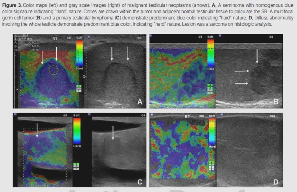
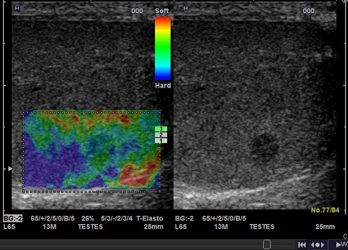
5 Testicular Elastography Strain Elastography of Normal Testis The blue rim on the outside of the normal testis is referred to as a boundary effect. It is thought to arise because the soft tissue is more tethered at the testicular margin and so suffers less deformation when stress is applied. This is demonstrated in an exaggerated fashion in a long standing atrophied testis. Normal Missed Torsion Infertility Atrophy

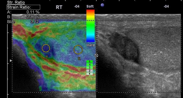
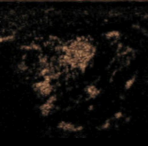
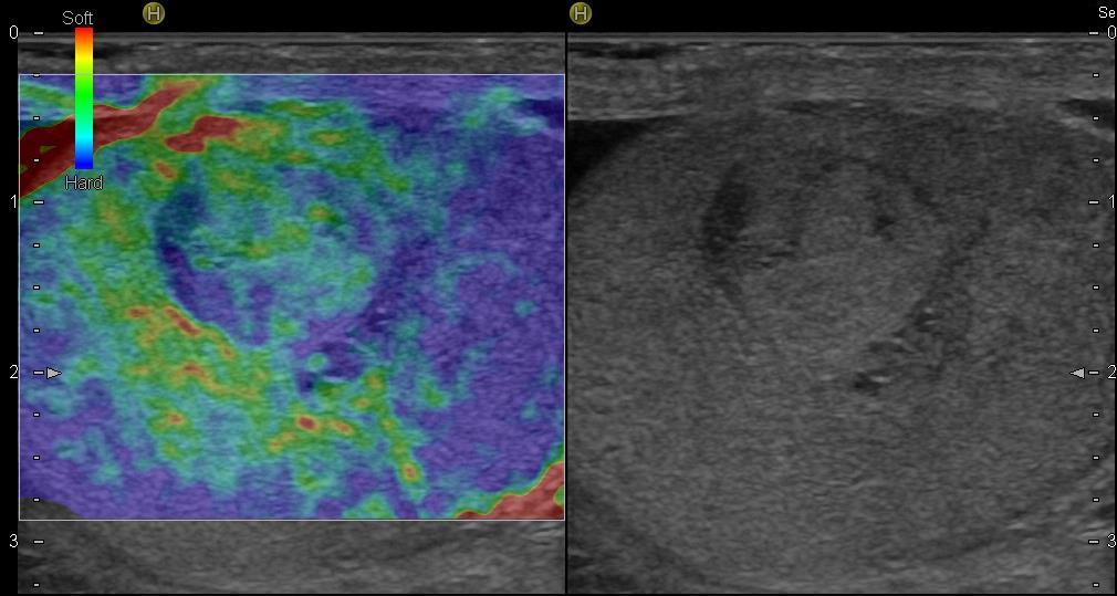
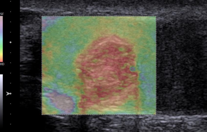
6 Testicular Elastography Normal Strain Elastography of Normal Testis The blue rim on the outside of the normal testis is referred to as a boundary effect. It is thought to arise because the soft tissue is more tethered at the testicular margin and so suffers less deformation when stress is applied. This is demonstrated in an exaggerated fashion in a long standing atrophied testis. Germ Cell Tumour Focal lesion in the testis that of a seminoma, with blue indicating hardness and strain ratio of 43.72
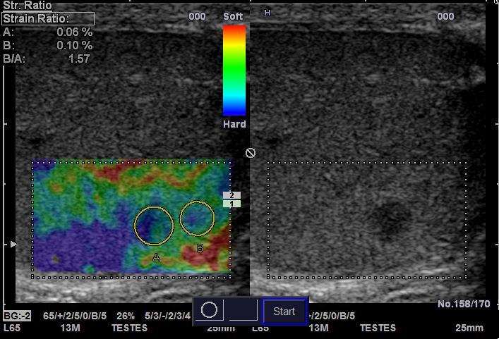
7 Strain Elastography
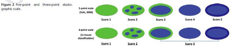
 Manually drawn ROI within the lesion and normal tissue Scoring system created")
8 Testicular Elastography Light vertical manual compression until optimised 256 colour scale (Red = Soft, Blue = Hard) Manually drawn ROI within the lesion and normal tissue Scoring system created (1-6)
9 Testicular Elastography
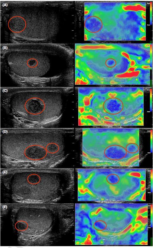
 50 Neoplasms Vs nonneoplasms Goddi, 2012 (9) 144 Malignant Vs Benign 87.5% 98.")
 68 Neoplasms Vs nonneoplasms Pozza, 2016 (18) 106 Malignant Vs Benign 81.1% 79.7% 59.4% 66.")
10 Testicular Elastography Study Lesions (n) Comparison VES VES SR SR (Reference) Sensitivity Specificity Sensitivity Sensitivity 100% 81% N/A N/A Aigner, 2012 (10) 50 Neoplasms Vs nonneoplasms Goddi, 2012 (9) 144 Malignant Vs Benign 87.5% 98.2% N/A N/A lesions Marsaud, 2015 (19) 34 Malignant Vs Benign 96.2% 37.5% N/A N/A lesions 98% 25% 90% 45% Schroder, 2016 (20) 68 Neoplasms Vs nonneoplasms Pozza, 2016 (18) 106 Malignant Vs Benign 81.1% 79.7% 59.4% 66.6% lesions 58.7% 100% 69.3% 61.3% Neoplasms Vs Nonneoplasms Auer, 2017 (16) 55 Malignant Vs Benign 100% 72.1% N/A N/A lesions
11 Testicular Elastography

12 Testicular Elastography
13 Testicular Elastography Extra-testicular Lesion Adenomatoid Lesion
14
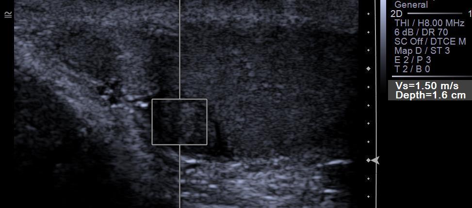
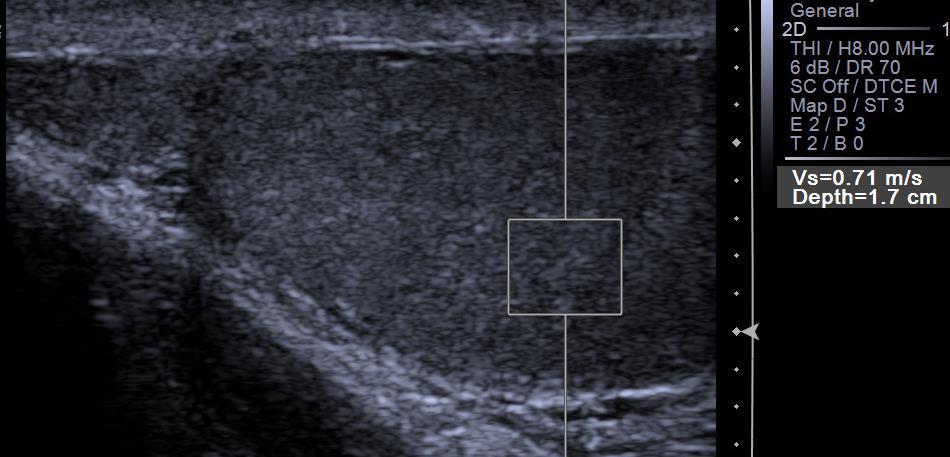
15 Testicular Elastography
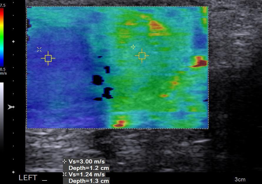
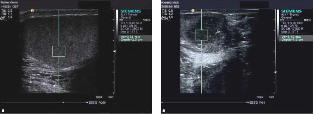
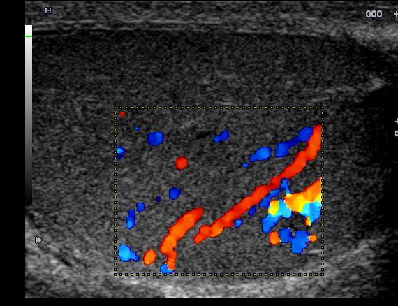
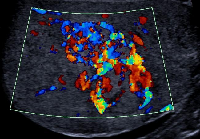
16 Testicular Elastography Colour coded shear wave elastography
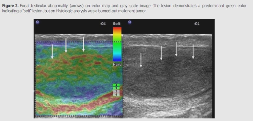
17 Testicular Elastography EFSUMB GUIDELINES ELASTOGRAPHY NON- LIVER APPLICATIONS 2018 GET-TOGETHER MEETING IN FRANKFURT ON 17 TH FEBRUARY STEERING COMMITTEE ADRIAN SĂFTOIU ODD HELGE GILJA PAUL SIDHU CHRISTOPH F. DIETRICH VITO CANTISANI Strain elastography 1.Strain elastography has been the most employed technique in assessing testicular lesions. 2. Although early studies have commented on the possibility of differentiating malignant from benign lesions with certainty using SE and a strain ratio (SR) these findings have not been confirmed in recent studies. 3. A number of case series detailing the use of SE and SR (some in combination with CEUS) have described the findings in Leydig cell tumours, epidermoid cysts, haematoma, lymphoma, focal infarction, capillary haemangioma, adrenal rest cells and in extra-testicular lesions, without comparison between the findings of these different lesions. Shear wave elastography 1. There is limited information regarding the use of shear wave elastography in the evaluation of testicular lesions. 2. SWE in the overall assessment of background parenchyma have suggested values may be raised with testicular microlithiasis, infertility, undescended testis, and has the potential to differentiate seminomas from non-seminomatous lesions and has been evaluated in burnt-out tumours. 3. No prospective study reporting the differences in SWE in focal testicular lesions has been published. Recommendation The application of strain or shear wave elastography techniques in the evaluation of focal testicular lesions are recommended only in conjunction with other ultrasound techniques, as there is overlap in values from benign and malignant neoplasms. (Level of Evidence;3A Grade of Evidence; B)
 to be performed, providing additional information: CEUS: dynamic functional imaging, including demonstration of presence or absence of vascularity SE: a measure of")

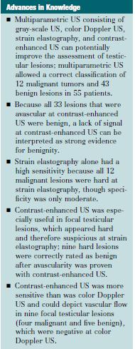
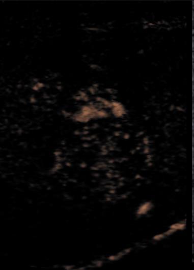
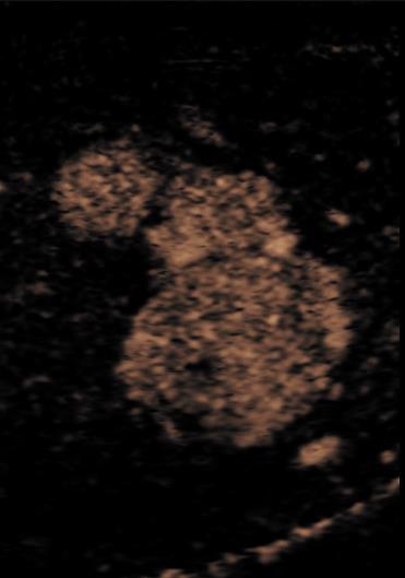
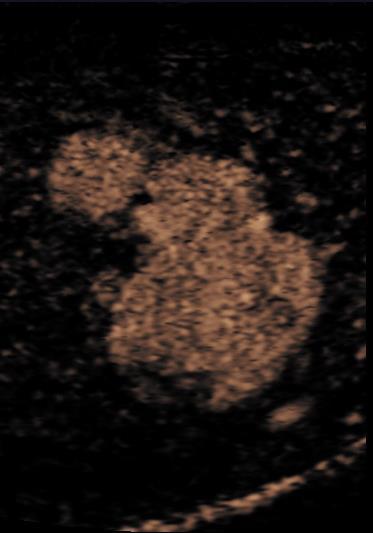
18 Multiparametric Ultrasound (MP-US) The addition of contrast-enhanced ultrasound (CEUS) and strain elastography (SE) to the established B-mode and colour Doppler US, facilitates testicular multiparametric ultrasound (MP-US) to be performed, providing additional information: CEUS: dynamic functional imaging, including demonstration of presence or absence of vascularity SE: a measure of tissue stiffness Classic seminoma: CEUS and SE appearances Huang DY, Sidhu PS. Focal testicular lesions: color Doppler ultrasound, contrast-enhanced ultrasound and tissue elastography as adjuvants to the diagnosis. Br J Radiol. 2012;85 Spec No 1:S Konstantatou E, Huang DY, Derchi LE, Bertolotto M, Valentino M, Sidhu PS. Focal testicular lesions: Use of contrastenhanced ultrasound (CEUS) and real-time Tissue Elastography (RTE) as adjuvant Sonographic techniques in determining clinical management. RSNA 2014 De Zordo T, Stronegger D, Pallwein-Prettner L, Harvey CJ, Pinggera G, Jaschke W, et al. Multiparametric ultrasonography of the testicles. Nature reviews Urology

19 MPUS of Scrotal Diseases

20 MPUS - Haematoma


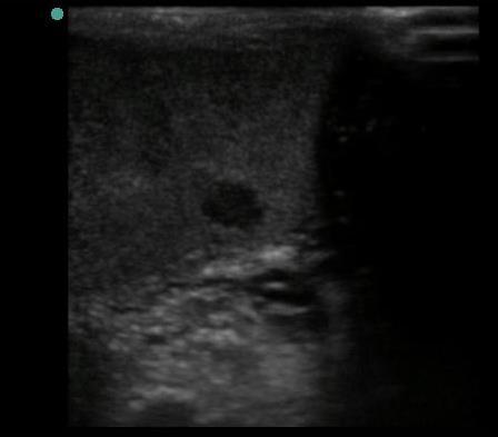
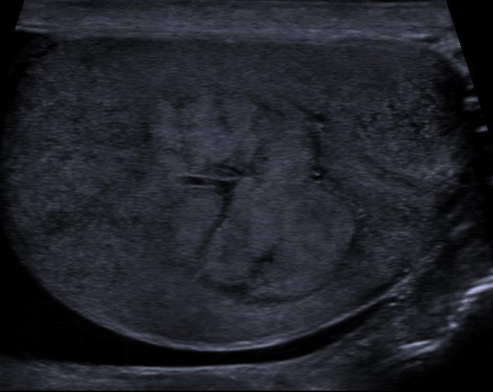
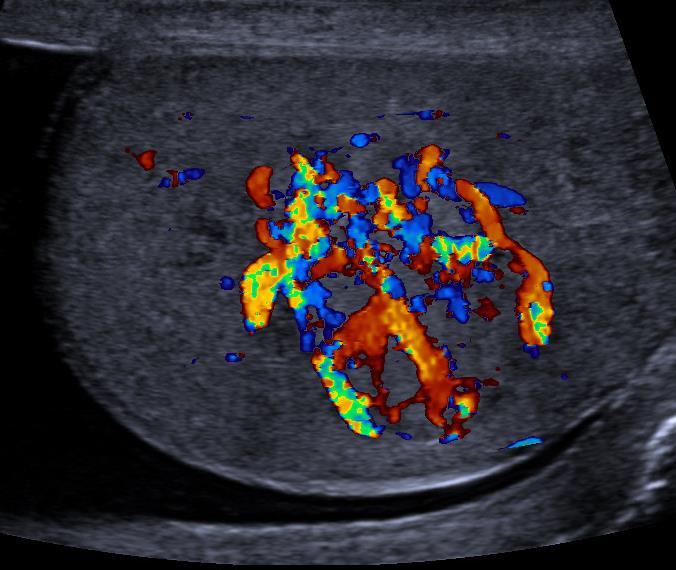
21 Testicular Indeterminate Tumours: Can CEUS and Elastography Help? Leydig Cell Tumour Heterogenous Highly vascular on CDUS Rapid and longer lasting enhancement on CEUS Strain ratio 1.56 Seminoma More homogenous Less vascular on CDUS Rapid and shorter lasting enhancement on CEUS Strain ratio 43.72


:115-22. Huang DY, Sidhu PS.")
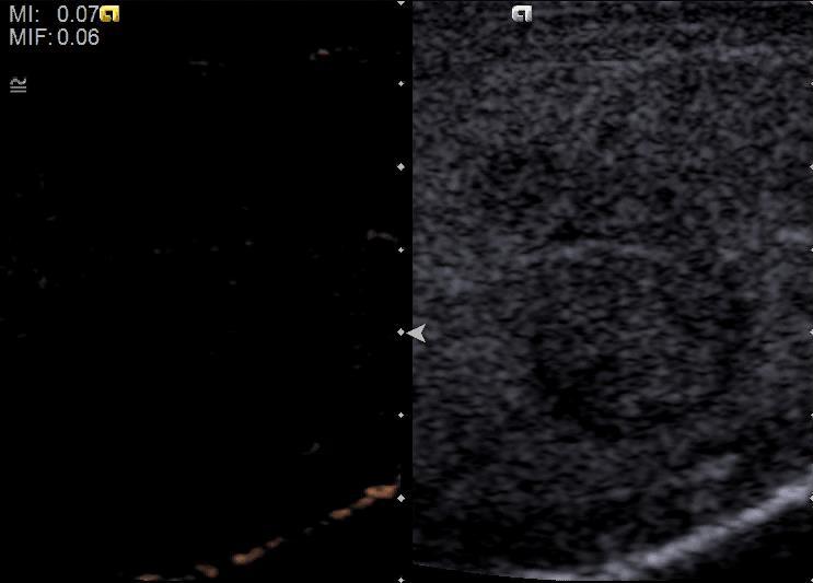
22 CEUS: Where CEUS is useful? The presence or absence of vascularity can be visualized with CEUS CEUS can conclusively demonstrate of lack of vascularity The absence of vascularity is an important indicator of benign disease. MP-US of an epidermoid cyst: CEUS demonstrates conclusively there is no internal vascular enhancement. Patel K, Sellars ME, Clarke JL, Sidhu PS. Features of testicular epidermoid cysts on contrast-enhanced sonography and real-time tissue elastography. J Ultrasound Med. 2012;31(1): Huang DY, Sidhu PS. Focal testicular lesions: color Doppler ultrasound, contrast-enhanced ultrasound and tissue elastography as adjuvants to the diagnosis. Br J Radiol. 2012;85 Spec No 1:S41-53.


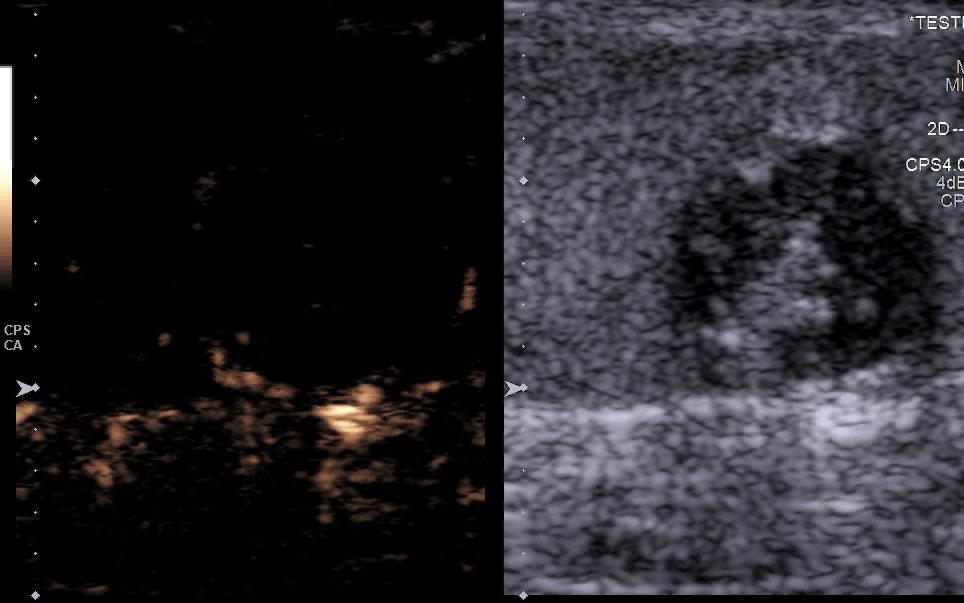
23 Where CEUS is useful? The pattern of vascularity can be visualised with CEUS. Marked hyper-vascularization is frequently observed in Leydig cell tumors CEUS improves identification Leydig cell tumors by this features for TSS consideration. Split screen images of CEUS and grey - scale US Huang DY, Eckersley RJ, Sellars ME, Sidhu PS. Differentiation Between Benign Leydig Cell and Malignant Germ Cell Testicular Tumors with Qualitative and Quantitative Contrast - Enhanced Ultrasound Assessments. European Congress of Radiology. 2013; Scientific presentation Lock G, Schroder C, Schmidt C, Anheuser P, Loening T, Dieckmann KP. Contrast-Enhanced Ultrasound and Real-Time Elastography for the Diagnosis of Benign Leydig Cell Tumors of the Testis - A Single Center Report on 13 Cases. Ultraschall Med. 2014
24 Indeterminate lesion - Pre-operative assessment (a) (b)

25 Indeterminate lesion - Pre-operative assessment
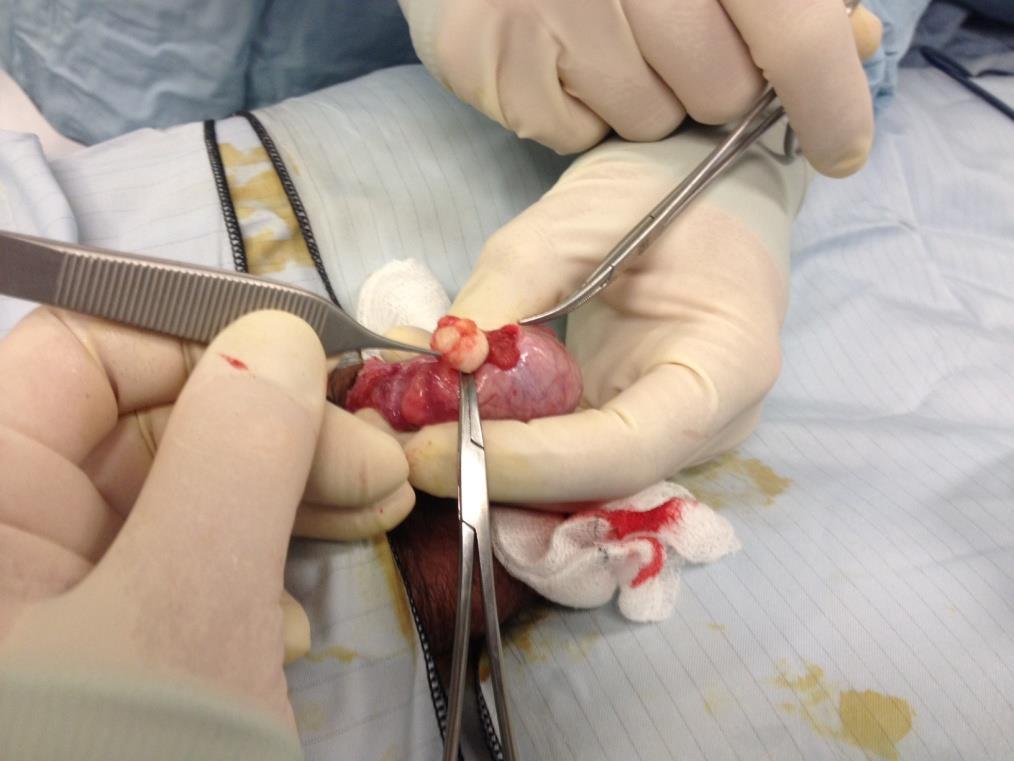
26 Indeterminate lesion - Intraoperative localisation

27 MPUS of Scrotal Diseases Seminoma

28 MPUS of Scrotal Diseases Epidermoid Cyst

29 MPUS of Scrotal Diseases Embryonal Cell Carcinoma

30 MPUS of Scrotal Diseases Haematoma
31 Segmental Infarction MPUS of Scrotal Diseases
32 Testicular Indeterminate Tumours: Can CEUS and Elastography Help?

33 Testicular Indeterminate Tumours: Can CEUS and Elastography Help?
34 Testicular Indeterminate Tumours: Can CEUS and Elastography Help?
35 Thank you
Strain histogram analysis for elastography in breast cancer diagnosis
 Strain histogram analysis for elastography in breast cancer diagnosis Poster No.: C-1854 Congress: ECR 2015 Type: Scientific Exhibit Authors: J. F. Carlsen, C. Ewertsen, S. Sletting, M. B. Nielsen; Copenhagen/DK
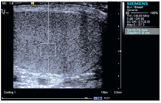
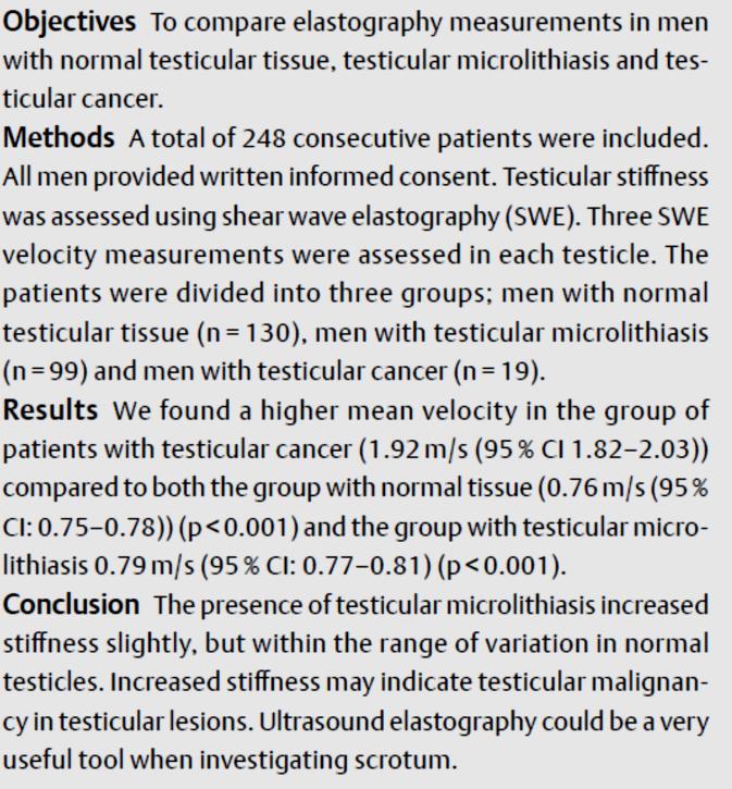
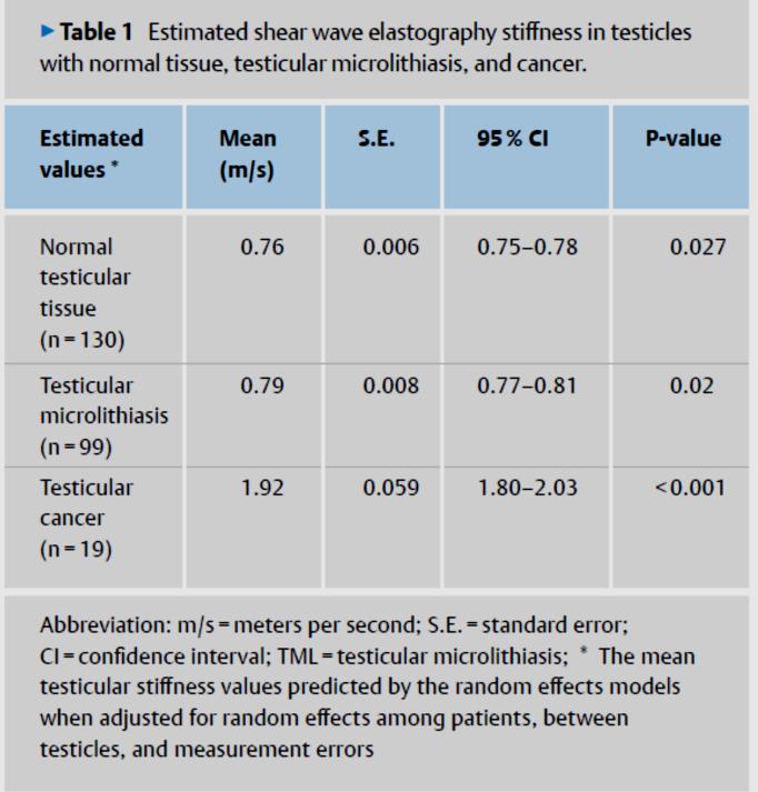
Comparison of Tissue Stiffness Using Shear Wave Elastography in Men with Normal Testicular Tissue, Testicular Microlithiasis and Testicular Cancer
 Original Article Comparison of Tissue Stiffness Using Shear Wave Elastography in Men with Normal Testicular Tissue, Testicular Microlithiasis and Testicular Cancer Authors Malene Roland Pedersen 1, 2,
Ultrasound of malignant testicular lesions. Arne Hørlyck Department of Radiology Aarhus University Hospital, Skejby
 Ultrasound of malignant testicular lesions Arne Hørlyck Department of Radiology Aarhus University Hospital, Skejby Testis Ultrasound is fantastic!! Scrotum Extratesticular mass: Benign Intratesticular
12th & 13th May 2016 Weston Education Centre, Kings College Hospital London
 EUROSON SCHOOL CEUS Course How to Incorporate CEUS into your Imaging Practice 12th & 13th Weston Education Centre, Kings College Hospital London CPD points for two days: EFSUMB 16 CME points BMUS 12 CPD
Contrast enhanced harmonic ultrasonography for the evaluation of acute scrotal pathology. Pictorial essay.
 Pictorial essay Med Ultrason 2016, Vol. 18, no. 1, 110-115 DOI: 10.11152/mu.2013.2066.181.esy Contrast enhanced harmonic ultrasonography for the evaluation of acute scrotal pathology. Pictorial essay.
COLOR DOPPLER ULTRASOUND IN EVALUATION OF SCROTAL LESIONS
 COLOR DOPPLER ULTRASOUND IN EVALUATION OF SCROTAL LESIONS Desai Sanjay D Associate Professor, Department of Radiology, RCSM Govt. Medical College, Kolhapur. ABSTRACT: Color Doppler ultrasound is a non-invasive,
ULTRASOUND IN CHRONIC LIVER DISEASE
 ULTRASOUND IN CHRONIC LIVER DISEASE Prof. Ioan Sporea, MD, PhD Head of Department of Gastroenterology and Hepatology University of Medicine and Pharmacy, Timişoara, Romania WFUMB Center of Education (COE),
A Comparative Study of Shear-Wave Elastography and Strain Elastography on a Breast Phantom for Diagnosis of Tumor and Cyst
 ` Volume VOLUME 2 ISSUE No 3 A Comparative Study of Shear-Wave Elastography and Strain Elastography on a Breast Phantom for Diagnosis of Tumor and Cyst Mahdi Al-Qahtani 1, Eraj Humayun Mirza 2, Mubarak
Evaluation of Testicular Disease using Ultrasound
 Evaluation of Testicular Disease using Ultrasound Abdullah Hamdan 1, Alsafi Abdulla 2, Mohamed Yousef 3 1 Radiologic Technology Department, College of Applied Medical Science, Qassim University, Buraduh,
Real-time Sonoelastography for the Evaluation of Testicular Lesions 1
 Note: This copy is for your personal non-commercial use only. To order presentation-ready copies for distribution to your colleagues or clients, contact us at www.rsna.org/rsnarights. Original Research
Global Testicular Infarction in the Presence of Epididymitis
 CASE SERIES Global Testicular Infarction in the Presence of Epididymitis Clinical Features, Appearances on Grayscale, Color Doppler, and Contrast-Enhanced Sonography, and Histologic Correlation Gibran
ShearWave elastography in lymph nodes
 ShearWave elastography in lymph nodes Poster No.: B-0158 Congress: ECR 2015 Type: Authors: Keywords: DOI: Scientific Paper F. Houari, O. Lucidarme, J. Gabarre, F. Charlotte, C. Pellot- Barakat, M. Lefort,
Does elastography change the indication to biopsy? IBDC
 Does elastography change the indication to biopsy? A LEXANDRA A THANASIOU, M D DEPARTMENT OF RADIOLOGY CURIE INSTITUTE PARIS, FRANCE IBDC Ultrasound Detected Cancers Physician-performed ultrasound increases
Diagnostic TRUS Elastography of the Prostate
 Diagnostic TRUS Elastography of the Prostate George Zacharopoulos Department of Diagnostic Ultrasound Hygeia Hospital Athens, Greece Prostate HI-RTE Why we need Elastography Better Detection of possible
Simplifying liver assessment in internal medicine
 Ultrasound Customer story Simplifying liver assessment in internal medicine Philips Affiniti ultrasound for elastography and contrast-enhanced ultrasound (CEUS) Where Sonography Institute, Uster, Switzerland
Real-time elastography of parotid gland masses: the value of strain ratio for the differentiation of benign from malignant tumors
 Realtime elastography of parotid gland masses: the value of strain ratio for the differentiation of benign from malignant tumors Poster No.: C09 Congress: ECR 05 Type: Scientific Exhibit Authors: M. M.
Strain Elastography - How To Do It?
 Strain Elastography - How To Do It? Authors Christoph F. Dietrich 1, Richard G. Barr 2, André Farrokh 3, Manjiri Dighe 4, Michael Hocke 5, Christian Jenssen 6, Yi Dong 7, Adrian Saftoiu 8, Roald Flesland
Prevalence of testicular microlithiasis in boys aged 0 to 19 years referred for scrotal pathology
 Chapter 2.3 Prevalence of testicular microlithiasis in boys aged 0 to 19 years referred for scrotal pathology J Goede HA Hofman AM Wagenvoort FH Pierik WWM Hack Nephro-Urol Mon, in press 59 Chapter 2.3
An improved quantification tool for breast ElastoScan : E-Breast
 An improved quantification tool for breast ElastoScan : E-Breast Volker F. Duda, MD, Christine Köhler, MD Interdisciplinary working group Senological diagnostics, University Hospital Gießen and Marburg
*OPERATIVE PROCEDURE. Serum tumour markers within normal limits S1.04 PRINCIPAL CLINICIAN
 Neoplasia of the Testis - Orchidectomy Histopathology Reporting Proforma Includes the International Collaboration on Cancer reporting dataset denoted by * Family name Given name(s) Date of birth Indigenous
EUS Elastography: Advances in Diagnostic EUS of the Pancreas
 Review Article http://dx.doi.org/10.3348/kjr.2012.13.s1.s12 pissn 1229-6929 eissn 2005-8330 Korean J Radiol 2012;13(S1):S12-S16 EUS Elastography: Advances in Diagnostic EUS of the Pancreas Tae Hee Lee,
Testicular tumors; Ultrasonographic and Pathologic correlation
 Testicular tumors; Ultrasonographic and Pathologic correlation Poster No.: C-0106 Congress: ECR 2014 Type: Educational Exhibit Authors: Y. Kim, S. W. Shin, E. T. Kim, M. Y. Kim ; Kuri City/KR, 1 1 2 1
MRI OF TESTICULAR MALIGNANCIES
 ATHENS 4-6 October 2018 European Society of Urogenital Radiology MRI OF TESTICULAR MALIGNANCIES Effrosyni I. Styliara, Athina C. Tsili, Alexia Psichou, Nikolaos Sofikitis, Maria I. Argyropoulou Department
Role of Colour Doppler Ultrasonography in evaluation of scrotal pain and swelling
 Original Research Article Role of Colour Doppler Ultrasonography in evaluation of scrotal pain and swelling Assistant Professor, Department of Radiodiagnosis, Government Medical College, Rajnandgaon Chattisghar,
Correlation between semiquantitative sonoelastography and immunohistochemistry in the evaluation of testicular focal lesions
 Pastore et al. Cancer Imaging 2014, 14:29 RESEARCH ARTICLE Open Access Correlation between semiquantitative sonoelastography and immunohistochemistry in the evaluation of testicular focal lesions Antonio
Elastography predicts thyroid cancer: comparison of two methods
 Elastography predicts thyroid cancer: comparison of two methods Poster No.: C-1267 Congress: ECR 2014 Type: Scientific Exhibit Authors: O. Sommer 1, H. Lanz 1, J. Hutter 1, M. Eberwein 2, J. Pratschke
Shear Wave Elastography In Characterization Of Liver Tumours
 Shear Wave Elastography In Characterization Of Liver Tumours Poster No.: C-1921 Congress: ECR 2014 Type: Scientific Exhibit Authors: K. L. Choong 1, B. J. J. Abdullah 1, G. Kumar 2, C. H. Yeong 1, K. L.
Follow up of Testicular Microlithiasis
 ATHENS 4-6 October 2018 European Society of Urogenital Radiology Follow up of Testicular Microlithiasis ESUR Scrotal Imaging Group Jonathan Richenberg 2nd ESUR Teaching Course Multimodality Imaging Approach
Sonography of the scrotum: still the best!
 Sonography of the scrotum: still the best! Poster No.: C-1110 Congress: ECR 2013 Type: Educational Exhibit Authors: W. Mnari, A. Zrig, M. Maatouk, B. Hmida, R. Salem, W. HarzallahHizem, M. Golli; Monastir/TN
Role of high frequency ultrasound and color Doppler examination in scrotal lesions
 Original article: Role of high frequency ultrasound and color Doppler examination in scrotal lesions 1Sanjeev Sharma, 2 Monika Jindal, 3 Sakshi Sharma, 4 Rekha Goyal, 5 Subhash Goyal 1Assistant Professor
Vikram Dogra, M.D. Professor of Radiology, Urology & BME Department of Imaging Sciences University Of Rochester Medical Center
 Ultrasound of the Scrotum Vikram Dogra, M.D. Professor of Radiology, Urology & BME Department of Imaging Sciences University Of Rochester Medical Center Etiologies of Acute Scrotal Pain Epididymitis/Orchitis
Shear-wave sonoelastography evaluation of Achilles tendons after surgery
 Shear-wave sonoelastography evaluation of Achilles tendons after surgery Poster No.: C-1507 Congress: ECR 2012 Type: Authors: Keywords: DOI: Scientific Paper G. Ivanac, D. Lemac, M. Cavka, B. Brkljacic;
Tissue Strain Analytics Virtual Touch Tissue Imaging and Quantification
 Whitepaper Tissue Strain Analytics Virtual Touch Tissue Imaging and Quantification ACUSON S2000 Ultrasound System Answers for life. Page 1 Tissue Strain Analytics: Virtual Touch Tissue Imaging and Quantification
Hepatic Imaging: What Every Practitioner Should Know
 Hepatic Imaging: What Every Practitioner Should Know Shuchi K. Rodgers, MD Section Chief, Abdominal Imaging Director of Ultrasound Department of Radiology Einstein Medical Center rodgerss@einstein.edu
RESEARCH ARTICLE. Lester Chee Hao Leong 1 *, Llewellyn Shao-Jen Sim 1, Ana Richelia Jara-Lazaro 2, Puay Hoon Tan 3. Abstract.
 DOI:http://dx.doi.org/10.7314/APJCP.2016.17.5.2673 US Breast Elastographic Evaluation of Mass-Forming Ductal Carcinoma-in-situ : Novel Toothpaste Sign RESEARCH ARTICLE Ultrasound Breast Elastographic Evaluation
VI E Article Clinics in diagnostic imaging (114)
") 264 Medical Education Singapore Med.1 2007, 48 (3) : VI E Article Clinics in diagnostic imaging (114) Muttarak M, Thinyu S, Lojanapiwat B Fig. I Clinical photograph of the scrotum. T t o T Fig. 2a Longitudinal
264 Medical Education Singapore Med.1 2007, 48 (3) : VI E Article Clinics in diagnostic imaging (114) Muttarak M, Thinyu S, Lojanapiwat B Fig. I Clinical photograph of the scrotum. T t o T Fig. 2a Longitudinal
Comparison of color-doppler and qualitative and quantitative strain-elastography for differentiation of thyroid nodules in daily practice
 HORMONES 2016, 15(2):197-204 Research paper Comparison of color-doppler and qualitative and quantitative strain-elastography for differentiation of thyroid nodules in daily practice Manuela Götzberger,
Testicular Infarction and Rupture After Blunt Trauma Use of Diagnostic Ultrasound
 Short Review TheScientificWorldJOURNAL (2004) 4, 437 441 ISSN 1537-744X; DOI 10.1100/tsw.2004.101 Testicular Infarction and Rupture After Blunt Trauma Use of Diagnostic Ultrasound Alistair Pace 1, * and
Radiation reduction in the follow-up of abdominal trauma imaging using contrast-enhanced ultrasound
 Radiation reduction in the follow-up of abdominal trauma imaging using contrast-enhanced ultrasound Poster No.: B-0892 Congress: ECR 2015 Type: Authors: Keywords: DOI: Scientific Paper A. Deganello, E.
Exercise. Discharge Summary
 Exercise Discharge Summary A 32-year-old Brazilian male presented with a 6 month history of right-sided scrotal swelling. Backache was present for 2 months and a history of right epididymitis was present
Testicular ultrasound in acute scrotal pain - beyond testicular torsion
 Testicular ultrasound in acute scrotal pain - beyond testicular torsion Poster No.: C-1284 Congress: ECR 2015 Type: Educational Exhibit Authors: I. Rolla, M. Nogueira, M. J. Aguiar, D. S. Garrido, J. A.
Case Report Testicular Arteriovenous Malformation: Gray-Scale and Color Doppler Ultrasonography Features
 Volume 2011, Article ID 876206, 4 pages doi:10.11/2011/876206 Case Report Testicular Arteriovenous Malformation: Gray-Scale and Color Doppler Ultrasonography Features Fatih Gulsen, 1 Ismail Mihmanli, 1
Essentials of Clinical MR, 2 nd edition. 73. Urinary Bladder and Male Pelvis
 73. Urinary Bladder and Male Pelvis Urinary bladder carcinoma is best locally staged with MRI. It is important however to note that a thickened wall (> 5 mm) is a non-specific finding seen in an underfilled
Sonography of the Pediatric Scrotum: Emphasis on the Ts Torsion, Trauma, and Tumors
 Pediatric Imaging Clinical Perspective Sung et al. Ultrasound of Pediatric Scrotum Pediatric Imaging Clinical Perspective Downloaded from www.ajronline.org by 37.44.201.148 on 01/21/18 from IP address
Correlation CEUS & Sono Elastography for Malignant Thyroid Nodule Evaluation
 Correlation CEUS & Sono Elastography for Malignant Thyroid Nodule Evaluation N Kumaran MD, Md Ameen MD ULTRASOUND Department Of Radiodiagnosis, Velammal Teaching Hospital, MaduralTuticorin Ring Road, Anupandi,
Ultrasonography of intratesticular lesions: its role in
 The Ulster Medical Journal, Volume 68, No. 2, pp. 54-8, November 1999. Ultrasonography of intratesticular lesions: its role in clinical management P T Kennedy, J M Elliott, P F Rice, B E Kelly Accepted
Original Article Differential diagnostic value of ultrasound elasticity score and strain ratio in breast cancer
 Int J Clin Exp Med 2018;11(7):7018-7028 www.ijcem.com /ISSN:1940-5901/IJCEM0068924 Original Article Differential diagnostic value of ultrasound elasticity score and strain ratio in breast cancer Zhiyuan
STAGING AND FOLLOW-UP STRATEGIES
 ATHENS 4-6 October 2018 European Society of Urogenital Radiology STAGING AND FOLLOW-UP STRATEGIES Ahmet Tuncay Turgut, MD Professor of Radiology Hacettepe University, Faculty of Medicine Ankara 2nd ESUR
Shear-wave elastography for breast masses: local shear wave speed (m/sec) versus Young modulus (kpa)
 versus Young modulus (kpa)") Shear-wave elastography for breast masses: local shear wave speed (m/sec) versus Young modulus (kpa) Ji Hyun Youk, Eun Ju Son, Ah Young Park, Jeong-Ah Kim Department of Radiology, Gangnam Severance Hospital,
Shear-wave elastography for breast masses: local shear wave speed (m/sec) versus Young modulus (kpa) Ji Hyun Youk, Eun Ju Son, Ah Young Park, Jeong-Ah Kim Department of Radiology, Gangnam Severance Hospital,
Role of Frozen Section Examination in the Management of Testicular Nodules: A Useful Procedure to Identify Benign Lesions
 SPECIAL FEATURE Role of Frozen Section Examination in the Management of Testicular Nodules: A Useful Procedure to Identify Benign Lesions Giorgio Bozzini, 1 Barbara Rubino, 2 Serena Maruccia, 1 Carlo Marenghi,
Role of ultrasound and colour doppler in assessment of adult scrotal pathologies
 Original Research Article Role of ultrasound and colour doppler in assessment of adult scrotal pathologies Ajay Vare 1, Dayanand Kawade 2*, Varsha Rote Kakinalkar 3, Prashant Titare 4, Samruddhi Sonawane
Real-Time Elastography Applications in Liver Pathology between Expectations and Results
 CLINICAL IMAGING Real-Time Elastography Applications in Liver Pathology between Expectations and Results Larisa Sandulescu, Ion Rogoveanu, Ioana Andreea Gheonea, Sergiu Cazacu, Adrian Saftoiu Research
Contrast Enhanced Transabdominal Ultrasound in the Characterisation of Pancreatic Lesions with Cystic Appearance
 ORIGINAL ARTICLE Contrast Enhanced Transabdominal Ultrasound in the Characterisation of Pancreatic Lesions with Cystic Appearance Stefan A Beyer-Enke 1, Michael Hocke 2, Andre Ignee 1, Barbara Braden 3,
Elastography. White Paper
 Elastography White Paper Strain Image in DC-8 Diagnostic Ultrasound System Shuangshuang Li, Rui Fan Relationship between the stiffness of tumor and it s malignance has far been known since the ancient
Original Article Shear wave elastography (SWE) is reliable method for testicular spermatogenesis evaluation after torsion
 is reliable method for testicular spermatogenesis evaluation after torsion") Int J Clin Exp Med 2015;8(5):7089-7097 www.ijcem.com /ISSN:1940-5901/IJCEM0005228 Original Article Shear wave elastography (SWE) is reliable method for testicular spermatogenesis evaluation after torsion
Int J Clin Exp Med 2015;8(5):7089-7097 www.ijcem.com /ISSN:1940-5901/IJCEM0005228 Original Article Shear wave elastography (SWE) is reliable method for testicular spermatogenesis evaluation after torsion
Shear Wave Elastographic Characterization of Normal and Torn Achilles Tendons
 ORIGINAL RESEARCH Shear Wave Elastographic Characterization of Normal and Torn Achilles Tendons A Pilot Study Xiang-Mei Chen, MD, PhD, Li-Gang Cui, MD, PhD, Ping He, MD, PhD, Wei-Wei Shen, MD, MS, Ya-Jun
Pelvic tumor in childhood Classification, imaging approach and radiological findings
 Pelvic tumor in childhood Classification, imaging approach and radiological findings M. Mearadji International Foundation for Pediatric Imaging Aid Rotterdam, The Netherlands Solid pelvic masses in childhood
Research Article Utility of Shear Wave Elastography for Diagnosing Chronic Autoimmune Thyroiditis
 yroid Research Volume 2015, Article ID 164548, 5 pages http://dx.doi.org/10.1155/2015/164548 Research Article Utility of Shear Wave Elastography for Diagnosing Chronic Autoimmune Thyroiditis Takahiro Fukuhara,
MRI IN THE CHARACTERIZATION OF SEMINOMATOUS AND NONSEMINOMATOUS GERM CELL TUMORS OF THE TESTIS
 MRI IN THE CHARACTERIZATION OF SEMINOMATOUS AND NONSEMINOMATOUS GERM CELL TUMORS OF THE TESTIS Ambesh Deshar *, Gyanendra KC and Zhang Lopsang *Department of Medical Imaging and Nuclear Medicine, First
LOGIQ E9 Shear Wave Elastography
 LOGIQ E9 Shear Wave Elastography Introduction Tissue stiffness is often related to underlying disease. For millennia, physicians have used palpation as a diagnostic tool to detect various ailments such
The role of ultrasonography with colour Doppler in the acute scrotum
 The role of ultrasonography with colour Doppler in the acute scrotum Poster No.: C-1509 Congress: ECR 2016 Type: Educational Exhibit Authors: M. S. R. O. Faustino, A. L. Amado Costa, J. J. B. Leitão, I.
Shear Wave Elastography in diagnostics of supraspinatus tendon.
 Shear Wave Elastography in diagnostics of supraspinatus tendon. Poster No.: C-2168 Congress: ECR 2013 Type: Authors: Keywords: DOI: Scientific Exhibit V. Saltykova; Moscow/RU Musculoskeletal joint, Musculoskeletal
Testicular microlithiasis and preliminary experience of acoustic radiation force impulse imaging
 Syddansk Universitet Testicular microlithiasis and preliminary experience of acoustic radiation force impulse imaging Pedersen, Malene Roland Vils; Osther, Palle Jørn Sloth; Rafaelsen, Søren Rafael Published
The role for contrast-enhanced ultrasonography outside of focal liver lesions
 The role for contrast-enhanced ultrasonography outside of focal liver lesions Paul S. Sidhu King s College Hospital, London, UK Introduction Contrast-enhanced ultrasonography (US) of focal liver lesions
Byung Ihn Choi, M.D. Department of Radiology Seoul National University Hospital
 Byung Ihn Choi, M.D. Department of Radiology Seoul National University Hospital CEUS & US Elastography : Contents CEUS Introduction Contrast agents & imaging Clinical application US Video WS Summary US
TitleUltrasonic evaluation of scrotal sw. Author(s) MISAKI, Toshimitsu; HISAZUMI, Haruo. Citation 泌尿器科紀要 (1985), 31(7):
 MISAKI, Toshimitsu; HISAZUMI, Haruo. Citation 泌尿器科紀要 (1985), 31(7):") TitleUltrasonic evaluation of scrotal sw Author(s) MISAKI, Toshimitsu; HISAZUMI, Haruo Citation 泌尿器科紀要 (1985), 31(7): 1151-1158 Issue Date 1985-07 URL http://hdl.handle.net/2433/118548 Right Type Departmental
TitleUltrasonic evaluation of scrotal sw Author(s) MISAKI, Toshimitsu; HISAZUMI, Haruo Citation 泌尿器科紀要 (1985), 31(7): 1151-1158 Issue Date 1985-07 URL http://hdl.handle.net/2433/118548 Right Type Departmental
Sonography of the Scrotum: Case-Based Review
 AJR Integrative Imaging LIFELONG LEARNING FOR RADIOLOGY Sonography of the Scrotum: Case-Based Review Joseph W. Stengel 1,2 and Erick M. Remer 1 ABSTRACT Objective We discuss five scenarios in which sonography
Breast Elastography: Our Initial Experience with Strain and Shear Wave Elastography
 Human Journals Short Communication September 2018 Vol.:10, Issue: 3 All rights are reserved by Pankaj Sharma et al. Breast Elastography: Our Initial Experience with Strain and Shear Wave Elastography Keywords:
Liver Ultrasound - Beyond the Basics. Pamela Parker Lead Sonographer
 Liver Ultrasound - Beyond the Basics Pamela Parker Lead Sonographer Aims Review what we know about the liver Reasons for imaging Focal lesions Diffuse disease Can we do more? The Liver The Liver The Liver
Bilateral Segmental Testicular Infarction
 Case Study TheScientificWorldJOURNAL (2007) 7, 779 783 TSW Urology ISSN 1537-744X; DOI 10.1100/tsw.2007.146 Bilateral Segmental Testicular Infarction Aaron Bayne 1, Brad Koslin 2, and Siamak Daneshmand
Contrast-enhanced ultrasound (CEUS) of focal liver lesions. A useful, rapid and accessible tool.
 of focal liver lesions. A useful, rapid and accessible tool.") Contrast-enhanced ultrasound (CEUS) of focal liver lesions. A useful, rapid and accessible tool. Poster No.: C-2329 Congress: ECR 2012 Type: Educational Exhibit Authors: S. Santamaria Jareño, J. Carrero
Contrast-enhanced ultrasound (CEUS) of focal liver lesions. A useful, rapid and accessible tool. Poster No.: C-2329 Congress: ECR 2012 Type: Educational Exhibit Authors: S. Santamaria Jareño, J. Carrero
The role of US elastography in the evaluation of benign and malignant breast lesions in relation to histopathological examination
 The role of US elastography in the evaluation of benign and malignant breast lesions in relation to histopathological examination Poster No.: C-1802 Congress: ECR 2013 Type: Scientific Exhibit Authors:
Differentiating Benign from Malignant Cervical Lymph Nodes with Sonoelastography
 International Journal of Medical Imaging 2017; 5(4): 42-46 http://www.sciencepublishinggroup.com/j/ijmi doi: 10.11648/j.ijmi.20170504.11 ISSN: 2330-8303 (Print); ISSN: 2330-832X (Online) Differentiating
Role of sonoelastography in the evaluation of superficial soft tissue lesions: qualitative and quantitative study
 Role of sonoelastography in the evaluation of superficial soft tissue lesions: qualitative and quantitative study Poster No.: C-2016 Congress: ECR 2013 Type: Scientific Exhibit Authors: N. Magarelli, C.
U ltrasonography (US) is reported to be the primary
 is reported to be the primary") 401 LIVER Improved characterisation of histologically proven liver tumours by contrast enhanced ultrasonography during the portal venous and specific late phase of SHU 508A C F Dietrich, A Ignee, J Trojan,
401 LIVER Improved characterisation of histologically proven liver tumours by contrast enhanced ultrasonography during the portal venous and specific late phase of SHU 508A C F Dietrich, A Ignee, J Trojan,
US features of scrotal disorders: A pictorial essay
 US features of scrotal disorders: A pictorial essay Poster No.: C-0463 Congress: ECR 2014 Type: Educational Exhibit Authors: J. SAAD, F. Marrakchi ; nejran, sa/sa, Monastir/TN Keywords: Genital / Reproductive
The acute scrotum the two most difficult US problems. Simon Freeman Derriford Hospital, Plymouth. UK
 The acute scrotum the two most difficult US problems Simon Freeman Derriford Hospital, Plymouth. UK simonfreeman@nhs.net The Acute Scrotum 1. Ischaemia 1. Spermatic cord torsion 2. Torsion of a testicular
Acute scrotum. Acute Epididymo-orchitis. Phyllis Yan, APDR (QEH)
") Acute scrotum Acute Epididymo-orchitis Phyllis Yan, APDR (QEH) Conditions leading to acute pain Torsion Acute Epididymitis / Epididymoorchitis Scrotal trauma Inguinal hernias Testicular tumors Epididymitis/epididymo
Acute scrotum Acute Epididymo-orchitis Phyllis Yan, APDR (QEH) Conditions leading to acute pain Torsion Acute Epididymitis / Epididymoorchitis Scrotal trauma Inguinal hernias Testicular tumors Epididymitis/epididymo
Case Scenario 1 Discharge Summary Pathology Report Final Diagnosis: Oncology Consult
 Case Scenario 1 Discharge Summary A 31-year-old Brazilian male presented with a 6 month history of right-sided scrotal swelling. Backache was present for 2 months and a history of right epididymitis was
The Contribution of Contrast Enhanced Ultrasound for the characterization of benign liver lesions in clinical practice a monocentric experience
 Original papers Med Ultrason 2012, Vol. 14, no. 4, 283-287 The Contribution of Contrast Enhanced Ultrasound for the characterization of benign liver lesions in clinical practice a monocentric experience
Differential Diagnosis of Focal Epididymal Lesions With Gray Scale Sonographic, Color Doppler Sonographic, and Clinical Features
 Article Differential Diagnosis of Focal Epididymal Lesions With Gray Scale Sonographic, Color Doppler Sonographic, and Clinical Features Dal Mo Yang, MD, Sun Ho Kim, MD, Ha Na Kim, MD, Jee Hee Kang, MD,
J of Evolution of Med and Dent Sci/ eissn , pissn / Vol. 4/ Issue 39/ May 14, 2015 Page 6787
 ROLE OF HIGH RESOLUTION SONOGRAPHY IN CHARACTERIZATION OF SOLID SALIVARY GLAND TUMORS Sheetal Singh 1, Amlendu Nagar 2, Pramod Sakhi 3, Sachin Kataria 4, Kumud Julka 5, Anup Gupta 6 HOW TO CITE THIS ARTICLE:
A Practical Approach to Adnexal Masses
 A Practical Approach to Adnexal Masses Darcy J. Wolfman, MD Section Chief of Genitourinary Imaging American Institute for Radiologic Pathology Clinical Associate Johns Hopkins Community Radiology Division
Contrast-enhanced ultrasound evaluation of testicular interstitial cell tumours in conscious non-sedated dogs
 Veterinarni Medicina, 63, 2018 (03): 125 130 Original Paper Contrast-enhanced ultrasound evaluation of testicular interstitial cell tumours in conscious non-sedated dogs M. Quartuccio*, C. Mangano, F.
Elastosonography. Prof. Massimo Midiri Direttore Istituto di Radiologia Policlinico Universitario Paolo Giaccone Diagnostica per Immagini, - Palermo
 Elastosonography Prof. Massimo Midiri Direttore Istituto di Radiologia Policlinico Universitario Paolo Giaccone Diagnostica per Immagini, - Palermo everybody has certainly had, at least once in a lifetime,
Quiz 1. Assign Race 1, Race 2 and Spanish Hispanic Origin to the following scenarios.
 Quiz 1 Assign Race 1, Race 2 and Spanish Hispanic Origin to the following scenarios. 1. 62 year old Brazilian female Race 1 Race 2 Spanish/Hispanic Origin 2. 43 year old Asian male born in Japan Race 1
iu22 Liver Shear Wave ElastPQ
 iu22 Liver Shear Wave ElastPQ Clinical Case Study Lucy Wang Clinical Application Specialist ASEAN Case Study: History: 58-year-old male patient, hepatitis B virus (HBV) carrier, with non clinical symptoms
Case Scenario 1 Discharge Summary Pathology Report Final Diagnosis: Oncology Consult
 Case Scenario 1 Discharge Summary A 31-year-old Brazilian male presented with a 6 month history of right-sided scrotal swelling. Backache was present for 2 months and a history of right epididymitis was
Scrotal extratesticular schwannoma: a case report and review of the literature
 Palleschi et al. BMC Urology 2014, 14:32 CASE REPORT Open Access Scrotal extratesticular schwannoma: a case report and review of the literature Giovanni Palleschi 1,2, Antonio Carbone 1,2, Jessica Cacciotti
Role of ultrasound and color Doppler in the evaluation of acute scrotal pain
 Bang Med J Khulna 2017; 50 : 26-30 ORIGINAL ARTICLE Abstract Role of ultrasound and color Doppler in the evaluation of acute scrotal pain G Salahuddin 1, SMZ Nayeem 2, SM Hossain 3, S Parvin 4, MM Hossain
The Incidental Renal lesion
 The Incidental Renal lesion BACKGROUND Increase in abdominal CT/US in last 15 years Resulted in detection of many (small) renal lesions 50% > 50yrs has at least 1 lesion majority simple cysts Renal lesions
Genitourinary Radiology In-Training Test Questions for Diagnostic Radiology Residents
 Genitourinary Radiology In-Training Test Questions for Diagnostic Radiology Residents March, 2013 Sponsored by: Commission on Education Committee on Residency Training in Diagnostic Radiology 2013 by American
Emergent Pediatric Ultrasound. Katharine Dennis, RDMS/RVT Tiffany Schultz, RDMS UNC Health Care Dept of General Ultrasound
 Emergent Pediatric Ultrasound Katharine Dennis, RDMS/RVT Tiffany Schultz, RDMS UNC Health Care Dept of General Ultrasound Introduction Learning Objectives Review common pediatric emergent ultrasound exams
Role of US in acute scrotal pain
 World J Urol (2011) 29:639 643 DOI 10.1007/s00345-011-0698-8 TOPIC PAPER Role of US in acute scrotal pain G. Liguori S. Bucci A. Zordani S. Benvenuto G. Ollandini G. Mazzon M. Bertolotto F. Cacciato S.
Incidental Discovery of Testicular Microlithiasis: What Is the Importance of Ultrasound Surveillance? Two Case Reports
 Published online: October 24, 2013 1662 6575/13/0063 0520$38.00/0 This is an Open Access article licensed under the terms of the Creative Commons Attribution-NonCommercial 3.0 Unported license (CC BY-NC)
Segmental testicular infarction: role of imaging
 Segmental testicular infarction: role of imaging Poster No.: C-0583 Congress: ECR 2013 Type: Scientific Exhibit Authors: F. Albarello, S. Zago, P. Campioni, M. Giganti, G. Parenti ; 1 1 2 1 1 2 2 Ferrara/IT,
Vascular Related Torsion Venous compression Hemorrhagic infarct Young men At night Very painful Can be reduced Scrotal Masses Testicular Tumors (solid
 Pathology of the Male Reproductive System Testis and Epididymis Failure of Testis to Descend Testis are not always in scrotum at birth. Testes from in abdomen with kidneys Migrate to scrotum May get stuck
Surgery Illustrated Surgical Atlas Inguinal orchidectomy for testicular cancer
 Surgery Illustrated Focus on Details SURGERY ILLUSTRATED SURGICAL ATLASPIZZOCARO and GUARNERI PIZZOCARO and GUARNERI BJUI BJU INTERNATIONAL Surgery Illustrated Surgical Atlas Inguinal orchidectomy for