Digestive system (Systema digestorium/ alimentarium) Lecture #1
|
|
|
- Frederica Bates
- 6 years ago
- Views:
Transcription
1 Digestive system (Systema digestorium/ alimentarium) Lecture #1
2 Internal organs are grouped into 1. System - Have the same functions and development Digestive system Respiratory system 2. Apparatus - Have the same development but different functions Urogenital apparatus
3 Internal organs: Parenchymal organs Hollow (tubular) organs

4 Parenchymal organs - Parenchyma specialized tissue that carries out the specific functions of the organ - Parenchymal organs have stromal capsule, that gives off trabecules into the parenchyma - Stroma contains lymphatic and blood vessels, nerves
5 Tubular organs Layers of the wall: 1) Tunica mucosa 2) Tunica submucosa 3) Tunica musculosa (longitudinal and circular) 4) Tunica serosa/adventitia

6 Alimentary system is a complex of organs with the function of mechanical and chemical treatment of food, absorption of the treated nutrients, and excretion of undigested remnants.

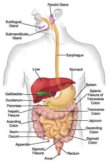
7 Overview of Digestive Anatomy
8
9 Development of digestive system
10
 Ectoderm Mesoderm")
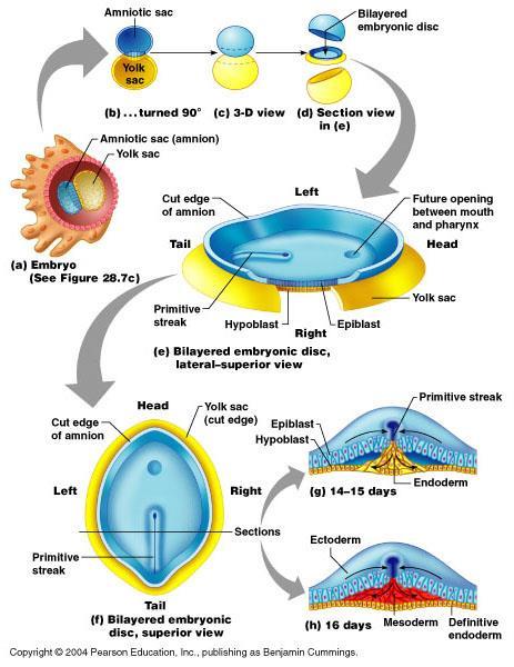
11 Gastrulation formation of germ layers (4 th week) Ectoderm Mesoderm Endoderm
 appendicular skeleton and")
12 Intraembryonic mesoderm plates: Paraxial (dorsal) mesoderm axial skeleton (somites) Intermediate mesoderm urogenital apparatus Lateral mesoderm (somatic and splanchnic) appendicular skeleton and internal organs
13 Lateral mesoderm forms two plates: somatic and splanchnic EEM, extraembryonic mesoderm; YS, Yolk sac; NP, neural plate.
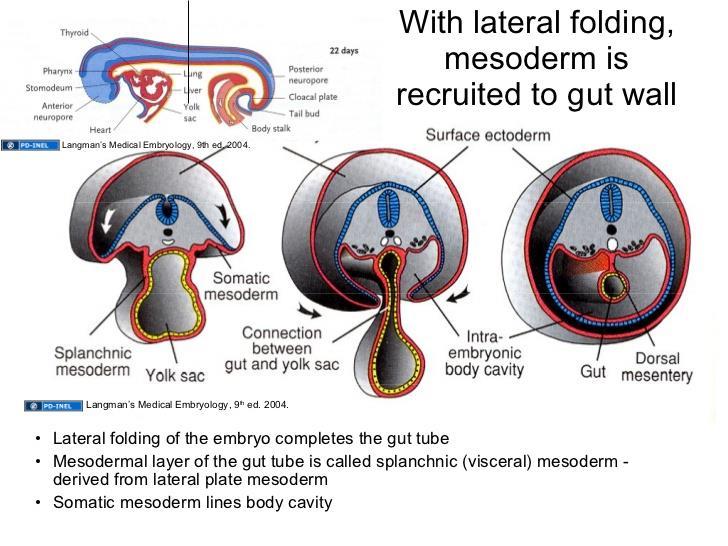
14 Coelom
15 Tubular organ layers development - Mucosa Epithelial lining and glands Lamina propria Muscularis mucosae - Derived from endoderm - Submucosa Derived from visceral mesoderm - Muscularis externa - Adventitia/Serosa
16 Intraembryonic body cavity(coelom) Primitive gut tube Dorsal mesentery Peritoneal cavity Tubular organs (gut) Mesentery
17 Primitive gut subdivisions: Stomodeum cranial entrance of the gut tube Foregut Omphalomesenteric duct=vitelline duct (joins yolk sac and midgut lumen) Proctodeum caudal exit of the gut tube
18 From foregut develop: - Esophagus - Stomach - Duodenum (proximal part) - Liver, pancreas, gall bladder - Respiratory tube Blood supply truncus coeliacus Sympathetic innervation n. splanchnicus major Parasympathetic innervation n.vagus Foregut

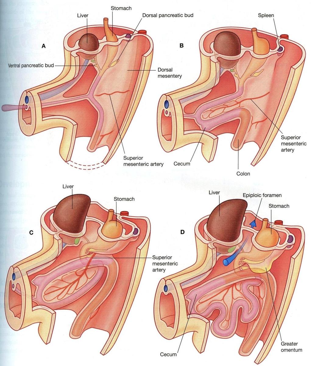
19 1. The stomach form fusiform dilatation of the foregut, suspended from body wall by a dorsal and ventral gastric mesentery. 2. The dorsal portion grows more rapidly formation of the greater curvature. 3. The primitive stomach rotates 90 degrees clockwise around longitudinal axis. 4. Ventral gastric mesentery lesser omentum. 5. Dorsal gastric mesentery greater omentum.
20
 in response to signals from nearby")
21 Development of the liver Liver bud arise from foregut endoderm towards septum transversum (developing diaphragm) in response to signals from nearby mesoderm

22 Development of the liver Endoderm Mesoderm Primitive ventral mesentery Primitive dorsal mesentery Liver parenchyma Capsule, stroma Falciform ligament Lesser omentum
23

24 Development of the pancreas - Ventral pancreatic bud outgrowth of liver bud - Dorsal pancreatic bud outgrowth of duodenal bud into the stomach mesentery. Dorsal pancreatic bud accessory pancreatic duct. - The rotation of the duodenum to the right carries the ventral pancreatic bud dorsally, where it fuses with the dorsal pancreatic bud.

25 Retroperitoneal position of the pancreas and duodenum
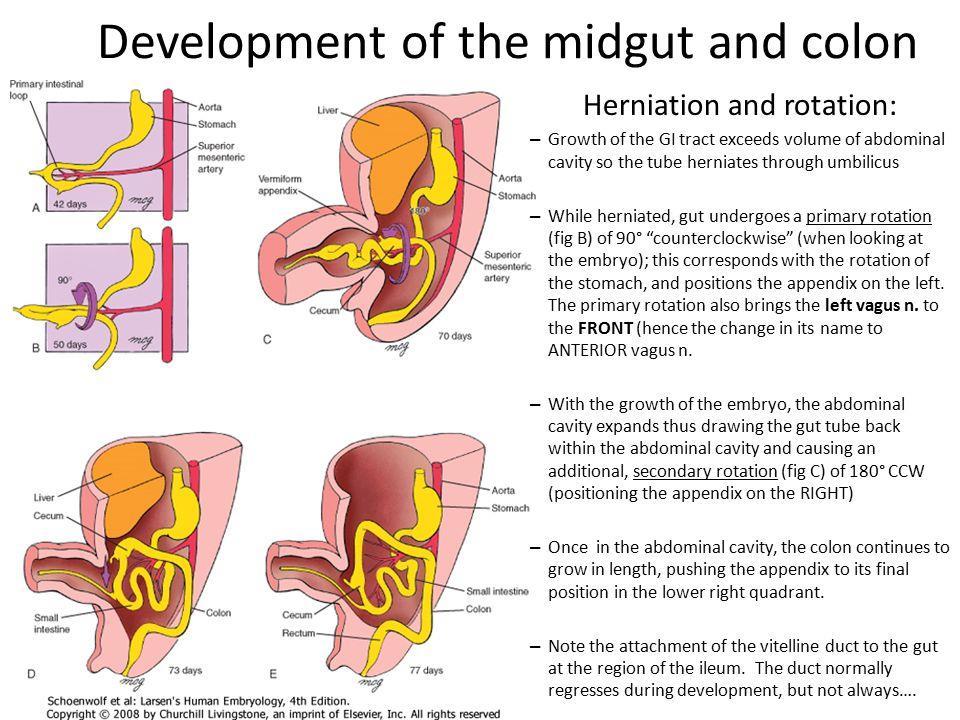
26 From midgut develop: - Duodenum distal to the opening of the bile duct - The rest of intestine (ileum, jejunum) - Cecum and appendix - Ascending and proximal 2/3 of transverse colon Blood supply a. mesenterica superior Sympathetic innervation n. splanchnicus minor Parasympathetic innervation n.vagus
27 Development of the midgut and colon Vitelline duct Omphalomesenteric duct = vitelline duct (joins yolk sac and midgut lumen) axis for rotation
28

29
30


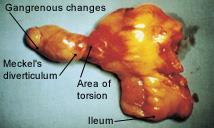
31 Meckel`s diverticulum - remnant of the omphalomesenteric duct (the vitelline duct)


32 Premature birth disorders of the digestive system Variety of appendix position Intestinal malrotation
33 Cecum and appendix
34 From hindgut develop: - The rest 1/3 of transverse colon - Descending and sigmoid colon, rectum Blood supply a. mesenterica inferior Sympathetic innervation nn. splanchnici lumbales et pelvini Parasympathetic innervation nn. splanchnici pelvini


35 Caudal end of the hindgut proctodeum (9 th week)


36 Cloacal membrane is not ruptured Atresia of rectum

37 Superior to that line the intestine derives from the embryonic hindgut and the epithelium derives from endoderm. - a. et v. mesenterica inferior Inferior to that line the epithelium derives from ectoderm. - a. et v. iliaca interna
 level of internal (involuntary) muscular anal sphincter.")
38 Anorectal line (upper end of columns) (lower end of columns) Pectinate line - anatomical border between rectum and anal canal Anal pecten 1) place between pectinate and anocutaneous lines; 2) level of internal (involuntary) muscular anal sphincter. Anocutaneous line ( white line ) 1) lower border of internal anal sphincter; 2) mucous layer changes to skin.
39
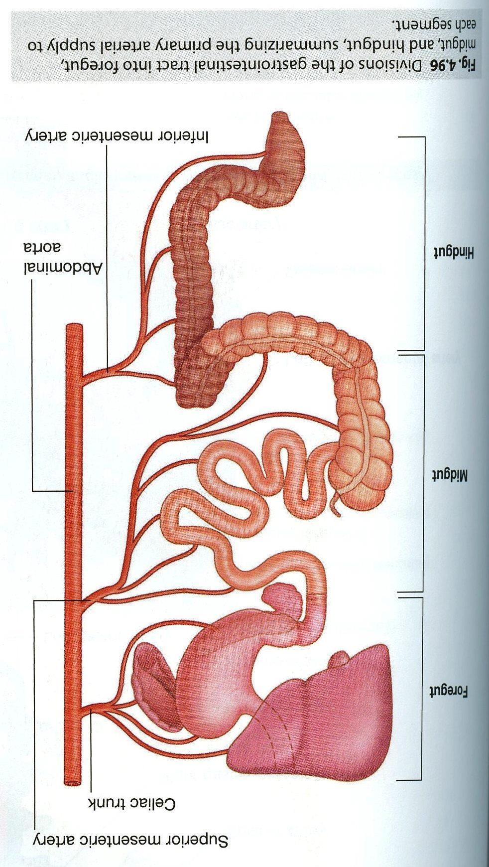
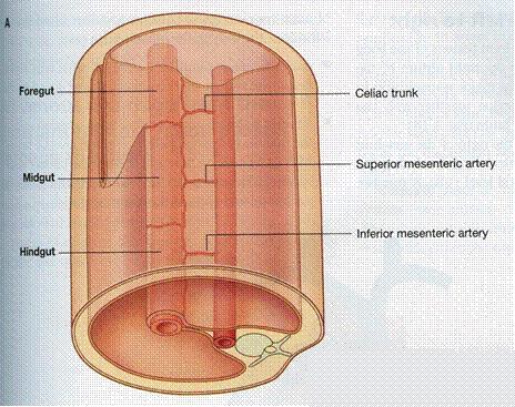
40 Development of GI and blood supply
41 Foregut derivatives include which of the following? 1) The esophagus 2) The stomach 3) The proximal duodenum 4) The ileum 5) The liver 6) The descending colon 1,2,3,5
42 Midgut derivatives include which of the following? 1) The stomach 2) The distal duodenum 3) The ileum 4) The jejunum 5) The appendix 6) The descending colon 2,3,4,5
43 Which of the following cells are endodermal derivatives? 1) Pancreatic acinar cells 2) Alpha and beta cells in the islets of Langerhans 3) Liver parenchymal cells 4) Cells lining the lumen of the gallbladder 5) Cells lining the lumen of intestine 6) Muscular layer of intestinal wall 7) Peritoneum 8) Connective tissue and blood vessels of the organs 1,2,3,4,5
44

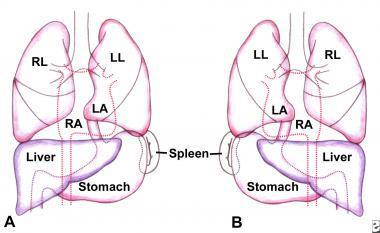
45 Situs viscerum inversus partialis/totalis Normal position Inverse position
46 Development of peritoneum and its derivatives Peritoneum serose membrane lining organs and walls of the abdominal cavity - is derived from intraembryonic mesoderm
 Layers continue one to another without")
47 Layers of peritoneum: Peritoneal cavity Parietal peritoneum (covers abdominal walls) Visceral peritoneum (forms serous covering of the internal organs) Layers continue one to another without any gap

48 Lateral mesoderm forms two plates: somatic and splanchnic EEM, extraembryonic mesoderm; YS, Yolk sac; NP, neural plate.
49 Intraembryonic body cavity(coelom) Peritoneal cavity Somatic mesoderm Parietal peritoneum Splanchnic mesoderm Visceral peritoneum
50 Peritoneal cavity Peritoneal cavity cavity between parietal and visceral layers of peritoneum - Female it communicates with external environment through fallopian tubes-uterine-vagina - Male it is closed serous sac
51 Abdominal cavity vs Peritoneal cavity
52 Abdominal cavity - Space in the trunk below diaphragm Walls: - Superior diaphragm - Anterior and lateral broad muscles of abdomen - Posterior the lumbar segment of the spine and muscles (m. psoas major, m. quadratus lumborum) - Inferior iliac bones and pelvic diaphragm
53 Position of the organs Intraperitoneal - totally covered by peritoneum Hepar Stomach (++) Spleen Jejunum (+) Ileum (+) Caecum Appendix vermiformis (+) Sigmoid colon (+) Superior part of rectum (+) Uterine (+) + - presence of mesenterium
54 Mesoperitoneal Position of the organs - 3 sides are covered (the 4 th side is covered by adventitia) Ascending and descending colon Middle part of the rectum Full gallbladder and urinary bladder
 Duodenum Pancreas Kidney Ureter Empty gallbladder and urinary")
55 Position of the organs Extraperitoneal / retroperitoneal - not covered (or only 1 side) Duodenum Pancreas Kidney Ureter Empty gallbladder and urinary bladder
56

57 Kidney access surgery
58 Derivatives of the peritoneum Ligaments Mesentery (mesenterium and mesolon) Omentum (majus and minus)

59 Derivatives of the peritoneum Ligaments = 2 layers of peritoneum Lig. falciforme Lig. coronarium Lig. hepatogastrica Lig. hepatoduodenale Lig. hepatorenale etc.
60 Derivatives of the peritoneum Mesentery = = 2 layers of peritoneum + vessels and nerves Structures that suspend visceral organs from the body wall Carry arteries, veins, lymphatic vessels and nerves


61 Mesenteries are established as the visceral organs grow into the intraembryonic coelom and carry their mesothelial covering and vessels with them
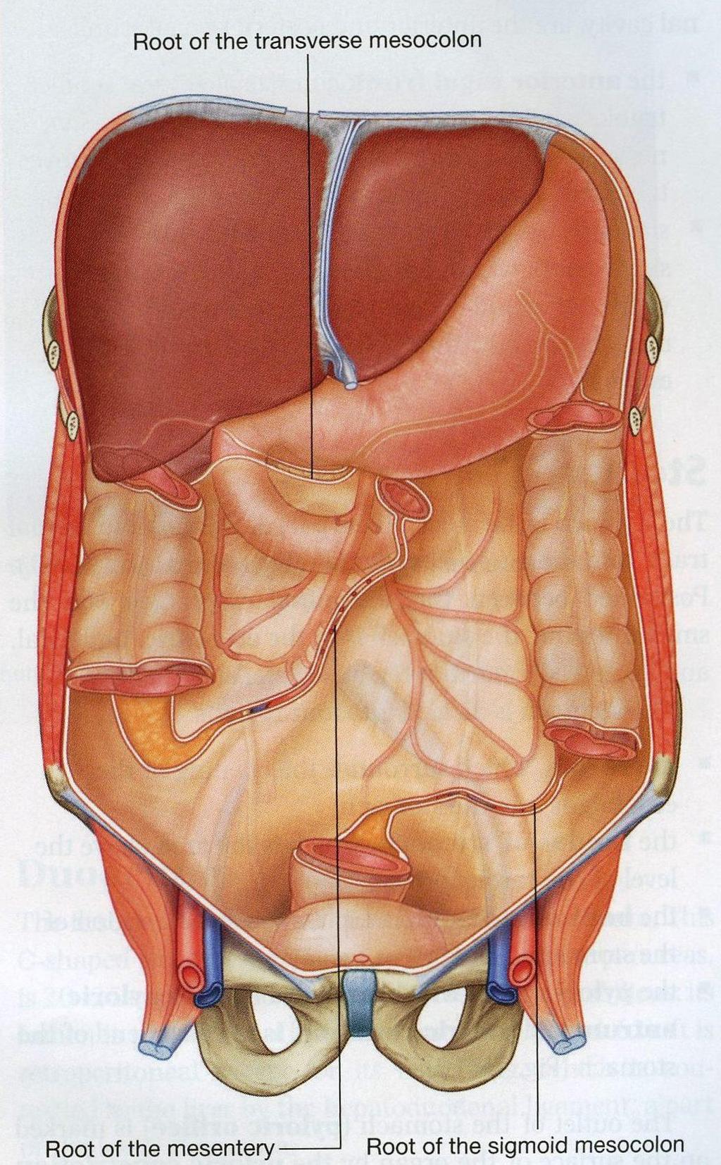
62 Derivatives of the peritoneum mesenteries Meso + Greek name of the organ The mesenterium (small intestine) The mesoappendix The transverse mesocolon The sigmoid mesocolon
63
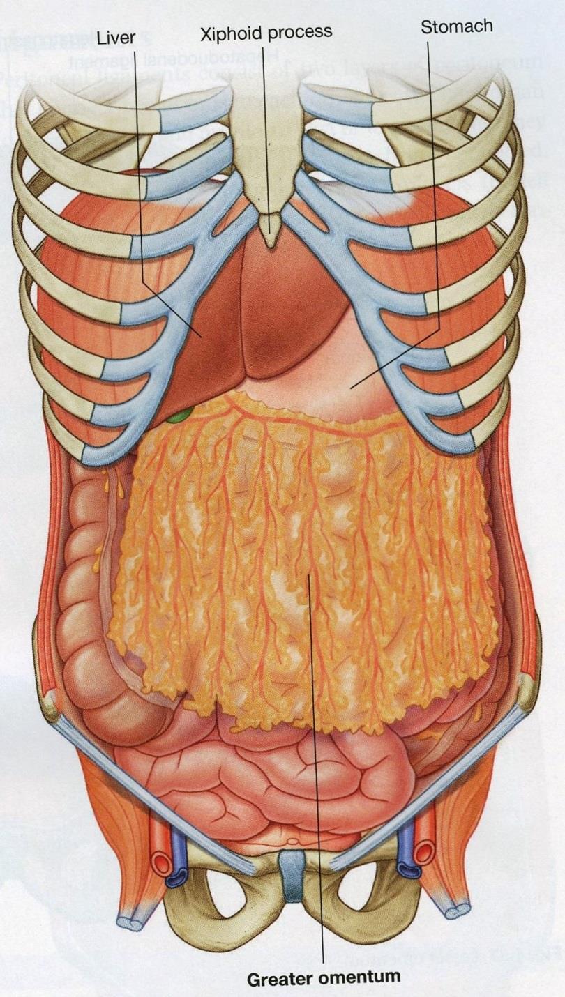
64 Derivatives of the peritoneum Omentum= 2 layers of peritoneum + fatty tissue
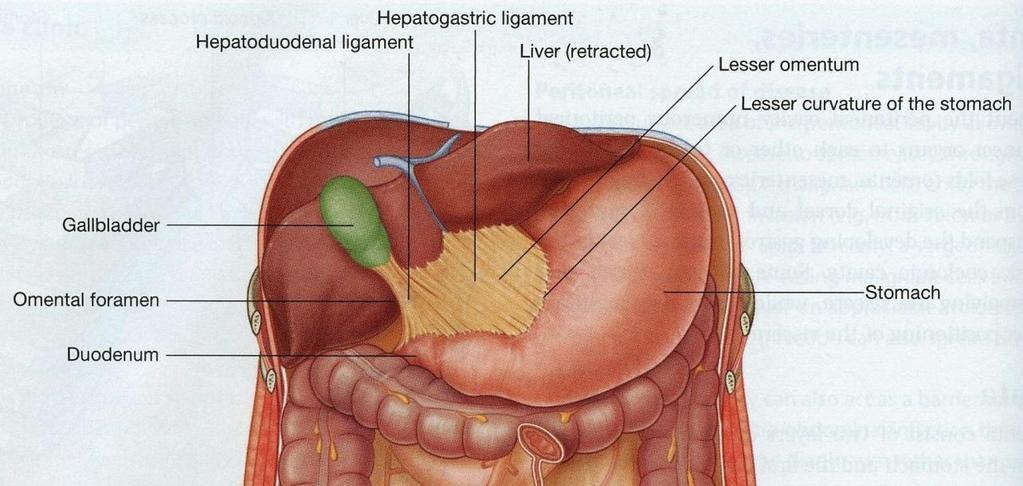
65 Lig. hepatogastricum Lig. hepatoduodenale Contain: - Ductus hepaticus communis - A. hepatica - V. porta Omentum minus Omentum minus - ventral mesentery of the stomach
 Omentum majus - dorsal mesentery of the")
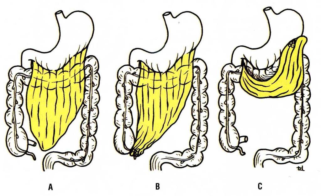
66 Omentum majus (4 layers of peritoneum) Anterior lamina (2 layers of peritoneum) + Posterior lamina (2 layers of peritoneum) Omentum majus - dorsal mesentery of the stomach

67 Omentum majus Lig. gastrophrenicum Lig. gastrosplenicum Lig. splenophrenicum Lig. gastrocolicum

68 Bursa omentalis

69 Omental foramen (Winslow`s foramen) Connects busra omentalis with the whole peritoneal cavity Walls: Superior lobus caudatus hepatis Anterior lig. hepatoduodenum Inferior duodenum Posterior parietal peritoneum

70
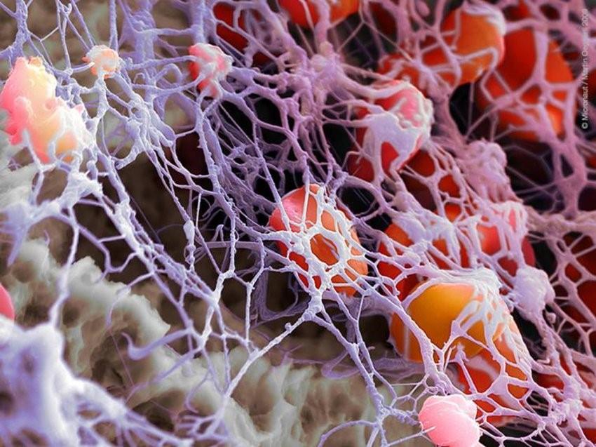
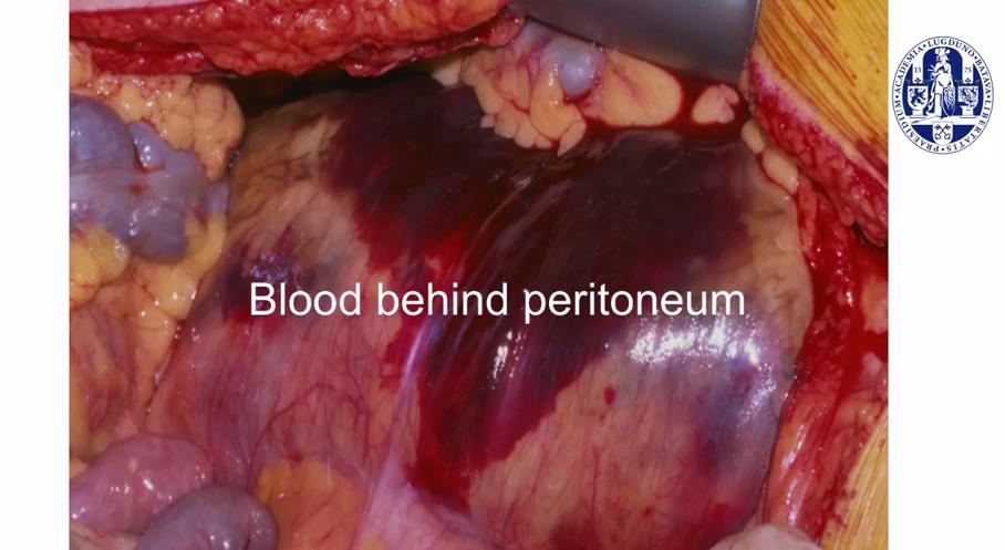
71 Fibrin
72
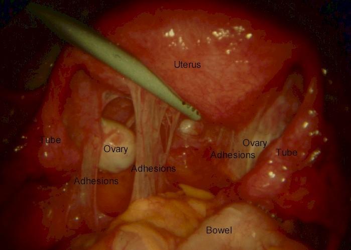
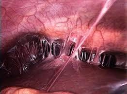
73 Adhesions
74 Sinuses (infracolic spaces, right and left)

75 Paracolic gutter, right and left

76 Recesses of peritoneal cavity
 Ligament of Treitz Recessus")

77 Recesses of peritoneal cavity (near duodenum) Ligament of Treitz Recessus duodenalis superior et inferior Recessus paraduodenalis Recessus supraduodenalis

78 Recesses of peritoneal cavity (near caecum) Recessus ileocaecalis superior 2 - Recessus ileocaecalis inferior 3 - Recessus retrocaecalis

79 Recesses of peritoneal cavity (near sigmoid colon)

80 Internal surface, the lower part of the anterior abdominal wall 1 plica umbilicalis mediana (obliterated urachus) 2 plica umbilicalis medialis 3 plica umbilicalis lateralis 4 fossa supravesicalis 5 fossa inguinalis medialis 6 fossa inguinalis lateralis

 Excavatio")
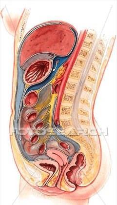
81 Peritoneum in pelvic cavity Excavatio rectovesicalis Excavatio rectouterina (Douglas pouch) Excavatio vesicouterina


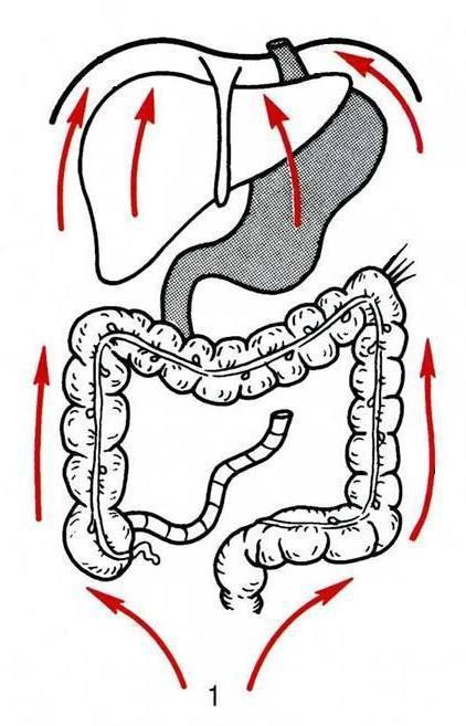
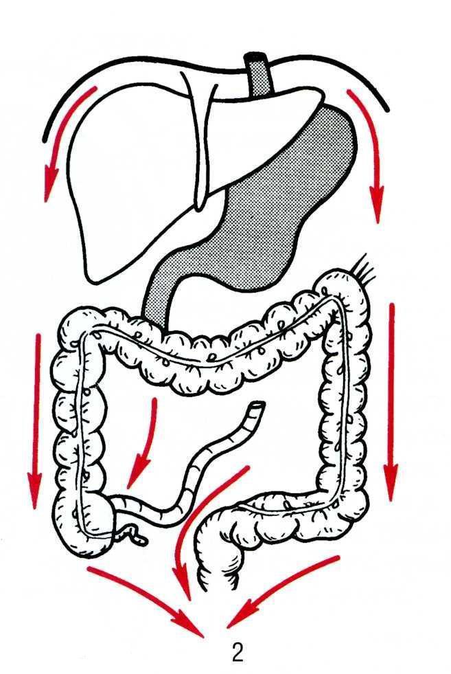
82 Peritoneal fluid - serose fluid, 75ml Functions: - moistens the peritoneum - decreases friction between organs Excavatio hepatorenalis Excavatio rectovesicalis Pelvic cavity
")

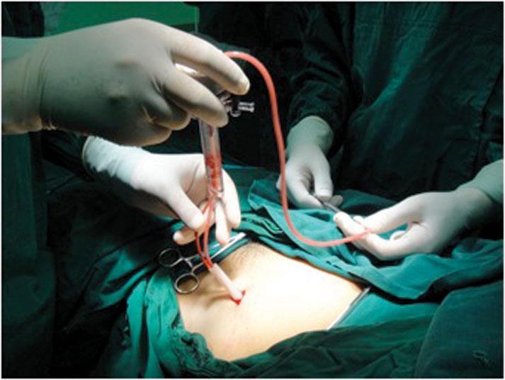
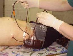
83 Ascite (abdominal dropsy) Accumulation of fluid in peritoneal cavity (liver cirrhosis, hepatic cancer or heart insufficiency) Charaf-ed-Din. Surgical puncture of the abdominal cavity of the aspiration of peritoneal fluid with a canula on a patient suffering from dropsy, 1466
84
85
86 THANK YOU FOR YOUR ATTENTION!
Development of the Digestive System. W.S. O The University of Hong Kong
 Development of the Digestive System W.S. O The University of Hong Kong Plan for the GI system Then GI system in the abdomen first develops as a tube suspended by dorsal and ventral mesenteries. Blood
Development of the Digestive System. W.S. O School of Biomedical Sciences, University of Hong Kong.
 Development of the Digestive System W.S. O School of Biomedical Sciences, University of Hong Kong. Organization of the GI tract: Foregut (abdominal part) supplied by coeliac trunk; derivatives include
Embryology of the Midgut and Hind gut
 Embryology of the Midgut and Hind gut Prof. Abdulameer Al-Nuaimi E-mail: a.al-nuaimi@sheffield.ac.uk E-mail: abdulameerh@yahoo.com Abdominal organs www.google.co.uk/search? Development of Duodenum The
The embryonic endoderm initially is widely connected with the yolk sac. As a consequence of cephalocaudal and lateral folding, a portion of the
 DIGESTIVE SYSTEM The embryonic endoderm initially is widely connected with the yolk sac. As a consequence of cephalocaudal and lateral folding, a portion of the endoderm-lined yolk sac cavity is incorporated
Exploring Anatomy: the Human Abdomen
 Exploring Anatomy: the Human Abdomen PERITONEUM AND PERITONEAL CAVITY PERITONEUM The peritoneum is a thin serous membrane that lines the abdominal cavity and covers, in variable amounts, the viscera within
The Foregut. At first the esophagus is short. but with descent of the heart and lungs it lengthens rapidly
 GI embryology 2 The Foregut At first the esophagus is short but with descent of the heart and lungs it lengthens rapidly The muscular coat, which is formed by surrounding splanchnic mesenchyme, is striated
Development of pancreas and Small Intestine. ANATOMY DEPARTMENT DR.SANAA AL-AlSHAARAWY DR.ESSAM Eldin Salama
 Development of pancreas and Small Intestine ANATOMY DEPARTMENT DR.SANAA AL-AlSHAARAWY DR.ESSAM Eldin Salama OBJECTIVES At the end of the lecture, the students should be able to : Describe the development
In the name ofgod. Abdomen 3. Dr. Zahiri
 In the name ofgod Abdomen 3 Dr. Zahiri Peritoneum Peritoneum It is the serous membrane(a type of loose connective tissue and is covered by mesothelium) that lines the abdominal cavity. Extensions of the
The Abdomen. Surface Anatomy, Vessels, Muscles, and Peritoneum
 The Abdomen Surface Anatomy, Vessels, Muscles, and Peritoneum Surface Anatomy Anterior abdominal wall extends from costal margin to inferior boundaries: Iliac crest Anterior superior iliac spine Inguinal
Fareed Khdair, MD Assistant Professor Chief, Section of Pediatric Gastroenterology, Hepatology, and Nutrition University of Jordan School of Medicine
 Fareed Khdair, MD Assistant Professor Chief, Section of Pediatric Gastroenterology, Hepatology, and Nutrition University of Jordan School of Medicine Outline Lecture one : Gut formation Foregut: esophagus,
Embryology - GIT - Lecture 2
 Embryology - GIT - Lecture 2 Last time we talked about embryology of the GIT. We said that the development of the stomach is accompanied with the development of the duodenum and the pancreas. Also we talked
Midgut. Over its entire length the midgut is supplied by the superior mesenteric artery
 Gi Embryology 3 Midgut the midgut is suspended from the dorsal abdominal wall by a short mesentery and communicates with the yolk sac by way of the vitelline duct or yolk stalk Over its entire length the
The peritoneum. Prof. Oluwadiya KS, MBBS, FMCS(Orthop) Website:
 Website:") The peritoneum Prof. Oluwadiya KS, MBBS, FMCS(Orthop) Website: http://oluwadiya.com The peritoneum Serous membrane that lines the abdominopelvic cavity and invests the viscera The largest serous membrane
The peritoneum Prof. Oluwadiya KS, MBBS, FMCS(Orthop) Website: http://oluwadiya.com The peritoneum Serous membrane that lines the abdominopelvic cavity and invests the viscera The largest serous membrane
Peritoneum: Def. : It is a thin serous membrane that lines the walls of the abdominal and pelvic cavities and clothes the viscera.
 Peritoneum: Def. : It is a thin serous membrane that lines the walls of the abdominal and pelvic cavities and clothes the viscera. Layers of the peritoneum: 1. Outer Layer ( Parietal Peritoneum) : lines
Anatomy: Know Your Abdomen
 Anatomy: Know Your Abdomen Glossary Abdomen - part of the body below the thorax (chest cavity); separated by the diaphragm. Anterior - towards the front of the body. For example, the umbilicus is anterior
2/2/2011. Primitive Gut Tube Proctodeum and Stomodeum Stomach Duodenum Pancreas Liver and Biliary Apparatus Spleen Midgut
 DEVELOPMENT OF THE DIGESTIVE SYSTEM Development of Endodermal Organs Primitive Gut Tube Proctodeum and Stomodeum Stomach Duodenum Pancreas Liver and Biliary Apparatus Spleen Midgut Wednesday, February
- Tamara Wahbeh. - Fareed Khdair. 0 P a g e
 -1 - Tamara Wahbeh - - Fareed Khdair 0 P a g e GI Embryology Note: I included everything in the records and slides; anything in the slide not included in this sheet was not mentioned by the doctor during
Preview from Notesale.co.uk Page 1 of 34
 Abdominal viscera and digestive tract Digestive tract Abdominal viscera comprise majority of the alimentary system o Terminal oesophagus, stomach, pancreas, spleen, liver, gallbladder, kidneys, suprarenal
Anatomy of the Large Intestine
 Large intestine Anatomy of the Large Intestine 2 Large Intestine Extends from ileocecal valve to anus Length = 1.5-2.5m = 5 feet Regions Cecum = 2.5-3 inch Appendix= 3-5 inch Colon Ascending= 5 inch Transverse=
SUBJECTS 2nd year, 1st semester I. 1. Primitive gut - limits, derivatives 2. Foregut -limits, evolution, derivatives 3. Midgut -limits, evolution,
 SUBJECTS 2nd year, 1st semester I. 1. Primitive gut - limits, derivatives 2. Foregut -limits, evolution, derivatives 3. Midgut -limits, evolution, derivatives 4. Hindgut- limits, evolution, derivatives
Duodenum retroperitoneal
 Duodenum retroperitoneal C shaped Initial region out of stomach into small intestine RETROperitoneal viscus Superior 1 st part duodenal cap ; moves upwards and backwards to lie on the R crura medial to
BLOCK IV: OFFICIAL BODY PARTS LIST FOR ANTERIOR ABDOMINAL WALL AND ABDOMINAL CONTENTS
 BLOCK IV: OFFICIAL BODY PARTS LIST FOR ANTERIOR ABDOMINAL WALL AND ABDOMINAL CONTENTS External oblique muscle Muscular portion Aponeurotic portion Superficial inguinal ring Lateral (inferior) crus Medial
Block 3: DISSECTION 2 CELIAC TRUNK, JEJUNUM/ILEUM, LARGE INTESTINE, DUODENUM, PANCREAS, PORTAL VEIN; MOBILIZATION OF THE LIVER
 1 Block 3: DISSECTION 2 CELIAC TRUNK, JEJUNUM/ILEUM, LARGE INTESTINE, DUODENUM, PANCREAS, PORTAL VEIN; MOBILIZATION OF THE LIVER Attempt to complete as much as you can of the dissection explained in the
ANATOMY OF THE SMALL & LARGE INTESTINES. Semester 1, 2011 A. Mwakikunga
 ANATOMY OF THE SMALL & LARGE INTESTINES Semester 1, 2011 A. Mwakikunga LEARNING OBJECTIVES 1. List the parts and anatomical regions of the small and large intestines 2. State anatomical relations of the
Biology Human Anatomy Abdominal and Pelvic Cavities
 Biology 351 - Human Anatomy Abdominal and Pelvic Cavities Please place your name and I.D. number on the back of the last page of this exam. You must answer all questions on this exam. Because statistics
Mousa Salah. Dr. Mohammad Al. Mohtasib. 1 P a g e
 8 Mousa Salah Dr. Mohammad Al. Mohtasib 1 P a g e In the previous lecture we talked about the peritoneum, and we said that the peritonium is a serous sac, and it consists of two layers, visceral and parietal.
MICROSCOPIC STRUCTURE OF LIVER, GALLBLADDER, GALL DUCTS, AND PANCREAS OVERVIEW OF DEVELOPMENT OF THE ALIMENTARY CANAL
 Lecture 2 ESS_3rd semester MICROSCOPIC STRUCTURE OF LIVER, GALLBLADDER, GALL DUCTS, AND PANCREAS OVERVIEW OF DEVELOPMENT OF THE ALIMENTARY CANAL MICROSCOPIC STRUCTURE OF LIVER - is the largest gland of
Small Plicae Circularis. Short Closely packed together. Sparse, completely absent at distal part Lymphoid Nodule
 Intestines Differences Between Jejunum and Ileum Types Jejunum Ileum Color Deeper red Paler pink Calibre Bigger Smaller Thickness of wall Thick and Heavy Thin and Lighter Vascularity Highly vascularised
Lectures of Human Embryology
 Lectures of Human Embryology "Body Cavities & GIT" By DR. ABDEL-MONEM AWAD HEGAZY M.B. with honor 1983, Dipl."Gynecology and Obstetrics "1989, Master "Anatomy and Embryology" 1994, M.D. "Anatomy and Embryology"
THE ORAL CAVITY
 THE ORAL CAVITY WALL OF ABDOMEN (ANTERIOR) The paraumbilical vein drains into the portal vein and then through the liver. This is an important clinical connection. THE ABDOMINAL VISCERA The small
The Digestive System and Body Metabolism
 14 PART B The Digestive System and Body Metabolism PowerPoint Lecture Slide Presentation by Jerry L. Cook, Sam Houston University ESSENTIALS OF HUMAN ANATOMY & PHYSIOLOGY EIGHTH EDITION ELAINE N. MARIEB
This lab activity is aligned with Visible Body s Human Anatomy Atlas app. Learn more at visiblebody.com/professors
 1 This lab activity is aligned with Visible Body s Human Anatomy Atlas app. Learn more at visiblebody.com/professors 2 A. Digestive System Overview To Start: Go to the Views menu and scroll down to the
Overview of the Digestive
 Overview of the Digestive System Bởi: OpenStaxCollege The function of the digestive system is to break down the foods you eat, release their nutrients, and absorb those nutrients into the body. Although
Jhia Anjela D. Rivera 1 1. BS Biology, Department of Biology, College of Science, Polytechnic University of the Philippines
 DIGESTIVE SYSTEM Jhia Anjela D. Rivera 1 1 BS Biology, Department of Biology, College of Science, Polytechnic University of the Philippines DIGESTIVE SYSTEM Consists of the digestive tract (gastrointestinal
Netter's Anatomy Flash Cards Section 4 List 4 th Edition
 Netter's Anatomy Flash Cards Section 4 List 4 th Edition https://www.memrise.com/course/1577335/ Section 4 Abdomen (31 cards) Plate 4-1 Bony Framework of Abdomen 1.1 Costal cartilages 1.2 Iliac crest 1.3
ANATOMY OF THE DIGESTIVE SYSTEM PART II
 ANATOMY OF THE DIGESTIVE SYSTEM PART II 9.12.2014 Kaan Yücel M.D., Ph.D. http://fhs121.org Dr.Kaan Yücel http://fhs121.org Digestive system Part II 1. LIVER The liver is the largest gland in the body and,
Anatomical Considerations for Lab Practical II
 Anatomical Considerations for Lab Practical II For each of the following please be prepared to provide: Identification System Organ(s) or ducts to Function(s) location which it is attached Use your lecture
Al-Mohtaseb. Saba Alfayoumi. Mo Alfarra
 8 Al-Mohtaseb Saba Alfayoumi Mo Alfarra For the comparison purposes refer to the last page where you can find a table that summarizes them. Enjoy Jejunum and Ileum -They're intraperitoneal and freely mobile
STRUCTURAL BASIS OF MEDICAL PRACTICE EXAMINATION 3. October 16, 2015
 STRUCTURAL BASIS OF MEDICAL PRACTICE EXAMINATION 3 October 16, 2015 PART l. Answer in the space provided. (12 pts) 1. Identify the structures. (2 pts) A. B. A B C. D. C D 2. Identify the structures. (2
The abdominal Esophagus, Stomach and the Duodenum. Prof. Oluwadiya KS
 The abdominal Esophagus, Stomach and the Duodenum Prof. Oluwadiya KS www.oluwadiya.com Viscera of the abdomen Abdominal esophagus: Terminal part of the esophagus The stomach Intestines: Small and Large
Development of the Liver and Pancreas
 Development of the Liver and Pancreas Professor Alfred Cuschieri Department of Anatomy University of Malta Three glandular buds arise from the distal end of the foregut during the fourth week Day 22 -The
The Human Body: An Overview of Anatomy. Anatomy. Physiology. Anatomy - Study of internal and external body structures
 C H A P T E R 1 The Human Body: An Orientation An Overview of Anatomy Anatomy The study of the structure of the human body Physiology The study of body function Anatomy - Study of internal and external
The Digestive System
 The Digestive System Identify the Structure and Function. Mesentery of the Large Intestine The mesentery functions to connect the visceral organs to the abdominal wall. Identify the Structure. Nasal Cavity
Bushra Arafa Zayed & Hanan Jamal. - Dana AF
 - 10 - Bushra Arafa Zayed & Hanan Jamal - Dana AF - Mohammad Al Muhtaseb Notes: This sheet was written in the same order as the slides, and everything in the slides is mentioned in this sheet. Pictures
Digestive System. In one end and out the other.
 Digestive System In one end and out the other. Overview Every cell in the body needs nourishment, yet most cells cannot leave their position in the body and travel to a food source, so the food must be
Lab Monitor Images Dissection of the Abdominal Vasculature + Lower Digestive System
 Lab Monitor Images Dissection of the Abdominal Vasculature + Lower Digestive System Stomach & Duodenum Frontal (AP) View Nasogastric tube 2 1 3 4 Stomach Pylorus Duodenum 1 Duodenum 2 Duodenum 3 Duodenum
- Digestion occurs during periods of low activity - Produces more energy than it uses. - Mucosa
 Introduction Digestive System Chapter 29 Provides processes to break down molecules into a state easily used by cells - A disassembly line: Starts at the mouth and ends at the anus Digestive functions
Digestive System 7/15/2015. Outline Digestive System. Digestive System
 Digestive System Biology 105 Lecture 18 Chapter 15 Outline Digestive System I. Functions II. Layers of the GI tract III. Major parts: mouth, pharynx, esophagus, stomach, small intestine, large intestine,
Dr. Zahiri. In the name of God
 Dr. Zahiri In the name of God small intestine = small bowel is the part of the gastrointestinal tract Boundaries: Pylorus Ileosecal junction Function: digestion and absorption of food It receives bile
8. Development of digestive system II. Rotation if intestine. Liver, pancreas, spleen. Development of respiratory passages and lung.
 8. Development of digestive system II. Rotation if intestine. Liver, pancreas, spleen. Development of respiratory passages and lung. Duodenum originates partially from the terminal part of the foregut,
-Ensherah Mokheemer. -Shatha Al-Jaberi محمد المحتسب- 1 P a g e
 9-9 -Ensherah Mokheemer -Shatha Al-Jaberi محمد المحتسب- 1 P a g e Small intestine has three regions: ( االثني عشر( The duodenum The jejunum The ileum Small intestine Duodenum: -c-shaped -The concavity
ABDOMEN - GI. Duodenum
 TALA SALEH ABDOMEN - GI Duodenum - Notice the shape of the duodenum, it looks like capital G shape tube which extends from the pyloroduodenal junction to the duodenojejunal junction. - It is 10 inches
Pancreas & Biliary System. Dr. Vohra & Dr. Jamila
 Pancreas & Biliary System Dr. Vohra & Dr. Jamila 1 Objectives At the end of the lecture, the student should be able to describe the: Location, surface anatomy, parts, relations & peritoneal reflection
Exercise. Digestive System. Digestive system function. 1. Define the following terms: a. Chemical digestionb. Mechanical digestionc.
 Exercise 7 The Digestive System NAME: DATE: INSTRUCTOR: SECTION: Digestive system function 1. Define the following terms: a. Chemical digestionb. Mechanical digestionc. Ingestiond. Digestione. Absorptionf.
DIGESTIVE SYSTEM ALIMENTARY CANAL / GI TRACT & ACCESSORY ORGANS. Mar 16 10:34 PM
 DIGESTIVE SYSTEM ALIMENTARY CANAL / GI TRACT & ACCESSORY ORGANS Mar 16 10:34 PM 1 I. Digestive System Functions > Ingestion the taking in of food > Propulsion movement caused by force > Digestion breakdown
Accessory Glands of Digestive System
 Accessory Glands of Digestive System The liver The liver is soft and pliable and occupies the upper part of the abdominal cavity just beneath the diaphragm. The greater part of the liver is situated under
Lecture 1. Introduction to splanhnology. Digestive system
 Lecture 1 Introduction to splanhnology. Digestive system SPLANCHNOLOGY (THE SCIENCE OF THE VISCERA) SOMATOLOGY SPLANCHNOLOGY The general cover body (skin) The skeletal system The connection system The
Lab 9 Abdomen MUSCLES
 Lab 9 Abdomen MUSCLES External abdominal oblique continuous with the external intercostal muscle; its fibers point in a caudal direction as it moves anteriorly until it inserts on the linea alba via its
STRUCTURAL BASIS OF MEDICAL PRACTICE EXAMINATION 3. October 17, 2014
 STRUCTURAL BASIS OF MEDICAL PRACTICE EXAMINATION 3 October 17, 2014 PART l. Answer in the space provided. (12 pts) 1. Identify the structures. (2 pts) A. B. A B C. D. C D 2. Identify the structures. (2
The stomach is formed of three parts: -
 The stomach is formed of three parts: - (a) CARDIAC STOMACH: - It receives the oesophagus through Cardiac aperture guarded by a cardiac sphincter which prevents regurgitation of food. (b) FUNDIC PART:
Lab 8: Digestive System
 BIOL 221 A&P II Lab 8: Digestive System Become familiar with the gross anatomy of the digestive system (Exercise 38) using the models, Fig. 38.1 (Activity 1), and the rat. Recognize and know the functions
To describe the liver. To list main structures in porta hepatis.
 GI anatomy Lecture: 6 د. عصام طارق Objectives: To describe the liver. To list main structures in porta hepatis. To define portal system & portosystemic anastomosis. To list parts of biliary system. To
- Digestion occurs during periods of low activity - Produces more energy than it uses. 3 Copyright 2016 by Elsevier Inc. All rights reserved.
 Introduction Digestive System Chapter 29 Provides processes to break down molecules into a state easily used by cells - A disassembly line: Starts at the mouth and ends at the anus Digestive functions
Embryology of the Heart
 *Page 1A: Embryology of the Heart Human embryonic disc is divided into three layers: ectoderm, intraembryonic mesoderm, and endoderm. The embryonic disc lies between the amniotic cavity and the primary
Bio 104 Digestive System
 13 Lecture Outline: Digestive System Hole s HAP [Chapters 17 & 18] General Characteristics of the Alimentary Canal A. Functions 1. Ingestion 2. Mechanical digestion 3. Chemical digestion 4. Propulsion
Surface Anatomy. Location Shape Weight Role of Five Surfaces Borders Fissures Lobes Peritoneal Lig
 The Liver Functions Bile production and secretion Detoxification Storage of glycogen Protein synthesis Production of heparin and bile pigments Erythropoiesis (in fetus) Surface Anatomy Location Shape Weight
Embryology: Development of digestive system
 Embryology: Development of digestive system Embryo folding incorporation of endoderm to form primitive gut. Outside of embryo yolk sac and allantois. Vitelline duct Stomodeum (primitive mouth) the oral
ANATOMY AND BASIC FUNCTION OF THE ENDOCRINE GLANDS
 ANATOMY AND BASIC FUNCTION OF THE ENDOCRINE GLANDS Know these endocrine organs of the cat: thymus, thyroid, pancreas, adrenal glands, ovaries, and testes. Review and know microslides, hormones, and structures
Midterm 2 is Tuesday 5/28/13
 Business Reminder: No class Monday (Memorial Day) Midterm 2 is Tuesday 5/28/13 Optional review session tomorrow @ 5pm Homework due in Lab 1. PreLab 8 (1pt) 2. Replace a Missing Assignment (4 pts) Homework
ACTIVITY 11: RESPIRATORY AND DIGESTIVE SYSTEMS RESPIRATORY SYSTEM
 ACTIVITY 11: RESPIRATORY AND DIGESTIVE SYSTEMS OBJECTIVES: 1) How to get ready: Read Chapters 25 and 26, McKinley et al., Human Anatomy, 4e. All text references are for this textbook. 2) Identify structures
Anatomy of the SMALL INTESTINE. Dr. Noman Ullah Wazir PMC
 Anatomy of the SMALL INTESTINE Dr. Noman Ullah Wazir PMC SMALL INTESTINE The small intestine, consists of the duodenum, jejunum, and illium. It extends from the pylorus to the ileocecal junction were the
-12. -Renad Habahbeh. -Dr Mohammad mohtasib
 -12 -Renad Habahbeh - -Dr Mohammad mohtasib The Gallbladder -The gallbladder has a body, a fundus (a rounded end), a neck, Hartmann s pouch before the neck and a cystic duct that meets the common hepatic
Organizational Overview of Thorax, Abdomen, Pelvis. Introduction to Blood
 Biology 224 Human Anatomy and Physiology - II Week 1; Lecture 2; Wednesday Dr. Stuart S. Sumida Organizational Overview of Thorax, Abdomen, Pelvis Introduction to Blood Organizational Overview of Thorax,
Development of Gastrointestinal Tract
 11 Development of Gastrointestinal Tract Learning Objectives At the end of this chapter, students would be able to define and understand the following: Development of the esophagus and stomach Rotation
د. عصام طارق. Objectives:
 GI anatomy Lecture: 5 د. عصام طارق Objectives: To describe anatomy of stomach, duodenum & pancreas. To list their main relations. To define their blood & nerve supply. To list their lymph drainage. To
The Digestive System Laboratory
 The Digestive System Laboratory 1 The Digestive Tract The alimentary canal is a continuous tube stretching from the mouth to the anus. Liver Gallbladder Small intestine Anus Parotid, sublingual, and submaxillary
RESPIRATORY SYSTEM. described: pp. 744,746 fig. 25.1, described: p. 746 fig described: p. 776 fig. 26.3
 ACTIVITY 11: RESPIRATORY AND DIGESTIVE SYSTEMS OBJECTIVES: 1) How to get ready: Read Chapters 25 and 26, McKinley et al., Human Anatomy, 5e. All text references are for this textbook. 2) Identify structures
Group B: Organ systems (digestive, respiratory, urinary, genital system, heart, glands and skin) green
 green") Group B: Organ systems (digestive, respiratory, urinary, genital system, heart, glands and skin) green Digestive system 1. Teeth Main points: external and internal structure of a tooth, fixation of a tooth
Group B: Organ systems (digestive, respiratory, urinary, genital system, heart, glands and skin) green Digestive system 1. Teeth Main points: external and internal structure of a tooth, fixation of a tooth
OVARIES URETER FALLOPIAN TUBES BLADDER UROGENITAL OPENINGS (BOTH SEXES) PENIS VAGINA UTERUS
 PENIS VAGINA UTERUS") URETER OVARIES FALLOPIAN TUBES BLADDER UROGENITAL OPENINGS (BOTH SEXES) PENIS VAGINA UTERUS REPRODUCTIVE PRODUCE FEMALE HORMONES EXCRETORY FROM KIDNEY TO BLADDER EXCRETORY STORES URINE REPRODUCTIVE TRANSPORTS
URETER OVARIES FALLOPIAN TUBES BLADDER UROGENITAL OPENINGS (BOTH SEXES) PENIS VAGINA UTERUS REPRODUCTIVE PRODUCE FEMALE HORMONES EXCRETORY FROM KIDNEY TO BLADDER EXCRETORY STORES URINE REPRODUCTIVE TRANSPORTS
Ex. 1 :Language of Anatomy
 Collin College BIOL 2401 : Human Anatomy & Physiology Ex. 1 :Language of Anatomy The Anatomical Position Used as a reference point when referring to specific areas of the human body Body erect Head and
GASTROINTESTINAL SYSTEM
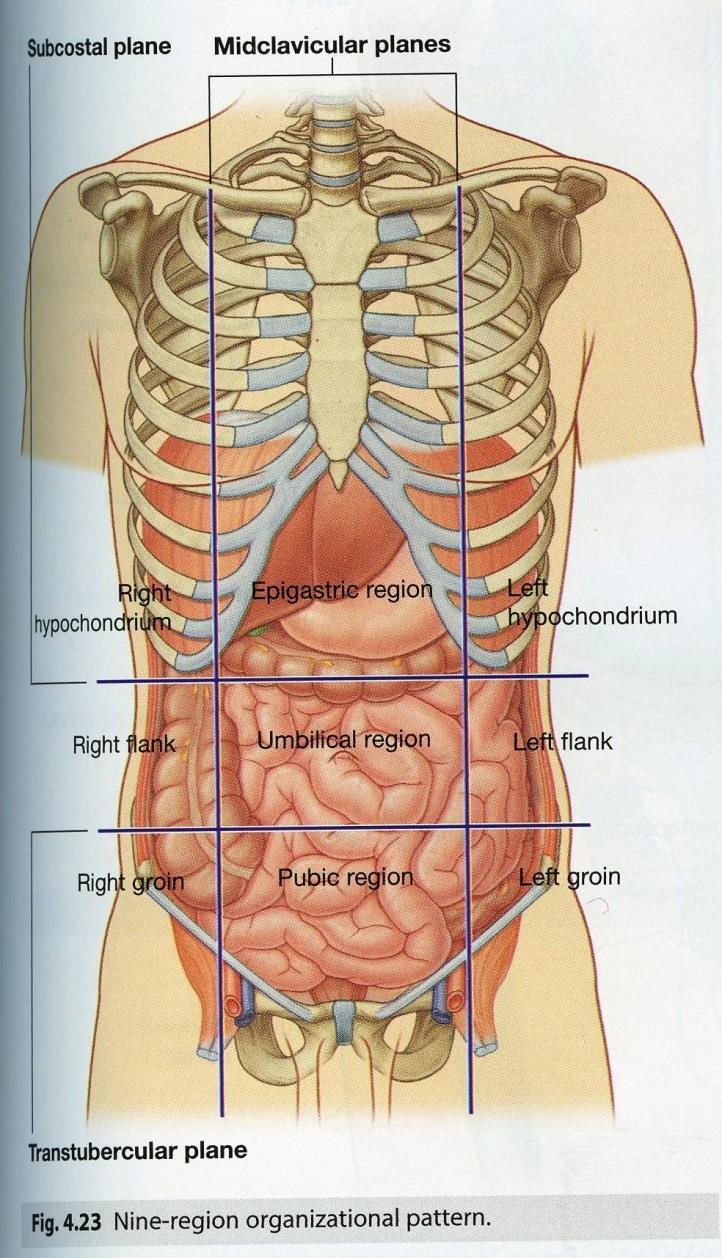
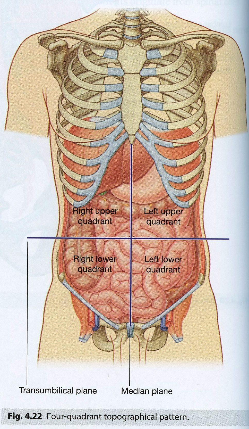
 GASTROINTESTINAL SYSTEM Topographic Anatomy of the Abdomen Surface Landmarks Xiphoid process T9/T10 Inferior costal margin L2/L3 Iliac Crest L4 level ASIS L5/S1 level Pubic symphysis level of greater trochanter
Respiratory & Digestive Organs of the Head and Neck, Human;
 Name Date Lab Exercise 5: Lab Exercise 6: Lab Exercise 7: Lab Exercise 8: Respiratory & Digestive Organs of the Head and Neck, Human; Histology of the Respiratory System Digestive System Models, Human
DIGESTIVE SYSTEM. Chapter 25
 DIGESTIVE SYSTEM Chapter 25 DIGESTIVE SYSTEM Digestive Tract Mouth Pharynx Esophagus Stomach Small intestines Large intestines Anus Accessory Organs Teeth Tongue Salivary glands Pancreas Liver Gallbladder
Body Regions Review. Anatomical Position. Anatomical Planes. Supine versus Prone 9/9/2009
 Body Regions Review The fundamental divisions of the human body Christine Sparks Anatomy / Physiology I Sept. 9, 2009 Anatomical Position Universal terms are used to describe the body accurately and result
Abdomen. Retroperitoneal space
 Abdomen. Retroperitoneal space Abdominal cavity The space bounded by: Anterolateral abdominal wall Posterior abdominal wall Diaphragm Pelvic walls and pelvic floor. Subdivided into: True abdominal cavity
Peritoneal cavity. Infracolic compartment. Assoc. prof. dr. S. Delchev, MD, PhD
 Peritoneal cavity. Infracolic compartment Assoc. prof. dr. S. Delchev, MD, PhD Infracolic compartment The infracolic compartment lies inferior to the transverse mesocolon and posterior to the greater omentum
Lecture #2. Respiratory system. Development
 Lecture #2 Respiratory system. Development Respiratory system - is a biological system consisting of specific organs and structures used for the process of respiration in an organism Breathing and Respiration
The Digestive System. Chapter 25
 The Digestive System Chapter 25 Introduction Structure of the digestive system A tube that extends from mouth to anus Accessory organs are attached Functions include Ingestion Movement Digestion Absorption
Done by: nisreen obeidat
 Sheet: liver and pancreas Done by: nisreen obeidat Embryology of the liver The liver develops in the ventral mesentery of the foregut and divides the ventral mesentery :into 1)lesser omentum (between the
Biology 340 Comparative Embryology Lecture 10 Dr. Stuart Sumida. Further Development of the Mesoderm (and Endoderm)
") Biology 340 Comparative Embryology Lecture 10 Dr. Stuart Sumida Further Development of the Mesoderm (and Endoderm) Further Development: Digestive System Foregut, Midgut, Hindgut Heart and Aortic Arches
Biology 340 Comparative Embryology Lecture 10 Dr. Stuart Sumida Further Development of the Mesoderm (and Endoderm) Further Development: Digestive System Foregut, Midgut, Hindgut Heart and Aortic Arches
Chapter 7 The digestive system
 41 Chapter 7 The digestive system Primitive gut tube 41 Foregut 42 Other foregut derivatives 44 Midgut 45 Hindgut 47 4 Fig. 7.1 The gut tube in a 4-week embryo. Pharynx Foregut Respiratory diverticulum
Anatomy of the spleen. Oluwadiya KS
 Anatomy of the spleen Oluwadiya KS www.oluwadiya.com Introduction The spleen is an ovoid, usually purplish, pulpy mass about the size and shape of one's fist. It is the largest lymphoid tissue in the body
Overview of the Digestive System *
 OpenStax-CNX module: m46506 1 Overview of the Digestive System * OpenStax This work is produced by OpenStax-CNX and licensed under the Creative Commons Attribution License 4.0 By the end of this section,
Nasogastric tube. Stomach. Pylorus. Duodenum 1. Duodenum 2. Duodenum 3. Duodenum 4
 Esophagus Barium Swallow Stomach and Duodenum 4 year old Upper GI Nasogastric tube Stomach and Duodenum 4 year old Upper GI Nasogastric tube Stomach Pylorus Duodenum 1 Duodenum 2 Duodenum 3 Duodenum 4
The jejunum and the Ileum. Prof. Oluwadiya KS
 The jejunum and the Ileum Prof. Oluwadiya KS www.oluwadiya.siteled.com Introduction Introduction The small intestine (SI) comprises of the duodenum, jejunum and the ileum The jejunum is the second part
Embryology of the Gut and Mesenteries, slide 7 This is very similar to a cross-section we looked at when were talking about formation of the diaphragm
 Embryology of the Gut and Mesenteries, slide 2 This is a median sagittal section of a four week embryo, most of you can probably draw it in your sleep. I made a few modifications from some of the earlier
consists of: Muscular, hollow tube (= digestive tract ) + Various accessory organs
 + Various accessory organs") DIGESTIVE SYSTEM consists of: Muscular, hollow tube (= digestive tract ) + Various accessory organs FUNCTION Individual parts function in: ingestion mechanical digestion chemical and enzymatic digestion
DIGESTIVE SYSTEM consists of: Muscular, hollow tube (= digestive tract ) + Various accessory organs FUNCTION Individual parts function in: ingestion mechanical digestion chemical and enzymatic digestion
Two main groups Alimentary canal continuous coiled hollow tube Accessory digestive organs
 Digestion Breakdown of ingested food Absorption of nutrients into the blood Metabolism Production of cellular energy (ATP) Constructive and degradative cellular activities Two main groups Alimentary canal
Biology Human Anatomy Abdominal and Pelvic Cavities
 Biology 351 - Human Anatomy Abdominal and Pelvic Cavities You must answer all questions on this exam. Because statistics demonstrate that, on average, between 2-5 questions on every 100-point exam are
The Urinary System Pearson Education, Inc.
 26 The Urinary System Introduction The urinary system does more than just get rid of liquid waste. It also: Regulates plasma ion concentrations Regulates blood volume and blood pressure Stabilizes blood