Development of the Digestive System. W.S. O School of Biomedical Sciences, University of Hong Kong.
|
|
|
- Mervin Bailey
- 6 years ago
- Views:
Transcription
1 Development of the Digestive System W.S. O School of Biomedical Sciences, University of Hong Kong.
2
3 Organization of the GI tract: Foregut (abdominal part) supplied by coeliac trunk; derivatives include oral cavity, oesophagus, stomach, duodenum, liver, gall bladder and pancreas Midgut supplied by superior mesenteric artery; derivatives include duodenum, small intestine, caecum, anterior 2/3 transverse colon Hindgut supplied by inferior mesenteric artery; derivatives include distal 1/3 transverse colon, descending colon, sigmoid colon, rectum and upper part of anal canal
4
5

6 Development of the foregut, midgut & hindgut
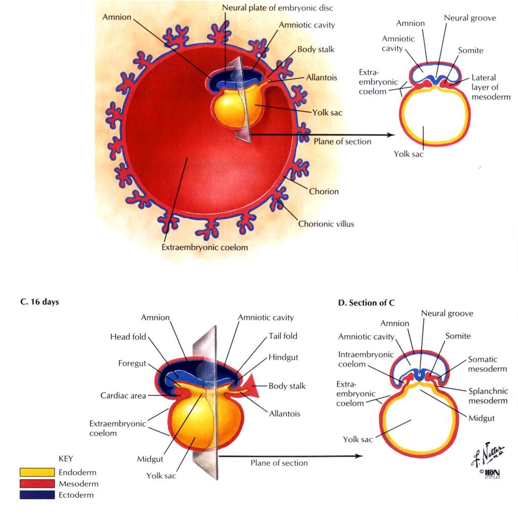
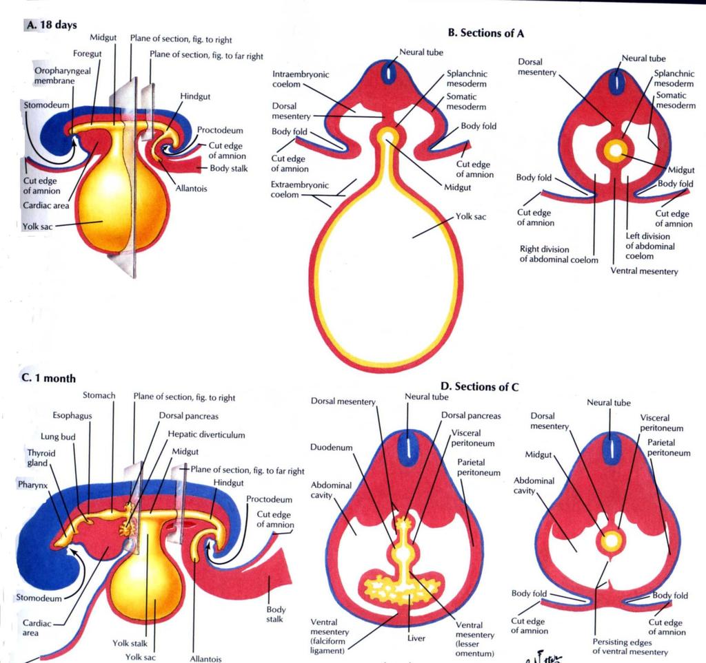
7 Early gut development Coelom formation Buccopharyngeal membrane (perforates around 4 th week) and cloacal membrane (~ 7 th week) Primitive gut wall: epithelial lining & associated glands endoderm Connective tissue & smooth muscle surrounding mesoderm
8 Development of the distal foregut-1 Oesophagus no coelomic cavity in thorax slow growth Stomach Rotate 90 o (clockwise, longitudinal axis) Rotate 90 o (clockwise, AP axis) Differential growth : ventral border slow growth (lesser curvature) dorsal border rapid growth (greater curvature)

9 vacuoles Rotation of the stomach around its longitudinal axis

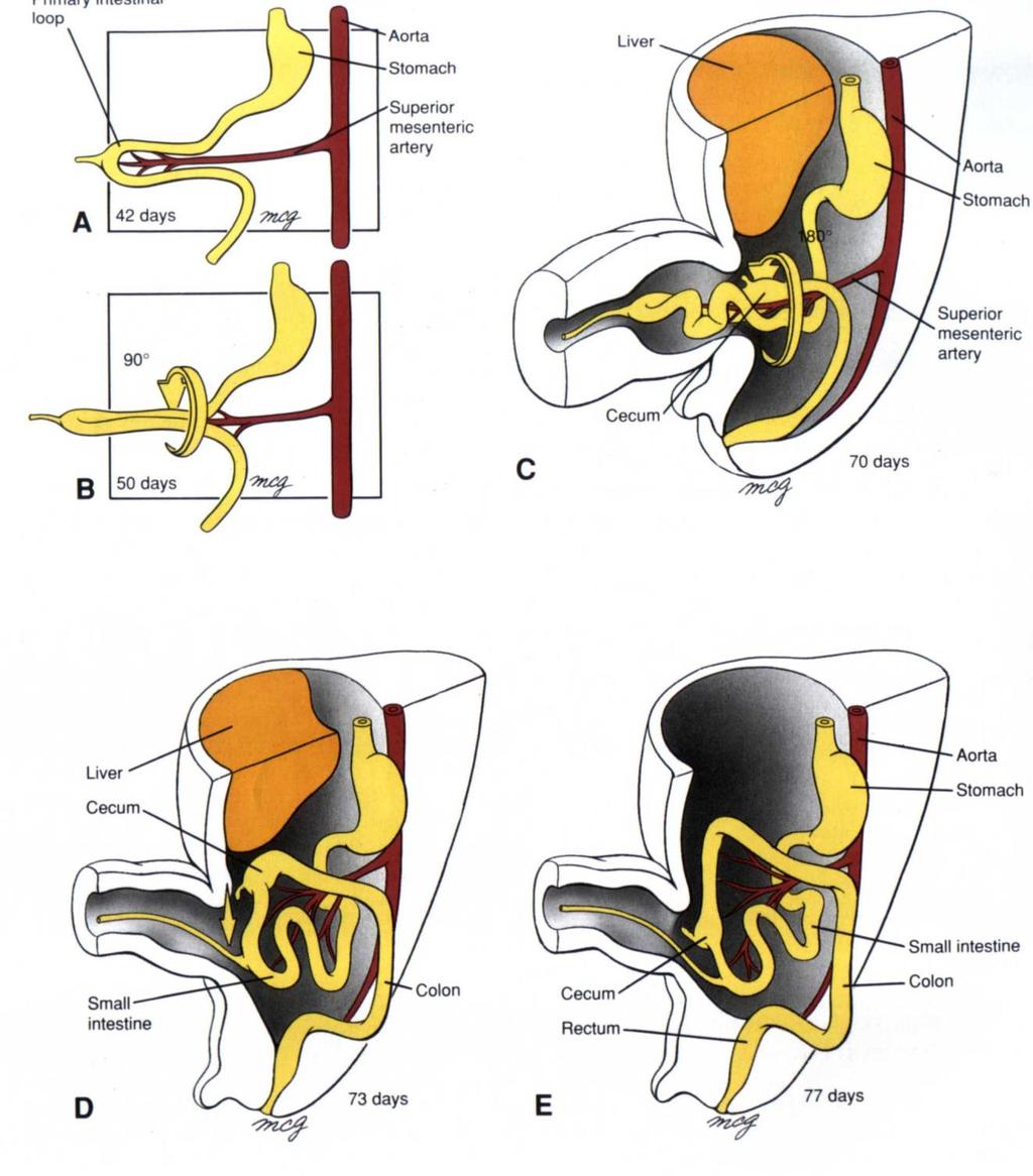
10 Rotates at longitudinal axis Rotates at AP axis D 56 days Rotations of the stomach
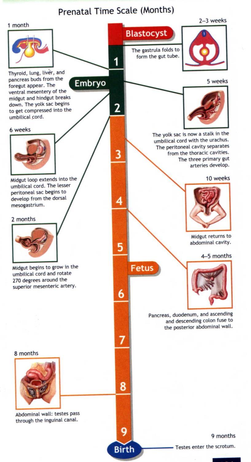
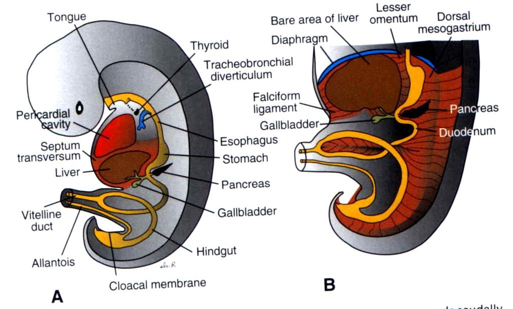
11 Development of the distal foregut-2 Duodenum pushed to the right and becomes secondarily retroperitoneal. Liver & gall bladder ventral outgrowth from duodenum: hepatic diverticulum and cystic diverticulum Pancreas dorsal pancreatic bud (main gland with head, body and tail) and ventral pancreatic bud (uncinate process); the two buds fuse after the stomach rotates.

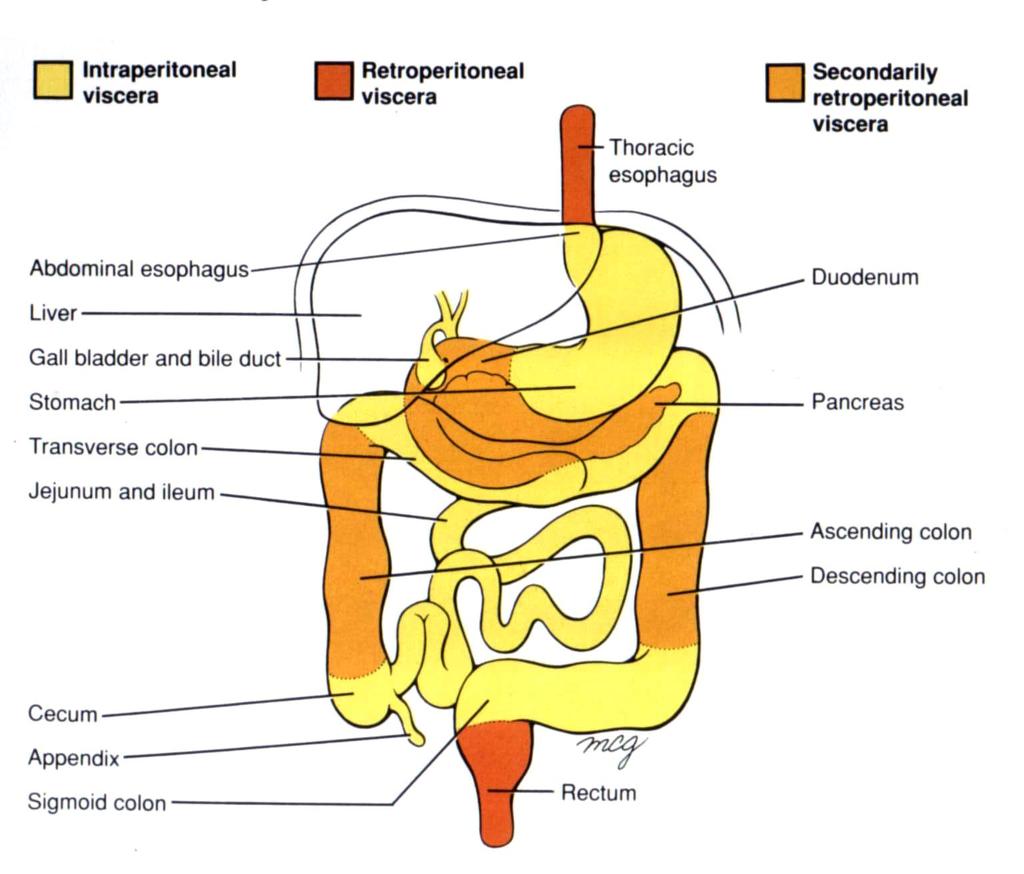
12 Mechanism by which portions of the gut become secondarily retroperitoneal

13 Development of the liver and pancreas
14 Anomalous pancreas

15 Formation of the liver and associated membranes

16 Development of the greater omentum and lesser sac
17 Development of the midgut - 1 Cranial limb of the midgut Characterized by rapid elongation and rotation Axis of rotation is around the superior mesenteric artery (dorsoventral axis) 90 o anticlockwise and herniates into the umbilical cord (~ 6-8 wk) Rapid elongation and retraction of herniated gut into abdominal cavity ~10 wk (further rotates 180 o ; i.e. a total of 270 o rotation)
18
19
20 Development of the midgut - 2 Mid point of midgut loop remains connected with yolk sac with a narrow vitelline duct embedded in the umbilicus Vitelline duct normally regresses between the 5-8 th week and later obliterates into a fibrous cord and degenerates complete. In 2% of the infants, abnormal remains of the vitelline duct forms Meckel s diverticulum; vitelline cyst or vitelline fistula.

21 Remnants of the vitelline duct
22 Development of the midgut - 3 Caudal limb of midgut is characterized by slow growth. Rotation of the cranial limb throws the caudal limb into an arch at the perimeter of the abdominal cavity. The caecum rests below the liver and later descends in the abdomen. The ascending and descending colon become secondarily retroperitoneal.
23
24
25 Development of the hindgut The distal end of the primitive gut expand to form the cloaca. Between the 4 th 6 th week, the cloaca is partitioned into a dorsal anorectal canal and a ventral primitive urogenital sinus by the growth of a coronal partition called the urorectal septum. The urorectal septum consists of a superior Tourneux fold and a pair of lateral folds called the Rathke folds.
26 Subdivision of the cloaca into an anterior primitve urogenital sinus and a posterior rectum (week 4-6).

27 Lower third of anorectal canal formed by ectodermal invagination
28 Inferior third of anorectal canal The superior two-third of the anorectal canal forms from the distal part of the hindgut. Inferior third formed from an ectodermal pit called the anal pit or proctodeum. The membrane separating the endoderm and ectoderm breaks in the 8 th week.(pectinate line) Superior to pectinate line supplied by branches of inferior mesenteric artery Inferior to pectinate line supplied by branches of the internal iliac arteries.
29 Congential malformations Imperforate anus anal membrane persists Imperforate anus with rectal atresia

30 Formation of definitive gut lumen

31 50% 20% 20% 5% The most common bowel atresias and stenoses Most are caused by vascular accidents; stenosis in the upper duodenum may be caused by a lack of recanalization.

32 Deficiency of anterior abdominal wall Congenital defect A & B omphalocele; C gastroschisis Combined incidents -1/2000 births
33 Reference: Sadler TW Langman s Medical Embryology 9 th edition, 2004, pp

34 Rotates 90 o anticlockwise without further 180 rotation Rotates 90 o clockwise Malformation during rotation of the gut Can you work out how these conditions come about?
Development of the Digestive System. W.S. O The University of Hong Kong
 Development of the Digestive System W.S. O The University of Hong Kong Plan for the GI system Then GI system in the abdomen first develops as a tube suspended by dorsal and ventral mesenteries. Blood
Midgut. Over its entire length the midgut is supplied by the superior mesenteric artery
 Gi Embryology 3 Midgut the midgut is suspended from the dorsal abdominal wall by a short mesentery and communicates with the yolk sac by way of the vitelline duct or yolk stalk Over its entire length the
Development of pancreas and Small Intestine. ANATOMY DEPARTMENT DR.SANAA AL-AlSHAARAWY DR.ESSAM Eldin Salama
 Development of pancreas and Small Intestine ANATOMY DEPARTMENT DR.SANAA AL-AlSHAARAWY DR.ESSAM Eldin Salama OBJECTIVES At the end of the lecture, the students should be able to : Describe the development
Embryology - GIT - Lecture 2
 Embryology - GIT - Lecture 2 Last time we talked about embryology of the GIT. We said that the development of the stomach is accompanied with the development of the duodenum and the pancreas. Also we talked
Embryology of the Midgut and Hind gut
 Embryology of the Midgut and Hind gut Prof. Abdulameer Al-Nuaimi E-mail: a.al-nuaimi@sheffield.ac.uk E-mail: abdulameerh@yahoo.com Abdominal organs www.google.co.uk/search? Development of Duodenum The
The embryonic endoderm initially is widely connected with the yolk sac. As a consequence of cephalocaudal and lateral folding, a portion of the
 DIGESTIVE SYSTEM The embryonic endoderm initially is widely connected with the yolk sac. As a consequence of cephalocaudal and lateral folding, a portion of the endoderm-lined yolk sac cavity is incorporated
Fareed Khdair, MD Assistant Professor Chief, Section of Pediatric Gastroenterology, Hepatology, and Nutrition University of Jordan School of Medicine
 Fareed Khdair, MD Assistant Professor Chief, Section of Pediatric Gastroenterology, Hepatology, and Nutrition University of Jordan School of Medicine Outline Lecture one : Gut formation Foregut: esophagus,
Development of Gastrointestinal Tract
 11 Development of Gastrointestinal Tract Learning Objectives At the end of this chapter, students would be able to define and understand the following: Development of the esophagus and stomach Rotation
- Tamara Wahbeh. - Fareed Khdair. 0 P a g e
 -1 - Tamara Wahbeh - - Fareed Khdair 0 P a g e GI Embryology Note: I included everything in the records and slides; anything in the slide not included in this sheet was not mentioned by the doctor during
Lectures of Human Embryology
 Lectures of Human Embryology "Body Cavities & GIT" By DR. ABDEL-MONEM AWAD HEGAZY M.B. with honor 1983, Dipl."Gynecology and Obstetrics "1989, Master "Anatomy and Embryology" 1994, M.D. "Anatomy and Embryology"
The Foregut. At first the esophagus is short. but with descent of the heart and lungs it lengthens rapidly
 GI embryology 2 The Foregut At first the esophagus is short but with descent of the heart and lungs it lengthens rapidly The muscular coat, which is formed by surrounding splanchnic mesenchyme, is striated
8. Development of digestive system II. Rotation if intestine. Liver, pancreas, spleen. Development of respiratory passages and lung.
 8. Development of digestive system II. Rotation if intestine. Liver, pancreas, spleen. Development of respiratory passages and lung. Duodenum originates partially from the terminal part of the foregut,
Chapter 7 The digestive system
 41 Chapter 7 The digestive system Primitive gut tube 41 Foregut 42 Other foregut derivatives 44 Midgut 45 Hindgut 47 4 Fig. 7.1 The gut tube in a 4-week embryo. Pharynx Foregut Respiratory diverticulum
Development of the urinary system
 Development of the urinary system WSO School of Biomedical Sciences, University of Hong Kong. 3 sets of kidneys developing in succession (temporally and spatially) : Pronephros ] Mesonephros ]- Intermediate
Development of the Liver and Pancreas
 Development of the Liver and Pancreas Professor Alfred Cuschieri Department of Anatomy University of Malta Three glandular buds arise from the distal end of the foregut during the fourth week Day 22 -The
Development of the Urinary System. 3 Distinct Embryonic Kidney Structures
 Development of the Urinary System Excretory portion of urinary system derived from intermediate mesoderm Week 4: 1 st nephrons/renal corpuscles form Nephrotomes form and develop hollow lumens to form nephric
Anatomy: Know Your Abdomen
 Anatomy: Know Your Abdomen Glossary Abdomen - part of the body below the thorax (chest cavity); separated by the diaphragm. Anterior - towards the front of the body. For example, the umbilicus is anterior
2/2/2011. Primitive Gut Tube Proctodeum and Stomodeum Stomach Duodenum Pancreas Liver and Biliary Apparatus Spleen Midgut
 DEVELOPMENT OF THE DIGESTIVE SYSTEM Development of Endodermal Organs Primitive Gut Tube Proctodeum and Stomodeum Stomach Duodenum Pancreas Liver and Biliary Apparatus Spleen Midgut Wednesday, February
Embryology: Development of digestive system
 Embryology: Development of digestive system Embryo folding incorporation of endoderm to form primitive gut. Outside of embryo yolk sac and allantois. Vitelline duct Stomodeum (primitive mouth) the oral
MICROSCOPIC STRUCTURE OF LIVER, GALLBLADDER, GALL DUCTS, AND PANCREAS OVERVIEW OF DEVELOPMENT OF THE ALIMENTARY CANAL
 Lecture 2 ESS_3rd semester MICROSCOPIC STRUCTURE OF LIVER, GALLBLADDER, GALL DUCTS, AND PANCREAS OVERVIEW OF DEVELOPMENT OF THE ALIMENTARY CANAL MICROSCOPIC STRUCTURE OF LIVER - is the largest gland of
Anatomy (embryo 2) Aseel Al- khader 29/11/2015. Mohammad al Haidari. 1 P a g e
 Aseel Al- khader 29/11/2015. Mohammad al Haidari. 1 P a g e") 14 Anatomy (embryo 2) Aseel Al- khader 29/11/2015 Mohammad al Haidari 1 P a g e بسم هللا الرحمن الرحيم *I advice you to watch this video ( from omar outom s post ) before studying this lecture for better
14 Anatomy (embryo 2) Aseel Al- khader 29/11/2015 Mohammad al Haidari 1 P a g e بسم هللا الرحمن الرحيم *I advice you to watch this video ( from omar outom s post ) before studying this lecture for better
Preview from Notesale.co.uk Page 1 of 34
 Abdominal viscera and digestive tract Digestive tract Abdominal viscera comprise majority of the alimentary system o Terminal oesophagus, stomach, pancreas, spleen, liver, gallbladder, kidneys, suprarenal
Small Plicae Circularis. Short Closely packed together. Sparse, completely absent at distal part Lymphoid Nodule
 Intestines Differences Between Jejunum and Ileum Types Jejunum Ileum Color Deeper red Paler pink Calibre Bigger Smaller Thickness of wall Thick and Heavy Thin and Lighter Vascularity Highly vascularised
Exploring Anatomy: the Human Abdomen
 Exploring Anatomy: the Human Abdomen PERITONEUM AND PERITONEAL CAVITY PERITONEUM The peritoneum is a thin serous membrane that lines the abdominal cavity and covers, in variable amounts, the viscera within
Duodenum retroperitoneal
 Duodenum retroperitoneal C shaped Initial region out of stomach into small intestine RETROperitoneal viscus Superior 1 st part duodenal cap ; moves upwards and backwards to lie on the R crura medial to
The stomach is formed of three parts: -
 The stomach is formed of three parts: - (a) CARDIAC STOMACH: - It receives the oesophagus through Cardiac aperture guarded by a cardiac sphincter which prevents regurgitation of food. (b) FUNDIC PART:
Lecture 21Development of respiratory system Dr. Rehan Asad At the end of session students should able to Describe formation of lung buds Describe
 Lecture 21Development of respiratory system Dr. Rehan Asad At the end of session students should able to Describe formation of lung buds Describe development of larynx, trachea and bronchi. Describe the
Biology 340 Comparative Embryology Lecture 10 Dr. Stuart Sumida. Further Development of the Mesoderm (and Endoderm)
") Biology 340 Comparative Embryology Lecture 10 Dr. Stuart Sumida Further Development of the Mesoderm (and Endoderm) Further Development: Digestive System Foregut, Midgut, Hindgut Heart and Aortic Arches
Biology 340 Comparative Embryology Lecture 10 Dr. Stuart Sumida Further Development of the Mesoderm (and Endoderm) Further Development: Digestive System Foregut, Midgut, Hindgut Heart and Aortic Arches
CEA (CARCINOEMBRYONIC ANTIGEN)
") (CARCINOEMBRYONIC ANTIGEN) 428 C15.3 Malignant neoplasm of upper third of esophagus C15.4 Malignant neoplasm of middle third of esophagus C15.5 Malignant neoplasm of lower third of esophagus C15.8 Malignant
(CARCINOEMBRYONIC ANTIGEN) 428 C15.3 Malignant neoplasm of upper third of esophagus C15.4 Malignant neoplasm of middle third of esophagus C15.5 Malignant neoplasm of lower third of esophagus C15.8 Malignant
Lab Monitor Images Dissection of the Abdominal Vasculature + Lower Digestive System
 Lab Monitor Images Dissection of the Abdominal Vasculature + Lower Digestive System Stomach & Duodenum Frontal (AP) View Nasogastric tube 2 1 3 4 Stomach Pylorus Duodenum 1 Duodenum 2 Duodenum 3 Duodenum
Pathology of Intestinal Obstruction. Dr. M. Madhavan, MBBS., MD., MIAC, Professor of Pathology Saveetha Medical College
 Pathology of Intestinal Obstruction Dr. M. Madhavan, MBBS., MD., MIAC, Professor of Pathology Saveetha Medical College Pathology of Intestinal Obstruction Objectives list the causes of intestinal obstruction
Development of the Urinary System
 Development of the Urinary System Lecture Objectives Understand the development of the kidney and related organs of the urinary system. Define the pronephrons, mesonephrons and metanephrons. Understand
Embryology of the Heart
 *Page 1A: Embryology of the Heart Human embryonic disc is divided into three layers: ectoderm, intraembryonic mesoderm, and endoderm. The embryonic disc lies between the amniotic cavity and the primary
Digestive system (Systema digestorium/ alimentarium) Lecture #1
 Lecture #1") Digestive system (Systema digestorium/ alimentarium) Lecture #1 Internal organs are grouped into 1. System - Have the same functions and development Digestive system Respiratory system 2. Apparatus - Have
Digestive system (Systema digestorium/ alimentarium) Lecture #1 Internal organs are grouped into 1. System - Have the same functions and development Digestive system Respiratory system 2. Apparatus - Have
FORMS OF EMBRYONIC PRIMORDIA
 FORMS OF EMBRYONIC PRIMORDIA BY PROF. ANTHONY OBIOMA NWAOPARA UNIVERSITY OF MEDICAL SCIENCES ONDO CITY, ONDO STATE LEARNING OBJECTIVES To recognise the different forms of embryonic primordia. To recognise
Lab 9 Abdomen MUSCLES
 Lab 9 Abdomen MUSCLES External abdominal oblique continuous with the external intercostal muscle; its fibers point in a caudal direction as it moves anteriorly until it inserts on the linea alba via its
SUBJECTS 2nd year, 1st semester I. 1. Primitive gut - limits, derivatives 2. Foregut -limits, evolution, derivatives 3. Midgut -limits, evolution,
 SUBJECTS 2nd year, 1st semester I. 1. Primitive gut - limits, derivatives 2. Foregut -limits, evolution, derivatives 3. Midgut -limits, evolution, derivatives 4. Hindgut- limits, evolution, derivatives
THE ORAL CAVITY
 THE ORAL CAVITY WALL OF ABDOMEN (ANTERIOR) The paraumbilical vein drains into the portal vein and then through the liver. This is an important clinical connection. THE ABDOMINAL VISCERA The small
OVARIES URETER FALLOPIAN TUBES BLADDER UROGENITAL OPENINGS (BOTH SEXES) PENIS VAGINA UTERUS
 PENIS VAGINA UTERUS") URETER OVARIES FALLOPIAN TUBES BLADDER UROGENITAL OPENINGS (BOTH SEXES) PENIS VAGINA UTERUS REPRODUCTIVE PRODUCE FEMALE HORMONES EXCRETORY FROM KIDNEY TO BLADDER EXCRETORY STORES URINE REPRODUCTIVE TRANSPORTS
URETER OVARIES FALLOPIAN TUBES BLADDER UROGENITAL OPENINGS (BOTH SEXES) PENIS VAGINA UTERUS REPRODUCTIVE PRODUCE FEMALE HORMONES EXCRETORY FROM KIDNEY TO BLADDER EXCRETORY STORES URINE REPRODUCTIVE TRANSPORTS
Embryology of the Gut and Mesenteries, slide 7 This is very similar to a cross-section we looked at when were talking about formation of the diaphragm
 Embryology of the Gut and Mesenteries, slide 2 This is a median sagittal section of a four week embryo, most of you can probably draw it in your sleep. I made a few modifications from some of the earlier
Urinary System. J. H. Lue. intermediate mesoderm cloaca coelomic epithelium
 Urinary System J. H. Lue Primordium: intermediate mesoderm cloaca coelomic epithelium 1 3w(18d) 3 w 4w (24d) 4w (26d) 2 Intermediate mesoderm 3 Intermediate mesoderm intermediate mesoderm urogenital ridge
The Human Body: An Overview of Anatomy. Anatomy. Physiology. Anatomy - Study of internal and external body structures
 C H A P T E R 1 The Human Body: An Orientation An Overview of Anatomy Anatomy The study of the structure of the human body Physiology The study of body function Anatomy - Study of internal and external
The jejunum and the Ileum. Prof. Oluwadiya KS
 The jejunum and the Ileum Prof. Oluwadiya KS www.oluwadiya.siteled.com Introduction Introduction The small intestine (SI) comprises of the duodenum, jejunum and the ileum The jejunum is the second part
A. Incorrect! Think of a therapy that reduces prostaglandin synthesis. B. Incorrect! Think of a therapy that reduces prostaglandin synthesis.
 USMLE Step 1 - Problem Drill 02: Embryology Question No. 1 of 10 1. A premature infant is born with a patent ductus arteriosis. Which of the following treatments may be used as part of the treatment regimen?
Heart & vascular system I. Dawei Dong
 Heart & vascular system I Dawei Dong Lecture goal Learn the basics of heart and vascular development. Development of Heart, Blood, and Blood Vessels LEARNING GOALS: 1. explain the early development of
This lab activity is aligned with Visible Body s Human Anatomy Atlas app. Learn more at visiblebody.com/professors
 1 This lab activity is aligned with Visible Body s Human Anatomy Atlas app. Learn more at visiblebody.com/professors 2 A. Digestive System Overview To Start: Go to the Views menu and scroll down to the
Development of Respiratory System. Dr. Sanaa Alshaarawy& Dr. Saeed Vohra
 Development of Respiratory System Dr. Sanaa Alshaarawy& Dr. Saeed Vohra OBJECTIVES At the end of the lecture the students should be able to: Identify the development of the laryngeotracheal (respiratory)
Biology Human Anatomy Abdominal and Pelvic Cavities
 Biology 351 - Human Anatomy Abdominal and Pelvic Cavities Please place your name and I.D. number on the back of the last page of this exam. You must answer all questions on this exam. Because statistics
Cardiovascular System Anatomy & Embryology
 بسم رلاهللا Cardiovascular System Anatomy & Embryology Portal circulation: Portal vessel: a vessel or a vein running between two sets of capillaries, the portal vein of the liver starts from a set of capillaries
Accessory Glands of Digestive System
 Accessory Glands of Digestive System The liver The liver is soft and pliable and occupies the upper part of the abdominal cavity just beneath the diaphragm. The greater part of the liver is situated under
Embryo#1. Mohammad Hisham Al-Mohtaseb باشق جهاد. 0 P a g e
 Embryo#1 Mohammad Hisham Al-Mohtaseb باشق جهاد 0 P a g e Before you start, it is important to link what you learn in gross anatomy with developmental stages discussed in embryology. Cells that form organs
Jhia Anjela D. Rivera 1 1. BS Biology, Department of Biology, College of Science, Polytechnic University of the Philippines
 DIGESTIVE SYSTEM Jhia Anjela D. Rivera 1 1 BS Biology, Department of Biology, College of Science, Polytechnic University of the Philippines DIGESTIVE SYSTEM Consists of the digestive tract (gastrointestinal
-Ensherah Mokheemer. -Shatha Al-Jaberi محمد المحتسب- 1 P a g e
 9-9 -Ensherah Mokheemer -Shatha Al-Jaberi محمد المحتسب- 1 P a g e Small intestine has three regions: ( االثني عشر( The duodenum The jejunum The ileum Small intestine Duodenum: -c-shaped -The concavity
Pancreas & Biliary System. Dr. Vohra & Dr. Jamila
 Pancreas & Biliary System Dr. Vohra & Dr. Jamila 1 Objectives At the end of the lecture, the student should be able to describe the: Location, surface anatomy, parts, relations & peritoneal reflection
The peritoneum. Prof. Oluwadiya KS, MBBS, FMCS(Orthop) Website:
 Website:") The peritoneum Prof. Oluwadiya KS, MBBS, FMCS(Orthop) Website: http://oluwadiya.com The peritoneum Serous membrane that lines the abdominopelvic cavity and invests the viscera The largest serous membrane
The peritoneum Prof. Oluwadiya KS, MBBS, FMCS(Orthop) Website: http://oluwadiya.com The peritoneum Serous membrane that lines the abdominopelvic cavity and invests the viscera The largest serous membrane
The sinus venosus represent the venous end of the heart It receives 3 veins: 1- Common cardinal vein body wall 2- Umbilical vein from placenta 3-
 1 2 The sinus venosus represent the venous end of the heart It receives 3 veins: 1- Common cardinal vein body wall 2- Umbilical vein from placenta 3- Vitelline vein from yolk sac 3 However!!!!! The left
Block 3: DISSECTION 2 CELIAC TRUNK, JEJUNUM/ILEUM, LARGE INTESTINE, DUODENUM, PANCREAS, PORTAL VEIN; MOBILIZATION OF THE LIVER
 1 Block 3: DISSECTION 2 CELIAC TRUNK, JEJUNUM/ILEUM, LARGE INTESTINE, DUODENUM, PANCREAS, PORTAL VEIN; MOBILIZATION OF THE LIVER Attempt to complete as much as you can of the dissection explained in the
LECTURE 11 & 12: ABDOMINAL VISCERA ABDOMINAL CONTENTS DIVISION. The location of abdominal viscera is divided into 4 quadrants:
 LECTURE 11 & 12: ABDOMINAL VISCERA ABDOMINAL CONTENTS DIVISION The location of abdominal viscera is divided into 4 quadrants: - horizontal line across the umbilicus divides the upper quadrants from the
Home FAQ Archives ABP Topics NeoReviews.org My Bookmarks CME Information Help. Print this Page Add to my Bookmarks Page 3 of 10
 Welcome Kristin Ingstrup [ Logout ] SEARCH Home FAQ Archives ABP Topics NeoReviews.org My Bookmarks CME Information Help Overview Editorial Board My Learning Plan January February March May June July August
Pharyngeal apparatus. - At the third week, it is a 3 layered structure: ectoderm, mesoderm and endoderm. This is called trilaminar disc
 Pharyngeal apparatus Remember from the first year embryology - The embryo was disc shaped in the second week of development (this is called embryonic disc) and it is a 2 layered disc (composed of two layers)---bilaminar
When you see this diagram, remember that you are looking at the embryo from above, through the amniotic cavity, where the epiblast appears as an oval
 When you see this diagram, remember that you are looking at the embryo from above, through the amniotic cavity, where the epiblast appears as an oval disc 2 Why the embryo needs the vascular system? When
د. عصام طارق. Objectives:
 GI anatomy Lecture: 5 د. عصام طارق Objectives: To describe anatomy of stomach, duodenum & pancreas. To list their main relations. To define their blood & nerve supply. To list their lymph drainage. To
Ex. 1 :Language of Anatomy
 Collin College BIOL 2401 : Human Anatomy & Physiology Ex. 1 :Language of Anatomy The Anatomical Position Used as a reference point when referring to specific areas of the human body Body erect Head and
Respiratory & Digestive Organs of the Head and Neck, Human;
 Name Date Lab Exercise 5: Lab Exercise 6: Lab Exercise 7: Lab Exercise 8: Respiratory & Digestive Organs of the Head and Neck, Human; Histology of the Respiratory System Digestive System Models, Human
بسم االه الرحمن الرحيم
 MAY 3, 2012 [POSTERIOR ABDOMINAL WALL] LECTURE 26 ANATOMY Quick Revision: بسم االه الرحمن الرحيم Last time we started with the anterior abdominal wall and said that: 1. Diaphragm is the root of the abdomen.
The abdominal Esophagus, Stomach and the Duodenum. Prof. Oluwadiya KS
 The abdominal Esophagus, Stomach and the Duodenum Prof. Oluwadiya KS www.oluwadiya.com Viscera of the abdomen Abdominal esophagus: Terminal part of the esophagus The stomach Intestines: Small and Large
STRUCTURAL BASIS OF MEDICAL PRACTICE EXAMINATION 3. October 16, 2015
 STRUCTURAL BASIS OF MEDICAL PRACTICE EXAMINATION 3 October 16, 2015 PART l. Answer in the space provided. (12 pts) 1. Identify the structures. (2 pts) A. B. A B C. D. C D 2. Identify the structures. (2
Al-Mohtaseb. Saba Alfayoumi. Mo Alfarra
 8 Al-Mohtaseb Saba Alfayoumi Mo Alfarra For the comparison purposes refer to the last page where you can find a table that summarizes them. Enjoy Jejunum and Ileum -They're intraperitoneal and freely mobile
GASTROINTESTINAL SYSTEM
 GASTROINTESTINAL SYSTEM Topographic Anatomy of the Abdomen Surface Landmarks Xiphoid process T9/T10 Inferior costal margin L2/L3 Iliac Crest L4 level ASIS L5/S1 level Pubic symphysis level of greater trochanter
ABDOMEN - GI. Duodenum
 TALA SALEH ABDOMEN - GI Duodenum - Notice the shape of the duodenum, it looks like capital G shape tube which extends from the pyloroduodenal junction to the duodenojejunal junction. - It is 10 inches
Obstetrics Content Outline Obstetrics - Fetal Abnormalities
 Obstetrics Content Outline Obstetrics - Fetal Abnormalities Effective February 2007 10 16% renal agenesis complete absence of the kidneys occurs when ureteric buds fail to develop Or degenerate before
Nasogastric tube. Stomach. Pylorus. Duodenum 1. Duodenum 2. Duodenum 3. Duodenum 4
 Esophagus Barium Swallow Stomach and Duodenum 4 year old Upper GI Nasogastric tube Stomach and Duodenum 4 year old Upper GI Nasogastric tube Stomach Pylorus Duodenum 1 Duodenum 2 Duodenum 3 Duodenum 4
The Digestive System
 The Digestive System Identify the Structure and Function. Mesentery of the Large Intestine The mesentery functions to connect the visceral organs to the abdominal wall. Identify the Structure. Nasal Cavity
Anatomical Considerations for Lab Practical II
 Anatomical Considerations for Lab Practical II For each of the following please be prepared to provide: Identification System Organ(s) or ducts to Function(s) location which it is attached Use your lecture
The Cardiovascular System (Part II)
") The Cardiovascular System (Part II) 黃敏銓 mchuang@ntu.edu.tw 解剖學暨細胞生物學研究所 1 Development of veins Three paired veins drain into the tubular heart of a 4-week embryo Vitelline veins: poorly oxygenated blood
The Cardiovascular System (Part II) 黃敏銓 mchuang@ntu.edu.tw 解剖學暨細胞生物學研究所 1 Development of veins Three paired veins drain into the tubular heart of a 4-week embryo Vitelline veins: poorly oxygenated blood
Dissection Lab Manuals: Required Content
 Dissection Lab Manuals: Required Content 1. Introduction a. Basic terminology (directions) b. External features of the cat c. Adaptations to predatory niche d. How to skin a cat e. How to make the incisions
Embryology, comparative anatomy, and congenital malformations of the gastrointestinal tract
 Danowitz et al. 39 review article Peer Reviewed OPEN ACCESS Embryology, comparative anatomy, and congenital malformations of the gastrointestinal tract Melinda Danowitz, Nikos Solounias Abstract Evolutionary
RECTAL PROLAPSE objectives
 RECTAL PROLAPSE objectives 1.Classify rectal prolapse 2. Enumerate the causes of rectal prolapse 3. Differentiate between complete rectal prolapse and intussusception 4. List the modalities of treatment
BLOCK IV: OFFICIAL BODY PARTS LIST FOR ANTERIOR ABDOMINAL WALL AND ABDOMINAL CONTENTS
 BLOCK IV: OFFICIAL BODY PARTS LIST FOR ANTERIOR ABDOMINAL WALL AND ABDOMINAL CONTENTS External oblique muscle Muscular portion Aponeurotic portion Superficial inguinal ring Lateral (inferior) crus Medial
Body Regions Review. Anatomical Position. Anatomical Planes. Supine versus Prone 9/9/2009
 Body Regions Review The fundamental divisions of the human body Christine Sparks Anatomy / Physiology I Sept. 9, 2009 Anatomical Position Universal terms are used to describe the body accurately and result
To describe the liver. To list main structures in porta hepatis.
 GI anatomy Lecture: 6 د. عصام طارق Objectives: To describe the liver. To list main structures in porta hepatis. To define portal system & portosystemic anastomosis. To list parts of biliary system. To
Anorectal Malformations
 CHAPTER Anorectal Malformations P. Stephen Almond Incidence The incidence of imperforate anus is one in every 5,000 live births, with cloaca malformations accounting for 10%. Males (58%) are more commonly
ANATOMY OF THE DIGESTIVE SYSTEM PART II
 ANATOMY OF THE DIGESTIVE SYSTEM PART II 9.12.2014 Kaan Yücel M.D., Ph.D. http://fhs121.org Dr.Kaan Yücel http://fhs121.org Digestive system Part II 1. LIVER The liver is the largest gland in the body and,
Digestive System. In one end and out the other.
 Digestive System In one end and out the other. Overview Every cell in the body needs nourishment, yet most cells cannot leave their position in the body and travel to a food source, so the food must be
Exercise. Digestive System. Digestive system function. 1. Define the following terms: a. Chemical digestionb. Mechanical digestionc.
 Exercise 7 The Digestive System NAME: DATE: INSTRUCTOR: SECTION: Digestive system function 1. Define the following terms: a. Chemical digestionb. Mechanical digestionc. Ingestiond. Digestione. Absorptionf.
Fetal Pig Visual Dissection Guide
 Fetal Pig Visual Dissection Guide WARD470156-776 Orientation Cranial Anterior Sagittal plane Frontal plane Ventral Dorsal Transverse plane Caudal Posterior 1 Incisions 1 Gender Key Male Female Both 4 3
Portal System & Lymphatic System. When the vein of any organ of the body does not open in the caval vein or heart.
 1. Introduction of portal system 2. Renal portal system 3. Hepatic portal system 4. Hypophysial portal system 5. Introduction of lymphatic system 6. The lymph 7. Lymph vessels 8. Lymph nodes 9. Lymphoid
Welcome to ANAT 10A! What is Anatomy? Different levels of Anatomy The Language of Anatomy Pearson Education, Inc.
 Welcome to ANAT 10A! What is Anatomy? Different levels of Anatomy The Language of Anatomy Introduction Anatomy means to dissect: (ANAT 10A) The study of internal & external body structures The study of
Organogenesis Part 2. V. Lateral Plate Mesoderm VI. Endoderm VII. Development of the Tetrapod Limb VIII. Sex Determination. V. Lateral Plate Mesoderm
 Organogenesis Part 2 V. Lateral Plate Mesoderm VI. Endoderm VII. Development of the Tetrapod Limb VIII. Sex Determination V. Lateral Plate Mesoderm chordamesoderm paraxial mesoderm intermediate mesoderm
Variations In Branching Pattern Of Coeliac Trunk
 IOSR Journal of Dental and Medical Sciences (IOSR-JDMS) e-issn: 2279-0853, p-issn: 2279-0861.Volume 14, Issue 11 Ver. IV (Nov. 2015), PP 54-58 www.iosrjournals.org Variations In Branching Pattern Of Coeliac
In the name ofgod. Abdomen 3. Dr. Zahiri
 In the name ofgod Abdomen 3 Dr. Zahiri Peritoneum Peritoneum It is the serous membrane(a type of loose connective tissue and is covered by mesothelium) that lines the abdominal cavity. Extensions of the
CHAPTER 2 Terms Pertaining to the Body as a Whole
 CHAPTER 2 Terms Pertaining to the Body as a Whole OBJECTIVES 1. Define terms that apply to the structural organization of the body. 2. Identify the body cavities and the organs contained within the cavities.
Anatomy of the SMALL INTESTINE. Dr. Noman Ullah Wazir PMC
 Anatomy of the SMALL INTESTINE Dr. Noman Ullah Wazir PMC SMALL INTESTINE The small intestine, consists of the duodenum, jejunum, and illium. It extends from the pylorus to the ileocecal junction were the
Intestinal Obstruction Clinical Presentation & Causes
 Intestinal Obstruction Clinical Presentation & Causes V Chidambaram-Nathan Consultant Transplant and General Surgeon Sheffield Kidney Institute Northern General Hospital Intestinal Obstruction One of the
JINNAH SINDH MEDICAL UNIVERSITY
 Module Title Introduction Target Students Duration Module Outcomes Departments DEPARTMENT/ SUBJECTS ANATOMY Spiral One Study Guide GIT & Hepatobiliary-1 Module This module is connected with and builds
DEVELOPMENT OF THE CIRCULATORY SYSTEM L E C T U R E 5
 DEVELOPMENT OF THE CIRCULATORY SYSTEM L E C T U R E 5 REVIEW OF CARDIAC ANATOMY Heart 4 chambers Base and apex Valves Pericardial sac 3 layers: epi, myo, endo cardium Major blood vessels Aorta and its
Organizational Overview of Thorax, Abdomen, Pelvis. Introduction to Blood
 Biology 224 Human Anatomy and Physiology - II Week 1; Lecture 2; Wednesday Dr. Stuart S. Sumida Organizational Overview of Thorax, Abdomen, Pelvis Introduction to Blood Organizational Overview of Thorax,
STRUCTURAL BASIS OF MEDICAL PRACTICE EXAMINATION 3. October 17, 2014
 STRUCTURAL BASIS OF MEDICAL PRACTICE EXAMINATION 3 October 17, 2014 PART l. Answer in the space provided. (12 pts) 1. Identify the structures. (2 pts) A. B. A B C. D. C D 2. Identify the structures. (2
Introduction to Anatomical Terms. Packet #3
 Introduction to Anatomical Terms Packet #3 Directional Terms Directional terms describe the positions of structures relative to other structures or locations in the body. Introduction Superior vs. Inferior