Dr Sanjiv Manek Oxford. Oxford Pathology Course 2010 for FRCPath Illustration-Cellular Pathology. Oxford Radcliffe NHS Trust
|
|
|
- Jade Palmer
- 5 years ago
- Views:
Transcription


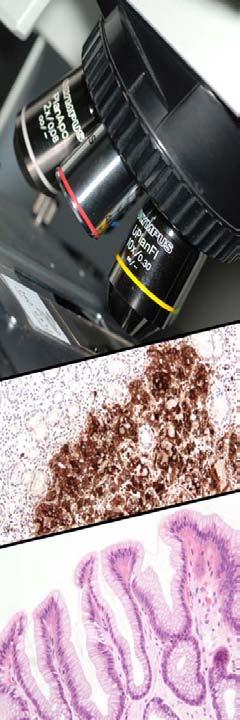
1 Dr Sanjiv Manek Oxford Oxford Pathology Course 2010 for FRCPath Illustration-Cellular Pathology. Oxford Radcliffe NHS Trust
2 Ovarian Endometrial Vulvo-vaginal Cervical Illustration-Cellular Pathology. Oxford Radcliffe NHS Trust
3 Primary Germ cell tumours Sex-cord/stromal tumours Epithelial/stromal tumours Metastatic Illustration-Cellular Pathology. Oxford Radcliffe NHS Trust
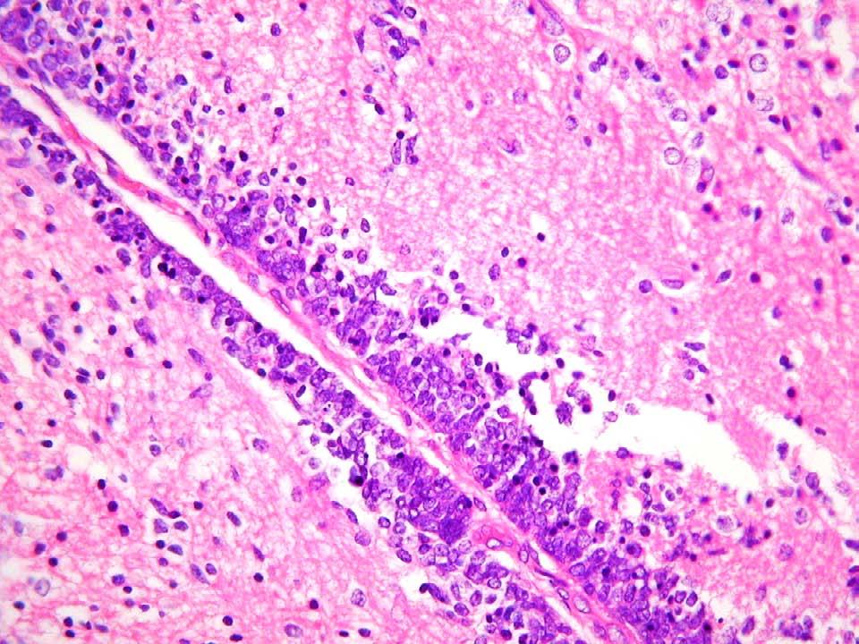
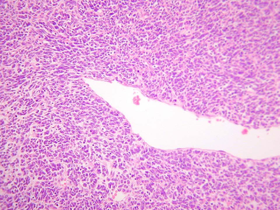
4 Young Dirty-messy, haphazard growth pattern Bizarre cells Haemorrhage and necrosis Inflammatory background Often no organoid structure Illustration-Cellular Pathology. Oxford Radcliffe NHS Trust
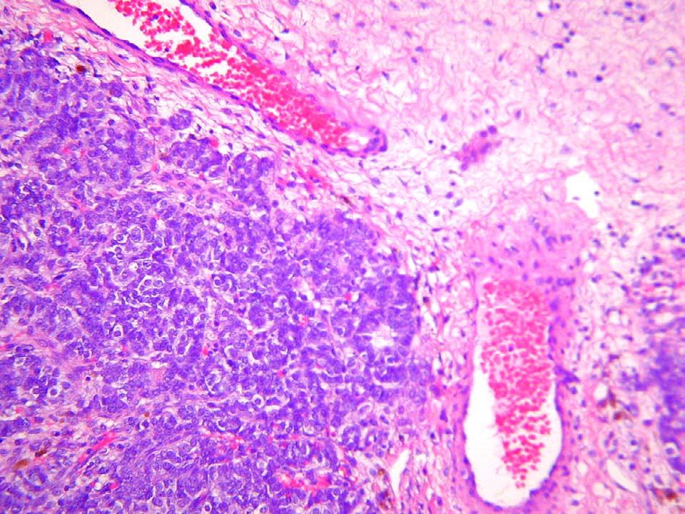
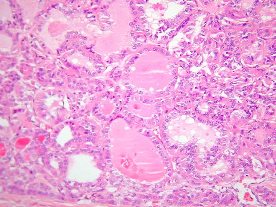
5 Specific features Schiller Duvall bodies (yolk sac tumour) Eosinophilic droplets (yolk sac tumour) Lacey pattern (yolk sac tumour) Nodules (dysgerminoma) Lymphocytic background (dysgerminoma) Dark tubules (immature teratoma) Thyroid-like follicles (struma ovarii) Illustration-Cellular Pathology. Oxford Radcliffe NHS Trust
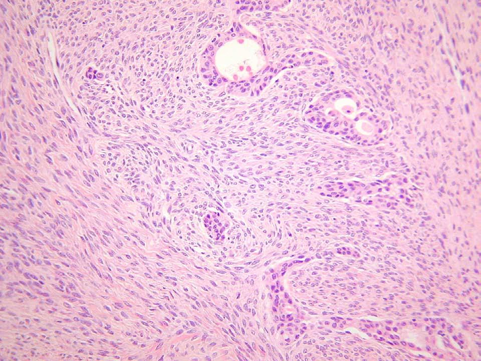
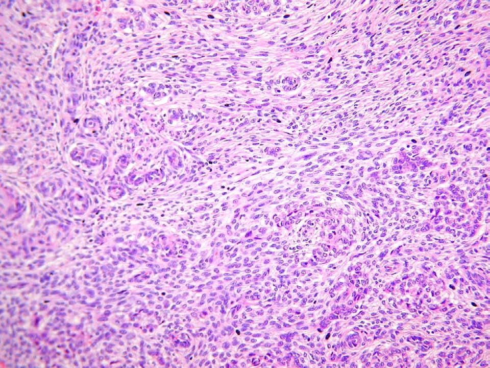
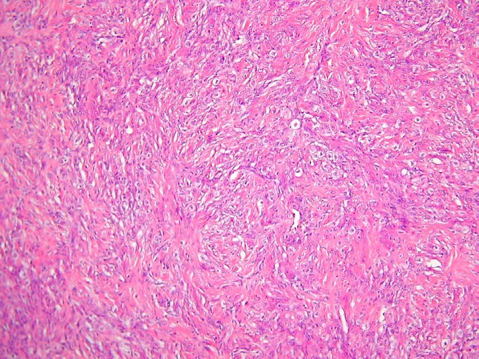
6 Generally uniform and diffuse Often spindly appearance Occasionally nodular and/or organoid Rare haemorrhage and necrosis Atypia not readily apparent Illustration-Cellular Pathology. Oxford Radcliffe NHS Trust
7 Specific features Call-Exner bodies (granulosa cell tumours) Macro and micro -follicles (granulosa cell tumours) Nuclear grooves (granulosa cell tumours) Mosaic/moire-silk pattern (granulosa cell tumours) Tubules and eosinophilic cells (Sertoli-Leydig cell tumours) Large irregular follicles with secretions (juvenile granulosa cell tumours) Atypia and mitoses (poorly differentiated Sertoli- Leydig cell tumours) Illustration-Cellular Pathology. Oxford Radcliffe NHS Trust
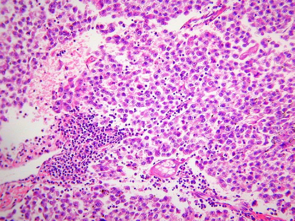
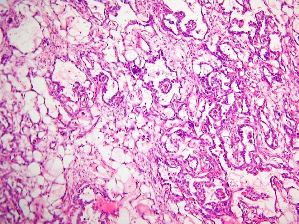
8 Many subtypes Glandular with various growth patterns Soild or cystic Variable amounts of stroma (+/- desmoplasia) Haemorrhage and necrosis Lymphovascular invasion often obvious Illustration-Cellular Pathology. Oxford Radcliffe NHS Trust
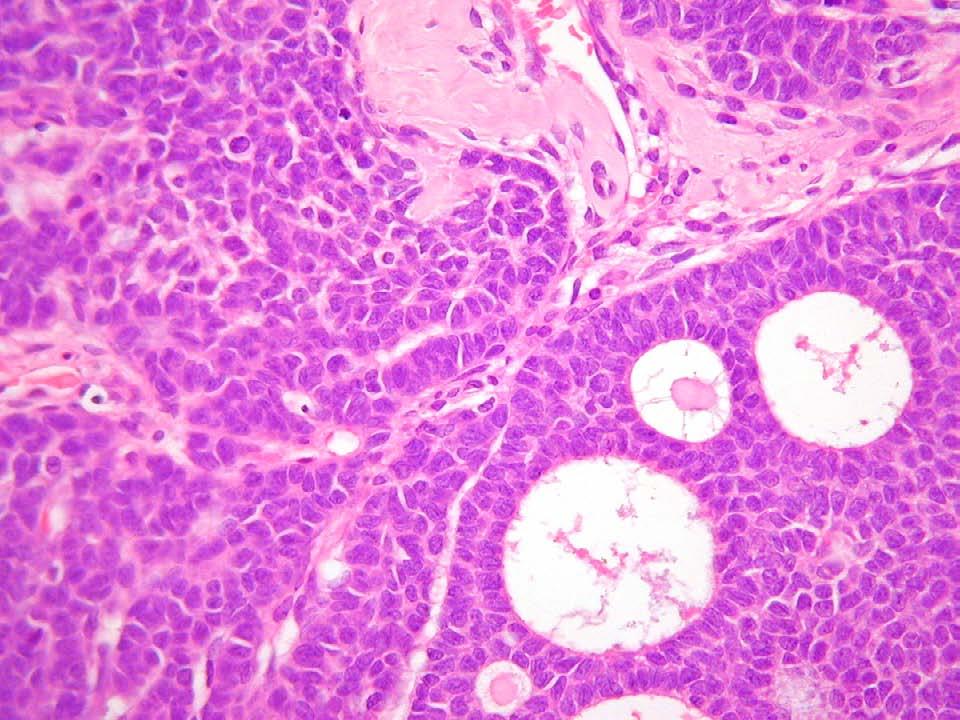
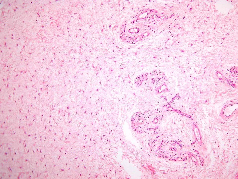
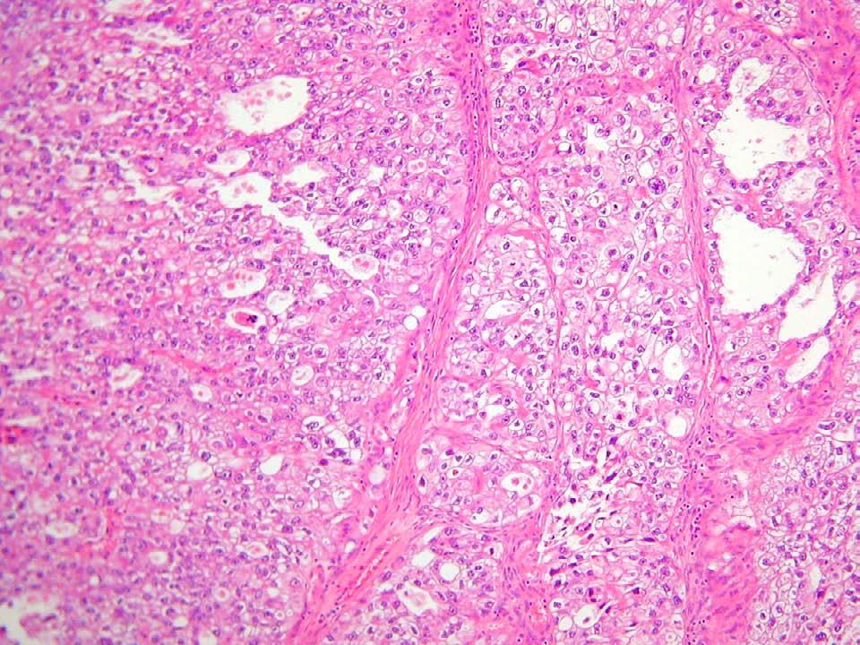
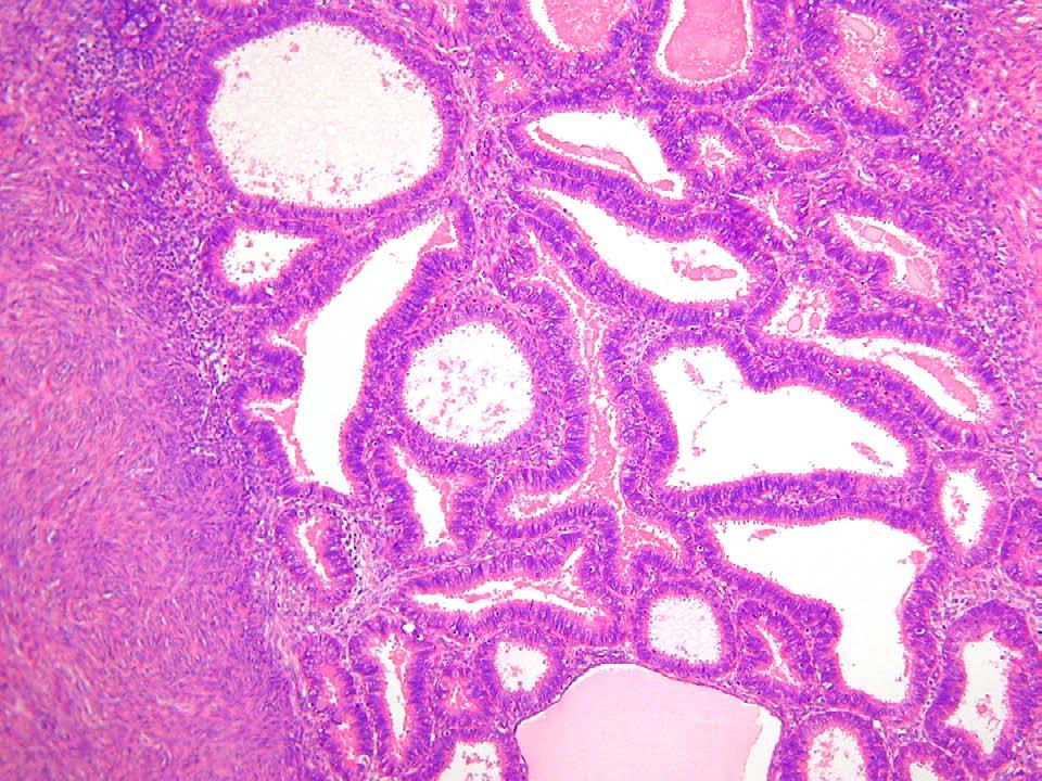
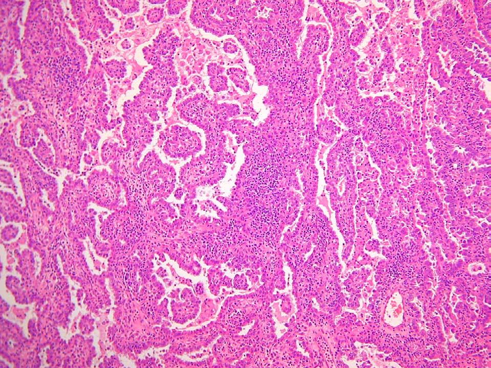
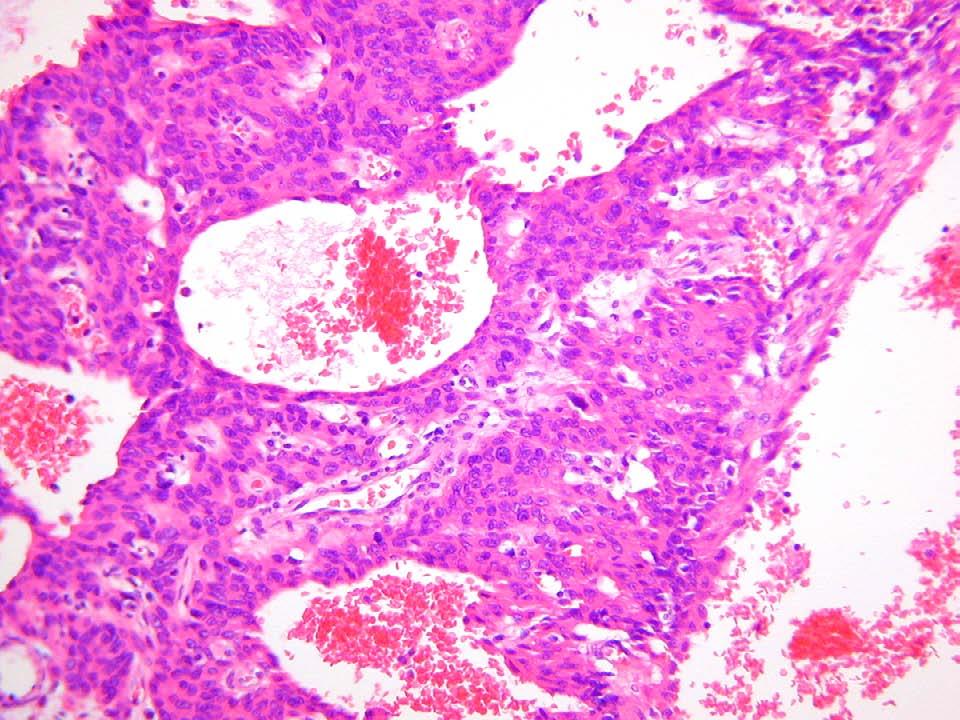
9 Specific features slit-like gland spaces = serous papillary = serous tubulo-cystic, cystic, solid = clear cell carcinoma clear or pink cells with hob-nail appearance and dark nuclei = clear cell carcinoma nests = Brenner papillomatous = TCC rounded glands = endometrioid (often sertoliform) bizzare cells = serous small atypical moulding cells with cystic areas = small cell carcinoma Illustration-Cellular Pathology. Oxford Radcliffe NHS Trust
10 Borderline Ovarian Tumours Cellular atypia Proliferation (glandular crowding and back to back arrangement, papillae, solid islands, multiple small cords and trabeculae, hyperplastic features) No stromal invasion Illustration-Cellular Pathology. Oxford Radcliffe NHS Trust
11 Bilaterality Surface tumour Nodular pattern Lymphatic invasion Desmoplasia +++ Necrosis ( especially colorectal = garland effect ) Single cell infiltration (e.g.signet ring cells or lobular carcinoma cells) Illustration-Cellular Pathology. Oxford Radcliffe NHS Trust
12 Granulosa cell tumours Carcinoid tumours Carcinomas (small cell, endometrioid) Sertoli-Leydig cell tumours Fibro-thecoma Endometrioid carcinoma Sertoli-Leydig cell tumours Clear cell carcinoma Dysgerminoma Illustration-Cellular Pathology. Oxford Radcliffe NHS Trust
13 Carcinomas Papillary serous, endometrioid, clear cell or mixed Background hyperplasia Metaplasias Mixed-Mullerian Stromal lesions Nodular and proliferative stroma-like = Stromal Nodule or ESS Lymphatic invasion = ESS For ESN or ESS - look for small arterioles and foam cells Mast cells in leiomyomas with larger blood vessels Illustration-Cellular Pathology. Oxford Radcliffe NHS Trust
14 Leiomyomas and variants Mitotically active leiomyomas Symplastic leiomyomas Leiomyosarcomas Look for cytological atypia, coagulative tumour necrosis and excess mitotic activity and look at the margins Illustration-Cellular Pathology. Oxford Radcliffe NHS Trust
15 Villoglandular adenocarcinoma - well differentiated Mesonephric adenocarcinoma - dense secretions in glands lined by cuboidal cells on a background of hyperplasia Endocervical adenocarcinoma and background CGIN and/or CIN Small cell carcinoma (neuroendocrine features and large cell variant) Illustration-Cellular Pathology. Oxford Radcliffe NHS Trust
16 Soft tissue lesions (see vulva) Carcinomas and VAIN Melanomas Endometriosis or adenosis related carcinomas GISTs Illustration-Cellular Pathology. Oxford Radcliffe NHS Trust
17 Paget s (large clear cells in epithelium). Look out for invasion or underlying adenocarcinoma Melanoma Verrucous carcinoma VIN +/- Lichen sclerosus Soft tissue lesions aggressive angiomyxoma (watery, scanty cellularity, arterioles, infiltrative) cellular angiofibroma (thick vessels with hyalinised walls, spindled cells intrlacing, mitotic angiomyofibroblastoma (clustered cells, cellular and acellular areas) Illustration-Cellular Pathology. Oxford Radcliffe NHS Trust
18 CK7 (with CK20), EMA, SMO47, CA-125, WT1, p53 and PTEN. Inhibin, Vimentin, CD10, CD99, Calretinin, H- caldesmon,, Chromogranin, CD56, Desmin, Smooth muscle actin, AFP, PLAP, HCG, p57 Illustration-Cellular Pathology. Oxford Radcliffe NHS Trust
19
20
21
22
23
24
25
26
27
28
29
30
31
32
33
34
35
36
37
38
39
40
41
42
43
44
45
46
47
48
Mody. AIS vs. Invasive Adenocarcinoma of the Cervix
 Common Problems in Gynecologic Pathology Michael T. Deavers, M.D. Houston Methodist Hospital, Houston, Texas Common Problems in Gynecologic Pathology Adenocarcinoma in-situ (AIS) of the Cervix vs. Invasive
Pathology of Ovarian Tumours. Dr. Jyothi Ranganathan MD ( Path) AFMC Pune PDCC (Cytopathology) PGI Chandigarh
 AFMC Pune PDCC (Cytopathology) PGI Chandigarh") Pathology of Ovarian Tumours Dr. Jyothi Ranganathan MD ( Path) AFMC Pune PDCC (Cytopathology) PGI Chandigarh Outline Incidence Risk factors Classification Pathology of tumours Tumour markers Prevention
Pathology of Ovarian Tumours Dr. Jyothi Ranganathan MD ( Path) AFMC Pune PDCC (Cytopathology) PGI Chandigarh Outline Incidence Risk factors Classification Pathology of tumours Tumour markers Prevention
Pathology of the female genital tract
 Pathology of the female genital tract Common illnesses of the female genital tract Before menarche Developmental anomalies Tumors (ovarial teratoma) Amenorrhea Fertile years PCOS, ovarian cysts Endometriosis
Gynaecological Malignancies
 Gynaecological Malignancies Dr Rodney Itaki Lecturer Anatomical Pathology Discipline University of Papua New Guinea Division of Pathology School of Medicine & Health Sciences Overview Genital tract tumors
Normal endometrium: A, proliferative. B, secretory.
 Normal endometrium: A, proliferative. B, secretory. Nội mạc tử cung Nội mạc tử cung Cyclic changes in endometrium.. Approximate relationship of useful microscopic changes. Arias-Stella reaction in endometrial
05/07/2018. Types of challenges. Challenging cases in uterine pathology. Case 1 ` 65 year old female Post menopausal bleeding Uterine Polyp
 Types of challenges Challenging cases in uterine pathology Nafisa Wilkinson Gynaecological Pathologist UCLH London Lack of complete history often, NO clinical history at all! Cases from other centres often
Vulva Inflammatory Disorders Lichen Planus Fixed Drug Eruption Erythema Multiforme Plasmacytosis Mucosae (Zoon) Lichen Sclerosus Allergic Contact
 Lichen Sclerosus Allergic Contact") Vulva Inflammatory Disorders Lichen Planus Fixed Drug Eruption Erythema Multiforme Plasmacytosis Mucosae (Zoon) Lichen Sclerosus Allergic Contact Dermatitis Psoriasis Lichen Simplex Chronicus Foreign Body
Vulva Inflammatory Disorders Lichen Planus Fixed Drug Eruption Erythema Multiforme Plasmacytosis Mucosae (Zoon) Lichen Sclerosus Allergic Contact Dermatitis Psoriasis Lichen Simplex Chronicus Foreign Body
Note: The cause of testicular neoplasms remains unknown
 - In the 15- to 34-year-old age group, they are the most common tumors of men. - Tumors of the testis are a heterogeneous group of neoplasms that include: I. Germ cell tumors : 95%; all are malignant.
Adenocarcinoma of the Cervix
 Question 1. Each of the following statements about cervical adenocarcinoma is true except: Adenocarcinoma of the Cervix SAMS a) A majority of women with cervical adenocarcinoma have stage I tumors at diagnosis.
CONTINUING EDUCATION IN TOXICOLOGIC PATHOLOGY REPRODUCTIVE SYSTEM
 CONTINUING EDUCATION IN TOXICOLOGIC PATHOLOGY REPRODUCTIVE SYSTEM ORGANIZED BY SOCIETY FOR TOXICOLOGIC PATHOLOGY IN INDIA (STPI) OCTOBER 29-31, 2010 The Atria Hotel, # 1, Palace Road, Bangalore - 560 001
Diagnostically Challenging Cases in Gynecologic Pathology
 Diagnostically Challenging Cases in Gynecologic Pathology Eric C. Huang, M.D., Ph.D. Department of Pathology and Laboratory Medicine University of California, Davis Medical Center Case 1 Presentation 38
CASE 4 21/07/2017. Ectopic Prostatic Tissue in Cervix. Female 31. LLETZ for borderline nuclear abnormalities
 Female 31 CASE 4 LLETZ for borderline nuclear abnormalities PSA Ectopic Prostatic Tissue in Cervix AJSP 2006;30;209-215 usually incidental microscopic finding usually in ectocervical stroma? developmental
Circulation: X Case number: 501 Number of responses: 84 Date: 4 MAY 12
 Circulation: X Case number: 500 Number of responses: 81 Date: 4 MAY 12 Female, aged 65 TAH and BSO for G1 endometrioid adenocarcinoma. Tumour positive with inhibin, vimentin, CD56 and SMA. Negative with
Ovarian Clear Cell Carcinoma
 Ovarian Clear Cell Carcinoma Rouba Ali-Fehmi, MD Professor of Pathology The Karmanos Cancer Institute, Wayne State University School of Medicine 50 year old woman with chief complaint of shortness of breath
2/9/2015. Bartholin Cyst. Vulva: Squamous epithelium skin. Vagina: Squamous epithelium mucosa. Cervix: Ectocervix: squamous Endocervix: glandular
 Vulva: Squamous epithelium skin Bartholin Cyst Vagina: Squamous epithelium mucosa Cervix: Ectocervix: squamous Endocervix: glandular Slide courtesy of Dr. Lodge Rigal Slide courtesy of Dr. Lodge Rigal
Endometrial Stromal Tumors
 Endometrial Stromal Tumors WHO Categories: Endometrial Stromal Nodule (ESN) Endometrial Stromal Sarcoma, low grade (LGESS) Endometrial Stromal Sarcoma, high grade (HGESS) Undifferentiated Uterine Sarcoma
Interesting Cases in Gynecologic Pathology. Michael Ward, MD Surgical Pathology Fellow University of Utah Health Sciences Center Salt Lake City, UT
 Interesting Cases in Gynecologic Pathology Michael Ward, MD Surgical Pathology Fellow University of Utah Health Sciences Center Salt Lake City, UT Case 1 History: 50 year old woman with a uterine mass
3 cell types in the normal ovary
 Ovarian tumors 3 cell types in the normal ovary Surface (coelomic epithelium) the origin of the great majority of ovarian tumors (neoplasms) 90% of malignant ovarian tumors Totipotent germ cells Sex cord-stromal
When Immunostains Can Get You in Trouble: Gynecologic Pathology p16: Panacea or Pandora s Box?
 When Immunostains Can Get You in Trouble: Gynecologic Pathology p16: Panacea or Pandora s Box? Teri A. Longacre, MD Stanford Medicine Stanford California pi6 in Gynecologic Pathology: Panacea or Pandora
Type I. Type II. Excess estrogen Lynch Endometrioid adenocarcinoma PTEN. High grade More aggressive Serous, Clear Cell p53
 Type I Excess estrogen Lynch Endometrioid adenocarcinoma PTEN Type II High grade More aggressive Serous, Clear Cell p53 Stage I IA IB Stage II Stage III IIIA IIIB IIIC IIIC1 IIIC2 Stage IV IVA IVB nodes
VULVAR CARCINOMA. Page 1 of 5
 VULVAR CARCINOMA EXAMPLE OF A VULVAR CARCINOMA USING PROPOSED TEMPLATE Case: Invasive squamous cell carcinoma arising in D-VIN Tumor in left labia major Left partial vaginectomy and sentinel lymph node
Annual report of the Committee on Gynecologic Oncology, the Japan Society of Obstetrics and Gynecology
 bs_bs_banner doi:10.1111/jog.12596 J. Obstet. Gynaecol. Res. Vol. 41, No. 2: 167 177, February 2015 Annual report of the Committee on Gynecologic Oncology, the Japan Society of Obstetrics and Gynecology
3 cell types in the normal ovary
 Ovarian tumors 3 cell types in the normal ovary Surface (coelomic epithelium) the origin of the great majority of ovarian tumors 90% of malignant ovarian tumors Totipotent germ cells Sex cord-stromal cells
ENODMETRIAL CARCINOMA: SPECIAL & NOT SO SPECIAL VARIANTS
 ENODMETRIAL CARCINOMA: SPECIAL & NOT SO SPECIAL VARIANTS Pacific Northwest Society of Pathologists Vancouver, B.C. September 26, 2015 Teri A. Longacre, M.D. longacre@stanford.edu Stanford University, Stanford,
Case E1. Female aged 63 years Right Nephrectomy Two separate tumours Section of each tumour
 Case E1 Female aged 63 years Right Nephrectomy Two separate tumours Section of each tumour Tumour 1 Upper pole tumour 28mm macro diameter Circumscribed Friable cut surface Tumour 2 Middle pole Part solid
The role of immunohistochemistry in surgical pathology of the uterine corpus and cervix
 The role of immunohistochemistry in surgical pathology of the uterine corpus and cervix Prof. Ben Davidson, MD PhD Department of Pathology, Norwegian Radium Hospital, Oslo University Hospital, Oslo, Norway
Tinh hoàn
 Tinh hoàn Tinh hoàn Tinh hoàn Tiền liệt tuyến Tiền liệt tuyến Mào tinh hoàn Mào tinh hoàn Túi tinh Túi tinh Túi tinh Túi tinh So-called cystadenoma of seminal vesicle. Gross appearance of granulomatous
of 20 to 80 and subsequently declines [2].
![of 20 to 80 and subsequently declines [2].](/thumbs/80/81450506.jpg "of 20 to 80 and subsequently declines [2].") - - According to the 2014 World Health Organization (WHO) classification and tumor morphology, primary ovarian tumors are subdivided into three categories: epithelial (60%), germ cell (30%), and sex-cord
- - According to the 2014 World Health Organization (WHO) classification and tumor morphology, primary ovarian tumors are subdivided into three categories: epithelial (60%), germ cell (30%), and sex-cord
Male Genital Cancers in the US in Frequency of Types
 Germ Cell Tumors of the Testis Pathology, Immunohistochemistry, and the Often Confusing Appearance of Their Metastases Charles Zaloudek, MD Department of Pathology UCSF Male Genital Cancers in the US in
6/5/2010. Outline of Talk. Endometrial Alterations That Mimic Cancer & Vice Versa: Metaplastic / reactive changes. Problems in Biopsies/Curettages
 Outline of Talk Endometrial Alterations That Mimic Cancer & Vice Versa: Problems in Biopsies/Curettages Metaplastic / reactive changes Mucinous change Microglandular hyperplasia-like change Squamous metaplasia
Atypical Hyperplasia/EIN
 EIN Atypical Hyperplasia/EIN Based on scientific and diagnostic advances, in 2014 the WHO moved that the precursor lesion for endometrioid carcinoma be atypical hyperplasia/ein, rather than what was previously
Institute of Pathology First Faculty of Medicine Charles University. Ovary
 Ovary Barrett esophagus ph in vagina between 3.8 and 4.5 ph of stomach varies from 1-2 (hydrochloric acid) up to 4-5 BE probably results from upward migration of columnar cells from gastroesophageal junction
Demystifying Endometrial Hyperplasia
 Demystifying Endometrial Hyperplasia A review from Diagnostic Histopathology 19:7 Dr R Hadden ST5 Histopathology Derriford Hospital Plymouth Endometrium Target for sex-steroid hormones Glands Stroma Proliferate
PATHOLOGY OF THE FEMALE GENITAL TRACT
 MBBS 2 nd Yr. Lecture Dr. Annie Cheung September 30, 2002 9:30 am LT2,G/F, Academic and Administration Block Faculty of Medicine Building UROGENITAL SYSTEM PATHOLOGY OF THE FEMALE GENITAL TRACT Learning
Mucinous Tumors of the Ovary Beirut, Lebanon. Anaís Malpica, M.D. Professor Department of Pathology
 Mucinous Tumors of the Ovary Beirut, Lebanon Anaís Malpica, M.D. Professor Department of Pathology Primary Mucinous Tumors of the Ovary Cystadenoma Borderline (Tumor of Low Malignant Potential/Atypical
International Society of Gynecological Pathologists Symposium 2007
 International Society of Gynecological Pathologists Symposium 2007 Anais Malpica, M.D. Department of Pathology The University of Texas M.D. Anderson Cancer Center Grading of Ovarian Cancer Histologic grade
Case year female. Routine Pap smear
 Case 1 57 year female Routine Pap smear Diagnosis? 1. Atypical glandular cells of unknown significance (AGUS) 2. Endocervical AIS 3. Endocervical adenocarcinoma 4. Endometrial adenocarcinoma 5. Adenocarcinoma
Case Report Poorly Differentiated Thyroid Carcinoma Arising in Struma Ovarii
 Hindawi Publishing Corporation Case Reports in Pathology Volume 2015, Article ID 826978, 6 pages http://dx.doi.org/10.1155/2015/826978 Case Report Poorly Differentiated Thyroid Carcinoma Arising in Struma
Presenter: Yeh-Han Wang M.D.
 Korea-Taiwan-Japan Joint Meeting for Gynecological Pathology Mini-lecture Female Adnexal Tumor of Probable Wolffian Origin (FATWO) in Taiwan: A Small Case Series and Literature Review Presenter: Yeh-Han
Disclosure. Case. Mixed Tumors of the Uterine Corpus and Cervix. I have nothing to disclose
 Mixed Tumors of the Uterine Corpus and Cervix Marisa R. Nucci, M.D. Division of Women s and Perinatal Pathology Department of Pathology Brigham and Women s Hospital Boston, MA UCSF Current Issues in Anatomic
Scotland and Northern Ireland EQA Scheme. Circulation 46
 Scotland and Northern Ireland EQA Scheme Circulation 46 Special Educational Cases E1 and E2 Presented by Dr K Robertson Case E1 Female 42 year old with heavy menstrual and intermenstrual bleeding. IUS
How to Recognize Gynecologic Cancer Cells from Pelvic Washing and Ascetic Specimens
 How to Recognize Gynecologic Cancer Cells from Pelvic Washing and Ascetic Specimens Wenxin Zheng, M.D. Professor of Pathology and Gynecology University of Arizona zhengw@email.arizona.edu http://www.zheng.gynpath.medicine.arizona.edu/index.html
Macro- and microacinar proliferations of the prostate
 Macro- and microacinar proliferations of the prostate (with emphasis on cancer mimics) Rodolfo Montironi, MD (IT), FRCPath (UK), IFCAP (USA) Polytechnic University of Marche Region (Ancona) School of Medicine,
Appendix I. A Summary of Common Pitfalls in Diagnostic Gynecologic and Obstetric Pathology
 Appendix I. A Summary of Common Pitfalls in Diagnostic Gynecologic and Obstetric Pathology Section Chapter Mistaken for Comments Candidiasis A premalignant lesion (VIN) Chronically rubbed skin can produce
Endometrial Metaplasia, Hyperplasia & Other Cancer Mimics: a Consultant s Experience
 Endometrial Metaplasia, Hyperplasia & Other Cancer Mimics: a Consultant s Experience Pacific Northwest Society of Pathologists Vancouver, B.C. September 26, 2015 Teri A. Longacre, M.D. longacre@stanford.edu
Annual report of Gynecologic Oncology Committee, Japan Society of Obstetrics and Gynecology, 2013
 bs_bs_banner doi:10.1111/jog.12360 J. Obstet. Gynaecol. Res. Vol. 40, No. 2: 338 348, February 2014 Annual report of Gynecologic Oncology Committee, Japan Society of Obstetrics and Gynecology, 2013 Daisuke
Normal thyroid tissue
 Thyroid Pathology Overview Normal thyroid tissue Normal thyroid tissue with follicles filled with colloid. Thyroid cells form follicles, spheres of epithelial cells (always single layered in health, usually
South East England General Histopathology EQA Scheme
 South East England General Round g Final Case Analyses Cases 707 to 718 Circulated January February 2018 135 responses (85.44%) Prepared April 2018 Authorised by: Prof J Schofield Date: 23 rd April 2018
Endometrial pathology. Dr Tom Dodd and Dr Georgina England
 Endometrial pathology Dr Tom Dodd and Dr Georgina England Case 1 Female age 35 Case 1 Proliferative endometrium Case 2 Female age 38 Case 2 Secretory endometrium Dating endometrium Assessed on the
Review of the AP Part II Practical Examination. Dr David Clift Co Chief Examiner
 Review of the AP Part II Practical Examination Dr David Clift Co Chief Examiner General Remarks The part II practical examination involved 15 cases which were presented with sufficient clinical data to
Gross appearance of peritoneal cysts. They have a thin, translucent wall and contain a clear fluid.
 Gross appearance of peritoneal cysts. They have a thin, translucent wall and contain a clear fluid. So-called multicystic benign mesothelioma. A, Gross appearance. So-called multicystic benign mesothelioma.
Diseases of the breast (1 of 2)
") Diseases of the breast (1 of 2) Introduction A histology introduction Normal ducts and lobules of the breast are lined by two layers of cells a layer of luminal cells overlying a second layer of myoepithelial
Diseases of the breast (1 of 2) Introduction A histology introduction Normal ducts and lobules of the breast are lined by two layers of cells a layer of luminal cells overlying a second layer of myoepithelial
Gross appearance of nodular hyperplasia in material obtained from suprapubic prostatectomy. Note the multinodular appearance and the admixture of
 Tiền liệt tuyến Tiền liệt tuyến Gross appearance of nodular hyperplasia in material obtained from suprapubic prostatectomy. Note the multinodular appearance and the admixture of solid and microcystic areas.
The incidence of cervical adenocarcinoma (ADC) has
 has") Cervical Adenocarcinoma of Human Papillomavirus Positive and Human Papillomavirus Negative Tumors Edyta C. Pirog, MD, PhD Context. Cervical adenocarcinomas span a diverse group of tumors with several distinct
Cervical Adenocarcinoma of Human Papillomavirus Positive and Human Papillomavirus Negative Tumors Edyta C. Pirog, MD, PhD Context. Cervical adenocarcinomas span a diverse group of tumors with several distinct
-The cause of testicular neoplasms remains unknown
 - In the 15- to 34-year-old age group, they are the most common tumors of men. - include: I. Germ cell tumors : (95%); all are malignant. II. Sex cord-stromal tumors: from Sertoli or Leydig cells; usually
Pitfalls in thyroid tumor pathology. Prof.Valdi Pešutić-Pisac MD, PhD
 Pitfalls in thyroid tumor pathology Prof.Valdi Pešutić-Pisac MD, PhD Too many or... Tumour herniation through a torn capsule simulating capsular invasion fibrous capsule with a sharp discontinuity, suggestive
Female Reproduc.ve System. Kris.ne Kra7s, M.D.
 Female Reproduc.ve System Kris.ne Kra7s, M.D. Female Reproduc.ve System Outline Cervix Uterus Ovaries Breast Cervical Carcinoma Once the most common cancer in women now not even in top 10. Decrease due
Leydig cell tumour. Testis: non-germ cell tumours. Testis: sex cord-stromal tumours. Differential diagnosis of Leydig cell tumour TTAGS
 Non-germ cell s of the testis Dr Jonathan H Shanks The Christie NHS Foundation Trust, Manchester, UK Testis: non-germ cell s Sex cord-stromal s Haemolymphoid neoplasms Other neoplasms Tumour-like conditions
Dr Agata T Kochman Wishaw General Hospital
 Dr Agata T Kochman Wishaw General Hospital Case E1 84 year old male Symptoms: R shoulder pain CT = thymic mass and (R) LL nodules + (L) lung nodule Clinically metastatic lesions in lung with primary thymic
Breast pathology. 2nd Department of Pathology Semmelweis University
 Breast pathology 2nd Department of Pathology Semmelweis University Breast pathology - Summary - Benign lesions - Acute mastitis - Plasma cell mastitis / duct ectasia - Fat necrosis - Fibrocystic change/
Pathology Slides. [Pathology]
![Pathology Slides. [Pathology]](/thumbs/94/120604575.jpg "Pathology Slides. [Pathology]") Pathology Slides MedicoNotes provides real laboratory pathological slides to aid you to differentiate between different pathological structures under microscope. www.mediconotes.com Histology slides example
Pathology Slides MedicoNotes provides real laboratory pathological slides to aid you to differentiate between different pathological structures under microscope. www.mediconotes.com Histology slides example
Thyroid pathology Practical part
 Thyroid pathology Practical part My Algorithm After a good macroscopy and a microscopic overview of the lesion, I especially look at the capsule and the thyroid just above and just beneath the capsule.
Neoplasia 2018 Lecture 2. Dr Heyam Awad MD, FRCPath
 Neoplasia 2018 Lecture 2 Dr Heyam Awad MD, FRCPath ILOS 1. List the differences between benign and malignant tumors. 2. Recognize the histological features of malignancy. 3. Define dysplasia and understand
Hyperchromatic Crowded Groups: What is Your Diagnosis? Session 3000
 Hyperchromatic Crowded Groups: What is Your Diagnosis? Session 3000 Thomas A. Bonfiglio, M.D. Professor Emeritus, Pathology and Laboratory Medicine University of Rochester Disclosures In the past 12 months,
OVARIES. MLS Basic histological diagnosis MLS HIST 422 Semester 8- batch 7 L13 Dr: Ali Eltayb.
 OVARIES MLS Basic histological diagnosis MLS HIST 422 Semester 8- batch 7 L13 Dr: Ali Eltayb. OBJECTIVES Recognize different disease of ovaries Classify ovarian cyst Describe the pathogenesis, morphology
Pathology of the Thyroid
 Pathology of the Thyroid Thyroid Carcinoma Arising from Follicular Cells 2015-01-19 Prof. Dr. med. Katharina Glatz Pathologie Carcinomas Arising from Follicular Cells Differentiated Carcinoma Papillary
Female Reproduc.ve System. Kris.ne Kra7s, M.D.
 Female Reproduc.ve System Kris.ne Kra7s, M.D. Female Reproduc.ve System Outline Cervix Uterus Ovaries Breast Female Reproduc.ve System Outline Cervix Cervical carcinoma Cervical Carcinoma Once the most
Neoplasms of the Canine, Feline and Lemur Liver:
 Neoplasms of the Canine, Feline and Lemur Liver: Classification and Prognosis Annual Seminar of the French Society of Veterinary Pathology John M. Cullen VMD PhD DACVP North Carolina State University Primary
They Do Look Alike : Mimics of Prostate Cancer in Biopsy Samples
 They Do Look Alike : in Biopsy Samples Gladell P. Paner, MD Departments of Pathology and Surgery (Urology) University of Chicago, IL USA Gladell.paner@uchospitals.edu Benign in Needle Biopsy 1. Benign
Recently, there has been an increasing incidence among women and younger persons
 Bladder Tumors Benign Transitional Cell Papilloma 2-3% Inverted Papilloma Rare Malignant Transitional (Urothelial) Carcinoma 90% Carcinoma In-Situ (By Itself) 5-10% Squamous Cell Carcinoma 3-7% Adenocarcinoma
Prepared By Jocelyn Palao and Layla Faqih
 Prepared By Jocelyn Palao and Layla Faqih The structure of the suspected atypical cell should always be compared to the structure of other similar, benign, cells which are present in the smears. The diagnosis
Journal of Rawalpindi Medical College (JRMC); 2016;20(4):
; 2016;20(4):") Original Article Morphological Profile of Ovarian Tumours Bilquis Begum, Iram Nadeem Rana, Nadeem Ikram Department of Pathology, Rawalpindi Medical College, Rawalpindi Abstract Background: To study the
Original Article Morphological Profile of Ovarian Tumours Bilquis Begum, Iram Nadeem Rana, Nadeem Ikram Department of Pathology, Rawalpindi Medical College, Rawalpindi Abstract Background: To study the
In situ and Invasive Endocervical Carcinoma: Problems and Pitfalls in Diagnosis
 In situ and Invasive Endocervical Carcinoma: Problems and Pitfalls in Diagnosis Rouba Ali-Fehmi,MD The Karmanos Cancer Institute, Wayne State University School of Medicine Global incidence of cervical
May 2017 TSL workshops. EQA 012 Final analysis with Diagnoses
 May 2017 TSL workshops EQA 012 Final analysis with Diagnoses CASE 1 28 yr old female with menorrhagia. Well diff endometrioid adenocarcinoma (back ground complex hyperplasia) 4 Non-atypical endometrial
ESS: Pathologic Insights
 GEIS XVI INTERNATIONAL SYMPOSIUM Seville 4th October 2018 ESS: Pathologic Insights Sílvia Bagué The Royal Marsden Hospital London (United Kingdom) I have no conflicts of interest Endometrial stromal sarcoma
Special slide seminar
 Special slide seminar Tomáš Rozkoš The Fingerland Department of Pathology Charles University Medical Faculty and Faculty Hospital in Hradec Králové Czech Republic Case history, 33 years old resistance
ICD-O Morphology code. R=Rare Tier Tumour ICD-O Topography code C30.0, C31
 R=Rare Tier Tumour ICD-O Topography code ICD-O Morphology code EPITHELIAL TUMOURS OF NASAL CAVITY AND SINUSES R 2 Squamous cell carcinoma with variants of nasal cavity and sinuses C30.0, C3 C30.0, C3 8000,
Villoglandular adenocarcinoma of cervix a tumour with bland cytological features: report of a case missed on cytology
 Malaysian J Pathol 2003; 25(2) : CERVICAL 139 143 VILLOGLANDULAR ADENOCARCINOMA CYTOLOGY CASE REPORT Villoglandular adenocarcinoma of cervix a tumour with bland cytological features: report of a case missed
Objectives. Atypical Glandular Cells. Atypical Endocervical Cells. Reactive Endocervical Cells
 2013 California Society of Pathologists 66 th Annual Meeting San Francisco, CA Atypical Glandular Cells to Early Invasive Adenocarcinoma: Cervical Cytology and Histology Christina S. Kong, MD Associate
2% of all malignancies Male predominance Patients usually more than 60 years old
 Benign Bladder Tumors Transitional Cell Papilloma 2-3% Inverted Papilloma Rare Malignant Transitional (Urothelial) Carcinoma 90% Carcinoma In-Situ (By Itself) 5-10% Squamous Cell Carcinoma 3-7% Adenocarcinoma
What really matters When and Why. Pathology of Uterine Mesenchymal Lesions. Nafisa Wilkinson London
 What really matters When and Why Pathology of Uterine Mesenchymal Lesions Nafisa Wilkinson London Patient centred approach immunohistochemistry Histological diagnosis Next generation sequencing Genetic
Case 1: Verruciform Lichen Simplex Chronicus
 INTERACTIVE MICROSCOPY COURSE GYNECOLOGIC PATHOLOGY Marisa R. Nucci, M.D. Case 1: Verruciform Lichen Simplex Chronicus Lichen simplex chronicus (LSC) is defined as acanthosis, hyperkeratosis and hypergranulosis
7 Mousa. Obada Zalat. Mohammad Badi
 7 Mousa Obada Zalat Mohammad Badi Tumors of the ovaries Last lecture we talked about surface epithelial tumors of the ovaries (the most common type). But there are many other types of tumors of germ cell
Papillary Lesions of the breast
 Papillary Lesions of the breast Emad Rakha Professor of Breast Pathology The University of Nottingham Papillary lesions of the breast are a heterogeneous group of disease, which are characterised by neoplastic
Synonyms. Nephrogenic metaplasia Mesonephric adenoma
 Nephrogenic Adenoma Synonyms Nephrogenic metaplasia Mesonephric adenoma Definition Benign epithelial lesion of urinary tract with tubular, glandular, papillary growth pattern Most frequently in the urinary
BREAST PATHOLOGY. Fibrocystic Changes
 BREAST PATHOLOGY Lesions of the breast are very common, and they present as palpable, sometimes painful, nodules or masses. Most of these lesions are benign. Breast cancer is the 2 nd most common cause
Diagnostic problems in uterine smooth muscle tumors
 Diagnostic problems in uterine smooth muscle tumors Marina Kos Ljudevit Jurak Clinical Department of Pathology, Clinical Hospital Center Sestre milosrdnice, Zagreb Institute of Pathology, University of
SMOOTH MUSCLE TUMOURS
 SMOOTH MUSCLE TUMOURS NORMAL SMOOTH MUSCLE Cytology Immunohistochemistry Ultrastructure Masson Trichrome Smooth Muscle Ultrastructure Many myofilaments running parallel to the long axis of the smooth
Non-Invasive Follicular Thyroid Neoplasm with Papillary-like Nuclei (NIFTP)
") Papillary Thyroid Carcinoma: Follicular Variant Encapsulated Type Replaced by: Non-Invasive Follicular Thyroid Neoplasm with Papillary-like Nuclei (NIFTP) Lester D. R. Thompson www.lester-thompson.com
Papillary Thyroid Carcinoma: Follicular Variant Encapsulated Type Replaced by: Non-Invasive Follicular Thyroid Neoplasm with Papillary-like Nuclei (NIFTP) Lester D. R. Thompson www.lester-thompson.com
Case Report Sertoli-Leydig cell tumor with heterologous element: a case report and a review of the literature
 Int J Clin Exp Pathol 2014;7(3):1176-1181 www.ijcep.com /ISSN:1936-2625/IJCEP1401091 Case Report Sertoli-Leydig cell tumor with heterologous element: a case report and a review of the literature Longwen
Page # 1. Endometrium. Cellular Components. Anatomical Regions. Management of SIL Thomas C. Wright, Jr. Most common diseases:
 Endometrium Pathology of the Endometrium Thomas C. Wright Columbia University, New York, NY Most common diseases: Abnormal uterine bleeding Inflammatory conditions Benign neoplasms Endometrial cancer Anatomical
Selected miscellaneous ovarian lesions: small cell carcinomas, mesothelial lesions, mesenchymal and mixed neoplasms, and non-neoplastic lesions
 & 2005 USCAP, Inc All rights reserved 0893-3952/05 $30.00 www.modernpathology.org Selected miscellaneous ovarian lesions: small cell carcinomas, mesothelial lesions, mesenchymal and mixed neoplasms, and
Cytology and Surgical Pathology of Gynecologic Neoplasms
 Cytology and Surgical Pathology of Gynecologic Neoplasms Current Clinical Pathology ANTONIO GIORDANO, MD, PHD SERIES EDITOR For further titles published in this series, go to http://www.springer.com/springer/series/7632
2/24/19. Ovarian pathology: IOTA ADNEXAL MASSES. Content. IOTA terms for description of an adnexal mass. IOTA terms for description of an adnexal mass
 Content Ovarian pathology: IOTA ADNEXAL MASSES X SIMPLE COMPLEX Dr DESCRIBE WHAT YOU SEE FRANZCOG, MPH, DDU, COGU Sonologist Clinically useful Benign Malignant Communication between clinicians/research
New Developments in Immunohistochemistry for Gynecologic Pathology
 New Developments in Immunohistochemistry for Gynecologic Pathology Michael T. Deavers, M.D. Professor, Departments of Pathology and Gynecologic Oncology Immunohistochemistry in Gynecologic Pathology Majority
GOBLET CELL CARCINOID. Hanlin L. Wang, MD, PhD University of California Los Angeles
 GOBLET CELL CARCINOID Hanlin L. Wang, MD, PhD University of California Los Angeles Disclosure of Relevant Financial Relationships USCAP requires that all planners (Education Committee) in a position to
GOBLET CELL CARCINOID
 GOBLET CELL CARCINOID Hanlin L. Wang, MD, PhD University of California Los Angeles Disclosure of Relevant Financial Relationships USCAP requires that all planners (Education Committee) in a position to
Icd 10 ovarian stroma
 Icd 10 ovarian stroma Struma ovarii; Micrograph of a struma ovarii. Characteristic thyroid follicles are seen on the right, and ovarian stroma on the left. H&E stain. Classification and. Free, official
Histopathological diagnosis of CUP
 Histopathological diagnosis of CUP Dr Karin Oien karin.oien@glasgow.ac.uk Disclosure slide Dr Karin Oien has no financial interests in any company mentioned in this presentation. Dr Karin Oien is conducting