Testicular Tumors: What s New, True, Important Cristina Magi-Galluzzi, MD, PhD
|
|
|
- Christian Harrington
- 5 years ago
- Views:
Transcription

1 Testicular Tumors: What s New, True, Important Cristina Magi-Galluzzi, MD, PhD Director, Genitourinary Pathology R.J. Tomsich Pathology & Laboratory Medicine Institute Professor of Pathology, Lerner College of Medicine Cleveland Clinic
2 Testicular Cancer Uncommon (~1% cancer in male) 90-95% germ cell origin Most common cancer in white males age Incidence has in the last half century and is variable in different regions Risk factors for Germ Cell Tumors (GCTs) Cryptorchidism Prior testicular GCT Family history of GCT (brother > sons > fathers) Disorders of sex development (gonadal dysgenesis) (Infertility, Marijuana use)

3 WHO 2016: Tumors of the Testis Updated pathogenetic model for GCTs Restructuring of classification New entities - Germ cell tumors - Sex cord stromal tumors
4 Preinvasive lesion to malignant testicular germ cell tumors (GCTs): evolution of nomenclature CIS Skakkebaek, Lancet 1972 CIS had characteristics of primordial germ cells IGCNU IGCN GCNI GCNIS GCNIS IGCNU Scully, Rosai, Mostofi, Kurman, et al, 1980 WHO classification 2004 Germ Cell Neoplasia In Situ 2016 WHO classification
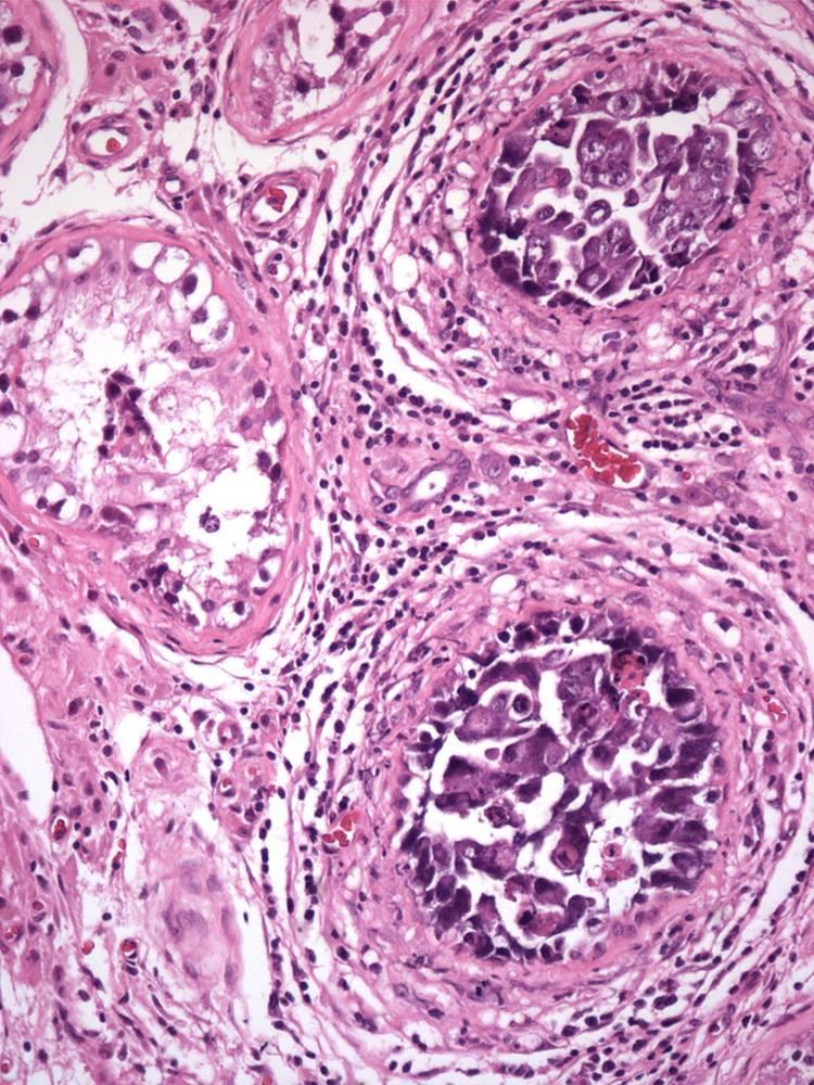
5 Germ Cell Neoplasia In situ (GCNIS) Malignant germ cells in spermatogonial niche Increased incidence in sex development disorders, up to 70% - Cryptorchidism - Gonadal dysgenesis - Androgen insensitivity syndrome 1-4% in subfertile/infertile men Seen in most seminomas and non-seminomas; 2-6% of testes contralateral to unilateral GCT - GCNIS supports a diagnosis of GCT 50% of men with GCNIS develop invasive GCT within 5 years
")


6 Germ Cell Neoplasia In situ (GCNIS) Gonocyte-like germ cells Single layer in basilar location Decreased or absent spermatogenesis

")

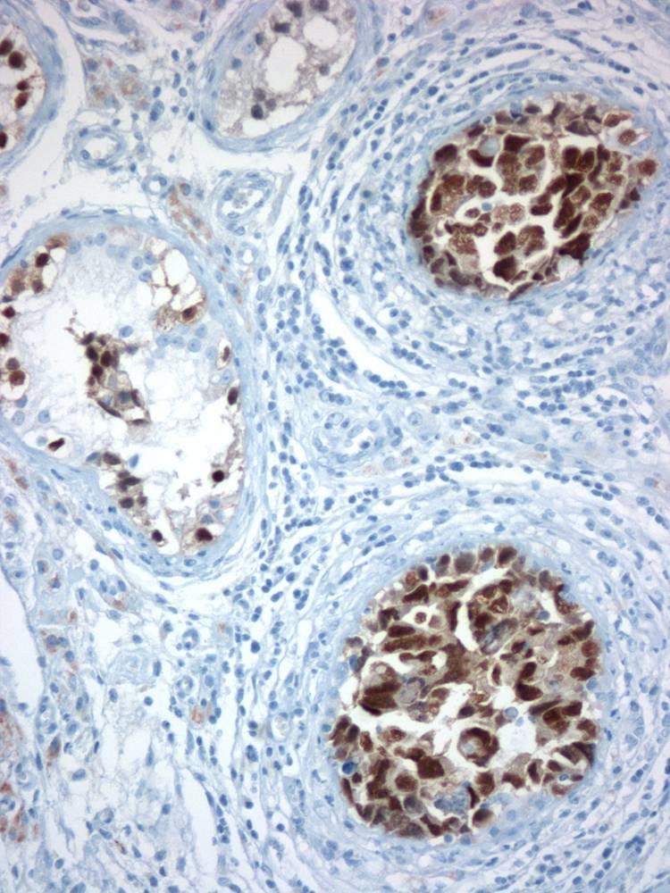
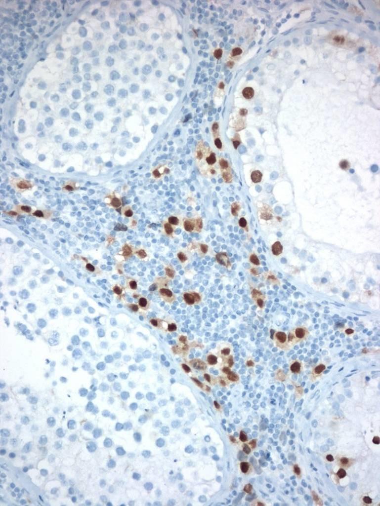
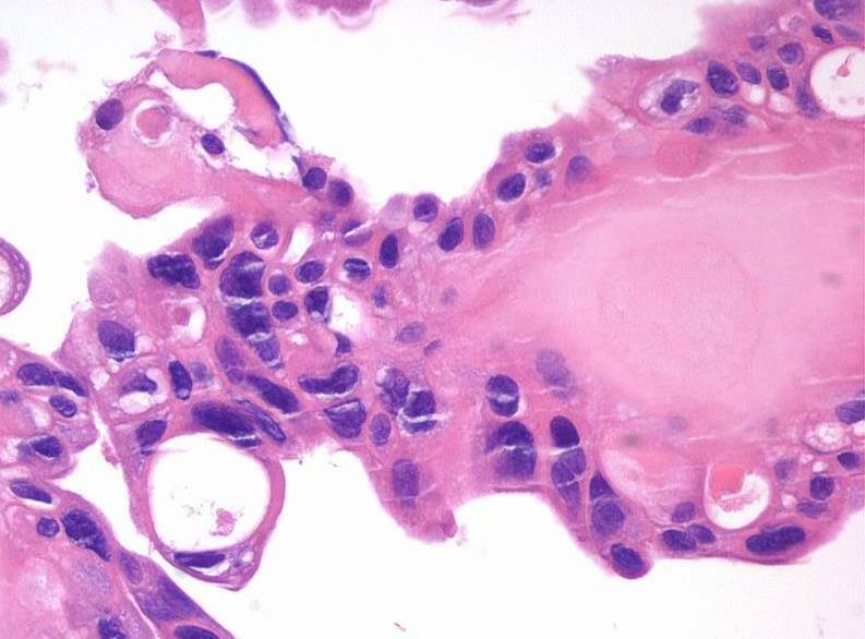
7 Germ Cell Neoplasia In situ (GCNIS) PLAP OCT3/4 CD117
 - Binucleation, OCT3/4- Specific forms of intratubular neoplasia - Intratubular seminoma - Intratubular non-seminoma (embryonal")
8 Differential Diagnosis of GCNIS Delayed maturation of gonocytes in prepubertal patients with sex development disorder (beyond 6 mo) - OCT3/4+, PLAP+; central tubular location Atypical germ cells due to perturbation of spermatogenesis (cryptorchidism, infertility) - Binucleation, OCT3/4- Specific forms of intratubular neoplasia - Intratubular seminoma - Intratubular non-seminoma (embryonal carcinoma, YST, teratoma)

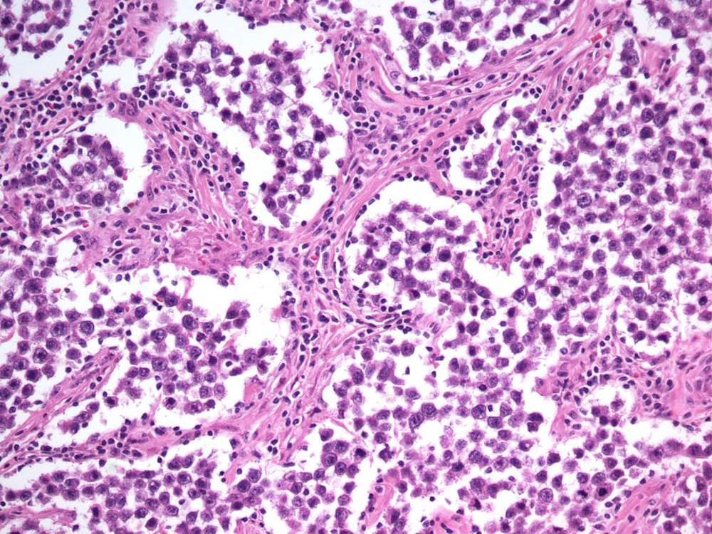
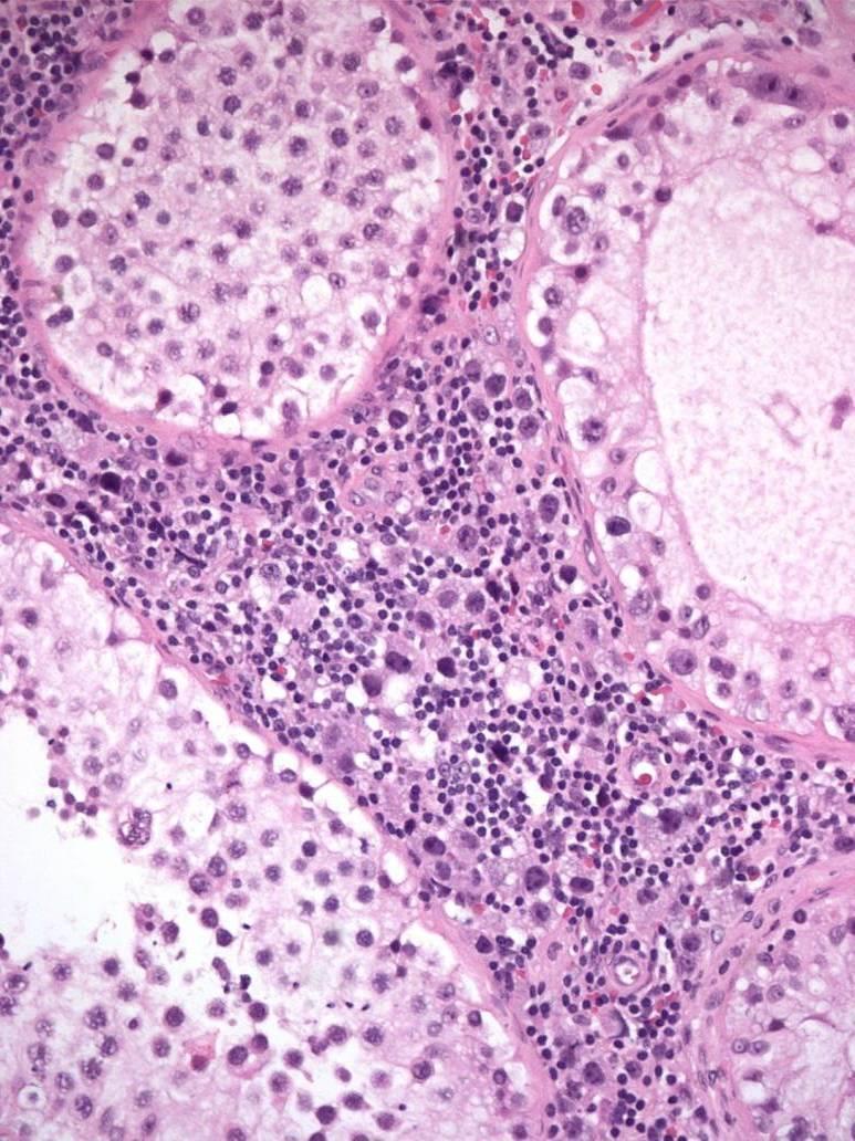
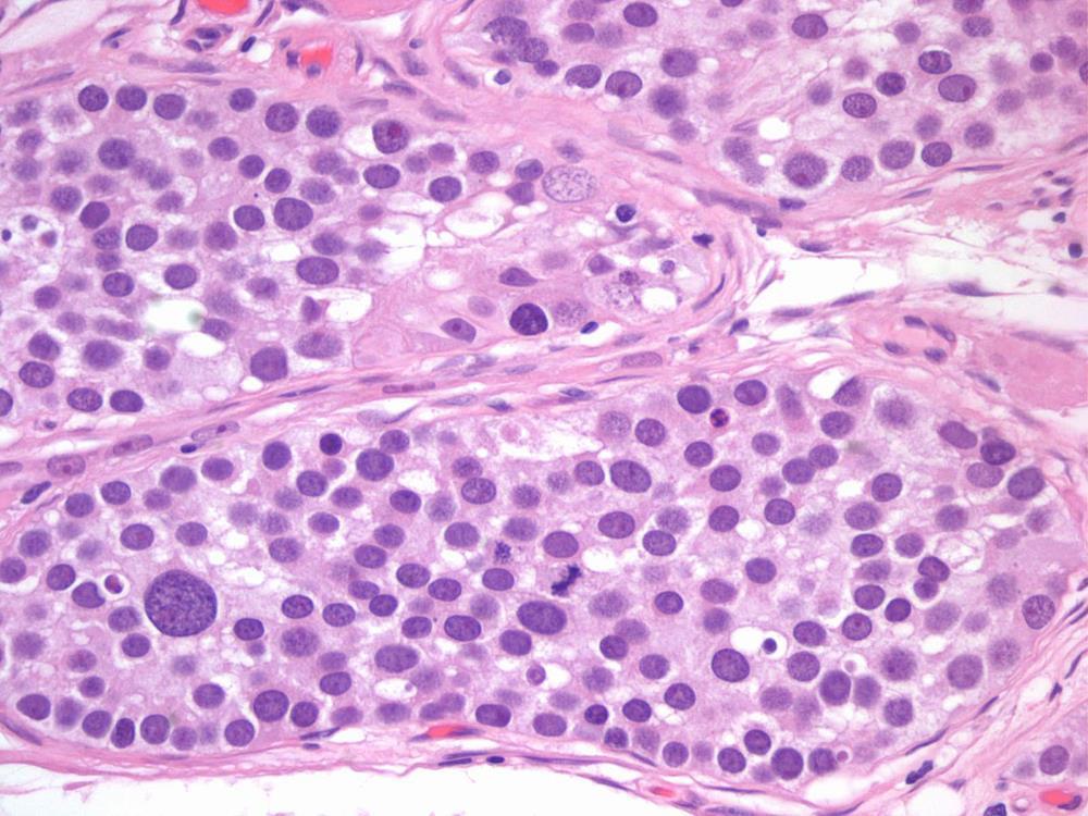
9 Intratubular Seminoma Expanded tubules, no residual Sertoli cells Tubules often contain lymphocytes IHC identical to seminoma
10 Intratubular Embryonal Carcinoma OCT3/4

11 GCNIS UNRELATED GCNIS RELATED Pathogenetic Model for Germ Cell Tumors
12 2016 WHO Germ Cell Tumor Classification Yolk sac tumor prepubertal Dermoid cyst, epidermoid cyst, carcinoid tumor Sarcomatoid YST/ sarcoma NOS Teratoma prepubertal Yolk sac tumor Somatic malignancy Teratoma postpubertal Embryonal carcinoma Spermatocytic tumor Not GCNIS-derived Germ Cell Tumors GCNIS-derived Seminoma Spermatocytic tumor with sarcoma Trophoblastic tumors i(12p) Choriocarcinoma Other trophoblastic tumors
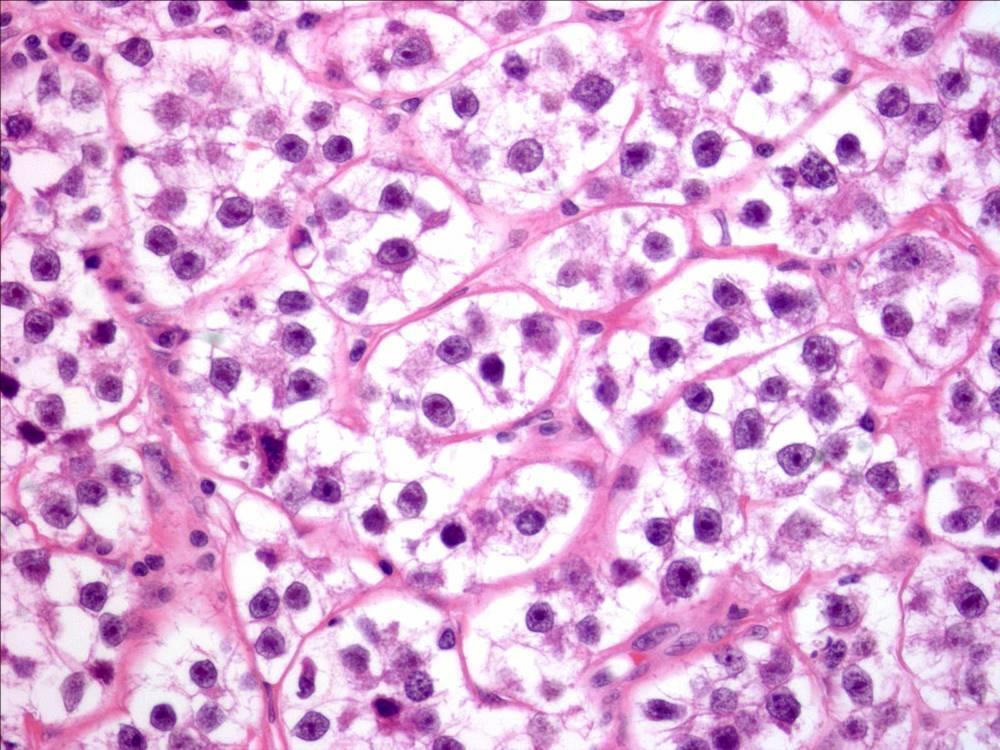
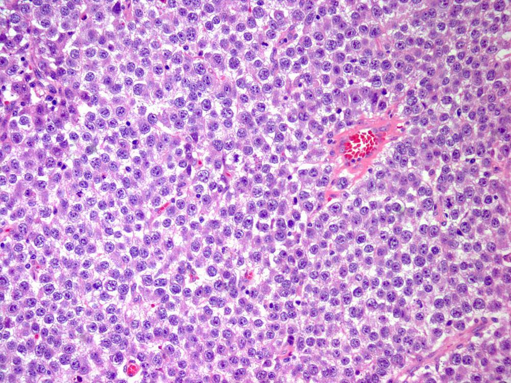
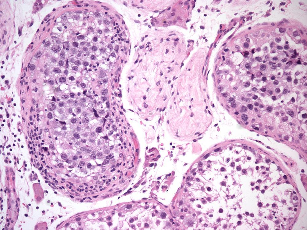
13 Seminoma Most common type of testicular GCT (up to 50%) Average age = 40.5 years (decade later than others GCT) Usually presents with testicular mass Pain or dull aching sensation A few present with metastatic disease - 75% limited to testis - 20% retroperitoneal involvement - 5% distant metastases - may have mild elevated HCG, AFP normal


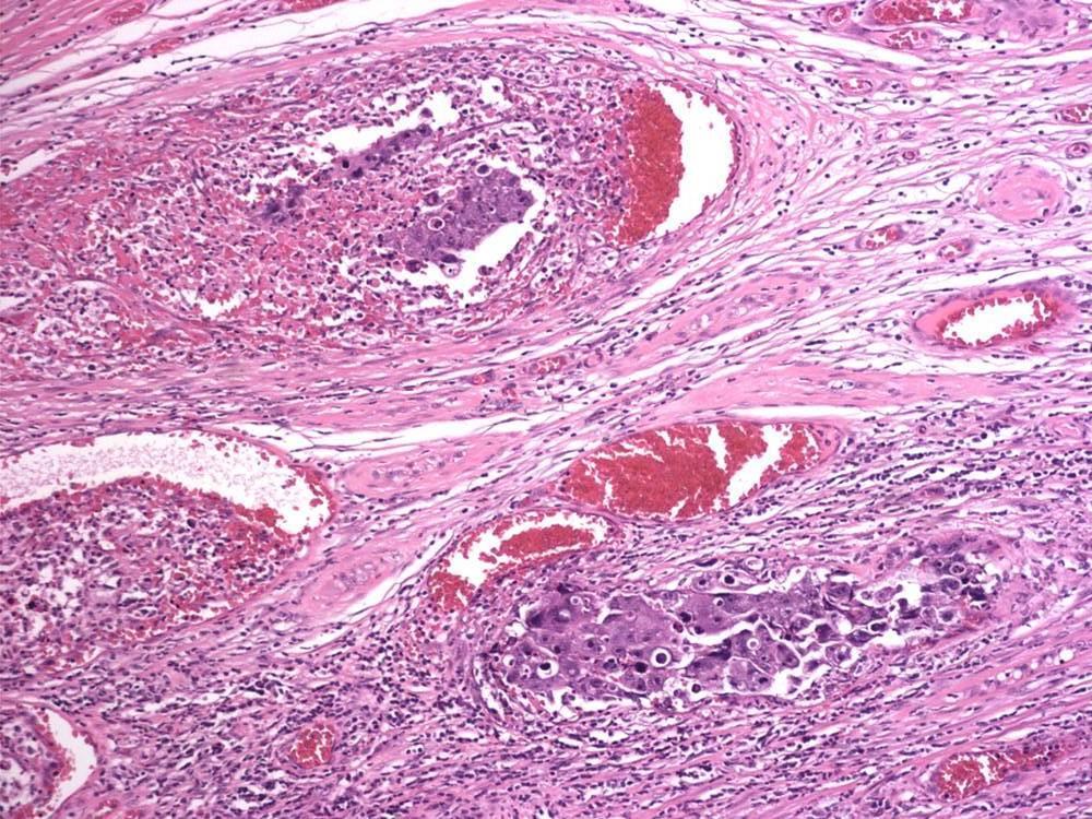
14 Seminoma Homogeneous light-tan nodular fleshy mass Hemorrhage & necrosis
15 Seminoma
16 Seminoma
17 Seminoma
18 Seminoma

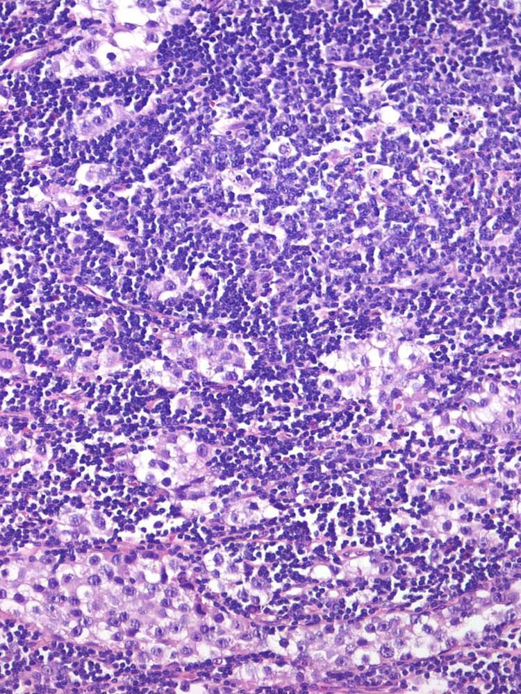
19 Seminoma with marked inflammatory infiltrate

20 Seminoma with granulomatous inflammation
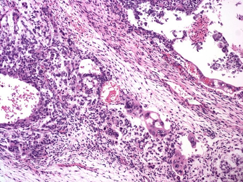
21 Seminoma: intertubular pattern of spread OCT3/4

22 Seminoma: intertubular pattern of spread
23 Intratubular Seminoma


24 Burnt-out germ cell tumor Lymphoplasmacytic infiltrate Fibrotic scar with calcification
![Spontaneous regression of gonadal GCT [so-called burnt-out germ cell tumor] No identifiable invasive neoplasm Dense, hyaline scarring,](/docs-images/82/85043980/images/25-0.jpg "sometimes with GCNIS in adjacent tubules Intratubular calcifications Lymphoplasmacytic infiltrate Hemosiderin-containing macrophages")
25 Spontaneous regression of gonadal GCT [so-called burnt-out germ cell tumor] No identifiable invasive neoplasm Dense, hyaline scarring, sometimes with GCNIS in adjacent tubules Intratubular calcifications Lymphoplasmacytic infiltrate Hemosiderin-containing macrophages Testicular atrophy
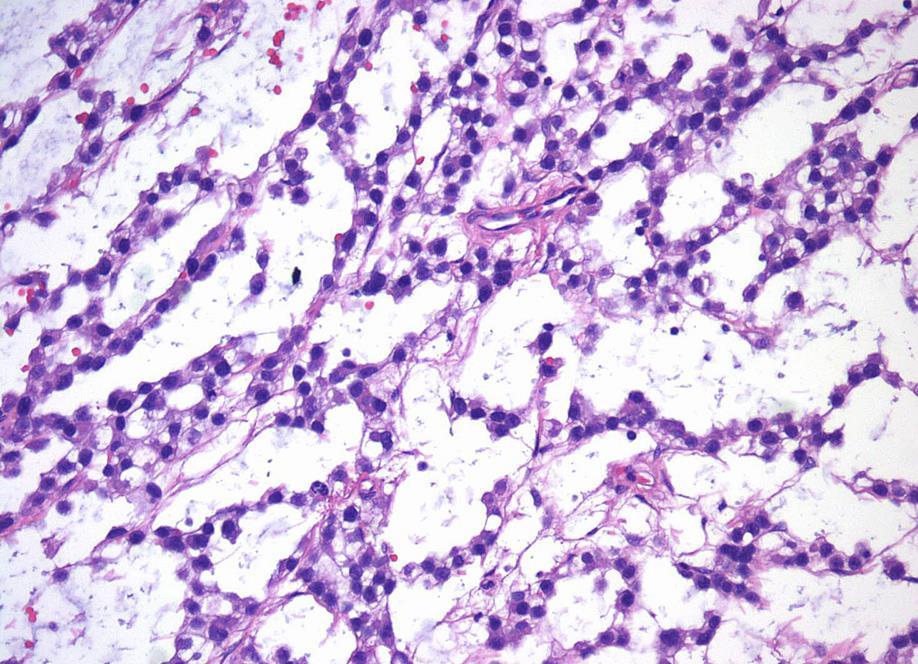
26 Seminoma: differential diagnosis PLAP OCT3/4 AE1/3 CD30 CD117 SALL4 CD45 AFP Seminoma + + focal _ + + Embryonal carcinoma (solid pattern) - Indistinct cell border and overlapping nuclei - Glandular structure only seen in EC - AE1/3 and CD OCT3/4 + Yolk sac tumor (solid pattern) - No fibrous septae - Solid YST is usually associated with other types - Edema in seminoma may resemble reticular YST - AE1/3+, Glypican 3, AFP +/-; OCT3/4, CD117
27 Seminoma: differential diagnosis Sertoli cell tumor - Tubular pattern may be confused with Sertoli cell tumor - Lipid (not glycogen) is responsible for clear cytoplasm - PLAP, OCT3/4, inhibin + Lymphoma - No fibrous septae - Older patients - CD Bilateral involvement more likely Choriocarcinoma (CC) - No biphasic pattern is seen in seminoma - HCG is markedly elevated in CC; modestly in seminoma - AE1/3+, EMA +; OCT3/4

28 Seminoma: tubular pattern

29 Seminoma: tubular pattern


30 Seminoma: tubular pattern OCT3/4 CD117 inhibin

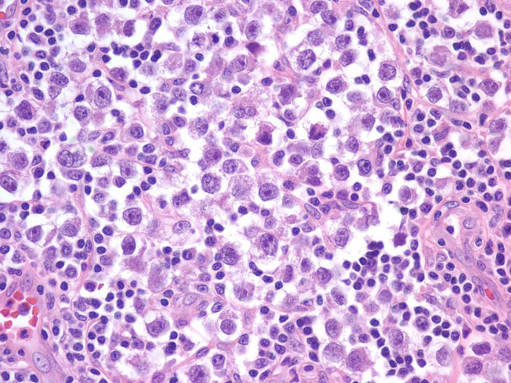
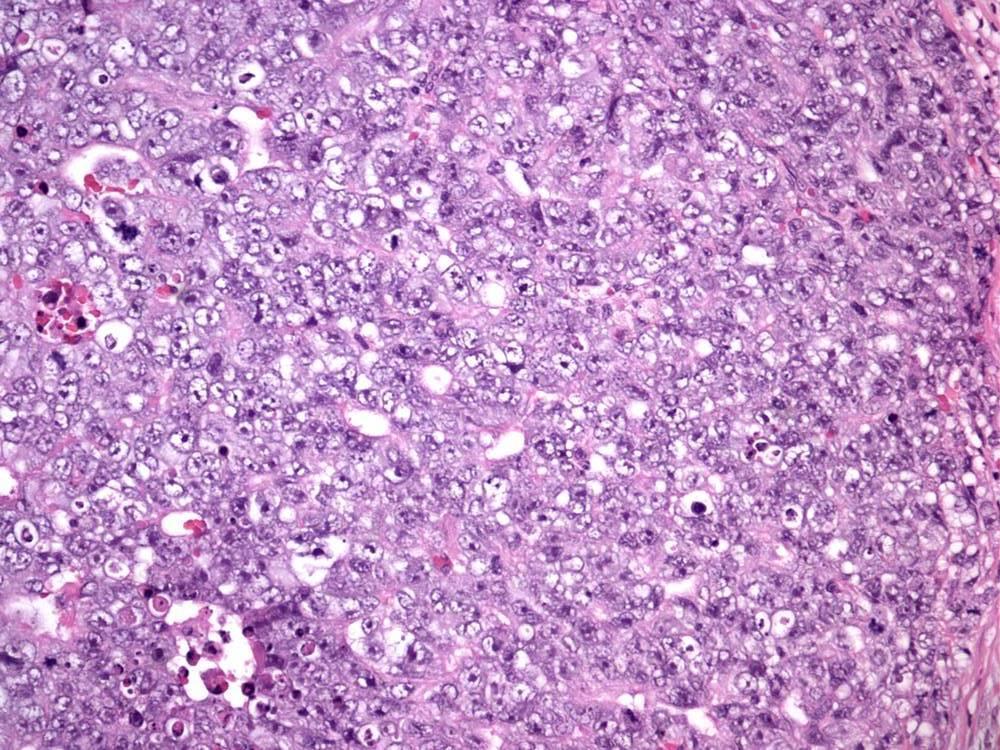
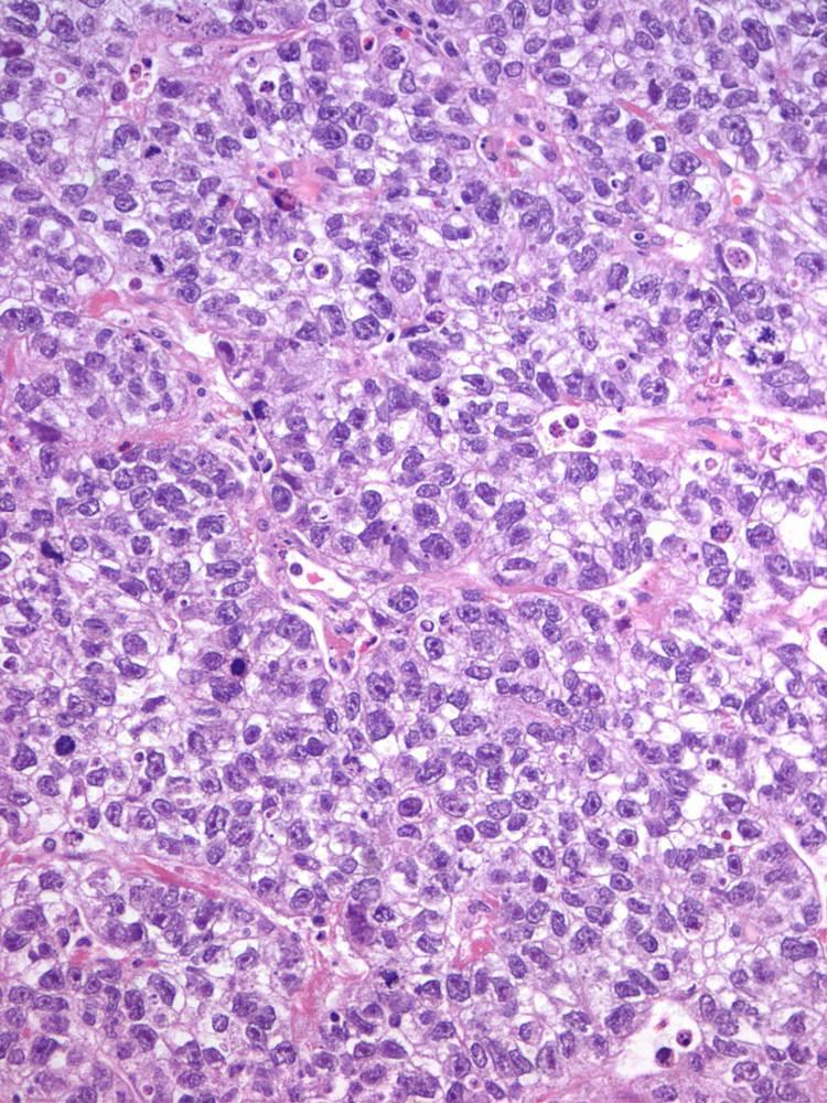
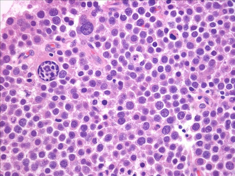
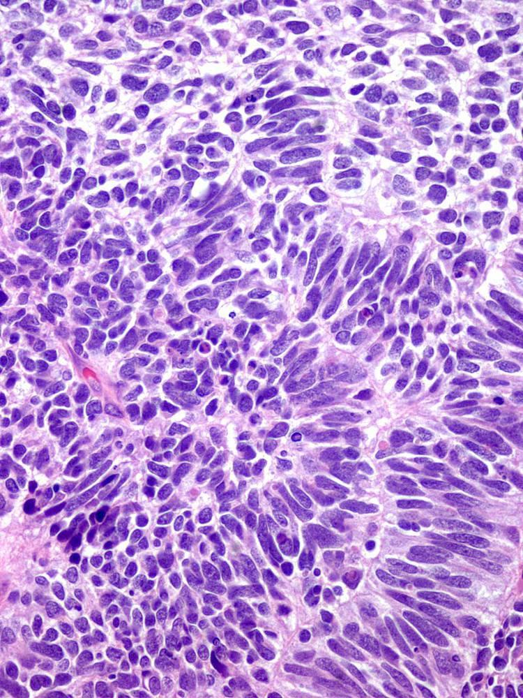
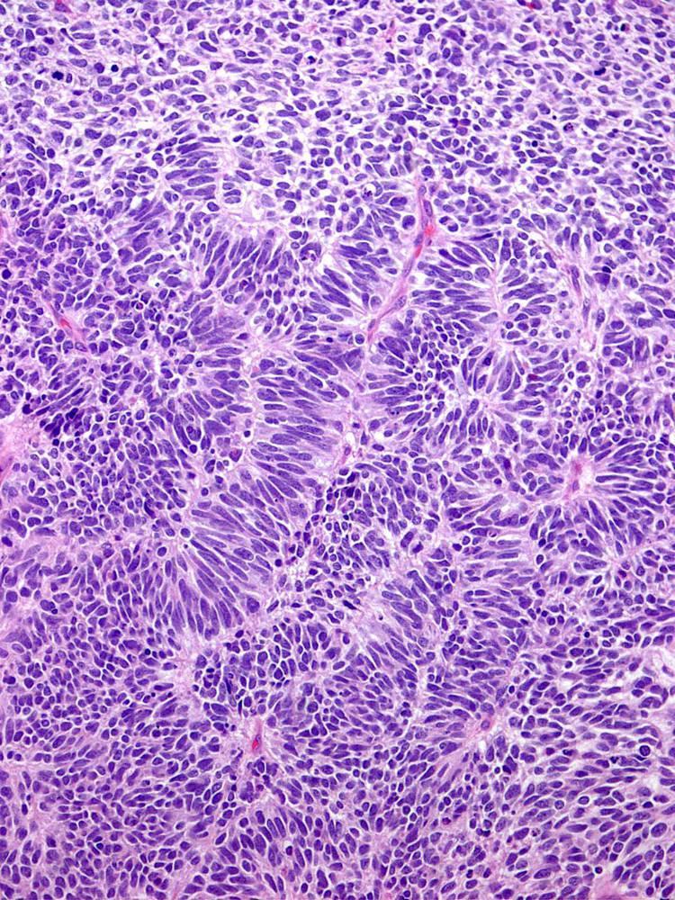
31 Embryonal Carcinoma Pure is rare (10%) Seen in 40% of TGCTs Mean age = 32 Only 40% have disease limited to testis at presentation 2/3 have metastatic disease upon staging Hemorrhage/necrosis common Not as well circumscribed as seminoma

32 Embryonal Carcinoma

33 Embryonal Carcinoma


34 Embryonal Carcinoma: growth pattern Papillary Glandular/tubular Solid

35 Embryonal Carcinoma
36 Seminoma EC: differential diagnosis PLAP OCT3/4 AE1/3 CD30 CD117 SALL4 EMA CEA AFP EC focal _ + focal - Previously discussed Yolk sac tumor - Cells are smaller and less pleomorphic - Hyaline globules are present - AFP is diffusely + - CD30 and OCT3/4 Choriocarcinoma - Syncytiotrophoblast cells are mixed with cytotrophoblast cell (biphasic pattern)
37 Yolk sac tumor (YST) Most common testicular neoplasm in children: 80% of pure YSTs occur in the first 2 years of life Pure YST is uncommon in adults (1.5% of GCTs); however YST is a component of ~40% of mixed GCT In adults present as a painless mass Serum alpha fetoprotein (AFP) levels are elevated in 90% of cases Patterns resemble portions of rat placenta


38 YST: gross appearance Typically solid and soft, white-gray, light yellow with cystic degeneration Large tumors may show necrosis and hemorrhage
39 Yolk sac tumor (YST) Histologic patterns Microcystic (reticular) Macrocystic Myxoid Endodermal sinus (festoon) Solid Polyvesicular vitelline Hepatoid Spindle cells (in post-chemotherapy tumors) Parietal (AFP -) Glandular (clear cells)

40 YST: microcystic variant

41 YST: microcystic and solid variant


42 YST: histologic patterns Microcystic Solid and microcystic


43 YST: histologic patterns Myxoid-spindle Endodermal sinus (festoon)

44 YST: solid variant

45 YST: solid variant
46 YST: differential diagnosis PLAP OCT3/4 AE1/3 CD30 Glypican-3 SALL4 EMA CEA AFP YST +/- _ + _ + + _ + + Seminoma (vs. solid YST) - No hyaline globules seen - Glypican 3, AFP, OCT3/4 + Embryonal carcinoma - Marked nuclear crowding not seen in YST - CK +, focally AFP + (similar to YST) - CD30 and OCT3/4 + Teratoma (vs. glandular YST) - AFP

47 Spermatocytic Seminoma Tumor Derived from postpubertal-type germ cells No relationship with seminoma years old patients More frequently bilateral than other CGTs (9%) Never described in any site other than testis No association with cryptorchidism; no racial predisposition Amplification of chr. 9 (DMRT1) is most consistent genetic abnormality
48 Spermatocytic Tumor

49 Intratubular Spermatocytic Tumor

50 Spermatocytic Tumor: Intratubular Growth
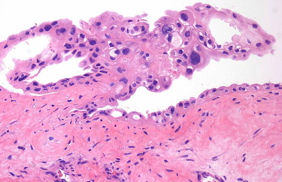
51 Spermatocytic Tumor It metastasizes only exceedingly rarely (2 cases) Treatment: orchiectomy without adjuvant treatment Sarcomatous transformation is a rare complication: ~50% of patients develop metastatic disease and die of it Differential diagnosis: - Classic seminoma - Embryonal carcinoma - Lymphoma Spermatocytic Tumor PLAP OCT3/4 AE1/3 CD30 CD117 SALL4 CD _

52 Teratoma: Post-Pubertal Type Most are mixed with other GCT elements; 4% are pure Capable of metastasis despite lack of malignant appearance May displays differentiation toward mature or immature somatic tissue Even patients with pure teratoma may develop metastases containing other GCT types


53 Teratoma Mature Immature Immature elements do NOT affect overall prognosis

54 Teratoma: immature elements
55 Teratoma: Malignant Transformation Carcinomatous transformation requires an overtly invasive growth pattern Somatic-type malignancy requires overgrowth of malignant-appearing mesenchymal or embryonic tissues to exclude other elements (at least a 4X low power field) Overgrowth of primitive neuroectodermal tissue should be recognized as primitive neuroectodermal tumor (PNET): - Limited to testis: most men are cured of the disease - In metastases: surgical resection is mainstay of therapy; outcome is generally poor
56 Teratoma: overgrowth of PNET

57 Teratoma: Prepubertal Type GCT usually seen in pre-pubertal testis Composed of elements resembling somatic tissues derived from one of more germinal layers NOT associated with: - GCNIS or atypia - Dysgenetic changes - Scarring - Chr. 12p amplification Conservative treatment
58 Changes in Trophoblastic Tumor WHO 2004 Trophoblastic Tumors Choriocarcinoma Trophoblastic neoplasms other than choriocarcinoma - Monophasic choriocarcinoma - Placental site trophoblastic tumor WHO 2016 Trophoblastic Tumors Choriocarcinoma - Monophasic choriocarcinoma Non-choriocarcinomatous trophoblastic tumors - Placental site trophoblastic tumor (PSTT) - Epithelioid trophoblastic tumor (ETT) - Cystic trophoblastic tumor
 Serum HCG is typically elevated (> 55,000 IU/L) Prognosis is worse than for other GCT")
59 Pure is quite rare (<1%); uncommon in mixed GCT (15%) Young patients (mean age years) Symptoms related to metastatic disease (lungs, brain, GI tract) Serum HCG is typically elevated (> 55,000 IU/L) Prognosis is worse than for other GCT Choriocarcinoma

60 Choriocarcinoma Cytotrophoblasts, intermediate trophoblasts Syncytiotrophoblast
61 Choriocarcinoma: Differential Diagnosis Other GCT may contain trophoblast cells, but they are scattered individual cells and lack biphasic pattern EC may show degenerate cells with a poorly defined syncytiotrophoblastic component: lack of hemorrhage, hcg+ and OCT3/4+ distinguish EC from chorio Monophasic chorio should be distinguished from seminoma and solid pattern YST: - diffuse hcg +, AFP -, OCT3/4 - greater pleomorphism than in seminoma PLAP OCT3/4 CK CD30 Inhibin GATA3 EMA hcg AFP Chorio +/- _ + _ + + +/- + _
")
62 Non-choriocarcinomatous trophoblastic tumors: Cystic Trophoblastic Tumors May evolve from choriocarcinoma with regression of highly proliferative elements Occur mostly in metastatic sites after chemotherapy Rare de novo tumors in testis Normal/slightly elevated hcg Clinical significance similar to residual teratoma Treat as post-chemo teratoma (surgical resection; no additional chemo)

63 Cystic Trophoblastic Tumors Non-infiltrative Lack biphasic growth Low mitotic rate


64 Changes in Sex Cord-Stromal Tumor Sclerosing Sertoli cell tumor - Variant of Sertoli cell tumor NOS - Similar CTNNB1 gene mutation and nuclear ß-catenin Intratubular large cell hyalinizing Sertoli cell tumor - Distinct entity associated with Peutz-Jeghers syndrome - STK11 gene mutation


65 Changes in Mixed Germ Cell Sex Cord-Stromal Tumors Gonadoblastoma (only entity) - Germ cells, similar to GCNIS - Sex cord cells resembling immature granulosa cells Rare, but seen in 50% of sex development disorders 70% diagnosed in neonatal period due to ambiguous genitalia May occur in dysgenetic testis: 40% bilateral If untreated, progresses to invasive GCT

66 8th AJCC/TNM Staging of Testicular Tumors In seminoma, T1 is subclassified to T1a and T1b according to size, using a 3 cm cutoff Size is independent predictor of disease recurrence


67 8th AJCC/TNM Staging of Testicular Tumors Hilar soft tissue invasion is T2

68 8th AJCC/TNM Staging of Testicular Tumors Epididymal invasion is T2 rather than T1


69 8th AJCC/TNM Staging: Spermatic Cord Invasion Vascular invasion in spermatic cord without stromal invasion: T2 Cord involvement continuous with primary tumor: T3 Cord involvement discontinuous with primary tumor: M1 T3 T3
70 Take Home Message Updated pathogenetic model for GCTs Restructuring of classification - GCNIS related - GCNIS unrelated New entities Changes in testicular tumor staging


71 Thank you!
Male Genital Cancers in the US in Frequency of Types
 Germ Cell Tumors of the Testis Pathology, Immunohistochemistry, and the Often Confusing Appearance of Their Metastases Charles Zaloudek, MD Department of Pathology UCSF Male Genital Cancers in the US in
Testicular Germ Cell Tumors; A Simplistic Approach
 Testicular Germ Cell Tumors; A Simplistic Approach Merce Jorda, MD, PhD, MBA Professor and Vice Chair, Director of Anatomic Pathology Director of Genitourinary Pathology Service Interim Director of Cytopathology
Note: The cause of testicular neoplasms remains unknown
 - In the 15- to 34-year-old age group, they are the most common tumors of men. - Tumors of the testis are a heterogeneous group of neoplasms that include: I. Germ cell tumors : 95%; all are malignant.
-The cause of testicular neoplasms remains unknown
 - In the 15- to 34-year-old age group, they are the most common tumors of men. - include: I. Germ cell tumors : (95%); all are malignant. II. Sex cord-stromal tumors: from Sertoli or Leydig cells; usually
Tinh hoàn
 Tinh hoàn Tinh hoàn Tinh hoàn Tiền liệt tuyến Tiền liệt tuyến Mào tinh hoàn Mào tinh hoàn Túi tinh Túi tinh Túi tinh Túi tinh So-called cystadenoma of seminal vesicle. Gross appearance of granulomatous
Disclosure of Relevant Financial Relationships
 Evening Specialty Conference - Genitourinary Pathology Case 2 Disclosure of Relevant Financial Relationships Sean R Williamson, MD Henry Ford Health System, Detroit, MI @Williamson_SR USCAP requires that
Testis. Protocol applies to all malignant germ cell and malignant sex cord-stromal tumors of the testis, exclusive of paratesticular malignancies.
 Testis Protocol applies to all malignant germ cell and malignant sex cord-stromal tumors of the testis, exclusive of paratesticular malignancies. Protocol revision date: January 2005 Based on AJCC/UICC
2% of all malignancies Male predominance Patients usually more than 60 years old
 Benign Bladder Tumors Transitional Cell Papilloma 2-3% Inverted Papilloma Rare Malignant Transitional (Urothelial) Carcinoma 90% Carcinoma In-Situ (By Itself) 5-10% Squamous Cell Carcinoma 3-7% Adenocarcinoma
Germ cell tumours UK SH. Ivo Leuschner. Kiel Pediatric Tumor Registry, Institute of Pathology University Hospital of Schleswig-Holstein Campus Kiel
 Germ cell tumours Ivo Leuschner Kiel Pediatric Tumor Registry, Institute of Pathology University Hospital of Schleswig-Holstein Campus Kiel UK SH Old histogenetic Concept of Germ cell tumours Pluripotent
Recently, there has been an increasing incidence among women and younger persons
 Bladder Tumors Benign Transitional Cell Papilloma 2-3% Inverted Papilloma Rare Malignant Transitional (Urothelial) Carcinoma 90% Carcinoma In-Situ (By Itself) 5-10% Squamous Cell Carcinoma 3-7% Adenocarcinoma
Cardiff MRCS OSCE Courses Testicular Cancer
 Testicular Cancer Scenario: A 40-year-old male presents to the surgical out-patient clinic with a 6-8 week history of a painless lump in his left scrotum. He however complains of a dull ache in the scrotum
Protocol for the Examination of Lymphadenectomy Specimens From Patients With Malignant Germ Cell and Sex Cord-Stromal Tumors of the Testis
 Protocol for the Examination of Specimens From Patients With Malignant Germ Cell and Sex Cord-Stromal Tumors of the Testis Version: Testis 4.0.1.1 Protocol Posting Date: February 2019 Accreditation Requirements
Transitional Cell Papilloma 2-3% Inverted Papilloma Rare
 BLADDER TUMORS Benign Transitional Cell Papilloma 2-3% Inverted Papilloma Rare Malignant Transitional (Urothelial) Carcinoma90% Carcinoma In-Situ (By Itself) 5-10% Squamous Cell Carcinoma 3-7% Adenocarcinoma
Morphologic Aspects Aspects
 Testicular Testicular Germ Germ Cell Cell Neoplasia: Neoplasia: Molecular Molecular Pathways Pathways & Challenging Morphologic Aspects Aspects George George J. J. Netto, Netto, M.D. M.D. Johns Johns Hopkins
Diseases of the penis & testis
 Diseases of the penis & testis Done by : Saef B AL-Abbadi Diseases of penis, Condyloma Acuminatum A benign tumor *Tend to recur but only rarely progress into in situ or invasive cancers read this = genital
The Pathology of Germ Cell Tumours of the Ovary
 The Pathology of Germ Cell Tumours of the Ovary Professor Mike Wells University of Sheffield Amman, Jordan November 2013 Professor Francisco Paco Nogales I. Primitive germ cell tumors A. Dysgerminoma
Case E1. Female aged 63 years Right Nephrectomy Two separate tumours Section of each tumour
 Case E1 Female aged 63 years Right Nephrectomy Two separate tumours Section of each tumour Tumour 1 Upper pole tumour 28mm macro diameter Circumscribed Friable cut surface Tumour 2 Middle pole Part solid
International Journal of Research and Review E-ISSN: ; P-ISSN:
 International Journal of Research and Review www.ijrrjournal.com E-ISSN: 2349-9788; P-ISSN: 2454-2237 Case Report Rare Combinations of Testicular Mixed Germ Cell Tumors - 2 Case Reports Dr. Byna Syam Sundara
International Journal of Medical and Health Sciences
 International Journal of Medical and Health Sciences Journal Home Page: http://www.ijmhs.net ISSN:2277-4505 Case Report Histomorphological Specrtum of Malignant Germ Cell Tumours: An Overview and Report
Adenocarcinoma of the Cervix
 Question 1. Each of the following statements about cervical adenocarcinoma is true except: Adenocarcinoma of the Cervix SAMS a) A majority of women with cervical adenocarcinoma have stage I tumors at diagnosis.
Ovarian Clear Cell Carcinoma
 Ovarian Clear Cell Carcinoma Rouba Ali-Fehmi, MD Professor of Pathology The Karmanos Cancer Institute, Wayne State University School of Medicine 50 year old woman with chief complaint of shortness of breath
Prof. Dr. med. Beata BODE-LESNIEWSKA Institute of Pathology and Molecular Pathology University Hospital; Zurich
 Prof. Dr. med. Beata BODE-LESNIEWSKA Institute of Pathology and Molecular Pathology University Hospital; Zurich 32 year old man 2 months history of growing left supraclavicular lymph nodes Antibiotic treatment
GUIDELINES ON TESTICULAR CANCER
 38 (Text updated March 2005) P. Albers (chairman), W. Albrecht, F. Algaba, C. Bokemeyer, G. Cohn-Cedermark, A. Horwich, O. Klepp, M.P. Laguna, G. Pizzocaro Introduction Compared with other types of cancer
3 cell types in the normal ovary
 Ovarian tumors 3 cell types in the normal ovary Surface (coelomic epithelium) the origin of the great majority of ovarian tumors (neoplasms) 90% of malignant ovarian tumors Totipotent germ cells Sex cord-stromal
Mediastinal Germ Cell Tumors
 Mediastinal Germ Cell Tumors Anja C. Roden, M.D. Department of Laboratory Medicine and Pathology, Mayo Clinic, Rochester, MN, USA 2018 MFMER slide-1 Disclosure I have no relevant financial relationships
Updates in Urologic Pathology WHO Made Those Changes?! Peyman Tavassoli Pathology Department BC Cancer Agency
 Updates in Urologic Pathology WHO Made Those Changes?! Peyman Tavassoli Pathology Department BC Cancer Agency World Health Organization Available in Feb 2016 Frame work for reporting Major contributing
Pathology of Ovarian Tumours. Dr. Jyothi Ranganathan MD ( Path) AFMC Pune PDCC (Cytopathology) PGI Chandigarh
 AFMC Pune PDCC (Cytopathology) PGI Chandigarh") Pathology of Ovarian Tumours Dr. Jyothi Ranganathan MD ( Path) AFMC Pune PDCC (Cytopathology) PGI Chandigarh Outline Incidence Risk factors Classification Pathology of tumours Tumour markers Prevention
Pathology of Ovarian Tumours Dr. Jyothi Ranganathan MD ( Path) AFMC Pune PDCC (Cytopathology) PGI Chandigarh Outline Incidence Risk factors Classification Pathology of tumours Tumour markers Prevention
Leukaemia 35% Lymphoma 14%
 Distribution ib ti of Cancers in Children under 15 years Leukaemia 35% Lymphoma 14% Neuroblastoma 9% Other 5% Liver 1% Retinoblastoma 3% Bone and STS 15% CNS 20% Wilms' 8% 30-40% Mortality Germ Cell Tumours
Exercise. Discharge Summary
 Exercise Discharge Summary A 32-year-old Brazilian male presented with a 6 month history of right-sided scrotal swelling. Backache was present for 2 months and a history of right epididymitis was present
Intratubular Germ Cell Neoplasia of the Testis
 Intratubular Germ Cell Neoplasia of the Testis KS Ngoo Department of Urology Hospital Selayang Advanced Urology Course 15 Aug 2014 MUA Office Clinical scenario A 33 years old man has bilateral testicular
DISORDERS OF MALE GENITALS
 Wit JM, Ranke MB, Kelnar CJH (eds): ESPE classification of paediatric endocrine diagnosis. 9. Testicular disorders/disorders of male genitals. Horm Res 2007;68(suppl 2):63 66 ESPE Code Diagnosis OMIM ICD10
3 cell types in the normal ovary
 Ovarian tumors 3 cell types in the normal ovary Surface (coelomic epithelium) the origin of the great majority of ovarian tumors 90% of malignant ovarian tumors Totipotent germ cells Sex cord-stromal cells
CNS TUMORS. D r. Ali Eltayb ( U. of Omdurman. I ). M. Path (U. of Alexandria)
. M. Path (U. of Alexandria)") CNS TUMORS D r. Ali Eltayb ( U. of Omdurman. I ). M. Path (U. of Alexandria) CNS TUMORS The annual incidence of intracranial tumors of the CNS ISmore than intraspinal tumors May be Primary or Secondary
CNS TUMORS D r. Ali Eltayb ( U. of Omdurman. I ). M. Path (U. of Alexandria) CNS TUMORS The annual incidence of intracranial tumors of the CNS ISmore than intraspinal tumors May be Primary or Secondary
Testicular Tumors Including Secondary and Unusual Tumors of the Testis
 Testicular Tumors Including Secondary and Unusual Tumors of the Testis Milton W. Datta Partner, Hospital Pathology Associates University of Minnesota Minneapolis, MN Topics Review Features of Germ Cell
04/09/2018. Salivary Gland Pathology in the Molecular Era Old Friends, Old Foes, & New Acquaintances
 Salivary Gland Pathology in the Molecular Era Old Friends, Old Foes, & New Acquaintances Jennifer L. Hunt, MD, MEd Aubrey J. Hough Jr, MD, Endowed Professor of Pathology Chair of Pathology and Laboratory
Leydig cell tumour. Testis: non-germ cell tumours. Testis: sex cord-stromal tumours. Differential diagnosis of Leydig cell tumour TTAGS
 Non-germ cell s of the testis Dr Jonathan H Shanks The Christie NHS Foundation Trust, Manchester, UK Testis: non-germ cell s Sex cord-stromal s Haemolymphoid neoplasms Other neoplasms Tumour-like conditions
Fellow GU Lecture Series, Testicular Cancer. Asit Paul, MD, PhD 02/06/2018
 Fellow GU Lecture Series, 2018 Testicular Cancer Asit Paul, MD, PhD 02/06/2018 Rare cancer worldwide, approximately 1% of all male cancers There is a large difference among ethnic/racial groups. Rates
*OPERATIVE PROCEDURE. Serum tumour markers within normal limits S1.04 PRINCIPAL CLINICIAN
 Neoplasia of the Testis - Orchidectomy Histopathology Reporting Proforma Includes the International Collaboration on Cancer reporting dataset denoted by * Family name Given name(s) Date of birth Indigenous
Tumours of the Prostate and Testis update on WHO classification, IHC and molecular Morphology
 Tumours of the Prostate and Testis update on WHO classification, IHC and molecular Morphology Glen Kristiansen Professor and Chairman Institute of Pathology, University Hospital Bonn January 2016 WHO GU-Pathology
N-cadherin Expression in Testicular Germ Cell and Gonadal Stromal Tumors
 381 Ivyspring International Publisher Research Paper Journal of Cancer 2012; 3: 381-389. doi: 10.7150/jca.5017 N-cadherin Expression in Testicular Germ Cell and Gonadal Stromal Tumors Daniel J. Heidenberg
TRAPDOORS IN TESTICULAR PATHOLOGY
 TRAPDOORS IN TESTICULAR PATHOLOGY Daniel Berney, M.B.B.S. FRCPath Consultant Histopathologist Department of Cellular Pathology The Orchid Tissue Laboratory, Barts and The London NHS Trust The Royal London
Dr Sanjiv Manek Oxford. Oxford Pathology Course 2010 for FRCPath Illustration-Cellular Pathology. Oxford Radcliffe NHS Trust
 Dr Sanjiv Manek Oxford Oxford Pathology Course 2010 for FRCPath Illustration-Cellular Pathology. Oxford Radcliffe NHS Trust Ovarian Endometrial Vulvo-vaginal Cervical Illustration-Cellular Pathology. Oxford
Trophoblastic tumors
 Trophoblastic tumors Uterus tumor course Oslo, 21-22/1/16 Prof. Ben Davidson, MD PhD Department of Pathology, Norwegian Radium Hospital, Oslo University Hospital, Oslo, Norway Cases 45 38 39 4 Case 45
Uncommon secondary tumour of the stomach
 Uncommon secondary tumour of the stomach B. Bancel, Hôpital CROIX ROUSSE LYON Bucharest Nov 2013 Case report 33-year old man Profound mental retardation and motor disturbances (sequelae of neonatal meningeal
Disclosure. Relevant Financial Relationship(s) None. Off Label Usage None MFMER slide-1
 None. Off Label Usage None MFMER slide-1") Disclosure Relevant Financial Relationship(s) None Off Label Usage None 2013 MFMER slide-1 Case Presentation A 43 year old male, with partial nephrectomy for a right kidney mass 2013 MFMER slide-2 2013
Disclosure Relevant Financial Relationship(s) None Off Label Usage None 2013 MFMER slide-1 Case Presentation A 43 year old male, with partial nephrectomy for a right kidney mass 2013 MFMER slide-2 2013
Case Scenario 1 Discharge Summary Pathology Report Final Diagnosis: Oncology Consult
 Case Scenario 1 Discharge Summary A 31-year-old Brazilian male presented with a 6 month history of right-sided scrotal swelling. Backache was present for 2 months and a history of right epididymitis was
57th Annual HSCP Spring Symposium 4/16/2016
 An Unusual Malignant Spindle Cell Lesion to Involve the Breast Erinn Downs-Kelly, D.O. Associate Professor of Pathology University of Utah & ARUP Laboratories No disclosures Case 39 y/o female with no
EAU GUIDELINES ON TESTICULAR CANCER
 EAU GUIDELINES ON TESTICULAR CANCER (Limited text update March 2018) P. Albers (Chair), W. Albrecht, F. Algaba, C. Bokemeyer, G. Cohn-Cedermark, K. Fizazi, A. Horwich, M.P. Laguna (Vice-chair), N. Nicolai,
More than 90% testicular tumors are of germ cell
 ORIGINAL ARTICLE SALL4 Is a Novel Diagnostic Marker for Testicular Germ Cell Tumors Dengfeng Cao, MD, PhD,* Jianping Li, BS,* Charles C. Guo, MD,w Robert W. Allan, MD,z and Peter A. Humphrey, MD, PhD*
Fellow GU Lecture Series, Testicular Cancer. Asit Paul, MD, PhD 02/06/2018
 Fellow GU Lecture Series, 2018 Testicular Cancer Asit Paul, MD, PhD 02/06/2018 Rare cancer worldwide, approximately 1% of all male cancers There is a large difference among ethnic/racial groups. Rates
Case Scenario 1 Discharge Summary Pathology Report Final Diagnosis: Oncology Consult
 Case Scenario 1 Discharge Summary A 31-year-old Brazilian male presented with a 6 month history of right-sided scrotal swelling. Backache was present for 2 months and a history of right epididymitis was
Pelvic tumor in childhood Classification, imaging approach and radiological findings
 Pelvic tumor in childhood Classification, imaging approach and radiological findings M. Mearadji International Foundation for Pediatric Imaging Aid Rotterdam, The Netherlands Solid pelvic masses in childhood
Salivary Glands 3/7/2017
 Salivary Glands 3/7/2017 Goals and objectives Focus on the entities unique to H&N Common board type facts Information for your future practice Salivary Glands Salivary Glands Major gland. Paratid. Submandibular.
Testicular tumors; Ultrasonographic and Pathologic correlation
 Testicular tumors; Ultrasonographic and Pathologic correlation Poster No.: C-0106 Congress: ECR 2014 Type: Educational Exhibit Authors: Y. Kim, S. W. Shin, E. T. Kim, M. Y. Kim ; Kuri City/KR, 1 1 2 1
EAU GUIDELINES ON TESTICULAR CANCER
 EU GUIDELINES ON TESTICULR CNCER (Limited text update March 2017) P. lbers (Chair), W. lbrecht, F. lgaba, C. Bokemeyer, G. Cohn-Cedermark, K. Fizazi,. Horwich, M.P. Laguna, N. Nicolai, J. Oldenburg Introduction
Ovarian Malignant Germ Cell Tumors: Cellular Classification and Clinical and Imaging Features 1
 Note: This copy is for your personal non-commercial use only. To order presentation-ready copies for distribution to your colleagues or clients, contact us at www.rsna.org/rsnarights. Ovarian Malignant
Quiz 1. Assign Race 1, Race 2 and Spanish Hispanic Origin to the following scenarios.
 Quiz 1 Assign Race 1, Race 2 and Spanish Hispanic Origin to the following scenarios. 1. 62 year old Brazilian female Race 1 Race 2 Spanish/Hispanic Origin 2. 43 year old Asian male born in Japan Race 1
Normal endometrium: A, proliferative. B, secretory.
 Normal endometrium: A, proliferative. B, secretory. Nội mạc tử cung Nội mạc tử cung Cyclic changes in endometrium.. Approximate relationship of useful microscopic changes. Arias-Stella reaction in endometrial
Lesions Mimicking Adenoid Cystic Carcinoma. Diagnostic Problems in Salivary Gland Pathology An Update 5/29/2009
 Diagnostic Problems in Salivary Gland Pathology An Update Lesions Mimicking Adenoid Cystic Carcinoma Stacey E. Mills, M.D. W.S. Royster Professor of Pathology Director of Surgical and Cytopathology University
Case Presentation. Maha Akkawi, MD, Fatima Obeidat, MD, Tariq Aladily, MD. Department of Pathology Jordan University Hospital Amman, Jordan
 Case Presentation Maha Akkawi, MD, Fatima Obeidat, MD, Tariq Aladily, MD Department of Pathology Jordan University Hospital Amman, Jordan The 25th Annual Congress of the ADIAP The 8/11/2013 1 5th International
Immunohistochemistry in Bone and Soft Tissue Tumors. Sahar Rassi Zankoul, MD
 Immunohistochemistry in Bone and Soft Tissue Tumors Sahar Rassi Zankoul, MD Introduction Bone tumors represent a wide variety of tumors of various origins and malignant potentials. These different tumor
EAU GUIDELINES ON TESTICULAR CANCER
 EAU GUIDELINES ON TESTICULAR CANCER (Limited text update March 2015) P. Albers (Chair), W. Albrecht, F. Algaba, C. Bokemeyer, G. Cohn-Cedermark, K. Fizazi, A. Horwich, M.P. Laguna, N. Nicolai, J. Oldenburg
Pathology of the lower urinary tract and male genital system
 Pathology of the lower urinary tract and male genital system Neoplasms of the lower urinary tract Incidence: Urinary bladder > upper urinary tract; male:female=3:1 Symptoms: painless hematuria, hydronephrosis
Citation for published version (APA): Lutke Holzik, M. F. (2007). Genetic predisposition to testicular cancer s.n.
: Lutke Holzik, M. F. (2007). Genetic predisposition to testicular cancer s.n.") University of Groningen Genetic predisposition to testicular cancer Lutke Holzik, Martijn Frederik IMPORTANT NOTE: You are advised to consult the publisher's version (publisher's PDF) if you wish to cite
University of Groningen Genetic predisposition to testicular cancer Lutke Holzik, Martijn Frederik IMPORTANT NOTE: You are advised to consult the publisher's version (publisher's PDF) if you wish to cite
Unilateral ovarian metastases of gastric adenocarcinoma simulating primary ovarian yolk sac tumours
 Unilateral ovarian metastases of gastric adenocarcinoma simulating primary ovarian yolk sac tumours Francisco F Nogales, Elvira Stacher (*), Ovidiu Preda, Pablo Goyenaga, José Aneiros-Fernandez, Farid
Pathological Classification of Hepatocellular Carcinoma
 3 rd APASL Single Topic Conference: HCC in 3D Pathological Classification of Hepatocellular Carcinoma Glenda Lyn Y. Pua, M.D. HCC Primary liver cancer is the 2 nd most common cancer in Asia HCC is the
What is Testicular cancer?
 Testicular Cancer What is Testicular cancer? Testicular cancer is a disease in which cancer cells form in the tissues of one or both testicles. The testicles are 2 egg-shaped glands located inside the
Teratocarcinoma In A Young Boy- An Unusual Presentation
 Human Journals Case Report November 2015 Vol.:2, Issue:1 All rights are reserved by Atia Zaka-ur-Rab et al. Teratocarcinoma In A Young Boy- An Unusual Presentation Keywords: Boy, Testicular Mass, Teratocarcinoma
CHAPTER NINE PATHOLOGY OF THE MALE GENITAL SYSTEM
 CHAPTER NINE PATHOLOGY OF THE MALE GENITAL SYSTEM THE PENIS Malformations of the Penis Hypospadias is the most common malformation (1 in 250 live male births). It refers to the abnormal location of the
Doppler ultrasound of the abdomen and pelvis, and color Doppler
 - - - - - - - - - - - - - Testicular tumors are rare in children. They account for only 1% of all pediatric solid tumors and 3% of all testicular tumors [1,2]. The annual incidence of testicular tumors
Genetic Studies of Dysgerminoma
 Genetic Studies of Chromosome 12p abnormalities are characteristic of germ cell tumors isochromosome 12p and 12p overrepresentation Some can be detected by karyotyping FISH study of 21 dysgerminomas showed
CONTINUING EDUCATION IN TOXICOLOGIC PATHOLOGY REPRODUCTIVE SYSTEM
 CONTINUING EDUCATION IN TOXICOLOGIC PATHOLOGY REPRODUCTIVE SYSTEM ORGANIZED BY SOCIETY FOR TOXICOLOGIC PATHOLOGY IN INDIA (STPI) OCTOBER 29-31, 2010 The Atria Hotel, # 1, Palace Road, Bangalore - 560 001
7 Mousa. Obada Zalat. Mohammad Badi
 7 Mousa Obada Zalat Mohammad Badi Tumors of the ovaries Last lecture we talked about surface epithelial tumors of the ovaries (the most common type). But there are many other types of tumors of germ cell
Selected Pseudomalignant Soft Tissue Tumors of the Skin and Subcutis
 Selected Pseudomalignant Soft Tissue Tumors of the Skin and Subcutis Andrew L. Folpe, M.D. Professor of Laboratory Medicine and Pathology Mayo Clinic, Rochester, MN folpe.andrew@mayo.edu 2016 MFMER slide-1
Testicular Malignancies /8/15
 Collecting Cancer Data: Testis 2014-2015 NAACCR Webinar Series January 8, 2015 Q&A Please submit all questions concerning webinar content through the Q&A panel. Reminder: If you have participants watching
Diseases of the breast (1 of 2)
") Diseases of the breast (1 of 2) Introduction A histology introduction Normal ducts and lobules of the breast are lined by two layers of cells a layer of luminal cells overlying a second layer of myoepithelial
Diseases of the breast (1 of 2) Introduction A histology introduction Normal ducts and lobules of the breast are lined by two layers of cells a layer of luminal cells overlying a second layer of myoepithelial
Synonyms. Nephrogenic metaplasia Mesonephric adenoma
 Nephrogenic Adenoma Synonyms Nephrogenic metaplasia Mesonephric adenoma Definition Benign epithelial lesion of urinary tract with tubular, glandular, papillary growth pattern Most frequently in the urinary
Tumour Markers. For these reasons, only a handful of tumour markers are commonly used by most doctors.
 Tumour Markers What are Tumour Markers? Tumour markers are substances that can be found in the body when cancer is present. They are usually found in the blood or urine. They can be products of cancer
CYSTIC TUMORS OF THE KIDNEY JOHN N. EBLE, M.D. CYSTIC NEPHROMA
 Page 1 CYSTIC TUMORS OF THE KIDNEY JOHN N. EBLE, M.D. Department of Pathology & Laboratory Medicine Phone (317) 274-4806 Medical Science A-128 FAX: (317) 278-2018 635 Barnhill Drive jeble @iupui.edu Indianapolis,
2 to 3% of All New Visceral Cancers Peak Incidence is 6th Decade M:F = 2:1 Grossly is a Bright Yellow, Necrotic Mass with a Pseudocapsule
 GENITOURINARY PATHOLOGY Kathleen M. O Toole, M.D. Renal Cell Carcinoma 2 to 3% of All New Visceral Cancers Peak Incidence is 6th Decade M:F = 2:1 Grossly is a Bright Yellow Necrotic Mass Grossly is a Bright
New Developments in Immunohistochemistry for Gynecologic Pathology
 New Developments in Immunohistochemistry for Gynecologic Pathology Michael T. Deavers, M.D. Professor, Departments of Pathology and Gynecologic Oncology Immunohistochemistry in Gynecologic Pathology Majority
An Overview of Genital Stromal Tumors
 An Overview of Genital Stromal Tumors By Konstantinos Linos MD, FCAP, FASDP Bone, Soft Tissue and Dermatopathology Assistant Professor of Pathology Dartmouth-Hitchcock Medical Center Geisel School of Medicine
Diplomate of the American Board of Pathology in Anatomic and Clinical Pathology
 A 33-year-old male with a left lower leg mass. Contributed by Shaoxiong Chen, MD, PhD Assistant Professor Indiana University School of Medicine/ IU Health Partners Department of Pathology and Laboratory
CNS pathology Third year medical students. Dr Heyam Awad 2018 Lecture 12: CNS tumours 2/3
 CNS pathology Third year medical students Dr Heyam Awad 2018 Lecture 12: CNS tumours 2/3 Pilocytic astrocytoma Relatively benign ( WHO grade 1) Occurs in children and young adults Mostly: in the cerebellum
Gynaecological Malignancies
 Gynaecological Malignancies Dr Rodney Itaki Lecturer Anatomical Pathology Discipline University of Papua New Guinea Division of Pathology School of Medicine & Health Sciences Overview Genital tract tumors
Pathology Slides. [Pathology]
![Pathology Slides. [Pathology]](/thumbs/94/120604575.jpg "Pathology Slides. [Pathology]") Pathology Slides MedicoNotes provides real laboratory pathological slides to aid you to differentiate between different pathological structures under microscope. www.mediconotes.com Histology slides example
Pathology Slides MedicoNotes provides real laboratory pathological slides to aid you to differentiate between different pathological structures under microscope. www.mediconotes.com Histology slides example
The Effects of Chemotherapy on Metastatic Testicular Germ Cell Tumors
 The Open Pathology Journal, 2009, 3, 45-52 45 Open Access The Effects of Chemotherapy on Metastatic Testicular Germ Cell Tumors Ivan Damjanov *,1 and Ondrej Hes 2 1 Department of Pathology and Laboratory
Springer Healthcare. Understanding and Diagnosing Ovarian Cancer. Concise Reference: Krishnansu S Tewari, Bradley J Monk
 Concise Reference: Understanding and Diagnosing Ovarian Cancer Krishnansu S Tewari, Bradley J Monk Extracted from: The 21 st Century Handbook of Clinical Ovarian Cancer Published by Springer Healthcare
New Immunohistochemical Markers in the Evaluation of Primary Non-Glial Central Nervous System Tumors
 New Immunohistochemical Markers in the Evaluation of Primary Non-Glial Central Nervous System Tumors Suzanne Z. Powell, M.D. The Methodist Hospital Houston, TX The Methodist Hospital Research Institute
Special slide seminar
 Special slide seminar Tomáš Rozkoš The Fingerland Department of Pathology Charles University Medical Faculty and Faculty Hospital in Hradec Králové Czech Republic Case history, 33 years old resistance
Protocol for the Examination of Specimens From Patients With Malignant Germ Cell and Sex Cord-Stromal Tumors of the Testis
 Protocol for the Examination of Specimens From Patients With Malignant Germ Cell and Sex Cord-Stromal Tumors of the Testis Version: Protocol Posting Date: June 2017 Includes ptnm requirements from the
Breast pathology. 2nd Department of Pathology Semmelweis University
 Breast pathology 2nd Department of Pathology Semmelweis University Breast pathology - Summary - Benign lesions - Acute mastitis - Plasma cell mastitis / duct ectasia - Fat necrosis - Fibrocystic change/
BENIGN & MALIGNANT TESTIS DISEASES. Gary J. Faerber, M.D. Associate Professor, Dept of Urology March 2009 OBJECTIVES
 BENIGN & MALIGNANT TESTIS DISEASES Gary J. Faerber, M.D. Associate Professor, Dept of Urology March 2009 OBJECTIVES 1. Become familiar with the scrotal contents and their anatomical relationship with each
Neoplasms of the Canine, Feline and Lemur Liver:
 Neoplasms of the Canine, Feline and Lemur Liver: Classification and Prognosis Annual Seminar of the French Society of Veterinary Pathology John M. Cullen VMD PhD DACVP North Carolina State University Primary
DAX1, testes development role 7, 8 DFFRY, spermatogenesis role 49 DMRT genes, male sex differentiation role 15
 Subject Index N-Acetylcysteine, sperm quality effects 71 Ambiguous genitalia, origins 1, 2 Anti-Müllerian hormone function 13 receptors 13 Sertoli cell secretion 10, 38 Apoptosis assays in testes 73, 74
Regressed Testicular Seminoma with Extensive Metastases. S Andhavarapu, B Low, J Raj, S Skinner, J Armenta-Corona
 ISPUB.COM The Internet Journal of Oncology Volume 5 Number 1 S Andhavarapu, B Low, J Raj, S Skinner, J Armenta-Corona Citation S Andhavarapu, B Low, J Raj, S Skinner, J Armenta-Corona.. The Internet Journal
Ultrasound of malignant testicular lesions. Arne Hørlyck Department of Radiology Aarhus University Hospital, Skejby
 Ultrasound of malignant testicular lesions Arne Hørlyck Department of Radiology Aarhus University Hospital, Skejby Testis Ultrasound is fantastic!! Scrotum Extratesticular mass: Benign Intratesticular