PSOAS ABSCESS. Dr Noman Ullah Wazir
|
|
|
- Melanie Dixon
- 5 years ago
- Views:
Transcription
1
2 PSOAS ABSCESS Dr Noman Ullah Wazir

3 Psoas Major muscle The psoas major is a long fusiform muscle located on the side of the lumbar region of the vertebral column and brim of the lesser pelvis.

4 Psoas Major muscle Origin:- The psoas muscle arises from the transverse processes and the lateral aspects of the vertebral bodies of T12 - L5 vertebrae.
5 Psoas Major muscle Insertion:- courses downward across the pelvic brim, passes deep to the inguinal ligament and anterior to the hip joint capsule to form a tendon that inserts into the lesser trochanter of the femur.


6 Psoas Major muscle It joins the iliacus muscle to form the iliopsoas muscle and insert via the same tendon. The tendon is separated from the hip capsule by the iliopsoas bursa. Iliopsoas muscle is located in an extra peritoneal space called the iliopsoas compartment.

7 Psoas Major muscle Innervation:- Innervation of the psoas major is through lumber plexus via anterior rami of L1 to L3 nerves..

8 Psoas Major muscle Action:- As part of the iliopsoas, psoas major contributes to flexion in the hip joint.
9 Psoas Minor muscle The psoas minor is a long, slender muscle, only being present in about 27% of humans. is located anterior to the psoas major.
10 Psoas Abscess Psoas (or iliopsoas) abscess : Is a collection of pus in the iliopsoas muscle compartment. Pathogenesis : Psoas abscesses may be divided into primary and secondary abscesses according to the pathogenesis.
11 Primary psoas abscess It may arise by the hematogenous or lymphatic seeding from a distant site Most frequently due to infection with a single organism.
12 Primary psoas abscess In regions where Mycobacterium tuberculosis is endemic, this is a frequent cause of psoas abscess. The most common bacterial cause is Staphylococcus aureus, including methicillinresistant Staphylococcus aureus (MRSA).
13 Primary abscess Risk factors include : Diabetes Intravenous drug use Human immunodeficiency virus (HIV) infection Renal failure And other forms of immunosuppression.
14 Primary Psoas Abscess Primary psoas abscesses tend to occur in children and young adults. They are more common in tropical and developing countries. It may be difficult to distinguish between primary and secondary abscesses in some circumstances
15 Secondary Psoas abscess Secondary psoas abscess occurs as a result of direct spread of infection to the psoas muscle from an adjacent structure.
16 Secondary Psoas abscess Risk factors for secondary abscess include: Trauma And instrumentation in the inguinal region, lumbar spine, or hip region. Secondary psoas abscess may be monomicrobial or polymicrobial and frequently consist of enteric organisms (both aerobic and anaerobic bacteria
17 Clinical Manifestation Psoas abscesses are more common in males than females. The median age 44 to 58 years in developed countries.
18 Clinical Manifestation Psoas abscess occurs on the right and left sides with roughly equal frequency. Bilateral psoas abscesses are uncommon. In most cases the frequency of bilateral abscesses is 1 to 5 %

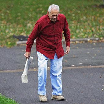
19 Signs and symptoms Signs and symptoms of psoas abscess include : Back or flank pain Fever Inguinal or back mass Limping Anorexia Weight loss
20
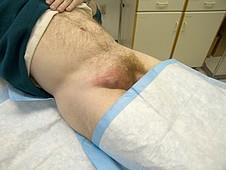
21 Signs and symptoms Psoas abscesses occasionally extend distally and present as a painful or painless mass below the inguinal ligament. When an inguinal mass in a patient with a psoas abscess is painless (ie, a cold abscess), tuberculosis is a more likely cause than a bacterial infection The mass may rarely mimic inguinal lymphadenopathy or a femoral hernia
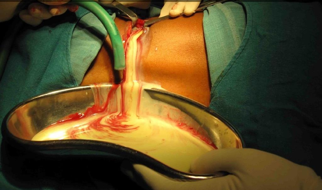
22
23 Signs and symptoms Pain is exacerbated when performing movements in which the psoas muscle is stretched or extended The "psoas sign" is pain brought on by extension of the hip. Limitation of hip movement is common and patients frequently prefer to be in a position of less discomfort that includes hip flexion and lumbar lordosis.

24 Psoas sign
25 Laboratory tests Leukocytosis (>10,000/mL) is observed in up to 83 % of cases Anemia:- HB <11 g/l is frequent Thrombocytosis is observed less frequently. An elevated ESR may be observed (>50 in 73 % of cases) The C-reactive protein is often elevated Elevated aspartate aminotransferase has also been described

26 Complications Complications of psoas abscess include: Septic shock Deep venous thrombosis due to extrinsic compression of the iliac vein Paralytic bowel ileus Hydronephrosis due to ureteric compression
27 Differential diagnosis Retroperitoneal and intraperitoneal lesions including inflammation, hematoma, or tumor of the psoas muscle can mimic a psoas abscess Retrocecal appendicitis, Enlargement of the iliopsoas bursa Infection of adjacent structures not involving the psoas muscle (eg, septic hip arthritis)


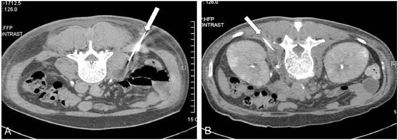
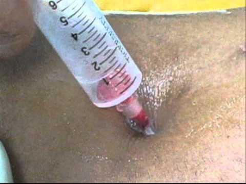
28 DIAGNOSIS CT Scan MRI Ultra sound Culture Blood tests
29 Treatment Most cases require percutaneous (PCD) or open surgical drainage with parenteral antibiotic treatment. CT-guided PCD is the initial procedure of choice. PCD is associated with a shorter hospital stay. Suitable for patients intolerant of general anesthesia
30

31 Open or Surgical drainage Open drainage indicated for large, complex, or multiloculated abscesses, significant involvement of adjacent structures or if PCD fails. Primary surgical intervention is more likely to be successful in patients with gas-forming abscess.
32 Surgical Drainage Psoas abscesses associated with inflammatory bowel disease, ruptured appendicitis, or infected aortic ruptures are effectively managed with open drainage and surgical treatment of the underlying disease
33
34
Lecture 08 THIGH MUSCLES ANTERIOR COMPARTMENT. Dr Farooq Khan Aurakzai. Dated:
 Lecture 08 THIGH MUSCLES ANTERIOR COMPARTMENT BY Dr Farooq Khan Aurakzai Dated: 11.02.2017 INTRODUCTION to the thigh Muscles. The musculature of the thigh can be split into three sections by intermuscular
The posterior abdominal wall. Prof. Oluwadiya KS
 The posterior abdominal wall Prof. Oluwadiya KS www.oluwadiya.sitesled.com Posterior Abdominal Wall Lumbar vertebrae and discs. Muscles opsoas, quadratus lumborum, iliacus, transverse, abdominal wall
ANATYOMY OF The thigh
 ANATYOMY OF The thigh 1- Lateral cutaneous nerve of the thigh Ι) Skin of the thigh Anterior view 2- Femoral branch of the genitofemoral nerve 5- Intermediate cutaneous nerve of the thigh 1, 2 and 3 are
lower limb Anterior Compartment: lecture 3 The deep fascia ( fascia lata) divides the thigh into 3 compartments:
 divides the thigh into 3 compartments:") lower limb lecture 3 The deep fascia ( fascia lata) divides the thigh into 3 compartments: 1. Anterior Extensor compartment 2. Medial Adductor compartment 3. Posterior Flexor compartment Anterior Compartment:
lower limb lecture 3 The deep fascia ( fascia lata) divides the thigh into 3 compartments: 1. Anterior Extensor compartment 2. Medial Adductor compartment 3. Posterior Flexor compartment Anterior Compartment:
ANATYOMY OF The thigh
 ANATYOMY OF The thigh 1- Lateral cutaneous nerve of the thigh Ι) Skin of the thigh Anterior view 2- Femoral branch of the genitofemoral nerve 5- Intermediate cutaneous nerve of the thigh 1, 2 and 3 are
ANATYOMY OF The thigh
 ANATYOMY OF The thigh 1- Lateral cutaneous nerve of the thigh Ι) Skin of the thigh Anterior view 2- Femoral branch of the genitofemoral nerve 1, 2 and 3 are From the lumber plexus 5- Intermediate cutaneous
Case Report: Arthroscopic Treatment of Psoas Abscess Concurrent with Septic Arthritis of the Hip Joint
 Case Report: Arthroscopic Treatment of Psoas Abscess Concurrent with Septic Arthritis of the Hip Joint Pil Whan Yoon, MD*, Jeong Joon Yoo, MD, Hee Joong Kim, MD, and Kang Sup Yoon, MD* Department of Orthopedic
GI module Lecture: 9 د. عصام طارق. Objectives:
 GI module Lecture: 9 د. عصام طارق Objectives: To list structures forming posterior abdominal wall. To follow aorta & its main branches. To describe IVC & its main tributaries. To list nerves of posterior
UC Irvine Western Journal of Emergency Medicine: Integrating Emergency Care with Population Health
 UC Irvine Western Journal of Emergency Medicine: Integrating Emergency Care with Population Health Title Bilateral Psoas Abscess in the Emergency Department Permalink https://escholarship.org/uc/item/8296x9cx
Identify the muscles associated with the medial compartment of the thigh. Identify the attachment points of the medial thigh muscles.
 L 8 A B O R A T O R Y Thigh MEDIAL THIGH Identify the muscles associated with the medial compartment of the thigh. Identify the attachment points of the medial thigh muscles. Identify the actions of these
Baraa Ayed حسام أبو عوض. Ahmad Salman. 1 P a g e
 4 Baraa Ayed حسام أبو عوض Ahmad Salman 1 P a g e Today we are going to cover these concepts: Iliotibial tract Anterior compartment of the thigh and the hip Medial compartment of the thigh Femoral triangle
The thigh. Prof. Oluwadiya KS
 The thigh Prof. Oluwadiya KS www.oluwadiya.com The Thigh: Boundaries The thigh is the region of the lower limb that is approximately between the hip and knee joints Anteriorly, it is separated from the
RETROPERITONEAL ABSCESS FOLLOWING APPENDECTOMY: A CASE REPORT
 Bulletin of the Transilvania University of Braşov Series VI: Medical Sciences Vol. 7 (56) No. 1-2014 RETROPERITONEAL ABSCESS FOLLOWING APPENDECTOMY: A CASE REPORT L. VIDA 1 A. MIRONESCU 1 Abstract: Retroperitoneal
In athletes presenting with pain or injury, general medical
 665112SPHXXX10.1177/1941738116665112Moriarty and BakerSports Health research-article2016 Moriarty and Baker [ Primary Care ] A Pain in the Psoas: Groin Injury in a Collegiate Football Athlete Charlotte
rotation of the hip Flexion of the knee Iliac fossa of iliac Lesser trochanter Femoral nerve Flexion of the thigh at the hip shaft of tibia
 Anatomy of the lower limb Anterior & medial compartments of the thigh Dr. Hayder The fascia lata encloses the entire thigh like a sleeve/stocking. Three intramuscular fascial septa (lateral, medial, and
TitlePyogenic psoas muscle abscess: repo. Author(s) Takashi; Horita, Hiroki; Koroku, Mi. Citation 泌尿器科紀要 (1994), 40(8):
 Takashi; Horita, Hiroki; Koroku, Mi. Citation 泌尿器科紀要 (1994), 40(8):") TitlePyogenic psoas muscle abscess: repo Author(s) Takagi, Seiji; Tsukamoto, Taiji; Ku Takashi; Horita, Hiroki; Koroku, Mi Citation 泌尿器科紀要 (1994), 40(8): 699-702 Issue Date 1994-08 URL http://hdl.handle.net/2433/115330
TitlePyogenic psoas muscle abscess: repo Author(s) Takagi, Seiji; Tsukamoto, Taiji; Ku Takashi; Horita, Hiroki; Koroku, Mi Citation 泌尿器科紀要 (1994), 40(8): 699-702 Issue Date 1994-08 URL http://hdl.handle.net/2433/115330
musculoskeletal system anatomy nerves of the lower limb 1 done by: dina sawadha & mohammad abukabeer
 musculoskeletal system anatomy nerves of the lower limb 1 done by: dina sawadha & mohammad abukabeer What is the importance of plexuses? plexuses provides us the advantage of a phenomenon called convergence
Ultrasound Guided Lower Extremity Blocks
 Ultrasound Guided Lower Extremity Blocks CONTENTS: 1. Femoral Nerve Block 2. Popliteal Nerve Block Updated December 2017 1 1. Femoral Nerve Block Indications Surgery involving the knee, anterior thigh,
The Hip (Iliofemoral) Joint. Presented by: Rob, Rachel, Alina and Lisa
 Joint. Presented by: Rob, Rachel, Alina and Lisa") The Hip (Iliofemoral) Joint Presented by: Rob, Rachel, Alina and Lisa Surface Anatomy: Posterior Surface Anatomy: Anterior Bones: Os Coxae Consists of 3 Portions: Ilium Ischium Pubis Bones: Pubis Portion
The Hip (Iliofemoral) Joint Presented by: Rob, Rachel, Alina and Lisa Surface Anatomy: Posterior Surface Anatomy: Anterior Bones: Os Coxae Consists of 3 Portions: Ilium Ischium Pubis Bones: Pubis Portion
Anatomy of the renal system. Professor Nawfal K. Al-Hadithi
 Anatomy of the renal system Professor Nawfal K. Al-Hadithi Objectives To describe the posterior abdominal wall To identify the main anatomical landmarks of the kidneys & ureters To describe the suprarenal
Femoral Triangle and Adductor Canal. Dr. Heba Kalbouneh Associate Professor of Anatomy and Histology
 Femoral Triangle and Adductor Canal Dr. Heba Kalbouneh Associate Professor of Anatomy and Histology Femoral Triangle and Adductor Canal Femoral triangle Is a triangular depressed area located in the upper
The Iliopsoas compartment: A pictorial review of anatomy and common pathologies
 The Iliopsoas compartment: A pictorial review of anatomy and common pathologies Poster No.: C-456 Congress: ECR 2009 Type: Educational Exhibit Topic: Abdominal and Gastrointestinal Authors: M. Kaduthodil,
Spartan Medical Research Journal
 Spartan Medical Research Journal Research at Michigan State University College of Osteopathic Medicine Volume 2 Number 2 Winter, 2017 Pages 59-72 Title: Pelvic Abscess with Presentation as Inability to
The Lower Limb II. Anatomy RHS 241 Lecture 3 Dr. Einas Al-Eisa
 The Lower Limb II Anatomy RHS 241 Lecture 3 Dr. Einas Al-Eisa Tibia The larger & medial bone of the leg Functions: Attachment of muscles Transfer of weight from femur to skeleton of the foot Articulations
Muscles of the lower extremities. Dr. Nabil khouri MD, MSc, Ph.D
 Muscles of the lower extremities Dr. Nabil khouri MD, MSc, Ph.D Posterior leg Popliteal fossa Boundaries Biceps femoris (superior-lateral) Semitendinosis and semimembranosis (superior-medial) Gastrocnemius
Structure and Function of the Vertebral Column
 Structure and Function of the Vertebral Column Posture Vertebral Alignment Does it really matter? Yes it does! Postural Curves The vertebral column has a series of counterbalancing curves posterior anterior
Anterior and Medial compartments of the thigh. Dr. Heba Kalbouneh Associate Professor of Anatomy and Histology
 Anterior and Medial compartments of the thigh Dr. Heba Kalbouneh Associate Professor of Anatomy and Histology Terms Related to Movements Movement Flexion Extension Abduction Adduction Medial (internal)
CLINICAL MASSAGE THERAPY A Structural Approach to Pain Management
 World Massage Conference Presents: Massage to support Pregnancy, Pelvic Birth Stabilization and Postnatal Including Hip for mother Replacement and baby World Massage Conference Protocols June with 2013
Group of students. - Rawan almujabili د. محمد المحتسب - 1 P a g e
 - 14 - Group of students - Rawan almujabili د. محمد المحتسب - 1 P a g e Nerves of the posterior abdominal wall The spinal cord gives off spinal nerves between the vertebrae. In the abdomen, through the
Anatomy of the Large Intestine
 Large intestine Anatomy of the Large Intestine 2 Large Intestine Extends from ileocecal valve to anus Length = 1.5-2.5m = 5 feet Regions Cecum = 2.5-3 inch Appendix= 3-5 inch Colon Ascending= 5 inch Transverse=
Types of bone/joint infections. Bone and Joint Infections. Septic Arthritis. Pathogenesis. Pathogenesis. Bacterial arthritis: predisposing factors
 Bone and Joint Infections Types of bone/joint infections Arthritis (infective/septic) Osteomyelitis Prosthetic bone and joint infections Septic Arthritis Common destructive athroplasty Mono-articular Poly-articular
Abdomen: Introduction. Prof. Oluwadiya KS
 Abdomen: Introduction Prof. Oluwadiya KS www.oluwadiya.com Abdominopelvic Cavity Abdominal Cavity Pelvic Cavity Extends from the inferior margin of the thorax to the superior margin of the pelvis and the
Infected cardiac-implantable electronic devices: diagnosis, and treatment
 Infected cardiac-implantable electronic devices: diagnosis, and treatment The incidence of infection following implantation of cardiac implantable electronic devices (CIEDs) is increasing at a faster rate
Bushra Arafa Zayed & Hanan Jamal. - Dana AF
 - 10 - Bushra Arafa Zayed & Hanan Jamal - Dana AF - Mohammad Al Muhtaseb Notes: This sheet was written in the same order as the slides, and everything in the slides is mentioned in this sheet. Pictures
Always keep it in the differential
 Acute Appendicitis Lissa C. Sakata and Lindsey Perea 2 Always keep it in the differential Learning Objectives 1. The learner should be able to describe the etiology of acute appendicitis. 2. The learner
Regional Human Anatomy (HBA 461/561/540): Course Objectives
: Course Objectives") Regional Human Anatomy (HBA 461/561/540): Course Objectives This is a 5-credit course that consists of 1-hour lectures followed by 3-hour labs. It is organized into three modules (see syllabus): Module
Regional Human Anatomy (HBA 461/561/540): Course Objectives This is a 5-credit course that consists of 1-hour lectures followed by 3-hour labs. It is organized into three modules (see syllabus): Module
THE HIP. Cooler than cool, the pinnacle of what is "it". Beyond all trends and conventional coolness.
 THE HIP Cooler than cool, the pinnacle of what is "it". Beyond all trends and conventional coolness. Objectives Hip anatomy Causes of hip pain Hip exam Anatomy Bones Ilium Anterior Superior Iliac Spine
The hip: Built for endurance and mobility
 The hip: Built for endurance and mobility The hip joint Some anatomical landmarks Innominate Ilium, pubis, ischium Sacrum Iliac crests Asis Psis Pubic tubercle Acetabulum Femur Head of femur Neck of femur
Inferior Pelvic Border
 Pelvis + Perineum Pelvic Cavity Enclosed by bony, ligamentous and muscular wall Contains the urinary bladder, ureters, pelvic genital organs, rectum, blood vessels, lymphatics and nerves Pelvic inlet (superior
ANATOMY OF PELVICAYCEAL SYSTEM -DR. RAHUL BEVARA
 1 ANATOMY OF PELVICAYCEAL SYSTEM -DR. RAHUL BEVARA 2 KIDNEY:ANATOMY OVERVIEW Kidneys are retroperitoneal, in posterior abdominal region, extending from T12 L3 Bean-shaped Right kidney is lower than left
Nerves on the Posterior Abdominal Wall
 Nerves on the Posterior Abdominal Wall Lumbar Plexus The lumbar plexus, which is one of the main nervous pathways supplying the lower limb, is formed in the psoasmuscle from the anterior ramiof the upper
Hydronephrosis. What is hydronephrosis?
 What is hydronephrosis? Hydronephrosis Hydronephrosis describes the situation where the urine collecting system of the kidney is dilated. This may be a normal variant or it may be due to an underlying
Gluteal region DR. GITANJALI KHORWAL
 Gluteal region DR. GITANJALI KHORWAL Gluteal region The transitional area between the trunk and the lower extremity. The gluteal region includes the rounded, posterior buttocks and the laterally placed
[ANATOMY #12] April 28, 2013
![[ANATOMY #12] April 28, 2013](/thumbs/86/93473883.jpg "[ANATOMY #12] April 28, 2013") Sympathetic chain : Sympathetic chain is each of the pair of ganglionated longitudinal cords of the sympathetic nervous system; extend from level of atlas (base of skull) till coccyx. It is paravertebral
Sympathetic chain : Sympathetic chain is each of the pair of ganglionated longitudinal cords of the sympathetic nervous system; extend from level of atlas (base of skull) till coccyx. It is paravertebral
Epidemiology of Low back pain
 Low Back Pain Definition Pain felt in your lower back may come from the spine, muscles, nerves, or other structures in that region. It may also radiate from other areas like the mid or upper back, a inguinal
3 section of the Foot
 TERMINOLOGY 101 How many Bones 3 section of the Foot Bilateral Relating to both Plantar Relating to the bottom or sole Lateral Relating to the outside or farther from the median Medial Relating to the
e-anatomy Paper 2 Exam Monday, 4 April 2016
 e-anatomy Paper 2 Exam Monday, 4 Level 9, 51 Druitt Street, Sydney NSW 2000, Australia Ph: +61 2 9268 9777 Fax: +61 2 9268 9799 Web: www.ranzcr.edu.au Email: ranzcr@ranzcr.edu.au ABN 37 000 029 863 CASE
TUBERCULOSIS OF HIP AND KNEE JOINT
 TUBERCULOSIS OF HIP AND KNEE JOINT TUBERCULOSIS OF HIP JOINT Occurrence-15% of all osteo articular tuber culosis Next common after spinal TB AETIO PATHOGENESIS Most common cause Mycobacterium tuberculosis
Excretory urography (EU) or IVP US CT & radionuclide imaging
 or IVP US CT & radionuclide imaging") Excretory urography (EU) or IVP US CT & radionuclide imaging MRI arteriography studies requiring catherization or direct puncture of collecting system EU & to a lesser extent CT provide both functional
Excretory urography (EU) or IVP US CT & radionuclide imaging MRI arteriography studies requiring catherization or direct puncture of collecting system EU & to a lesser extent CT provide both functional
Index. Note: Page numbers of article titles are in boldface type.
 Note: Page numbers of article titles are in boldface type. A Abscess, epidural, 822 824 Achilles tendon rupture, 894 895, 981 982 Acromioclavicular separations, shoulder pain in, 751 753 Adhesive capsulitis,
The Kidneys. (L., ren; Gk, nephros; hence the adjectives renal and nephric) & Suprarenal (Adrenal) Glands. Dr Maan Al-Abbasi PhD, MBChB
 & Suprarenal (Adrenal) Glands. Dr Maan Al-Abbasi PhD, MBChB") The Kidneys (L., ren; Gk, nephros; hence the adjectives renal and nephric) & Suprarenal (Adrenal) Glands Dr Maan Al-Abbasi PhD, MBChB Functions of Urinary System Regulate electrolytes (K+, Na+, etc) Regulate
The Kidneys (L., ren; Gk, nephros; hence the adjectives renal and nephric) & Suprarenal (Adrenal) Glands Dr Maan Al-Abbasi PhD, MBChB Functions of Urinary System Regulate electrolytes (K+, Na+, etc) Regulate
Lumbar Plexus. Ventral rami L1 L4 Supplies: Major nerves.. Abdominal wall External genitalia Anteromedial thigh
 Lower Limb Nerves Lectures Objectives Describe the structure and relationships of the plexuses of the lower limb. Describe the course, relationships and structures supplied for the major nerves of the
Types of osteoarthritis
 ARTHRITIS Osteoarthritis is a degenerative joint disease is the most common joint disorder. It is a frequent part of aging and is an important cause of physical disability in persons older than 65 years
Adductor canal (Subsartorial) or Hunter s canal
 or Hunter s canal") Adductor canal (Subsartorial) or Hunter s canal John Hunter described the exposure and ligation of the femoral artery in this canal for aneurysm of the popliteal artery; this method has the advantage that
Adductor canal (Subsartorial) or Hunter s canal John Hunter described the exposure and ligation of the femoral artery in this canal for aneurysm of the popliteal artery; this method has the advantage that
Lower Extremity Ultrasound-Guided Regional Anesthesia. Stephanie Duffy, CRNA Regional Anesthesia Faculty Acute Pain Service NMCSD
 Lower Extremity Ultrasound-Guided Regional Anesthesia Stephanie Duffy, CRNA Regional Anesthesia Faculty Acute Pain Service NMCSD Objectives Review anatomy of lumbosacral plexus Lumbar plexus blocks Psoas
Appendix 5. EFSUMB Newsletter. Gastroenterological Ultrasound
 EFSUMB Newsletter 87 Examinations should encompass the full range of pathological conditions listed below A log book listing the types of examinations undertaken should be kept Training should usually
Surgery Under Regional Anesthesia
 Surgery Under Regional Anesthesia Jean Daniel Eloy, MD Assistant Professor Residency Program Director Rutgers-New Jersey Medical School Rutgers The State University of New Jersey Peripheral Nerve Block
The abdominal muscles:
 Core Body Strength A horse s natural centre of gravity is slightly behind the shoulder and approximately one third of the way down it s body. In order to accommodate the weight of the rider, we need to
International Journal of Case Reports in Medicine
 International Journal of Case Reports in Medicine Vol. 2013 (2013), Article ID 507024, 18 minipages. DOI:10.5171/2013.507024 www.ibimapublishing.com Copyright 2013 Makram Koubaa, Abir Aouam, Adnene Toumi,
The Muscular System. Chapter 10 Part D. PowerPoint Lecture Slides prepared by Karen Dunbar Kareiva Ivy Tech Community College
 Chapter 10 Part D The Muscular System Annie Leibovitz/Contact Press Images PowerPoint Lecture Slides prepared by Karen Dunbar Kareiva Ivy Tech Community College Table 10.14: Muscles Crossing the Hip and
The University Of Jordan Faculty Of Medicine THE LOWER LIMB. Dr.Ahmed Salman Assistant Prof. of Anatomy. The University Of Jordan
 The University Of Jordan Faculty Of Medicine THE LOWER LIMB Dr.Ahmed Salman Assistant Prof. of Anatomy. The University Of Jordan Gluteal Region Cutaneous nerve supply of (Gluteal region) 1. Lateral cutaneous
Psoas abscesses complicating colonic disease: imaging and therapy
 Ann R Coll Surg Engl 1998; 80: 405-409 Psoas abscesses complicating colonic disease: imaging and therapy D N Lobo MS FRCSEd(Gen Surg)' Specialist Surgical Registrar W K Dunn MB BS FRCR1 Consultant Radiologist
FUNCTIONAL ANATOMY AND EXAM OF THE HIP, GROIN AND THIGH
 FUNCTIONAL ANATOMY AND EXAM OF THE HIP, GROIN AND THIGH Peter G Gerbino, MD, FACSM Orthopedic Surgeon Monterey Joint Replacement and Sports Medicine Monterey, CA TPC, San Diego, 2017 The lecturer has no
Muscles of the Core. PSK 4U Mr. S. Kelly North Grenville DHS
 Muscles of the Core PSK 4U Mr. S. Kelly North Grenville DHS Intercostal Muscles Run between the ribs Provide shape and movement for chest wall External intercostals: aid in both quiet (passive) and forced
Abscess. A abscess is a localized collection of pus in the skin and may occur on any skin surface and be formed in any part of body.
 Abscess A abscess is a localized collection of pus in the skin and may occur on any skin surface and be formed in any part of body. Ethyology Bacteria causing cutaneous abscesses are typically indigenous
Case Report Femoral Nerve Injury as a Complication of Percutaneous Simple Renal Cyst Sclerotherapy with Ethanol: A Case Report
 Case Reports in Medicine Volume 2012, Article ID 589108, 4 pages doi:10.1155/2012/589108 Case Report Femoral Nerve Injury as a Complication of Percutaneous Simple Renal Cyst Sclerotherapy with Ethanol:
Find Medical Solutions to Your Problems HYDRONEPHROSIS. (Distension of Renal Calyces & Pelvis)
") HYDRONEPHROSIS (Distension of Renal Calyces & Pelvis) Hydronephrosis is the distension of the renal calyces and pelvis due to accumulation of the urine as a result of the obstruction to the outflow of
HYDRONEPHROSIS (Distension of Renal Calyces & Pelvis) Hydronephrosis is the distension of the renal calyces and pelvis due to accumulation of the urine as a result of the obstruction to the outflow of
Lectures of Human Anatomy
 Lectures of Human Anatomy Lower Limb Gluteal Region and Hip Joint By DR. ABDEL-MONEM AWAD HEGAZY M.B. with honor 1983, Dipl."Gynecology and Obstetrics "1989, Master "Anatomy and Embryology" 1994, M.D.
Case Report An Easily Overlooked Presentation of Malignant Psoas Abscess: Hip Pain
 Case Reports in Orthopedics Volume 2015, Article ID 410872, 4 pages http://dx.doi.org/10.1155/2015/410872 Case Report An Easily Overlooked Presentation of Malignant Psoas Abscess: Hip Pain Ayhan Askin,
Human Anatomy Biology 255
 Human Anatomy Biology 255 Exam #4 Please place your name and I.D. number on the back of the last page of this exam. You must answer all questions on this exam. Because statistics demonstrate that, on average,
Lumbar and Sacral Plexuses. Dr. Heba Kalbouneh Associate Professor of Anatomy and Histology
 Lumbar and Sacral Plexuses Dr. Heba Kalbouneh Associate Professor of Anatomy and Histology Structure of Spinal Nerves: Somatic Pathways dorsal root CNS interneuron spinal nerve dorsal ramus somatic sensory
Introduction. Paronychia. Hand Infections. Eponychium. Paronychia 1/26/2015. INFECTIONS OF THE UPPER EXTREMITY Mark Rekant M.D.
 Introduction INFECTIONS OF THE UPPER EXTREMITY Mark Rekant M.D. Thomas Jefferson University Philadelphia Hand Center South Jersey Hand Center Hand infections are frequent Taking into account the countless
9/21/15. Joshua Pruitt, MD, FAAEM Medical Director, LifeGuard Air Ambulance Iowa PA Society Fall CME Conference September 29, 2015
 Unless they prove otherwise. ~Every ED attending ever Joshua Pruitt, MD, FAAEM Medical Director, LifeGuard Air Ambulance Iowa PA Society Fall CME Conference September 29, 2015 AAA with rupture Mesenteric
ABDOMINAL WALL & RECTUS SHEATH
 ABDOMINAL WALL & RECTUS SHEATH Learning Objectives Describe the anatomy, innervation and functions of the muscles of the anterior, lateral and posterior abdominal walls. Discuss their functional relations
Case Discussion Splenic Abscess
 Case Discussion Splenic Abscess Personal Data Gender: male Birth Date: 1928/Mar/06th Allergy: Mefenamic Smoking: 0.5 PPD for 55 years Alcohol: negative (?) 4 Months Ago Abdominal pain: epigastric area
Ultrasound of the Hip: Anatomy, Pathology, and Procedures
 Ultrasound of the Hip: Anatomy, Pathology, and Procedures Jon A. Jacobson, M.D. Professor of Radiology Director, Division of Musculoskeletal Radiology University of Michigan Outline Hip Joint Native hip
ISPUB.COM. Spectrum Of MRI Findings In Musculoskeletal Tuberculosis: Pictoral Essay. P Chudgar INTRODUCTION SPINE
 ISPUB.COM The Internet Journal of Radiology Volume 8 Number 2 Spectrum Of MRI Findings In Musculoskeletal Tuberculosis: Pictoral Essay P Chudgar Citation P Chudgar.. The Internet Journal of Radiology.
LAB Notes#1. Ahmad Ar'ar. Eslam
 LAB Notes#1 Ahmad Ar'ar Eslam 1 P a g e Anatomy lab Notes Lower limb bones :- Pelvic girdle: It's the connection between the axial skeleton and the lower limb; it's made up of one bone called the HIP BONE
HUMAN BODY COURSE LOWER LIMB NERVES AND VESSELS
 HUMAN BODY COURSE LOWER LIMB NERVES AND VESSELS October 22, 2010 D. LOWER LIMB MUSCLES 2. Lower limb compartments ANTERIOR THIGH COMPARTMENT General lfunction: Hip flexion, knee extension, other motions
Neurologic complications - whom to blame? Benno Rehberg Médecin adjoint agrégé Unité d anesthésiologie gynéco-obstétricale, HUG
 Neurologic complications - whom to blame? Benno Rehberg Médecin adjoint agrégé Unité d anesthésiologie gynéco-obstétricale, HUG SAOA spring meeting 2015 The simple surgical answer: outline Epidemiology
Thoracic and Lumbar Spine Anatomy.
 Thoracic and Lumbar Spine Anatomy www.fisiokinesiterapia.biz Thoracic Vertebrae Bodies Pedicles Laminae Spinous Processes Transverse Processes Inferior & Superior Facets Distinguishing Feature Costal Fovea
Indian Journal of Basic & Applied Medical Research; June 2013: Issue-7, Vol.-2, P
 Case Report: An Unusual Formation of the Femoral Nerve - A Case Report 1Dr. Indrajit Gupta, 2 Dr. Sudeshna Majumdar*, 3 Dr. Santanu Bhattacharya, 4 Dr. Seikh Ali Amam, 5Dr. Susmita Ghosh, 6 Dr. Lopamudra
b. The Gluteal Complex - Gluteus maximus
 This session is based upon Dr. Osar s newest book: The Psoas Solution 1. What do we know about the psoas and glutes? a. We have been taught that the primary function of the psoas is hip flexion and it
MUSCULOSKELETAL INFECTIONS IN CHILDREN. Dr Caren Landes Alder Hey Children s NHS Foundation Trust Liverpool
 MUSCULOSKELETAL INFECTIONS IN CHILDREN Dr Caren Landes Alder Hey Children s NHS Foundation Trust Liverpool MUSCULOSKELETAL INFECTIONS Common and uncommon infections Common and uncommon presentations Imaging
Anatomy & Physiology Pelvic Girdles 10.1 General Information
 Anatomy & Physiology Pelvic Girdles 10.1 General Information ICan2Ed, Inc. In human anatomy, the pelvis (plural pelves or pelvises) is the lower part of. The area of the body that is between the abdomen
MANAGEMENT OF PYOGENIC LIVER ABSCESS BOYOUNG SONG, M.D. SUNY DOWNSTATE SURGERY 11/7/13
 MANAGEMENT OF PYOGENIC LIVER ABSCESS BOYOUNG SONG, M.D. SUNY DOWNSTATE SURGERY 11/7/13 CASE THE PATIENT IS A 79 YEAR OLD MALE WITH 3 DAY HISTORY OF LOWER ABDOMINAL PAIN, NAUSEA WITHOUT VOMITING, CHILLS
Anatomy of the SMALL INTESTINE. Dr. Noman Ullah Wazir PMC
 Anatomy of the SMALL INTESTINE Dr. Noman Ullah Wazir PMC SMALL INTESTINE The small intestine, consists of the duodenum, jejunum, and illium. It extends from the pylorus to the ileocecal junction were the
Lower limb summary. Anterior compartment of the thigh. Done By: Laith Qashou. Doctor_2016
 Lower limb summary Done By: Laith Qashou Doctor_2016 Anterior compartment of the thigh Sartorius Anterior superior iliac spine Upper medial surface of shaft of tibia 1. Flexes, abducts, laterally rotates
Doc, I've done my groin. Groin Pain. Peter Brukner. Doc, I've done my groin 1. acute chronic
 Doc, I've done my groin Peter Brukner Associate Professor in Sports Medicine Centre for Sports Medicine Research and Education School of Physiotherapy 9/22/2006 The University of Melbourne Groin Pain acute
Assessment of limping child (beware the child who does not weight bear at all):
:") Department of Paediatrics Clinical Guideline Acutely Limping Child and Septic Arthritis Assessment of limping child (beware the child who does not weight bear at all): History Careful history of any significant
Department of Paediatrics Clinical Guideline Acutely Limping Child and Septic Arthritis Assessment of limping child (beware the child who does not weight bear at all): History Careful history of any significant
Case Report Sacral Emphysematous Osteomyelitis Caused by Escherichia coli after Arthroscopy of the Knee
 Case Reports in Orthopedics Volume 2016, Article ID 1961287, 4 pages http://dx.doi.org/10.1155/2016/1961287 Case Report Sacral Emphysematous Osteomyelitis Caused by Escherichia coli after Arthroscopy of
2. List the 8 pelvic spaces: list one procedure or dissection which involves entering that space.
 Name: Anatomy Quiz: Pre / Post 1. In making a pfannensteil incision you would traverse through the following layers: a) Skin, Camper s fascia, Scarpa s fascia, external oblique aponeurosis, internal oblique
Thoracolumbar Anatomy Eric Shamus Catherine Patla Objectives
 1 2 Thoracolumbar Anatomy Eric Shamus Catherine Patla Objectives List the muscular and ligamentous attachments of the thoracic and lumbar spine Describe how the muscles affect the spine and upper extremity
Nuclear medicine and Prosthetic Joint Infections
 Nuclear medicine and Prosthetic Joint Infections Christophe Van de Wiele, M.D., Ph.D. Department of Nuclear Medicine, University Hospital Ghent, Belgium Orthopedic prostheses: world market 1996 Prosthetic
Main Menu. Joint and Pelvic Girdle click here. The Power is in Your Hands
 1 Hip Joint and Pelvic Girdle click here Main Menu K.6 http://www.handsonlineeducation.com/classes//k6entry.htm[3/23/18, 2:01:12 PM] Hip Joint (acetabular femoral) Relatively stable due to : Bony architecture
BONE AND JOINT INFECTION. Dr.Jónás Zoltán Dept.of Orthopaedics
 BONE AND JOINT INFECTION Dr.Jónás Zoltán Dept.of Orthopaedics www.ortopedia.dote.hu Order of verbal exams: The students are able to register for the exam on the Neptun system. The students pick two titles,
Unusual Lateral Presentation of Popliteal Cyst
 Unusual Lateral Presentation of Popliteal Cyst Tarek Hemmali,* Abstract: The most common cyst occurs in the popliteal region is the popliteal cyst and over the past years it has been received much clinical
SEPTIC ARTHRITIS. Dr Ahmed Husam Al Ahmed Rheumatologist SYRIA. University of Science and technology Hospital Sanaa Yemen 18/Dec/2014
 SEPTIC ARTHRITIS Dr Ahmed Husam Al Ahmed Rheumatologist SYRIA University of Science and technology Hospital Sanaa Yemen 18/Dec/2014 Objectives be able to define Septic Arthritis know what factors predispose
Lecture 09. Popliteal Fossa. BY Dr Farooq Khan Aurakzai
 Lecture 09 Popliteal Fossa BY Dr Farooq Khan Aurakzai Dated: 14.02.2018 What is popliteus? Introduction Anything relating to, or near the part of the leg behind the knee. From New Latin popliteus the muscle
The Thoracic wall including the diaphragm. Prof Oluwadiya KS
 The Thoracic wall including the diaphragm Prof Oluwadiya KS www.oluwadiya.com Components of the thoracic wall Skin Superficial fascia Chest wall muscles (see upper limb slides) Skeletal framework Intercostal