BIOMETRICAL AND MORPHOHISTOLOGICAL STUDY OF POSTPHARYNGEAL DIGESTIVE TUBE FOR SALAMANDRA SALAMANDRA SALAMANDRA L.
|
|
|
- Augustus Burke
- 5 years ago
- Views:
Transcription
1 Analele Ştiinţifice ale Universităţii Al. I. Cuza Iaşi, s. Biologie animală, Tom LIV, 2008 BIOMETRICAL AND MORPHOHISTOLOGICAL STUDY OF POSTPHARYNGEAL DIGESTIVE TUBE FOR SALAMANDRA SALAMANDRA SALAMANDRA L. Sergiu HAIMOVICI Al. I. Cuza University Iaşi, Faculty of Biology, Bd. Carol I 20A, Iaşi, Romania Abstract. Salamander, common nowadays as a species belongs to Urodels, one of the first species who moved from the aquatic to terrestrial environment 150 million years ago. They preserved primitive characters and added some others new as well as some specific characters. This is clearly evidentiated if we take into consideration the postpharyngeal digestive tube. So, we made a biometrical, morphological and histological study of this tube of 17 mature individuals, of which 10 males and 7 females. Measurements have been executed and morphological studies have been made on the four segments of the digestive tube: esophagus, stomach, small intestine and large intestine and than, on histological sections using nine coloration types we described the characteristics as we see on microscope for fragments coming from the four segments. Some data have been outlined the most important aspects being presented as conclusions and some others being included in the paper, for supporting our affirmations. Keywords: Salamander, digestive tube, biometrical morphological histological study. Rezumat. Studiu biometric şi morfohistologic al tubului digestiv postfaringial la Salamandra salamandra salamandra L. Salamandra, astăzi comună ca specie, aparţine urodelelor, care au fost printre primele care acum 150 milioane de ani au reuşit să treacă din mediul acvatic în mediul terestru. Ele au rămas cu caractere primitive dar au căpătat şi altele noi, mai evaluate, iar unele caracteristice lor. Acest lucru apare bine evidenţiat dacă considerăm tractusul digestiv postfaringian. Noi am lucrat pe 17 exemplare mature, zece de sex mascul şi şapte de sex femel. S-au executat măsurători şi studii de morfologie pe cele patru segmente ale acestui tub: esofag, stomac, intestin mediu şi intestin posterior, iar apoi, pe secţiuni histologice folosind nouă tipuri de colorare, am descris şi caracteristicile văzute prin microscop, pe porţiuni din fiecare cele patru segmente. Au fost puse în evidenţă o serie de date, dintre care cele mai importante sunt arătate în concluziile lucrării noastre, şi altele, ce sunt prezentate în text, ceea ce arată foarte bine ceea ce am susţinut mai sus. Cuvinte cheie: Salamandra, tub digestiv, studiu biometrico morfologico histologic. Introduction We chose to study a relatively common species for the Romanian high falling leaves woods, a well known species described even from Antiquity by Aristoteles who considered it immortal, immune to fire, maybe influenced by the stories of the ancient Greek story-tellers. His words were taken as good in the Middle Ages, especially on the space of nowadays Germany, a place where it used to be known as Foer Salamander; the common people did not experimented this but the wizards were often associated with the presence of this small animal. We studied the salamander s digestive tube from scientific reasons, because firstly the urodels represent a very old group from the paleontological point of view. They are closed to the first tetrapodals who left the aquatic area for the land 150 million years ago. The urodels have not evolved too much, still preserving some primitive characters, some of them even more primitive than those of the teleostean fish, younger from the paleontological point of view but more evolved, for these fish a great diversification being specific, caused by their diverse habitat. We could name them as living fossils. Secondly, although their digestive tube have been studied but not too thoroughly by varied authors, salamander was considered as a gender, without mentioning the species or subspecies; sometimes some characters are present only with some particular species
2 Sergiu Haimovici (or subspecies); more than these only a small number of individuals have been studied. We should mention that in Western Europe as well as in the Mediterranean area another species is common, We studied Salamandra salamandra, salamandra subspecies common on Romanian territory and mentioned by Linné (Fuhn, 1960). We should also say that Salamander is a carnivorous insectivorous species. Material and Methods The material was collected from the falling leaves forests around the biological research area Pângaraţi, on the middle course of Bistriţa River (Neamţ county) from early spring till late in autumn. There are 17 mature individuals, 10 males and 7 females (the sex was determined by abdominal section). Both external and internal measurements have been made on the individuals. The whole digestive tube as well as its fragments has been measured (in mm) using a thread to follow all the sinuosities. By cutting, the internal morphology of the digestive tube fragments was studied (using a 3x magnifying glass): the esophagus, stomach, the larger area we called fundic and the tight one we called pilorical, the small intestine and large intestine till the cloacum. Small fragments from the walls of every segment have been prepared according to histological technique for 87 slides. General colorants have been used to give color the histological fragments and selective colorants for histochemistry or evidence the presence or absence of some formations; nine color blends have been used. For studying the histological material we used a microscope with 10x15 or 15x15 magnifying indices and for some details with an increased magnifying index. Results and Discussion External morphology The digestive tube, immediately after the pharyngeal fragment, is made of a relatively short and thick esophagus which continues with the stomach, longer with a proximal part as thick as the esophagus and growing larger in the medium part (the fundus area) and then it becomes narrow again, so its final part (the pyloric area) is even tighter than the esophagus. This fragment makes a curve, as some authors say, a well circumscribed swelling but just on palpation it seems harder and its aspect is a little whiter (Pernkopf & Lehner, 1937). Next follows the middle intestine, much tighter with three sometimes four intestinal ansa well packed in order to get into the belly. We may observe that there is a slight narrowing from the proximal to distal area so we can distinguish three fragments: the proximal (let us call it duodenum), the middle and the distal ones. Then, we have identified the large intestine, much larger (similar to the fundus area of the stomach) with a tight area to the opening in the cloacum. Biometrical study The table contains measurements (in mm). There are 17 individuals, seven females and ten males; one of the males (number 5) is much smaller than the others. We may say that the digestive tube is three times longer than the body length (from the anterior part to the opening in the cloacum) an average of about mm. This indicates superiority as we said before salamander is a carnivorous species and as we know this animal group usually has a smaller digestive tube. By studying the postpharyngean digestive tract by segments we may present the following results: esophagus length is about 5.05%; stomach about 11.24%; middle intestine 75.45%; large intestine 7.87% of the digestive tube. We may say that the greatest percentage belongs to the middle intestine as a proof of its absorption function importance. This fragment as well as the whole digestive
3 Analele Ştiinţifice ale Universităţii Al. I. Cuza Iaşi, s. Biologie animală, Tom LIV, 2008 tube is a little bit longer for the females and this is not only because their body length is bigger. Internal morphology The internal morphology was studied for the whole digestive tube longitudinally sectioned. We shall take it fragment after fragment. The esophagus is relatively narrow and short longitudinally oriented and often placed towards the dorsal (back) part of the individual s body. We may observe 7-11 longitudinal prominent folds that may be seen without a microscope. Towards the inferior part of the esophagus they become smaller and less prominent. The passing through the stomach is gentle, with no sphincter. Stomach with its large lumen in the middle part has also 3 to 9 folds, some of them as a kind of elongation of the ones in the esophagus; the others originate in the stomach and they do not cover the whole stomach length. The stomach intestine passage is gentle and on a segment of about 3 mm, the color is whiter and the surface is harder, there are no more folds and a sphincter like structure appears but with no covering valva. The middle intestine has two to five folds not too prominent in its anterior part, and only 2 in its posterior part (the intestine gets narrower). The passing middle to large intestine is abrupt with no specific formation, from 1.5 mm in diameter to 6-7 mm. The large intestine with its representative diameter has many folds, sometimes 12 but very small distinguishable only with a magnifying glass. There is no gender characteristic evidenced by the internal morphology. Structure (microscopical) study We should say from the beginning that histological material belongs to three individuals, a male and two females. On the studied slides there was no characteristic specific to a certain gender. We shall follow the structure of each segment of the postpharyngeal tube. Esophagus. We took fragments from two different parts of the esophagus: the one noted with A, from the proximal area, immediately after the pharynx and the one noted with B from the distal area, closer to the stomach. For the A fragment (Fig. 1) we may see some folds mucous formation, some longer and sharp and sometimes blunt with shorter or longer, thinner or larger side ramifications, like a little pine tree along the folds axe with its leaves up. There are also smaller and simpler folds. So the section lumen appears multi-star shaped. The mucous formation is represented by a stratified and cylindrical epithelium with 3-5 cell rows on the basal membrane. There are cylindrical, narrow cells, with an elongated, oval nucleus. To the structure s apical end, they become cylindrical cubical, the nucleus shortens but it does not become round. The last cell row to the lumen, presents short ciliated cells, and starting with the epithelium middle bigger and oval goblet cells become more and more numerous, opening in the lumen among the ciliated cells; for some areas they are placed next to each other; there are no blood vessels in the epithelium. The corium of the mucous formation (lamina propria) consists of conjunctive tissue with a bigger quantity of conjunctive fibers, also fibrocytes (fiber cells) to the basal membrane as well as elastic fibers. The passing to the submucous formation is gentle, the number of fibers and cells diminishing slowly. There is no muscularis mucose in the mucous formation. The sub mucous formation consists of conjunctive tissue, almost lax with some bigger vessels surrounded by elastic fibers. Here and there lymphoid infiltrations come from lamina propria. The corium contains ductal - alveolar glands, slightly swell at the inferior part and not with a typical alveolus; they have cubic or cylindrical - cubical cells with round nucleus usually at the basis, with slightly acidophilus
4 Sergiu Haimovici grains cytoplasm (named by the German authors oxiphil granulierente zellen ); the cells become polymeric or oval shaped with flattened nuclei and the cytoplasm turns white, their apical end is larger and without membrane, their acid secretion leaking out. The muscle formation is made of a circle layer of long muscle fibers; the longitudinal one looks like discontinuous formations made of long fibers too. We have also found two striated muscle fibers. The so-called B fragment (Fig. 2) has a structure similar to the A fragment. The folds are lower, distally widened and less prominent. The glands in the corium of the mucous formation are more numerous white colored, with an acid secretion. The muscle formation is circular, integer with no striated fibers. The longitudinal one is discontinuous. Stomach. For the amphibians the stomach has a wide part, let us call it fundus (noted with C) (Fig. 3) and another part, tighter, placed just before the middle intestine, let us call it pyloric (noted with D) (Fig. 4). Like all the vertebrates, the mucous epithelium is made of a single cell type; here and there the epithelium goes back inside creating holes. The C part (fundus) The mucous formation folds are small and large and the epithelium is simple. There are prismatic, high cells packed together. The oval, elongated nucleus is placed to the cell basis, strongly colorable and with many nucleoli. The cytoplasm colors well too; about a third of it near the apex contains a mucous type with positive Schiff reaction that makes it quite distinct from the rest of the cell. The cell height and the layer thickness diminish to the end of those holes. The mucus drains by a kind of dialysis and not by the apical end breaking; that is why some authors consider them as closed mucous cells. The mucous formation corium is completely covered with dense tubular glands (very closed to each other) so lamina propria is represented by conjunctive inter - granular walls. All these glands are placed on line. They have two cell types: the main cells with a relatively round nucleus and a relatively granulated and acid cytoplasm ( oxyphil gekornte zellen, in German) at the basis; on top to the apical part to the gland opening there is a layer made of some cells (the so called throat cells cellules a mucus du collet, in French), slightly colored with allogenous blue; some authors consider that they replace the continuous destroying special cells from the epithelium in the holes; the main cells produce a white acid mucus. Under these glands there is a fiber layer that splits the corium from the submucous formation as there is no muscularis mucose; there are some muscle cells here and there. The submucous formation is made of lax conjunctive tissue with some bigger vessels surrounded by elastic fibers. This is very narrow. The muscle membrane is a circular, thicker strip (about 1/3 of mucous and sub mucous membrane); the longitudinal strip is so thin that it seems almost discontinuous. We identified a MAST cell (metachromatic coloration with toluidine blue) in a fold axes. The D (pyloric) part is shorter and tighter making connection with the intestine. On cross section, a very thick circle muscle tissue is observed, its thickness being similar to the mucous and submucous formation altogether. The mucous formation folds are of two types: some simple, elongated and some elongated but ramificated ones; all these folds continue to the central part so there is no more lumen. The mucous formation has a simple epithelium but those typical cells have their apical part narrower, almost undistinguishable, and the cells become very elongated and with high and thin nuclei; the glands are shorter and rare, some of them with an almost acinous formation; the so-called main cells do not turn into acid mucous anymore. The corium is better represented inside folds, but at their basis is rather thin. There is no
5 Analele Ştiinţifice ale Universităţii Al. I. Cuza Iaşi, s. Biologie animală, Tom LIV, 2008 muscularis mucose in the mucous formation. The submucous made of conjunctive tissue is thin too. The thicker part is represented by circle muscles that by constriction push the fundus stomach content into intestine. In the muscle formation there is a thinner but well represented longitudinal muscle structure. Middle intestine.this represents the most elongated part of the digestive tube with the same aspect all along from the histological point of view. So, we describe thoroughly the first part, a little larger ansa noted with E (Fig. 5), some authors naming it the duodenum. The folds are relatively long and narrow, very rare branched, leaving in the middle a quite large lumen. The mucous formation has a simple epithelium but different from the one in the stomach; this is also typical for the vertebrates. They are called cells with striated plate or enterocytes. The cells from the basal membrane are high and cylindrical, of about μ. The nucleus is oval and placed in the middle; in their apical area there is an obvious plate with a PNS positive. Among these enterocytes there are mucous producer goblet cells (rarely at the folds basis). The corium is well represented by a dense conjunctive tissue made of a connected anastomosed fibrocytes, with different leucocytes and collagen cells. In the corium, to the folds basis there are some big nuclei cells cell nests (Fig. 6) - each of them having three to five cells with big nuclei and full of chromatin splitting continuously; the nest is surrounded by thick conjunctive tissue with few elastic fibers. This is characteristic for amphibians, both urodels and anures (Andrew, 1959) even for tadpoles having the part of the epithelium cells substitution (Fig. 5); this is almost certitude as we discovered a cell turning into a goblet cell in a nest. Very rarely, to the folds basis there are some big cells of triangle shape with round nucleus and acidophili granulated cytoplasm maybe homologous to Paneth cells. It is no muscularis in the mucous formation yet. The submucous formation is very thin being made of lax conjunctive tissue, similar to the others that have been described so far. Although the muscle formation is very thin we still can distinguish the two formations, the longitudinal one being easily detectable. The second ansa (noted with F) (Fig. 7) has shorter and larger folds (maybe as a result of its fineness) but resembles to the E one. There is a parasite animal inside the lumen, a flat worm with a very thick cuticle for its protection. We could consider it a comensal as it does no harm to the salamander, as there are no lymphoid infiltrations in the corium. The immune system must have developed for the urodels. The last ansa is similar to the others. It seems the striated plate is a little bit thinner and narrower. Large intestine. The large intestine (noted with G) (Fig. 8) which is called terminal and due to its aspect we could also call it the thick intestine, has very thin walls. There are no prominent folds. The entorococytes are very low with an almost round nucleus and the striated plate is very thin or seems to disappear. The goblet cells are less than we could expect as for the mammals they are quite well represented. There is no clear distinction between lamina propria and the submucous membrane and there is no muscularis mucose. The muscle formation is very thin but the two stripes are distinguishable. The passing from the middle to the large intestine is very simple with no adjacent formation; the same thing is available for the cloacum too.


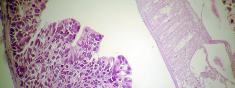
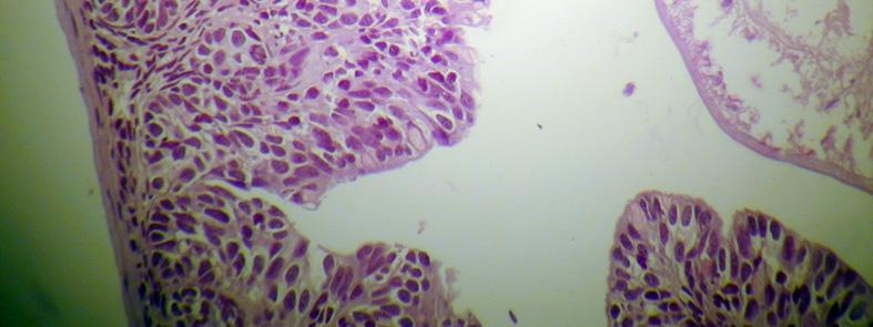
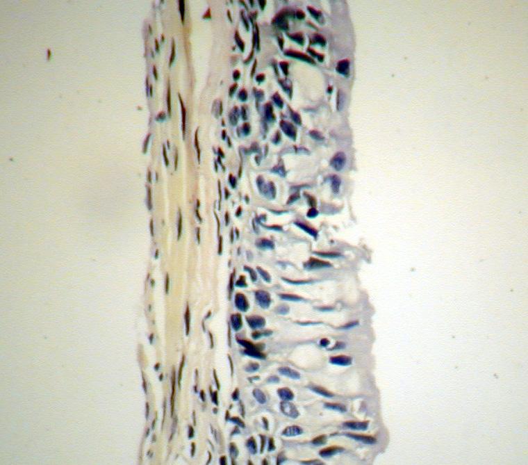
6 Sergiu Haimovici Figure 1. Esophagus, proximal area. Figure 2. Esophagus, distal area. Figure 3. Stomach, fundus area. Figure 4. Stomach, pyloric area. Figure 5. Middle intestine, general structure. Figure 6. Middle intestine, cell nests. Figure 7. Middle intestine, parasite inside lumen Figure 8. Large intestine, general structure.
7 Analele Ştiinţifice ale Universităţii Al. I. Cuza Iaşi, s. Biologie animală, Tom LIV, 2008 Table 1. Measurements (in mm) for Salamandra salamandra salamandra. Dimensions and indices Full length (1) Body length (2) Digestive tube length (3) Esophagus length (4) Stomach length (5) Middle intestine length (6) Large intestine length (7) (4x100)/3 Index (5x100)/3 Index (6x100)/3 Index (7x100)/3 Index Table 1 (sequel). Measurements (in mm) for Salamandra salamandra salamandra Variation Variation General Variation Average Average General Average Conclusions We may say that the females are a little bit longer than the males and their average digestive tube length is bigger too. The segments of the Salamander postpharyngeal digestive tube are well differentiated: esophagus, stomach, middle and large intestine. For all the levels the mucous, submucous and the muscle formation are relatively well represented but on the contrary to the superior vertebrates the muscularis mucouse is missing. Although for the esophagus the proximal and distal part are well outlined and for the stomach the fundic and piloric areas are different both by structure and aspect, for the middle intestine the ansa structure is unchanged. The internal folds are prominent except for the large intestine and the sphincters and valves between the digestive tube fragments are still missing. There are still ciliated cells in the esophagus, a primitive character. The fragments of the digestive tube have generally the same characteristics as for the other vertebrates, but for the salamander, as well as for all the amphibians, there are some cells placed in nest cells formations in the mucous corium of the middle intestine, cells meant to substitute the glands. Similar to superior vertebrates Mast cells have been
8 Sergiu Haimovici identified here and there and in the middle intestine the so called Paneth cells have been observed. By comparison with the superior vertebrates the defense activity of the white cells is rather low. Almost all cell secretions have an acid character. Translated by, Monica Popa References Andrew, W., Textbook of Comparative Histology, The Alimentary Tract of Vertebrates, Amphibiens, New York, Oxford University. Fuhn, I., Fauna Republicii Populare Române. Amfibieni, XIV (1), Editura Academiei Republicii Populare Române. Pernkopf, E., Lehner, J., E. Pernkopf und J. Lehner in Hanbuch der Vergleihenden Anatomie der Wibeltiere, III Banch, Darmsystem, 4 Amphibien, Urban et Scgwarzenberg Berlin und Wien.
General Structure of Digestive Tract
 Dr. Nabil Khouri General Structure of Digestive Tract Common Characteristics: Hollow tube composed of a lumen whose diameter varies. Surrounded by a wall made up of 4 principal layers: Mucosa Epithelial
(b) Stomach s function 1. Dilution of food materials 2. Acidification of food (absorption of dietary Fe in small intestine) 3. Partial chemical digest
 Stomach s function 1. Dilution of food materials 2. Acidification of food (absorption of dietary Fe in small intestine) 3. Partial chemical digest") (1) General features a) Stomach is widened portion of gut-tube: between tubular and spherical; Note arranged of smooth muscle tissue in muscularis externa. 1 (b) Stomach s function 1. Dilution of food
(1) General features a) Stomach is widened portion of gut-tube: between tubular and spherical; Note arranged of smooth muscle tissue in muscularis externa. 1 (b) Stomach s function 1. Dilution of food
Alimentary Canal (I)
") Alimentary Canal (I) Esophagus and Stomach (Objectives) By the end of this lecture, the student should be able to discuss the microscopic structure in correlation with the function of the following organs:
Alimentary Canal (I) Esophagus and Stomach (Objectives) By the end of this lecture, the student should be able to discuss the microscopic structure in correlation with the function of the following organs:
Tongue In the buccal cavity of the digestive system
 Tongue In the buccal cavity of the digestive system same layers as those of tubular organs Mucosa, submucosa, and muscularis muscularis = the muscularis externa no muscularis mucosa 1 Tongue ling = tongue
DIGESTIVE TRACT ESOPHAGUS
 DIGESTIVE TRACT From the lower esophagus to the lower rectum four fundamental layers comprise the wall of the digestive tube: mucosa, submucosa, muscularis propria (externa), and adventitia or serosa (see
HISTOLOGY. GIT Block 432 Histology Team. Lecture 1: Alimentary Canal (1) (Esophagus & Stomach) Done by: Ethar Alqarni Reviewed by: Ibrahim Alfuraih
 (Esophagus & Stomach) Done by: Ethar Alqarni Reviewed by: Ibrahim Alfuraih") HISTOLOGY Lecture 1: Alimentary Canal (1) (Esophagus & Stomach) Done by: Ethar Alqarni Reviewed by: Ibrahim Alfuraih Color Guide: Black: Slides. Red: Important. Green: Doctor s notes. Blue: Explanation.
HISTOLOGY Lecture 1: Alimentary Canal (1) (Esophagus & Stomach) Done by: Ethar Alqarni Reviewed by: Ibrahim Alfuraih Color Guide: Black: Slides. Red: Important. Green: Doctor s notes. Blue: Explanation.
Tissues. tissue = many cells w/ same structure and function. cell shape aids its function tissue shape aids its function
 Tissues tissue = many cells w/ same structure and function cell shape aids its function tissue shape aids its function Histology = study of tissues 4 types of tissues Epithelial coverings contact openings
Small intestine. Small intestine
 General features Tubular organ longest part; 5-6 m most of chemical digestion absorption of nutrients reabsorption of H2O occurs. Two structural features; maximize the lumenal surface area villi microvilli
Epithelia will be discussed according to the following scheme: Type Number of layers Shape Line drawing. Squamous Cuboidal Columnar
 Epithelia Epithelia will be discussed according to the following scheme: Type Number of layers Shape Line drawing Simple Squamous Cuboidal Columnar Covering and Lining epithelium Pseudostratified Stratified
Dana Alrafaiah. Dareen Abu Shalbak. Mohammad Almuhtaseb. 1 P a g e
 2 Dana Alrafaiah Dareen Abu Shalbak Mohammad Almuhtaseb 1 P a g e Esophagus: A muscular tube that is 25 cm long, but if measured from the incisors it would be 45cm long. Extends from C6 of cervical vertebra,
Tissues. tissue = many cells w/ same structure and function. cell shape aids function tissue shape aids function. Histology = study of tissues
 Tissues tissue = many cells w/ same structure and function cell shape aids function tissue shape aids function Histology = study of tissues 4 types of tissues Epithelial coverings contact openings Connective
ON THE STRUCTURE OF THE DIGESTIVE TRACTUS IN HYPOPHTHALMICHTHYS MOLITRIX
 Analele Ştiinţifice ale Universităţii Al. I. Cuza Iaşi, s. Biologie animală, Tom LIV, 2008 ON THE STRUCTURE OF THE DIGESTIVE TRACTUS IN HYPOPHTHALMICHTHYS MOLITRIX Gabriela VASILE, Gianina COMĂNESCU, Elena
Anatomy & Histology of The Small intestine
 Anatomy & Histology of The Small intestine Prof. Abdulameer Al-Nuaimi E-mail: a.al-nuaimi@sheffield.ac.uk E. mail: abdulameerh@yahoo.com Jejunum Ileum Histology: Duodenum, jejunum, and ileum
The Alimentary Canal of the Aphid Prociphilus Tesselata Fitch
 The Ohio State University Knowledge Bank kb.osu.edu Ohio Journal of Science (Ohio Academy of Science) Ohio Journal of Science: Volume 38, Issue 3 (May, 1938) 1938-05 The Alimentary Canal of the Aphid Prociphilus
The Digestive System Laboratory
 The Digestive System Laboratory 1 The Digestive Tract The alimentary canal is a continuous tube stretching from the mouth to the anus. Liver Gallbladder Small intestine Anus Parotid, sublingual, and submaxillary
Histology Notes -Part 1: Epithelial Tissues
 Introduction Group of cells w/ similar structure & function = TISSUE Four Basic Tissue Types 1. Epithelial-covers 2. Connective-supports 3. Muscular*-produces movement (will discuss in the muscular system
A deep groove encircles the body of the circumvallate papilla. Serous (von Ebner s) glands (serous) drain into the base of this groove.
 glands (serous) drain into the base of this groove.") By Dr. Raja Ali A deep groove encircles the body of the circumvallate papilla. Serous (von Ebner s) glands (serous) drain into the base of this groove. The flow of fluid from these glands serves to wash
By Dr. Raja Ali A deep groove encircles the body of the circumvallate papilla. Serous (von Ebner s) glands (serous) drain into the base of this groove. The flow of fluid from these glands serves to wash
Cell and Tissue Types. Epithelial, Connective, Muscle, Nerve
 Cell and Tissue Types Epithelial, Connective, Muscle, Nerve Objectives Explain the major stages of the cell cycle and cellular division (mitosis). Describe specific events occurring in each of the phases
Tissue: The Living Fabric
 PowerPoint Lecture Slide Presentation by Vince Austin Human Anatomy & Physiology FIFTH EDITION Elaine N. Marieb Chapter 4 Tissue: The Living Fabric Part A Tissues Groups of cells similar in structure and
Lec #2 histology. Bronchioles:
 Lec #2 histology. Last lecture we talked about the upper respiratory tract histology, this one is about the lower part histology. We will discuss the histology of: -bronchioles -respiratory bronchioles
Dr. Abeer.c.Yousif. Histology -2 nd stage. What is histology?
 What is histology? Histology is the science of microscopic anatomy of cells and tissues, in Greek language Histo= tissue and logos = study and it's tightly bounded to molecular biology, physiology, immunology
Epithelial Tissue. Functions include: 1. Protection 4. Absorption 2. Secretion 5. Filtration 3. Sensory reception
 Tissues There are 4 primary tissue types in the human body: 1. Epithelial (covering/lining) 2. Connective (support) 3. Muscle (movement) 4. Nervous (control) Epithelium Epithelial Tissue Covers the surface
Why are cells shaped the way they are?
 Why are cells shaped the way they are? # 1 Cheek Cells These cells were gently scraped from the inner surface of a person s cheek, and placed on a microscope slide. The cheek lining cells are thin and
Small Intestine, Large Intestine and anal cannel
 Small Intestine, Large Intestine and anal cannel 32409 Small intestine Large intestine Small intestine General Structure of the Digestive Tract rat 32409 Epithelium with goblet cells and absorptive cells
Basic Histology. By Mrs. Bailey
 Basic Histology By Mrs. Bailey Primary Tissues 1. Epithelial Tissue 2. Connective Tissue 3. Muscle Tissue 4. Nervous Tissue Very cellular Supported by underlying connective tissue Epithelial & connective
Tissue: The Living Fabric: Part A
 PowerPoint Lecture Slides prepared by Janice Meeking, Mount Royal College C H A P T E R 4 Tissue: The Living Fabric: Part A Tissues Groups of cells similar in structure and function Types of tissues Epithelial
Unit I Problem 9 Histology: Basic Tissues of The Body
 Unit I Problem 9 Histology: Basic Tissues of The Body - What is the difference between cytology and histology? Cytology: it is the study of the structure and functions of cells and their contents. Histology:
Epithelium. Four primary tissue types:
 Epithelium Four primary tissue types: Epithelial (covering) Connective (support) Nervous (control) Muscular (movement) Smooth muscle Cardiac muscle Skeletal muscle 1 Epithelial Tissue Features Epithelial
Slide 154: Pancreas, H&E
 Slide 154: Pancreas, H&E the pancreas, located adjacent to the duodenum, is a mixed exocrine and endocrine gland; it is usually readily identifiable by the presence of the interspersed endocrine pancreatic
Tissues 10/21/2016. Epithelial Tissue
 Tissues This is a generalized cell diagram. It shows the anatomy of a cell, but most cells do not actually look like this. Cells can have a wide variety of shapes and sizes, depending on their function.
Organs Histology D. Sahar AL-Sharqi. Respiratory system
 Respiratory system The respiratory system provides for exchange of O2 and CO2 to and from the blood. Respiratory organs include the lungs and a branching system of bronchial tubes that link the sites of
Epithelium Characteristics cont. 2. Apical Surface
 Epithelium Characteristics cont. 2. Apical Surface always has one exposed (apical) surface Some surfaces are smooth & slick, others may have: microvilli fingerlike extensions of the plasma membrane; increase
THE STUDY OF TOOTH DISEASES IN MAMMALS DISCOVERED ON FRAGMENTS BELONGING TO PRECUCUTENIAN CULTURE IN MOLDOVA
 Analele Ştiinţifice ale Universităţii AL. I. CUZA Iaşi, s. Biologie animală, Tom LII, 2006 THE STUDY OF TOOTH DISEASES IN MAMMALS DISCOVERED ON FRAGMENTS BELONGING TO PRECUCUTENIAN CULTURE IN MOLDOVA ANCA
Upper Respiratory Histology
 Upper Respiratory Histology - Today we ll discuss the histology of larynx, trachea, primary, secondary, and tertiary bronchus. *First: The Larynx: -The picture below represents a section in the larynx,
Section 1.1: What is the function of digestion?
 Section 1.1: What is the function of digestion? When you have completed this section, you should be able to: Describe the overall function of the GI tract. Describe the processes involved in digestion.
Lab Animal Tissue. LEARNING OBJECTIVES: To understand the relationship between the structure and function of different animal tissues
 Name: Bio A.P. PURPOSE: HYPOTHESIS: NONE Lab Animal Tissue BACKGROUND: In animals, groups of closely related cells specialized to perform the same function are called tissues. There are four general classes
Prelab #4 BLOOD; BONE MARROW; RESPIRATORY; INTEGUEMENT Page 1
 Prelab #4 BLOOD; BONE MARROW; RESPIRATORY; INTEGUEMENT Page 1 Blood Slide 101 This a classic slide of blood cells using a Wright stain. Inspect red blood cells and their appearance. Note the approximate
(A) Diarrhea. (B) Stomach cramps. (C) Dehydration due to excess fluid loss. (D) A, B, and C are correct. (E) Only answer B is correct.
 Diarrhea. (B) Stomach cramps. (C) Dehydration due to excess fluid loss. (D) A, B, and C are correct. (E) Only answer B is correct.") Human Anatomy - Problem Drill 21: The Digestive System Question No. 1 of 10 1. A 26-year-old male is treated in the emergency department for severe gastrointestinal disturbance. Which of the following
Human Anatomy - Problem Drill 21: The Digestive System Question No. 1 of 10 1. A 26-year-old male is treated in the emergency department for severe gastrointestinal disturbance. Which of the following
MICROSTRUCTURES LIPS TOOTH TONGUE OESOPHAGUS STOMACH, CARDIAC, PYLORIC FUNDIC GLANDS
 MICROSTRUCTURES LIPS TOOTH TONGUE OESOPHAGUS STOMACH, CARDIAC, PYLORIC FUNDIC GLANDS HUMAN ANATOMY: MICROSTRUCTURES CLASSIFICATION: LOCATION AND BOUNDARIES, FORM, FUNCTION, MICROSCOPIC STRUCTURE: A hollow
International Journal of Science, Environment and Technology, Vol. 7, No 5, 2018,
 International Journal of Science, Environment and Technology, Vol. 7, No 5, 2018, 1608 1614 ISSN 2278-3687 (O) 2277-663X (P) COMPARATIVE HISTOLOGICAL STUDIES OF DUEODENUM IN CATTLE SHEEP AND GOATS Thete
Dr Nadine Gravett School of Anatomical Sciences Room 2B10B
 Dr Nadine Gravett School of Anatomical Sciences Room 2B10B Nadine.Gravett@wits.ac.za Oral cavity Mechanical breakdown Formation of bolus Oesophagus Conduit from mouth to stomach Stomach Digestion Temporary
CHAPTER 05 Histology: EPITHELIUM
 BIO 211: ANATOMY & PHYSIOLOGY I 1 CHAPTER 05 Histology: EPITHELIUM Part 01: Brief Introduction Part 02: Survey of Types Dr. Lawrence G. G. Altman www.lawrencegaltman.com Some illustrations are courtesy
TISSUES TYPES. CHAPTER 05 Histology: EPITHELIUM BIO 211: ANATOMY & PHYSIOLOGY I. HISTOLOGY = the study of tissues
 BIO 211: ANATOMY & PHYSIOLOGY I 1 CHAPTER 05 Histology: EPITHELIUM Part 01: Brief Introduction Part 02: Survey of Types Dr. Lawrence G. G. Altman www.lawrencegaltman.com Some illustrations are courtesy
Digestive System. The group of organs which performs the function of digestion constitute digestive system.
 Digestive System Definition:- The active biological process by which food materials impermeable to the cell membrane is converted into permeable to the cell membrane is called digestion. The group of organs
Histology and development of the respiratory system
 Histology and development of the respiratory system Árpád Dobolyi Semmelweis University, Department of Anatomy, Histology and Embryology Outline of the lecture 1. Structure of the trachea 2. Histology
Tissues. Tissues - Overview. Bio211 Laboratory 2. Epithelial and Connective Tissues
 Bio211 Laboratory 2 Epithelial and Connective Tissues 1 Tissues Tissues to be examined under the microscope Epithelial Tissue (p. 79 Lab Manual) [TODAY] Connective Tissue (p. 93 Lab Manual) [TODAY] Muscle/Nervous
Digestive system L 2. Lecturer Dr. Firdous M. Jaafar Department of Anatomy/Histology section
 Digestive system L 2 Lecturer Dr. Firdous M. Jaafar Department of Anatomy/Histology section objectives 1-Describe the general structure of digestive tract: a-mucosa. b-submucosa. c-muscularis externa d-adventitia
HISTOLOGY OF THE RESPIRATORY SYSTEM I. Introduction A. The respiratory system provides for gas exchange between the environment and the blood. B.
 HISTOLOGY OF THE RESPIRATORY SYSTEM I. Introduction A. The respiratory system provides for gas exchange between the environment and the blood. B. The human respiratory system may be subdivided into two
******************************************************************************************************* MUSCLE CYTOLOGY AND HISTOLOGY
 BIOLOGY 211: HUMAN ANATOMY & PHYSIOLOGY ******************************************************************************************************* MUSCLE CYTOLOGY AND HISTOLOGY *******************************************************************************************************
Lab activity manual - Histology of the digestive system. Lab activity 1: esophagus stomach - small intestines
 Lab activity manual - Histology of the digestive system Jeanne Adiwinata Pawitan Prerequisite: Histology of the 4 basic tissues In this module we learn about the histology of the digestive system, from
STRUCTURE OF THE DIGESTIVE TRACTUS IN ARISTICHTHYS NOBILIS
 Lucrări ştiinţifice - vol. 51 seria Zootehnie STRUCTURE OF THE DIGESTIVE TRACTUS IN ARISTICHTHYS NOBILIS Gabriela VASILE, Gianina COMĂNESCU, Elena CIORNEA The paper presents some histological aspects of
A adipose cells. B capillary. C epithelium
 EPITHELIA Objective The objective of this class is to observe how different epithelia vary in terms of cell shape, size and number of cell layers enabling them to be well adapted for functions in different
The Digestive System
 The Digestive System Identify the Structure and Function. Mesentery of the Large Intestine The mesentery functions to connect the visceral organs to the abdominal wall. Identify the Structure. Nasal Cavity
PRACTICAL HISTOLOGY LAB
 PRACTICAL HISTOLOGY LAB.1 ----------------------------------------------------------------------------- INTRODUCTION Cells are the smallest units of life, and are named according to their function. Cells
Esophagus. Transport is achieved by peristaltic contractions and relaxation of the esophageal sphincters (upper and lower)
") GI Histology 2 Esophagus is a muscular tube whose function is to transport foodstuffs from the mouth to the stomach and to prevent the retrograde flow of gastric contents Transport is achieved by peristaltic
GI Histology 2 Esophagus is a muscular tube whose function is to transport foodstuffs from the mouth to the stomach and to prevent the retrograde flow of gastric contents Transport is achieved by peristaltic
Sinusoids and venous sinuses
 LYMPHOID SYSTEM General aspects Consists of organs that are made of lymphoid tissue; Immune defense Breakdown of red blood cells. 1 Sinusoids In place of capillaries Endothelium; often fenestrated More
Histology. There are four basic tissue types in the body are :-
 Histology Lab.I There are four basic tissue types in the body are :- 1- Epithelial tissues (Epithelium) 2- Connective tissues 3- Muscular tissues 4- Nervous tissues 1-Epithelial tissues epithelial tissues
Respiratory System. Organization of the Respiratory System
 Respiratory System In addition to the provision of oxygen and elimination of carbon dioxide, the respiratory system serves other functions, as listed in (Table 15 1). Respiration has two quite different
HOLE S ANATOMY CHAPTER 5, PART II Lecture notes
 HOLE S ANATOMY CHAPTER 5, PART II Lecture notes I. Connective Tissue A. Structure 1. have few cells that are spaced apart and can divide; two categories: a. fixed cells cells that are present in tissue
A Study on the Lymphatic Apparatus in the Pancreas of Macaca cyclopis, with Special Reference to the Development
 Okajimas Fol. anat. jap., 47: 433-444, 1971 A Study on the Lymphatic Apparatus in the Pancreas of Macaca cyclopis, with Special Reference to the Development By Hsi-Kuei Tsai Department of Anatomy, College
Zoology Exercise #10: Phylum Nematoda Lab Guide
 Zoology Exercise #10: Phylum Nematoda Lab Guide All animals with bilateral symmetry, except the acoelomates, have a body cavity. They are either true coelomates (where peritoneum covers both the inner
Anatomy PHL 212. Dr. Dina A. A. Hassan. -
 Anatomy PHL 212 Dr. Dina A. A. Hassan Associate Professor College of Pharmacy (Female Section) Sattam Bin Abdulaziz University Al kharj / Kingdom of Saudi Arabia Email :- da.hassan@psau.edu.sa 1 Anatomy
Chapter 05. *Lecture Outline. PowerPoints prepared by Melanie Waite-Altringer Biology Faculty Member of Anoka-Ramsey Community College
 Chapter 05 *Lecture Outline *See separate Image PowerPoint slides for all figures and tables pre-inserted into PowerPoint without notes. PowerPoints prepared by Melanie Waite-Altringer Biology Faculty
川北医学院讲稿. Under low power note the testis is enclosed by a strong fibrous. layer of serous epithelium. These fibrous tissue
 川北医学院讲稿 Experiment 5: Male and Female Reproductive System Hello, everybody, class is begin,keep quiet, please. And this is the last experimental class. Today we will learn 5 slices and review all structures
Lecture Overview. Chapter 4 Epithelial Tissues Lecture 9. Introduction to Tissues. Epithelial Tissues. Glandular Epithelium
 Visual Anatomy & Physiology First Edition Martini & Ober Chapter 4 Lecture 9 Lecture Overview Introduction to Tissues Location General characteristics Functions Classification Glandular Epithelium 2 Where
Muscle Tissue. General concepts. Classification of muscle. I. Functional classification is based on the type of neural control.
 Muscle Tissue LEARNING OBJECTIVES 1. Identify the three types of muscle tissue at the light microscopic level. 2. List and compare the structural and functional features of each of the three muscle fiber
Digestive System 7/15/2015. Outline Digestive System. Digestive System
 Digestive System Biology 105 Lecture 18 Chapter 15 Outline Digestive System I. Functions II. Layers of the GI tract III. Major parts: mouth, pharynx, esophagus, stomach, small intestine, large intestine,
Histology Lab. looking at microscopic pictures of tissues, for more information use Junqueira book and you can use BlueHistolgy website
 Done By: Aseel Twaijer & Laith Sorour Histology Lab *These notes help in differentiating tissues and you must read them while looking at microscopic pictures of tissues, for more information use Junqueira
Lecture Overview. Marieb s Human Anatomy and Physiology. Chapter 4 Tissues: The Living Fabric Epithelial Tissues Lecture 9. Introduction to Tissues
 Marieb s Human Anatomy and Physiology Marieb Hoehn Chapter 4 Tissues: The Living Fabric Epithelial Tissues Lecture 9 Lecture Overview Introduction to Tissues Epithelial Tissues Location General characteristics
Epithelial Lecture Test Questions
 Epithelial Lecture Test Questions 1. Which of the following free surfaces lack(s) epithelia: a. lung alveoli (air sacs) b. hard palate c. joint cavities d. abdominal cavity e. salivary gland ducts 2. Which
Lab 1 ANIMAL TISSUES
 Lab 1 ANIMAL TISSUES Levels of Organization Animals are multicellular heterotrophs whose cells lack cell walls. Most animals exhibit a hierarchical level of organization: Cells are organized into tissues
Body Tissues Pearson Education, Inc.
 Body Tissues Tissues Groups of cells with similar structure and function Four primary types: Epithelial tissue (epithelium).1 Connective tissue.2 Muscle tissue.3 Nervous tissue.4 Epithelial Tissues Locations:
5 Dr. Heba Kalbouneh
 5 Dr. Heba Kalbouneh Glandular epithelium Gland: Is a collection of epithelial cells the secrets a certain product, like: proteins, lipids and carbohydrates. Secretion : A certain material that is produced
This booklet belongs to: Spring Page 1 of 10
 This booklet belongs to: Spring 2013 Page 1 of 10 Frog Dissection Background Amphibians are studied in science for a variety of reasons. Amphibians are unique in many ways because their anatomy allows
Tissues. How do cells form tissues?
 Tissues How do cells form tissues? Using cell junctions Tissues Epithelial tissue Connective tissue Muscle tissue Nervous tissue Epithelial Tissue Closely packed cells in continuous sheets connected by
Epithelium tissue system
 Epithelium tissue system Histology : is the study of the microscopic anatomy (microanatomy) of cells and tissues of plants and animals. It is commonly performed by examining cells and tissues under a light
Dr. Heba Kalbouneh. Dr. Heba Kalbouneh. Dr. Heba Kalbouneh
 Dr. Heba Kalbouneh Dr. Heba Kalbouneh Dr. Heba Kalbouneh Basement membrane: What is the basement membrane? - It is a layer of ECM separating the epithelial cells from the underlying connective tissue Basement
Histology review. Histology. Slides. Epithelial tissue. Another example - kidney. Simple cuboidal epithelium. What to look for
 Histology review Histology What to look for Histology Practical = 50 pts Some slides set up on scopes (~10) Some Powerpoint pictures on the projector Questions I will ask: What kind of tissue? General
Tissue Outline (chapter 4) Tissues group of cells that perform structural and roles. List the 4 types:
 Tissues group of cells that perform structural and roles. List the 4 types:") Tissue Outline (chapter 4) Tissues group of cells that perform structural and roles. List the 4 types: 1. 2. 3. 4. I. Epithelial Tissue covers all the surfaces, inside & out. Are the major tissues of,
Tissue Outline (chapter 4) Tissues group of cells that perform structural and roles. List the 4 types: 1. 2. 3. 4. I. Epithelial Tissue covers all the surfaces, inside & out. Are the major tissues of,
Outline. Bio 105: Tissues Laboratory. Organization of the Human Body. Tissue - Epithelium. Tissues 3/2/ Copyright 2009 Pearson Education, Inc
 Outline Bio 105: Tissues Laboratory Laboratory 5 Reading: Chapter 4 I. Cell to cell contact II. Body Cavities III. Membranes IV. Homeostasis V. Integumentary System I. Includes skin, hair and nails 1 2
Cardiac Muscle Tissue. Cardiac Muscle Tissue
 Walls of the heart (cardia: heart); myocardium. Cardiac muscle fibers not as densely packed as skeletal cardiac muscle tissue is highly vascularized Other components; dense C.T. septa, larger blood vessels,
Chapter 1: Cells and Tissues
 Chapter 1: Cells and Tissues Cells and Tissues Carry out all chemical activities needed to sustain life Cells are the building blocks of all living things Tissues are groups of cells that are similar in
A Rough look at the tonsils and adenoids, for Bonny Peppa!
 A Rough look at the tonsils and adenoids, for Bonny Peppa! tonsils (two oval masses in the back of the throat) Lymphoid organs include: adenoids (two glands located at the back of the nasal passage) appendix
MAST-CELLS are present in the digestive tract of all classes of vertebrates
 The Distribution of Mast-Cells in the Digestive Tract of Laboratory Animals: Its Bearings on the Problem of the Location of Histamine in Tissues By I. MOTA, A. G. FERRI, AND S. YONEDA 251 (From the Laboratory
Biology. Dr. Khalida Ibrahim
 Dr. Khalida Ibrahim Biology Histology: Histology: is the study of the tissues of the body. Tissue: group of similar cells combined to perform a common function. The human body is composed of only 4 basic
Lesson 9A Tissues in Animals
 Lesson 9A Tissues in Animals Levels of Organization in the Human Body Similar types of cells Different types of tissues Different organs Many organ systems cell tissue organ organ system organism Levels
This booklet belongs to: Spring Page 1 of 10
 This booklet belongs to: Spring 2017 Page 1 of 10 Frog Dissection Background Amphibians are studied in science for a variety of reasons. Amphibians are unique in many ways because their anatomy allows
Lesson Overview The Digestive System
 30.3 THINK ABOUT IT The only system in the body that food actually enters is the digestive system. So how does food get to the rest of the body after the process of digestion? Functions of the Digestive
Chapter 5. Tissues. 4 Types of Body Tissues. Tissues
 Chapter 5 Tissues Tissues Tissues - groups of cells that are similar in structure & function RBC, WBC, & platelets are a group of cells working together to form BLOOD tissue Histology Pathohistology study
Unit II: Tissues and Integumentary System
 Unit II: Tissues and Integumentary System 2.1 - Tissues Chapter 4 Written Response #1 1. What is a tissue? 2. What are four major types of tissues? Tissue Definition: a group or mass of similar cells working
Respiratory System. Functional Anatomy of the Respiratory System
 Respiratory System Overview of the Respiratory System s Job Major Duty Respiration Other important aspects ph control Vocalization Processing incoming air Protection Metabolism (ACE) What structures allow
2. capillaries - allow exchange of materials between blood and tissue fluid
 Chapter 19 - Vascular System A. categories and general functions: 1. arteries - carry blood away from heart 2. capillaries - allow exchange of materials between blood and tissue fluid 3. veins - return
Epithelial tumors. Dr. F.F. Khuzin, PhD Dr. M.O. Mavlikeev
 Epithelial tumors Dr. F.F. Khuzin, PhD Dr. M.O. Mavlikeev Epithelial tumors Tumors from the epithelium are the most frequent among tumors. There are 2 group features of these tumors: The presence in most
MALE REPRODUCTIVE SYSTEM
 MALE REPRODUCTIVE SYSTEM The male reproductive system consists of primary sex organs (testes) and secondary or accessory sex organs. The secondary organs consist of a series of genital ducts (ductules
Tissues Chapter 5...Tissue - a group or mass of similar cells working together to perform certain common functions
 Tissues Chapter 5...Tissue - a group or mass of similar cells working together to perform certain common functions There are 4 major types of tissue Epithelial Connective Muscle Nervous 1. Epithelial Tissue
Tissues. Tissues - Overview. Bio 101 Laboratory 3. Epithelial Tissues and Integument
 Bio 101 Laboratory 3 Epithelial Tissues and Integument 1 Tissues Tissues to be examined under the microscope Epithelial Tissue Integument Connective Tissue **We will be doing muscle and nervous tissues
LYMPH GLAND. By : Group 1
 LYMPH GLAND By : Group 1 ANATOMY LYMPH NODE Lymphatic Organs Red bone marrow Thymus gland Lymph nodes Lymph nodules Spleen Primary organs Secondary organs Lymph Nodes Firm, smooth-surfaced, bean-shaped
The stomach is formed of three parts: -
 The stomach is formed of three parts: - (a) CARDIAC STOMACH: - It receives the oesophagus through Cardiac aperture guarded by a cardiac sphincter which prevents regurgitation of food. (b) FUNDIC PART:
GI Histology Lab 1. Prepared by: Zeina Kalaji
 GI Histology Lab 1 Prepared by: Zeina Kalaji Lip ORAL MUCOSA -Arrow shows labial salivary glands in the submucosa. VERMILLION transitional zone. SKIN Stratified Squamous epithelium, keratinized -Arrow
Anatomy and Physiology Tissue Review
 Anatomy and Physiology Tissue Review OVERVIEW Histology practicals can be rough, especially when access to slides is limited to the lab period. This resource provides an opportunity to learn or review