Nitrogen metabolism. I- Amino acids: disposal of nitrogen. Removal of nitrogen from amino acids: Overview:
|
|
|
- Helen Hodges
- 6 years ago
- Views:
Transcription
1 Nitrogen metabolism I- Amino acids: disposal of nitrogen Overview: Unlike fats and carbohydrates, amino acids are not stored by the body, that is, no protein exists whose sole function is to maintain a supply of amino acids for future use. Any amino acids in excess of the biosynthetic needs of the cell are rapidly degraded. - The first phase of catabolism involves the removal of the α-amino groups (usually by transamination and subsequent oxidative deamination)., forming ammonia and the corresponding α-keto acid the carbon skeletons of amino acids. - A portion of the free ammonia is excreted in the urine. - In the second phase of amino acid catabolism, the carbon skeletons of the α-ketoacids are converted to common intermediates of energy producing, metabolic pathways. Overall nitrogen metabolism: - The amount of protein in the body (about 12 kg in a 70-kg man). - Amino acid pool is defined as all the free amino acids are present in the body, for example, in cells, blood, and the extracellular fluids, is small ( g of amino acids). This pool is supplied by three sources: 1) Amino acids provided by the degradation of body proteins. 2) Amino acids derived from dietary protein. 3) Synthesis of nonessential amino acids from simple intermediates of metabolism. Conversely, the amino pool is depleted by three routes: 1) Synthesis of body protein. 2) Amino acids consumed as precursors of essential nitrogen-containing small molecules. 3) Conversion of amino acids to glucose, glycogen, fatty acids, ketone bodies, or CO2 + H2O Most proteins in the body are constantly being synthesized and then degraded, permitting the removal of abnormal or unneeded proteins. Removal of nitrogen from amino acids: The presence of the α-amino group keeps amino acids safely locked away from oxidative breakdown. Removing the α-amino group is essential for producing energy from any amino acid, and is an obligatory step in the catabolism of all amino acids. Once removed, this nitrogen can be incorporated into other compounds or excreted, with the carbon skeletons being metabolized. This section describes transamination and oxidative deamination reactions that ultimately provide ammonia and aspartate, the two sources of urea nitrogen.

2 2- Diagnostic value of plasma aminotransferases: A. Transamination: the funneling of amino groups to glutamate The first step in the catabolism of most amino acids is the transfer of their α-amino group to α-ketoglutarate. The products are an α-keto acid (derived from the original amino acid) and glutamate. α-ketoglutarate plays a pivotal role in amino acid metabolism by accepting the amino groups from most amino acids, thus becoming glutamate. This transfer of amino groups from one carbon skeleton to another is called transamination and catalyzed by a family of enzymes called aminotransferases (formerly called trans - aminases). These enzymes are found in the cytosol and mitochondria of cells throughout the body especially those of the liver, kidney, intestine, and muscle. All amino acids, with the exception of lysine and threonine, participate in transamination at some point in their catabolism. [Note: These two amino acids lose their α-amino groups by deamination).] 1- Substrate specificity of aminotransferases: Each aminotransferase is specific for one or, at most, a few amino group donors. Aminotransferases are named after the specific amino group donor, because the acceptor of the amino group is almost always α- ketoglutarate. The two most important aminotransferase reactions are catalyzed by alanine aminotransferase (ALT) and aspartate aminotransferase (AST)). Amino transferases are normally intracellular enzymes, with the low levels found in the plasma representing the release of cellular contents during normal cell turnover. The presence of elevated plasma levels of aminotransferases indicates damage to cells rich in these enzymes. a. Liver disease: Plasma AST and ALT are elevated in nearly all liver diseases, but are particularly high in conditions that cause extensive cell necrosis, such as severe viral hepatitis, toxic injury, and prolonged circulatory collapse. ALT is more specific than AST for liver disease, but the latter is more sensitive because the liver contains larger amounts of AST.


3 b. Nonhepatic disease: Aminotransferases may be elevated in nonhepatic disease, such as myocardial infarction and muscle disorders. However, these disorders can usually be distinguished clinically from liver disease. Alanine transaminase (ALT) also called as glutamate pyruvate transaminase (GPT) and Aspartate transaminase (AST) also called as glutamate oxaloacetate transaminase (GOT) are the two most important transaminases of clinical importance. B. Glutamate dehydrogenase: the oxidative deamination of amino acids In contrast to transamination reactions that transfer amino groups, oxidative deamination by glutamate dehydrogenase results in the liberation of the amino group as free ammonia (NH3) (Figure 19.11). These reactions occur primarily in the liver and kidney. They provide α-keto acids that can enter the central pathway of energy metabolism, and ammonia, which is a source of nitrogen in urea synthesis.
.")
 provide a pathway whereby the amino groups of most amino acids can be released as ammonia. C.")
4 1. Glutamate dehydrogenase: As described above, the amino groups of most amino acids are ultimately funneled to glutamate by means of transamination with α-ketoglutarate. Glutamate is unique in that it is the only amino acid that undergoes rapid oxidative deamination a reaction catalyzed by glutamate dehydrogenase (see Figure 19.11). Therefore, the sequential action of transamination (resulting in the collection of amino groups from most amino acids onto α-ketoglutarate to produce glutamate) and the oxidative deamination of that glutamate (regenerating α-ketoglutarate) provide a pathway whereby the amino groups of most amino acids can be released as ammonia. C. Transport of ammonia to the liver Two mechanisms are available in humans for the transport of ammonia from the peripheral tissues to the liver for its ultimate conversion to urea. - The first, found in most tissues, uses glutamine synthetase to combine ammonia (NH3) with glutamate to form glutamine a nontoxic transport form of ammonia (Figure 19.13). The glutamine is transported in the blood to the liver where it is cleaved by glutaminase to produce glutamate and free ammonia. - The second transport mechanism, used primarily by muscle, involves transamination of pyruvate (the end product of aerobic glycolysis) to form alanine (see Figure 19.8). Alanine is transported by the blood to the liver, where it is converted to pyruvate, again by transamination. In the liver, the pathway of gluconeogenesis can use the pyruvate to synthesize glucose, which can enter the blood and be used by muscle a pathway called the glucose-alanine cycle.

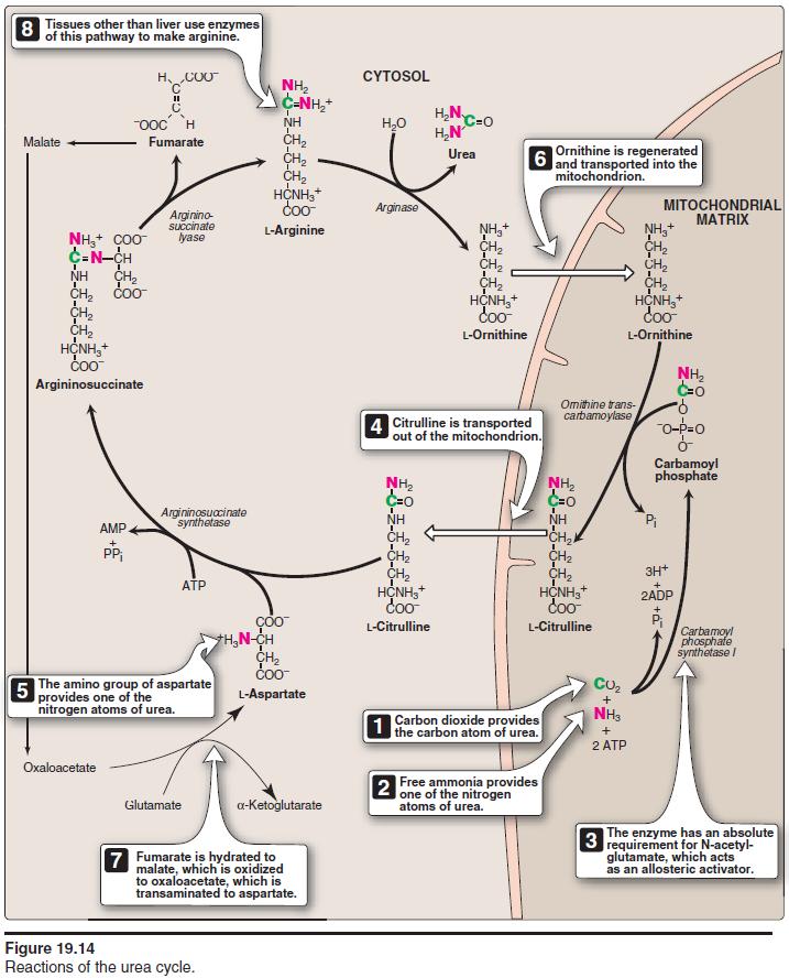
5 Urea cycle: Urea is the major disposal form of amino groups derived from amino acids, and accounts for about 90% of the nitrogen-containing components of urine. One nitrogen of the urea molecule is supplied by free ammonia and the other nitrogen by aspartate. [Note: Glutamate is the immediate precursor of both ammonia (through oxidative deamination by glutamate dehydrogenase) and aspartate nitrogen (through transamination of oxaloacetate by AST).] The carbon and oxygen of urea are derived from CO2. Urea is produced by the liver, and then is transported in the blood to the kidneys for excretion in the urine. The Urea Cycle: The first two reactions leading to the synthesis of urea occur in the mitochondria, whereas the remaining cycle enzymes are located in the cytosol 1. Formation of carbamoyl phosphate: Formation of carbamoyl phosphate by carbamoyl phosphate synthetase I is driven by cleavage of two molecules of ATP. Ammonia incorporated into carbamoyl phosphate is provided primarily by the oxidative deamination of glutamate by mitochondrial glutamate dehydrogenase (see Figure 19.11). Ultimately, the nitrogen atom derived from this ammonia becomes one of the nitrogens of urea. 2. Formation of citrulline: The carbamoyl portion of carbamoyl phosphate is transferred to ornithine by ornithine transcarbamoylase as the high-energy phosphate is released as Pi. The reaction product, citrulline, is transported to the cytosol. Ornithine is regenerated with each turn of the urea cycle, much in the same way that oxaloacetate is regenerated by the reactions of the citric acid cycle. 3. Synthesis of argininosuccinate: Argininosuccinate synthetase combines citrulline with aspartate to form argininosuccinate. The α- amino group of aspartate provides the second nitrogen that is ultimately incorporated into urea. The formation of argininosuccinate is driven by the cleavage of ATP to adenosine monophosphate (AMP) and pyrophosphate. This is the third and final molecule of ATP consumed in the formation of urea.

6 4. Cleavage of argininosuccinate: Argininosuccinate is cleaved by argininosuccinatelyase to yield arginine and fumarate. The arginine formed by this reaction serves as the immediate precursor of urea. Fumarate produced in the urea cycle is hydrated to malate, providing a link with several metabolic pathways. 5. Cleavage of arginine to ornithine and urea: Arginase cleaves arginine to ornithine and urea, and occurs almost exclusively in the liver. Thus, whereas other tissues, such as the kidney, can synthesize arginine by these reactions, only the liver can cleave arginine and, thereby, synthesize urea. 6. Fate of urea: Urea diffuses from the liver, and is transported in the blood to the kidneys, where it is filtered and excreted in the urine. A portion of the urea diffuses from the blood into the intestine, and is cleaved to CO2 and NH3 by bacterial urease. This ammonia is partly lost in the feces, and is partly reabsorbed into the blood. In patients with kidney failure, plasma urea levels are elevated, promoting a greater transfer of urea from blood into the gut. The intestinal action of urease on this urea becomes a clinically important source of ammonia, contributing to the hyperammonemia often seen in these patients.
7
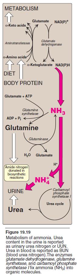
8 METABOLISM OF AMMONIA Ammonia is produced by all tissues during the metabolism of a variety of compounds, and it is disposed of primarily by formation of urea in the liver. However, the level of ammonia in the blood must be kept very low, because even slightly elevated concentrations (hyperammonemia) are toxic to the central nervous system (CNS). There must, therefore, be a metabolic mechanism by which nitrogen is moved from peripheral tissues to the liver for ultimate disposal as urea, while at the same time maintaining low levels of circulating ammonia. A. Sources of ammonia 1. Amino acids are quantitatively the most important source of ammonia. 1. From glutamine: The kidneys generate ammonia from glutamine by the actions of renal glutaminase (Figure 19.17) and glutamate dehydrogenase. Most of this ammonia is excreted into the urine as NH From bacterial action in the intestine: Ammonia is formed from urea by the action of bacterial urease in the lumen of the intestine. This ammonia is absorbed from the intestine by way of the portal vein and is almost quantitatively removed by the liver via conversion to urea. 3. From amines: Amines obtained from the diet, and monoamines that serve as hormones or neurotransmitters, give rise to ammonia by the action of amine oxidase. 4. From purines and pyrimidines: In the catabolism of purines and pyrimidines, amino groups attached to the rings are released as ammonia. B. Transport of ammonia in the circulation Although ammonia is constantly produced in the tissues, it is present at very low levels in blood. This is due both to the rapid removal of blood ammonia by the liver, and the fact that many tissues, particularly muscle, release amino acid nitrogen in the form of glutamine or alanine, rather than as free ammonia. 1. Urea: Formation of urea in the liver is quantitatively the most important disposal route for ammonia. Urea travels in the blood from the liver to the kidneys, where it passes into the glomerular filtrate. 2. Glutamine: This amide of glutamic acid provides a nontoxic storage and transport form of ammonia (Figure 19.18). The ATPrequiring formation of glutamine from glutamate and ammonia by glutamine synthetase occurs primarily in the muscle and liver, but is also important in the CNS where it is the major mechanism for the removal of ammonia in the brain. Glutamine is found in plasma at concentrations higher than other amino acids a finding consistent with its transport function. Circulating glutamine is removed by the liver and the kidneys and deaminated by glutaminase. In the liver, the NH3 produced is detoxified through conversion to urea, and in the kidney it can be used in the excretion of protons. The metabolism of ammonia is summarized in Figure C. Hyperammonemia The capacity of the hepatic urea cycle exceeds the normal rates of ammonia generation, and the levels of serum ammonia are normally low (5 35µmol/L). However, when liver function is compromised, due either to genetic defects of the urea cycle or liver disease, blood levels can rise above 1,000 µmol/l. Such hy perammonemia is a medical emergency, because ammonia has a direct neurotoxic effect on the CNS.


9 For example, elevated concentrations of ammonia in the blood cause the symptoms of ammonia intoxication, which include tremors, slurring of speech, somnolence, vomiting, cerebral edema, and blurring of vision. At high concentrations, ammonia can cause coma and death.
10

11 II-Synthesis and degradation of amino acids: GLUCOGENIC AND KETOGENIC AMINO ACIDS Amino acids can be classified as glucogenic, ketogenic, or both based on which of the seven intermediates are produced during their catabolism. A. Glucogenic amino acids Amino acids whose catabolism yields pyruvate or one of the intermediates of the citric acid cycle are termed glucogenic. These intermediates are substrates for gluconeogenesis and, therefore, can give rise to the net formation of glucose in the liver and kidney. B. Ketogenic amino acids Amino acids whose catabolism yields either acetoacetate or one of its precursors (acetyl CoA or acetoacetyl CoA) are termed ketogenic (see Figure 20.2). Aceto acetate is one of the ketone bodies. CATABOLISM OF THE CARBON SKELETONS OF AMINO ACIDS: A. Amino acids that form oxaloacetate B. Amino acids that form α-ketoglutarate via glutamate C. Amino acids that form pyruvate. D. Amino acids that form fumarate. E. Amino acids that form succinyl-coa. F. Amino acids that form acetyl CoA or acetoacetyl CoA.
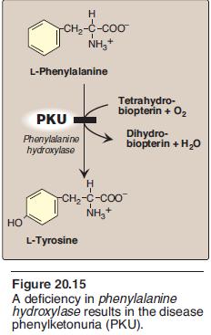
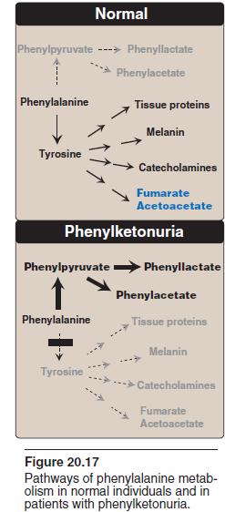
12 METABOLIC DEFECTS IN AMINO ACID METABOLISM A. Phenylketonuria Phenylketonuria (PKU), caused by a deficiency of phenylalanine hydroxylase (Figure 20.15), is the most common clinically encountered inborn error of amino acid metabolism (prevalence 1:15,000). Biochemically, it is characterized by accumulation of phenylalanine (and a deficiency of tyrosine). 1. Characteristics of classic PKU: a. Elevated phenylalanine: Phenylalanine is present in elevated concentrations in tissues, plasma, and urine. Phenyllactate, pheny lacetate, and phenylpyruvate, which are not normally produced in significant amounts in the presence of functional phenylalanine hydroxylase, are also elevated in PKU (Figure 20.17). These metabolites give urine a characteristic musty ( mousey ) odor. [Note: The disease acquired its name from the presence of a phenylketone (now known to be phenylpyruvate) in the urine.] b. CNS symptoms: Mental retardation, failure to walk or talk, seizures, hyperactivity, tremor, microcephaly, and failure to grow are characteristic findings in PKU. The patient with untreated PKU typically shows symptoms of mental retardation by the age of 1 year, and rarely achieves an IQ greater than 50 (Figure 20.18). [Note: These clinical manifestations are now rarely seen as a result of neonatal screening programs.] c. Hypopigmentation: Patients with phenylketonuria often show a deficiency of pigmentation (fair hair, light skin color, and blue eyes). The hydroxylation of tyrosine by tyrosinase, which is the first step in the formation of the pigment melanin, is competitively inhibited by the high levels of phenylalanine present in PKU. 2. Neonatal screening and diagnosis of PKU: Early diagnosis of phenylketonuria is important because the disease is treatable by dietary means. Because of the lack of neonatal symptoms, laboratory testing for elevated blood levels of phenylalanine is mandatory for detection. However, the infant with PKU frequently has normal blood levels of phenylalanine at birth because the mother clears increased blood phenylalanine in her affected fetus through the placenta. Normal levels of phenylalanine may persist until the newborn is exposed to hours of protein feeding. Thus, screening tests are typically done after this time to avoid false negatives. For newborns with a positive screening test, diagnosis is confirmed through quantitative determination of phenylalanine levels. B. Albinism Albinism refers to a group of conditions in which a defect in tyrosine metabolism results in a deficiency in the production of melanin. These defects result in the partial or full absence of pigment from the skin, hair, and eyes. Albinism appears in different forms, and it may be inherited.
13
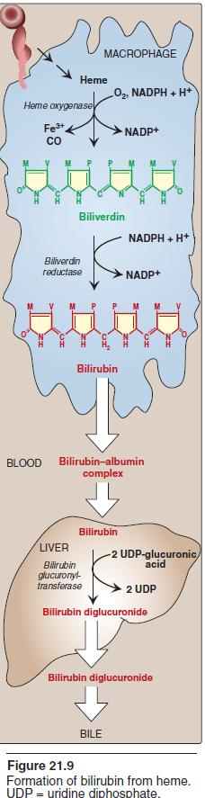
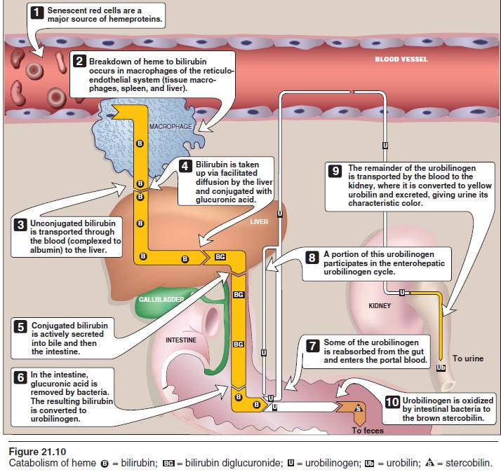
14 III- Conversion of amino acids to specialized products: In addition to serving as building blocks for proteins, amino acids are precursors of many nitrogencontaining compounds that have important physiologic functions. These molecules include porphyrins, neurotransmitters, hormones, purines, and pyrimidines. 1- PORPHYRIN METABOLISM Porphyrins are cyclic compounds that readily bind metal ions usually Fe2+ or Fe3+. The most prevalent metalloporphyrin in humans is heme, which consists of one ferrous (Fe2+) iron ion coordinated in the center of the tetrapyrrole ring of protoporphyrin. Heme is the prosthetic group for hemoglobin, myoglobin, the cytochromes, catalase, nitric oxide synthase, and peroxidase. These hemeproteins are rapidly synthesized and degraded. For example, 6 7 g of hemoglobin are synthesized each day to replace heme lost through the normal turnover of erythrocytes. Coordinated with the turnover of hemeproteins is the simultaneous synthesis and degradation of the associated porphyrins, and recycling of the bound iron ions. B. Biosynthesis of heme The major sites of heme biosynthesis are the liver, which synthesizes a number of heme proteins (particularly cytochrome P450 proteins), and the erythrocyte-producing cells of the bone marrow, which are active in hemoglobin synthesis. [Note: Over 85% of all heme synthesis occurs in erythroid tissue.] D. Degradation of heme After approximately 120 days in the circulation, red blood cells are taken up and degraded by the reticuloendothelial system, particularly in the liver and spleen (Figure 21.9). Approximately 85% of heme destined for degradation comes from senescent red blood cells, and 15% is from turnover of immature red blood cells and cytochromes from nonerythroid tissues. 1. Formation of bilirubin: The first step in the degradation of heme is catalyzed by the microsomal heme oxygenase system of the reticuloendothelial cells. The green pigment biliverdin is produced as ferric iron and CO are released (see Figure 21.9). [Note: The CO has biologic function, acting as a signaling molecule and vasodilator.] Biliverdin is reduced, forming the redorange bilirubin. Bilirubin and its derivatives are collectively termed bile pigments. [Note: The changing colors of a bruise reflect the varying pattern of intermediates that occurs during heme degradation.] 2. Uptake of bilirubin by the liver: Bilirubin is only slightly soluble in plasma and, therefore, is transported to the liver by binding noncovalently to albumin. [Note: Certain anionic drugs, such as salicylates and sulfonamides, can displace bilirubin from albumin, permitting bilirubin to enter the central nervous system. This causes the potential for neural damage in infants.] Bilirubin dissociates from the carrier albumin molecule, enters a hepatocyte via facilitated diffusion, and binds to intracellular proteins, particularly the protein ligand.
15
16

17 3. Formation of bilirubin diglucuronide: In the hepatocyte, the solubility of bilirubin is increased by the addition of two molecules of glucuronic acid. [Note: This process is referred to as conjugation.] 4. Secretion of bilirubin into bile: Bilirubin diglucuronide (conjugated bilirubin) is actively transported against a concentration gradient into the bile canaliculi and then into the bile. This energydependent, rate-limiting step is susceptible to impairment in liver disease. Unconjugated bilirubin is normally not secreted. 5. Formation of urobilins in the intestine: Bilirubin diglucuronide is hydrolyzed and reduced by bacteria in the gut to yield urobilinogen, a colorless compound. Most of the urobilinogen is oxidized by intestinal bacteria to stercobilin, which gives feces the characteristic brown color. However, some of the urobilinogen is reabsorbed from the gut and enters the portal blood. A portion of this urobilinogen participates in the enterohepatic urobilinogen cycle in which it is taken up by the liver, and then resecreted into the bile. The remainder of the urobilinogen is transported by the blood to the kidney, where it is converted to yellow urobilin and excreted, giving urine its characteristic color. The metabolism of bilirubin is summarized in Figure E. Jaundice Jaundice refers to the yellow color of skin, nail beds, and sclerae (whites of the eyes) caused by deposition of bilirubin, secondary to increased bilirubin levels in the blood (hyperbilirubinemia. Although not a disease, jaundice is usually a symptom of an underlying disorder. Jaundice in newborns: Newborn infants, particularly if premature, often accumulate bilirubin, because the activity of hepatic bilirubin glucuronyltransferase is low at birth it reaches adult levels in about 4 weeks. Elevated bilirubin, in excess of the binding capacity of albumin, can diffuse into the basal ganglia and cause toxic encephalopathy (kernicterus). Thus, newborns with significantly elevated bilirubin levels are treated with blue fluorescent light, which converts bilirubin to more polar and, hence, water-soluble isomers. These photoisomers can be excreted into the bile without conjugation to glucuronic acid. Catecholamines Dopamine, norepinephrine, and epinephrine are biologically active (biogenic) amines that are collectively termed catecholamines. Dopamine and norepinephrine are synthesized in the brain and function as neurotransmitters. Norepinephrine is also synthesized in the adrenal medulla, as is epinephrine.
.")

18 Histamine Histamine is a chemical messenger that mediates a wide range of cellular responses, including allergic and inflammatory reactions, gastric acid secretion, and possibly neurotransmission in parts of the brain. A powerful vasodilator, histamine is formed by decarboxylation of histidine in a reaction requiring PLP (Figure 21.17). It is secreted by mast cells as a result of allergic reactions or trauma. Histamine has no clinical applications, but agents that interfere with the action of histamine have important therapeutic applications. Creatine Creatine phosphate (also called phosphocreatine), the phosphorylated derivative of creatine found in muscle, is a high-energy compound that provides a small but rapidly mobilized reserve of high-energy phosphates that can be reversibly transferred to ADP (Figure 21.9) to maintain the intracellular level of ATP during the first few minutes of intense muscular contraction. [Note: The amount of creatine phosphate in the body is proportional to the muscle mass.]
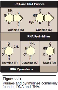
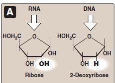
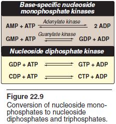
19 IV-Nucleotide Metabolism NUCLEOTIDE STRUCTURE Nucleotides are composed of a nitrogenous base, a pentose monosaccharide, and one, two, or three phosphate groups. The nitrogen containing bases belong to two families of compounds: the purines and the pyrimidines. A. Purine and pyrimidine structures Both DNA and RNA contain the same purine bases: adenine (A) and guanine (G). Both DNA and RNA contain the pyrimidine cytosine (C), but they differ in their second pyrimidine base: DNA contains thymine (T), whereas RNA contains uracil (U). T and U differ in that only T has a methyl group (Figure 22.1 B. Nucleosides The addition of a pentose sugar to a base produces a nucleoside. If the sugar is ribose, a ribonucleoside is produced; if the sugar is 2-deoxyribose, a deoxyribonucleoside is produced (Figure 22.3A). C. Nucleotides The addition of one or more phosphate groups to a nucleoside produces a nucleotide. If one phosphate group is attached to the 5'-carbon of the pentose, the structure is a nucleoside monophosphate, like adenosine monophosphate (AMP) (also called adenylate). If a second or third phosphate is added to the nucleoside, a nucleoside diphosphate (for example, adenosine diphosphate or ADP) or triphosphate (for example, adenosine triphosphate or ATP) results (Figure 22.4).
20 DEGRADATION OF PURINE NUCLEOTIDES
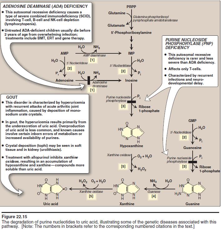
21 Degradation of dietary nucleic acids occurs in the small intestine, where a family of pancreatic enzymes hydrolyzes the nucleic acids to nucleotides. Inside the intestinal mucosal cells, purine nucleotides are sequentially degraded by specific enzymes to nucleosides and free bases, with uric acid as the end product of this pathway. A. Degradation of dietary nucleic acids in the small intestine Ribonucleases and deoxyribonucleases, secreted by the pancreas, hydrolyze dietary RNA and DNA primarily to oligonucleotides. Oligonucleotides are further hydrolyzed by pancreatic phosphodiesterases, producing a mixture of 3'- and 5'-mononucleotides. In the intestinal mucosal cells, a family of nucleotidases removes the phosphate groups hydrolytically, releasing nucleosides that are further degraded to free bases. Dietary purine bases are not used to any appreciable extent for the synthesis of tissue nucleic acids. Instead, they are generally converted to uric acid in intestinal mucosal cells. Most of the uric acid enters the blood, and is eventually excreted in the urine. B. Formation of uric acid A summary of the steps in the production of uric acid and genetic diseases associated with deficiencies of specific degradative enzymes are shown in Figure [Note: The bracketed numbers refer to specific reactions in the figure.] C. Diseases associated with purine degradation 1. Gout: Gout is a disorder characterized by high levels of uric acid the end product of purine catabolism in blood (hyperuricemia), as a result of either the overproduction or underexcretion of uric acid. The hyperuricemia can lead to the deposition of mono -sodium urate crystals in the joints, and an inflammatory response to the crystals, causing first acute and then progressing to chronic gouty arthritis. Formation of uric acid stones in the kidney (urolithiasis) may also be seen. a. Underexcretion of uric acid. b. Overproduction of uric acid.
22
Urea is the major end product of nitrogen catabolism in humans One nitrogen free NH3 other nitrogen aspartate. carbon oxygen CO2 liver,
 Urea is the major end product of nitrogen catabolism in humans Urea is the major disposal form of amino groups derived from amino acids, and accounts about 90% percent of the nitrogencontaining components
AMINO ACID METABOLISM
 AMINO ACID METABOLISM Synthesis of Urea in Liver The series of reactions that form urea is known as the Urea Cycle or the Krebs-Henseleit Cycle. The urea cycle operates only to eliminate excess nitrogen.
Amino acid oxidation and the production of urea
 Seminar 10 Urea cycle Amino acid oxidation and the production of urea Oxidation Waste or Reuse Ammonia has to be eliminated ammonia originates in the catabolism of amino acids that are primarily produced
AMINOACID METABOLISM FATE OF AMINOACIDS & UREA CYCLE
 AMINOACID METABOLISM FATE OF AMINOACIDS & UREA CYCLE SOURCE & FATE OF AA The aminoacids obtained from DIETARY SOURCE or BODY PROTEIN TURNOVER are utilized for protein biosynthesis and the production of
Biochemistry: A Short Course
 Tymoczko Berg Stryer Biochemistry: A Short Course Second Edition CHAPTER 30 Amino Acid Degradation and the Urea Cycle 2013 W. H. Freeman and Company In the cytosol of a cell amino groups from amino acids
Biochemistry: A Short Course
 Tymoczko Berg Stryer Biochemistry: A Short Course Second Edition CHAPTER 30 Amino Acid Degradation and the Urea Cycle 2013 W. H. Freeman and Company Chapter 30 Outline Amino acids are obtained from the
NITROGEN METABOLISM An Overview
 1 University of Papua New Guinea School of Medicine and Health Sciences Division of Basic Medical Sciences Discipline of Biochemistry and Molecular Biology PBL Seminar & Health Sciences NITROGEN METABOLISM
Amino Acid Oxidation and the Urea Cycle
 Amino Acid Oxidation and the Urea Cycle Amino Acids: Final class of biomolecules whose oxidation contributes significantly to the generation of energy Undergo oxidation in three metabolic circumstances
PROTEIN METABOLISM: SPECIFIC WAYS OF AMINO ACIDS CATABOLISM AND SYNTHESIS
 PROTEIN METABOLISM: SPECIFIC WAYS OF AMINO ACIDS CATABOLISM AND SYNTHESIS SPECIFIC WAYS OF AMINO ACID CATABOLISM After removing of amino group the carbon skeletons of amino acids are transformed into metabolic
Amino Acid Metabolism
 Amino Acid Metabolism The continuous degradation and synthesis of cellular proteins occur in all forms of life. Each day humans turn over 1 2% of their total body protein, principally muscle protein. Approximately
AMINO ACID METABOLISM. Sri Widia A Jusman Dept. of Biochemistry & Molecular Biology FMUI
 AMINO ACID METABOLISM Sri Widia A Jusman Dept. of Biochemistry & Molecular Biology FMUI Amino acids derived from dietary protein absorbed from intestine through blood taken up by tissues used for biosynthesis
Jana Novotná, Bruno Sopko. Department of the Medical Chemistry and Clinical Biochemistry The 2nd Faculty of Medicine, Charles Univ.
 Amino acid metabolism II. Urea cycle Jana Novotná, Bruno Sopko Department of the Medical Chemistry and Clinical Biochemistry The 2nd Faculty of Medicine, Charles Univ. Nitrogen balance Tissue proteins
NITROGEN METABOLISM: An Overview
 NITROGEN METABOLISM: An Overview University of PNG School of Medicine and Health Sciences Division of Basic Medical Sciences Discipline of Biochemistry & Molecular Biology VJ Temple 1 How are nitrogen-containing
Fate of Dietary Protein
 Fate of Dietary Protein Dietary protein Stomach: l, pepsin Denatured and partially hydrolyzed protein (large polypeptides) small intestine: proteases Amino acids and dipeptides intestinal lining: proteases
Amino Acid Metabolism
 Amino Acid Metabolism Fate of Dietary Protein Dietary protein Stomach: l, pepsin Denatured and partially hydrolyzed protein (large polypeptides) small intestine: proteases Amino acids and dipeptides intestinal
BIOCHEMISTRY Protein Metabolism
 BIOCHEMISTRY Protein Metabolism BIOB111 CHEMISTRY & BIOCHEMISTRY Session 25 Session Plan Digestion & Absorption of Proteins Amino Acid Utilization Amino Acid Degradation Transamination Oxidative Deamination
Lecture 10 - Protein Turnover and Amino Acid Catabolism
 Lecture 10 - Protein Turnover and Amino Acid Catabolism Chem 454: Regulatory Mechanisms in Biochemistry University of Wisconsin-Eau Claire 1 Introduction 2 Proteins are degraded into amino acids. Protein
Part III => METABOLISM and ENERGY. 3.5 Protein Catabolism 3.5a Protein Degradation 3.5b Amino Acid Breakdown 3.5c Urea Cycle
 Part III => METABOLISM and ENERGY 3.5 Protein Catabolism 3.5a Protein Degradation 3.5b Amino Acid Breakdown 3.5c Urea Cycle Section 3.5a: Protein Degradation Synopsis 3.5a - Dietary proteins are degraded
Metabolism of Nucleotides
 Metabolism of Nucleotides Outline Nucleotide degradation Components of Nucleobases Purine and pyrimidine biosynthesis Hyperuricemia Sources Nucleotide degradation The nucleotides are among the most complex
Lecture: Amino Acid catabolism: Nitrogen-The Urea cycle
 BIOC 423: Introductory Biochemistry Biochemistry Education Department of Biochemistry & Molecular Biology University of New Mexico Lecture: Amino Acid catabolism: Nitrogen-The Urea cycle OBJECTIVES Describe
Nitrogen Metabolism. Overview
 Nitrogen Metabolism Pratt and Cornely Chapter 18 Overview Nitrogen assimilation Amino acid biosynthesis Nonessential aa Essential aa Nucleotide biosynthesis Amino Acid Catabolism Urea Cycle Juicy Steak
Nitrogen Metabolism. Overview
 Nitrogen Metabolism Pratt and Cornely Chapter 18 Overview Nitrogen assimilation Amino acid biosynthesis Nonessential aa Essential aa Nucleotide biosynthesis Amino Acid Catabolism Urea Cycle Juicy Steak
Integration Of Metabolism
 Integration Of Metabolism Metabolism Consist of Highly Interconnected Pathways The basic strategy of catabolic metabolism is to form ATP, NADPH, and building blocks for biosyntheses. 1. ATP is the universal
Metabolism of amino acids. Vladimíra Kvasnicová
 Metabolism of amino acids Vladimíra Kvasnicová Classification of proteinogenic AAs -metabolic point of view 1) biosynthesis in a human body nonessential (are synthesized) essential (must be present in
Nitrogen Metabolism. Pratt and Cornely Chapter 18
 Nitrogen Metabolism Pratt and Cornely Chapter 18 Overview Nitrogen assimilation Amino acid biosynthesis Nonessential aa Essential aa Nucleotide biosynthesis Amino Acid Catabolism Urea Cycle Juicy Steak
Amino Acid Metabolism: Conversion of Amino Acids to Specialized Products
 Amino Acid Metabolism: Conversion of Amino Acids to Specialized Products Dr. Diala Abu-Hassan, DDS, PhD Medical students-first semester All images are taken from Lippincott s Biochemistry textbook except
Amino acid Catabolism
 Enzymatic digestion of dietary proteins in gastrointestinal-tract. Amino acid Catabolism Amino acids: 1. There are 20 different amino acid, they are monomeric constituents of proteins 2. They act as precursors
CONVERSION OF AMINO ACIDS TO SPECIALIZED PRODUCTS DR. A. TARAB DEPT. OF BIOCHEMISTRY HKMU
 CONVERSION OF AMINO ACIDS TO SPECIALIZED PRODUCTS DR. A. TARAB DEPT. OF BIOCHEMISTRY HKMU In addition to serving as building blocks for proteins, amino acids are precursors of many nitrogen-containing
Amino acid metabolism
 Amino acid metabolism The important reaction commonly employed in the breakdown of an amino acid is always the removal of its -amino group. The product ammonia is excreted after conversion to urea or other
Definition of bilirubin Bilirubin metabolism
 Definition of bilirubin Bilirubin metabolism obilirubin formation otransport of bilirubin in plasma ohepatic bilirubin transport oexcretion through intestine Other substances conjugated by glucuronyl transferase.
BIOB111 - Tutorial activity for Session 25
 BIOB111 - Tutorial activity for Session 25 General topics for week 14 Session 25 The metabolism of proteins Students are asked to draw the concept map showing all details of protein metabolism 1 Instructions:
Clinical Biochemistry department/ College of medicine / AL-Mustansiriyah University
 Clinical Biochemistry department/ College of medicine / AL-Mustansiriyah University Dr. Ali al-bayati NUCLEOTIDE METABOLISM Lec. 3 The salvage pathway of purine synthesis Purines that result from the normal
Energy Production In A Cell (Chapter 25 Metabolism)
") Energy Production In A Cell (Chapter 25 Metabolism) Large food molecules contain a lot of potential energy in the form of chemical bonds but it requires a lot of work to liberate the energy. Cells need
Energy Production In A Cell (Chapter 25 Metabolism) Large food molecules contain a lot of potential energy in the form of chemical bonds but it requires a lot of work to liberate the energy. Cells need
Amino acid metabolism: Disposal of Nitrogen
 Amino acid metabolism: Disposal of Nitrogen Dr. Diala Abu-Hassan, DDS, PhD All images were taken from Lippincott s Biochemistry textbook except where noted Textbook Amino acid metabolism: Biochemistry
Amino acid metabolism I
 Amino acid metabolism I Jana Novotná Department of the Medical Chemistry and Clinical Biochemistry The 2nd Faculty of Medicine, Charles Univ. Metabolic relationship of amino acids DIETARY PROTEINS GLYCOLYSIS
AMINO ACID METABOLISM
 AMINO ACID METABOLISM PHL-285 Biochemistry-2 Mahmoud N. Nagi, Ph.D. Professor of Biochemistry Overview of amino acid metabolism. Classification of amino acids. Biosynthesis of nonessential amino acids.
Amino Acid Metabolism
 Amino Acid Metabolism Last Week Most of the Animal Kingdom = Lazy - Most higher organisms in the animal kindom don t bother to make all of the amino acids. - Instead, we eat things that make the essential
SYNTHESIS OF NON-ESSENTIAL AMINO ACIDS [LIPPINCOTT S ] Deeba S. Jairajpuri
![SYNTHESIS OF NON-ESSENTIAL AMINO ACIDS [LIPPINCOTT S ] Deeba S. Jairajpuri](/thumbs/80/80689583.jpg "SYNTHESIS OF NON-ESSENTIAL AMINO ACIDS [LIPPINCOTT S ] Deeba S. Jairajpuri") SYNTHESIS OF NON-ESSENTIAL AMINO ACIDS [LIPPINCOTT S 267-274] Deeba S. Jairajpuri TYPES OF AMINO ACIDS Amino acids that cannot be synthesized by the human body and are required to be taken in the diet
SYNTHESIS OF NON-ESSENTIAL AMINO ACIDS [LIPPINCOTT S 267-274] Deeba S. Jairajpuri TYPES OF AMINO ACIDS Amino acids that cannot be synthesized by the human body and are required to be taken in the diet
Metabolism of proteins and amino acids
 BIOQUÍMICA E BIOLOGIA CELULAR António Ascensão, José Magalhães Metabolism of proteins and amino acids Faculdade de Desporto, Universidade do Porto, 1º Ciclo, 1º Ano 202_2013 Humans degradation of ingested
18 Amino Acid Oxidation and Production of Urea W. H. Freeman and Company
 18 Amino Acid Oxidation and Production of Urea 2013 W. H. Freeman and Company 1 Last Class of Biomolecules For Energy 1. Production of acetyl-coa. Glucose. To pyruvate via glycolysis. To acetyl-coa by
Welcome to Class 14! Class 14: Outline and Objectives. Overview of amino acid catabolism! Introductory Biochemistry!
 Welcome to Class 14 Introductory Biochemistry Class 14: Outline and Objectives Amino Acid Catabolism Fates of amino groups transamination urea cycle Fates of carbon skeletons important cofactors metabolic
Amino acid metabolism: Disposal of Nitrogen
 Amino acid metabolism: Disposal of Nitrogen Dr. Diala Abu-Hassan, DDS, PhD Medical students-first semester All images were taken from Lippincott s Biochemistry textbook except where noted Amino acids (AAs)
Integrative Metabolism: Significance
 Integrative Metabolism: Significance Energy Containing Nutrients Carbohydrates Fats Proteins Catabolism Energy Depleted End Products H 2 O NH 3 ADP + Pi NAD + NADP + FAD + Pi NADH+H + NADPH+H + FADH2 Cell
Chemistry 1120 Exam 4 Study Guide
 Chemistry 1120 Exam 4 Study Guide Chapter 12 12.1 Identify and differentiate between macronutrients (lipids, amino acids and saccharides) and micronutrients (vitamins and minerals). Master Tutor Section
Integration of Metabolism
 Integration of Metabolism Metabolism is a continuous process. Thousands of reactions occur simultaneously in order to maintain homeostasis. It ensures a supply of fuel, to tissues at all times, in fed
Midterm 2 Results. Standard Deviation:
 Midterm 2 Results High: Low: Mean: Standard Deviation: 97.5% 16% 58% 16.3 Lecture 17 Amino Acid Metabolism Urea Cycle N and S assimilation Last cofactors: THF and SAM Dietary (Exogenous) Proteins Hydrolyzed
Urea cycle: Urea cycle is discovered by Krebs andhanseleit(1932).
.") Urea cycle: Urea cycle is discovered by Krebs andhanseleit(1932). Urea cycle is the removal of excess of NH2 derived from amino acids catabolism in the tissues and excreted in urine. Site of synthesis:
Urea cycle: Urea cycle is discovered by Krebs andhanseleit(1932). Urea cycle is the removal of excess of NH2 derived from amino acids catabolism in the tissues and excreted in urine. Site of synthesis:
Lecture 17: Nitrogen metabolism 1. Urea cycle detoxification of NH 3 2. Amino acid degradation
 Lecture 17: Nitrogen metabolism 1. Urea cycle detoxification of NH 3 2. Amino acid degradation Reference material Biochemistry 4 th edition, Mathews, Van Holde, Appling, Anthony Cahill. Pearson ISBN:978
number Done by Corrected by Doctor Dr.Diala
 number 32 Done by Mousa Salah Corrected by Bahaa Najjar Doctor Dr.Diala 1 P a g e In the last lecture we talked about the common processes between all amino acids which are: transamination, deamination,
Conversion of amino acids الفريق الطبي األكاديمي
 الفريق الطبي األكاديمي لكية الطب البرشي البلقاء التطبيقية / املركز 2022 2016/ -the ones with * are the essential amino acids- - Histidine decarboxylation -> histamine Page 1 - Glutamine is a nitrogen carrier
SCBC203 Amino Acid Metabolism
 Breakdown of proteins Route I: Dietary protein breakdown SCBC203 Amino Acid Metabolism Dr Sarawut Jitrapakdee Professor of Biochemistry Department of Biochemistry Faculty of Science Mahidol University
Amino Acid Metabolism: Conversion of Amino Acids to Specialized Products
 Amino Acid Metabolism: Conversion of Amino Acids to Specialized Products Dr. Diala Abu-Hassan, DDS, PhD Medical students-first semester All images are taken from Lippincott s Biochemistry textbook except
Lipid and Amino Acid Metabolism
 CHEM 3331 Fundamentals of Biochemistry Chapter 14 Lipid and Amino Acid Metabolism Organic and Biochemistry for Today Spencer L. Seager / Michael R. Slabaugh Mr. Kevin A. Boudreaux Angelo State University
AMINO ACIDS NON-ESSENTIAL ESSENTIAL
 Edith Frederika Introduction A major component of food is PROTEIN The protein ingested as part of our diet are not the same protein required by the body Only 40 to 50 gr of protein is required by a normal
TCA CYCLE (Citric Acid Cycle)
") TCA CYCLE (Citric Acid Cycle) TCA CYCLE The Citric Acid Cycle is also known as: Kreb s cycle Sir Hans Krebs Nobel prize, 1953 TCA (tricarboxylic acid) cycle The citric acid cycle requires aerobic conditions!!!!
TCA CYCLE (Citric Acid Cycle) TCA CYCLE The Citric Acid Cycle is also known as: Kreb s cycle Sir Hans Krebs Nobel prize, 1953 TCA (tricarboxylic acid) cycle The citric acid cycle requires aerobic conditions!!!!
1 Digestion and absorption. Lecture #14 Lecturer: PhD Alexander N. Koval
 1 Digestion and absorption Lecture #14 Lecturer: PhD Alexander N. Koval Presentation of Protein 12/22/2016 A. Koval (C), 2016 2 Lectures plan 12/22/2016 A. Koval (C), 2016 3 Overview of Protein Metabolism
Non-Protein Nitrogenous Compounds. Non-Protein Nitrogenous Compounds. NPN s. Urea (BUN) Creatinine NH 3. University of Cincinnati MLS Program 1
 Creatinine NH 3. University of Cincinnati MLS Program 1") Non-Protein Nitrogenous Compounds NPN s Urea (BUN) Creatinine NH 3 Uric Acid Ammonia University of Cincinnati MLS Program 1 Urea Metabolic product derived from catabolism of proteins Proteolysis of proteins
Non-Protein Nitrogenous Compounds NPN s Urea (BUN) Creatinine NH 3 Uric Acid Ammonia University of Cincinnati MLS Program 1 Urea Metabolic product derived from catabolism of proteins Proteolysis of proteins
Dental Students Biochemistry Exam V Questions ( Note: In all cases, the only correct answer is the best answer)
") Dental Students Biochemistry Exam V Questions - 2006 ( Note: In all cases, the only correct answer is the best answer) 1. Essential fatty acids are: A. precursors of biotin B. precursors of tyrosine C.
Dental Students Biochemistry Exam V Questions - 2006 ( Note: In all cases, the only correct answer is the best answer) 1. Essential fatty acids are: A. precursors of biotin B. precursors of tyrosine C.
CLINICAL BIOCHEMISTRY 6 PLASMA PROTEINS AND PATHOLOGICAL IMPLICATIONS OF THEIR IMBALANCE
 LINIAL BIOHEMISTRY 6 PLASMA PROTEINS AND PATHOLOGIAL IMPLIATIONS OF THEIR IMBALANE DISTURBANES OF PROTEIN METABOLISM NPN result from the metabolism of aminoacids, proteins, nucleic acids 2.3.1. UREA (75%
MDSC 1102/VM1102 Cardiovascular and Renal. Purine nucleotide metabolism
 MDSC 1102/VM1102 Cardiovascular and Renal Purine nucleotide metabolism Dr. J. Foster Biochemistry Unit, Dept. Preclinical Sciences Faculty of Medical Sciences, U.W.I. Learning Objectives Discuss purineand
Urea Cycle Defects. Dr Mick Henderson. Biochemical Genetics Leeds Teaching Hospitals Trust. MetBioNet IEM Introductory Training
 Urea Cycle Defects Dr Mick Henderson Biochemical Genetics Leeds Teaching Hospitals Trust The Urea Cycle The urea cycle enables toxic ammonia molecules to be converted to the readily excreted and non toxic
Amino acid metabolism II
 Amino acid metabolism II Fates of amino acid carbon skeleton degradation to common intermediates pyruvate, intermediates of citric acid cycle, acetyl-coa Glucogenic AA precursors of glucose - degradation
Nucleotide Metabolism. Pyrimidine Met. Purine Met.
 Nucleotide Metabolism Pyrimidine Met. Purine Met. Learning Objectives 1. How Are Purines Synthesized? 2. How Are Purines Catabolized? 3. How Are Pyrimidines Synthesized and Catabolized? 4. How Are Ribonucleotides
Bio 366: Biological Chemistry II Test #2, 100 points total
 Bio 366: Biological Chemistry II Test #2, 100 points total Please neatly PRINT YOUR NAME on EACH PAGE. PRINT the l ast four digits of your SOCIAL SECURITY NUMBER on the BACK SIDE OF PAGE 11 of this test.
Bio 366: Biological Chemistry II Final Exam, 100 points total
 Bio 366: Biological Chemistry II Final Exam, 100 points total Please neatly print your name on the top of each page, and put the last four digits of your social security number and the sticker from your
Metabolism of amino acids I. Josef Fontana
 Metabolism of amino acids I Josef Fontana EC Overview of the lecture Introduction to protein and amino acids metabolism Metabolic pathways of amino acids Transamination Conversion glutamate - glutamine
Midterm 2. Low: 14 Mean: 61.3 High: 98. Standard Deviation: 17.7
 Midterm 2 Low: 14 Mean: 61.3 High: 98 Standard Deviation: 17.7 Lecture 17 Amino Acid Metabolism Review of Urea Cycle N and S assimilation Last cofactors: THF and SAM Synthesis of few amino acids Dietary
Find this material useful? You can help our team to keep this site up and bring you even more content consider donating via the link on our site.
 Find this material useful? You can help our team to keep this site up and bring you even more content consider donating via the link on our site. Still having trouble understanding the material? Check
Amino Acid Metabolism Parts I-III
 M1 - Biochemistry Amino Acid Metabolism Parts I-III Dr. Diegelmann 10/2, 10/3, 10/7 1 Biochemical Pathways; If you know all of this you can get an A 2 OBJECTIVES After studying the material presented in
CITRIC ACID CYCLE ERT106 BIOCHEMISTRY SEM /19 BY: MOHAMAD FAHRURRAZI TOMPANG
 CITRIC ACID CYCLE ERT106 BIOCHEMISTRY SEM 1 2018/19 BY: MOHAMAD FAHRURRAZI TOMPANG Chapter Outline (19-1) The central role of the citric acid cycle in metabolism (19-2) The overall pathway of the citric
Non-protein nitrogenous substances (NPN)
") Non-protein nitrogenous substances (NPN) A simple, inexpensive screening test a routine urinalysis is often the first test conducted if kidney problems are suspected. A small, randomly collected urine
Non-protein nitrogenous substances (NPN) A simple, inexpensive screening test a routine urinalysis is often the first test conducted if kidney problems are suspected. A small, randomly collected urine
Chapter 26. Outline. Nitrogen. Nitrogen and Amino Acid Metabolism. BCH 4054 Spring 2001 Chapter 26 Lecture Notes. Slide 1. Slide 2
 BCH 4054 Spring 2001 Chapter 26 Lecture Notes 1 Chapter 26 Nitrogen and Amino Acid Metabolism 2 utline No time to cover entire chapter, therefore concentrate on a few focal points Assimilation of inorganic
Oxidation of Long Chain Fatty Acids
 Oxidation of Long Chain Fatty Acids Dr NC Bird Oxidation of long chain fatty acids is the primary source of energy supply in man and animals. Hibernating animals utilise fat stores to maintain body heat,
III. 6. Test. Respiració cel lular
 III. 6. Test. Respiració cel lular Chapter Questions 1) What is the term for metabolic pathways that release stored energy by breaking down complex molecules? A) anabolic pathways B) catabolic pathways
Metabolism. Chapter 5. Catabolism Drives Anabolism 8/29/11. Complete Catabolism of Glucose
 8/29/11 Metabolism Chapter 5 All of the reactions in the body that require energy transfer. Can be divided into: Cell Respiration and Metabolism Anabolism: requires the input of energy to synthesize large
Physiology Unit 1 METABOLISM OF LIPIDS AND PROTEINS
 Physiology Unit 1 METABOLISM OF LIPIDS AND PROTEINS Alternate Fuel Sources When glucose levels are low Proteins and Triglycerides will be metabolized Tissues will use different fuel sources depending on:
Nucleotide Metabolism Biochemistry by Lippincott pp
 Nucleotide Metabolism Biochemistry by Lippincott pp 291-306 Deoxyribonucleotides Synthesis 2'-deoxyribonucleotides: Nucleotides required for DNA synthesis Produced from ribonucleoside diphosphates by ribonucleotide
Biologic Oxidation BIOMEDICAL IMPORTAN
 Biologic Oxidation BIOMEDICAL IMPORTAN Chemically, oxidation is defined as the removal of electrons and reduction as the gain of electrons. Thus, oxidation is always accompanied by reduction of an electron
If you ate a clown, would it taste funny? Oh, wait, that s cannibalism . Anabolism
 If you ate a clown, would it taste funny? Oh, wait, that s cannibalism. Anabolism is about putting things together. Anabolism: The Use of Energy in Biosynthesis Anabolism energy from catabolism is used
Adenosine triphosphate (ATP)
") Adenosine triphosphate (ATP) 1 High energy bonds ATP adenosine triphosphate N NH 2 N -O O P O O P O- O- O O P O- O CH 2 H O H N N adenine phosphoanhydride bonds (~) H OH ribose H OH Phosphoanhydride bonds
Adenosine triphosphate (ATP) 1 High energy bonds ATP adenosine triphosphate N NH 2 N -O O P O O P O- O- O O P O- O CH 2 H O H N N adenine phosphoanhydride bonds (~) H OH ribose H OH Phosphoanhydride bonds
Amino Acid Catabolism
 Amino Acid atabolism 3-1 Lec #8 To date we have considered the catabolism of carbohydrates and lipids with the object of generating energy in the form of ATP. Both give rise to AcoA which is fed through
Chapter 24 Lecture Outline
 Chapter 24 Lecture Outline Carbohydrate Lipid and Protein! Metabolism! In the catabolism of carbohydrates, glycolysis converts glucose into pyruvate, which is then metabolized into acetyl CoA. Prepared
4. Which step shows a split of one molecule into two smaller molecules? a. 2. d. 5
 1. Which of the following statements about NAD + is false? a. NAD + is reduced to NADH during both glycolysis and the citric acid cycle. b. NAD + has more chemical energy than NADH. c. NAD + is reduced
Marah Bitar. Faisal Nimri ... Nafeth Abu Tarboosh
 8 Marah Bitar Faisal Nimri... Nafeth Abu Tarboosh Summary of the 8 steps of citric acid cycle Step 1. Acetyl CoA joins with a four-carbon molecule, oxaloacetate, releasing the CoA group and forming a six-carbon
Lecture 11 - Biosynthesis of Amino Acids
 Lecture 11 - Biosynthesis of Amino Acids Chem 454: Regulatory Mechanisms in Biochemistry University of Wisconsin-Eau Claire 1 Introduction Biosynthetic pathways for amino acids, nucleotides and lipids
Liver function and clinical chemistry of liver
 INTRODUCTION Liver function and clinical chemistry of liver The liver plays a major role in carbohydrate, lipid and protein metabolism with the processes of glycolysis, the Krebs cycle,,homeostasis synthesis
Metabolism Energy Pathways Biosynthesis. Catabolism Anabolism Enzymes
 Topics Microbial Metabolism Metabolism Energy Pathways Biosynthesis 2 Metabolism Catabolism Catabolism Anabolism Enzymes Breakdown of complex organic molecules in order to extract energy and dform simpler
Protein & Amino Acid Metabolism
 Pathophysiology 101-823 Unit 4 Metabolism & Metabolic Disease Protein & Amino Acid Metabolism Paul Anderson FALL 2008 Learning Objectives 1. List the metabolic functions of proteins & amino acids. 2. Explain
Cellular Respiration
 Cellular Respiration 1. To perform cell work, cells require energy. a. A cell does three main kinds of work: i. Mechanical work, such as the beating of cilia, contraction of muscle cells, and movement
Integration of Metabolism 1. made by: Noor M. ALnairat. Sheet No. 18
 Integration of Metabolism 1 made by: Noor M. ALnairat Sheet No. 18 Data :24/11/2016 SLIDE 2: Metabolism Consist of Highly Interconnected Pathways The basic strategy of catabolic metabolism is to form ATP,
The molecule that serves as the major source of readily available body fuel is: a. fat. b. glucose. c. acetyl CoA. d. cellulose.
 The molecule that serves as the major source of readily available body fuel is: a. fat. b. glucose. c. acetyl CoA. d. cellulose. Dietary fats are important because: a. they keep blood pressure normal.
Glycolysis. Cellular Respiration
 Glucose is the preferred carbohydrate of cells. In solution, it can change from a linear chain to a ring. Energy is stored in the bonds of the carbohydrates. Breaking these bonds releases that energy.
MULTIPLE CHOICE QUESTIONS
 MULTIPLE CHOICE QUESTIONS 1. Which of the following statements concerning anabolic reactions is FALSE? A. They are generally endergonic. B. They usually require ATP. C. They are part of metabolism. D.
Coenzymes, vitamins and trace elements 209. Petr Tůma Eva Samcová
 Coenzymes, vitamins and trace elements 209 Petr Tůma Eva Samcová History and nomenclature of enzymes 1810, Gay-Lussac made an experiment with yeats alter saccharide to ethanol and CO 2 Fermentation From
Lujain Hamdan. Faisal Nimri ... Diala Abu-Hassan
 31 Lujain Hamdan Faisal Nimri... Diala Abu-Hassan Amino Acids Metabolism Amino acid has an alpha-carbon which is connected to Carboxyl group, Amino group, hydrogen atom and R group which differs from an
BY: RASAQ NURUDEEN OLAJIDE
 BY: RASAQ NURUDEEN OLAJIDE LECTURE CONTENT INTRODUCTION CITRIC ACID CYCLE (T.C.A) PRODUCTION OF ACETYL CoA REACTIONS OF THE CITIRC ACID CYCLE THE AMPHIBOLIC NATURE OF THE T.C.A CYCLE THE GLYOXYLATE CYCLE
BIOL 158: BIOLOGICAL CHEMISTRY II
 BIOL 158: BIOLOGICAL CHEMISTRY II Lecture 5: Vitamins and Coenzymes Lecturer: Christopher Larbie, PhD Introduction Cofactors bind to the active site and assist in the reaction mechanism Apoenzyme is an
Chemistry 3503 Final exam April 17, Student s name:
 Chemistry 3503 Final exam April 17, 2008 Student s name: THIS EXAM IS FOR STUDENTS IN D. CRAIG S SECTION. IF YOU ARE IN M. EZE S SECTION THIS EXAM IS NOT FOR YOU. Part I /40 Part II Question 1 /4 Question
2. When a muscle depletes its supply of ATP, the next molecule used as an energy source is: a) pyruvate b) muscle glycogen c) blood glucose d) GTP
 pyruvate b) muscle glycogen c) blood glucose d) GTP") Chapter 34: Carbohydrate Metabolism Multiple Choice 1. The synthesis of glycogen from glucose is known as: a) glycogenolysis b) gluconeogenesis c) glycogenesis d) the Embden-Myerhof pathway 2. When a muscle
Chapter 34: Carbohydrate Metabolism Multiple Choice 1. The synthesis of glycogen from glucose is known as: a) glycogenolysis b) gluconeogenesis c) glycogenesis d) the Embden-Myerhof pathway 2. When a muscle
Enzymes what are they?
 Topic 11 (ch8) Microbial Metabolism Topics Metabolism Energy Pathways Biosynthesis 1 Catabolism Anabolism Enzymes Metabolism 2 Metabolic balancing act Catabolism Enzymes involved in breakdown of complex