HISTOLOGICAL STUDIES OF EARTHWORMS ON THE TREATMENT OF INDUSTRIAL SLUDGE
|
|
|
- Barbara Peters
- 5 years ago
- Views:
Transcription
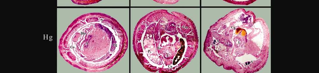
1 Chapter IX HISTOLOGICAL STUDIES OF EARTHWORMS ON THE TREATMENT OF INDUSTRIAL SLUDGE OVERVIEW Earthworms have been widely used in the breakdown of a wide range of industrial and organic waste including tannery, distillery sludge and poultry, municipal waste. The metabolic systems of earthworms transforming these waste into organic form as vermicompost and accumulate some heavy metals in their tissues, which cause damage or change in their organization and function of the same. Wide literature survey shows, only very few authors have undertaken research to assess the effect of vermicompost obtained from tannery and distillery sludge and their impact on histology of the animal (Filipek-Mazur et al., 2000). Therefore, the investigations aimed to study the effect of tannery and distillery sludge on the histology of the earthworms. Experiments were conducted in their direction and the change in the cell structure of the animal was explained with evidences of the cross section of the various regions of earthworms. INTRODUCTION Earthworms are regarded as one of the most suitable animals for testing the toxicity of chemicals in soils and have been adopted as standard organisms for eco toxicological testing. Acute and chronic toxicity tests have been used traditionally to asses the toxicity of contaminants, with mortality and changes in biomass, reproduction rates and behavioral responses representing endpoints. The uptake, accumulation, and elimination properties of metals by earthworm are the major part of toxicology, which is called toxicokinetics (Lee et al., 2008). However, limited works were carried out to assess the damages to tissues due to the effect of tannery and distillery sludge. So, the experiments were carried out to expose the changes to tissues with various regions and conditions Page 128
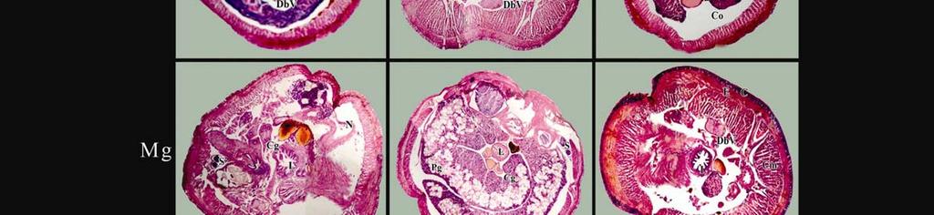
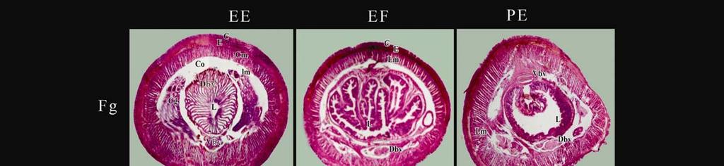
2 MATERIALS AND METHODS The histology of gut of earthworm was studied adopting the routine paraffin method (Humason, 1979).Gut of earthworm, dissected out from the control and experimental animals, were blotted free of mucus, washed thoroughly in physiological saline, cut into pieces of desired size and fixed in Bouins fluid fixative immediately after autopsy. Fixation was carried out at room temperature for 24 hr, after which the tissues were transferred to 70% alcohol. Several changes of 70% alcohol were given until the yellow colour disappeared from the tissues. The tissues were then dehydrated by passing through ascending grades of alcohol, cleared in xylene, infiltrated with molten paraffin, and finally embedded in paraffin wax (58 C MP). Tissue section of 5-μm thick transverse and longitudinal sections were obtained using a rotary microtome (Leica, Germany). The sections, thus obtained, were stained in Harris hematoxylene and eosin, dehydrated using alcohol, cleared in xylene and mounted using dihydroxy phthalate xylol (DPX). The stained slides were observed in a CarlZeiss (Germany) Axio-2 Plus research microscope (Chapter IV). RESULTS AND DISCUSSION The body wall consists of cuticle secreted by the epithelial cells, which underlines it. The epithelium has 4 types of cells. Most of the cells are tall columnar and epithelial cells, but between them are many mucus-secreting cells usually filled with granules (or) globules. In certain regions of the skin, sensory cells present some of which are photoreceptors, have a reticular cytoplasm containing a clear body, which seem to act as a lens. There found are short cells abundant on the prostomium and the first and last 2 (or) 3 segments others are small clusters of very tall, thin cells with minute sensory hairs. Under the epidermis is a layer of circular muscles and below this is much thicker layer of longitudinal muscles. The circular muscles contract to make the worm longer and thinner, the longitudinal to become short and stout. The pigment in the body wall is present in amoeboid cells in very young worms, but later only crowded granules can be found. Like hemoglobin and chlorophyll, this pigment, with a spectroscope, gives the absorption bands, which indicate that it is a porphyrin. The capacious coelom is lined by a peritoneal epithelium. It has two parts, the parietal peritoneum, which is a thin layer of flattened epithelial cells pressed against the Page 129
3 inner surface of the body wall, and the visceral peritoneum, which is pressed against the viscera, chiefly the intestine. While in a mammal, the visceral peritoneum is also of thin flattened cells, in earthworms it is composed of the very large chloragogen cells. The brown bodies often found with in the coelom of earthworms, especially in the hinder segments, are massed aggregations of these phagocytic cells and contain encysted nematodes, worm out setate, protozoan cysts and large amount of pigment granules form choragogen cells. In the coelomic fluid rounded cells called eleocytes present. These cells are bright yellow coloured and always filled with minute lipoid droplets. The adults of E. eugeniae, E. fetida and P.excavatus have a distinct swelling called clitellum. It is located about one third of the way down the earthworm. It produces most of the materials secreted to form earthworm cocoon. The clitellum forms a band that can be flared, non-flared, saddle shaped (or) annular. It is generally found between the segments The cuticle, epidermis, the circular muscles and longitudinal muscles are clearly on the left side. The ventral nerve cord, the dorsal and ventral blood vessel are clearly seen. The folding of the intestine the typlosole, the intestinal epithelium is definite. A large bright yellow tissue, which contains number of eleocytes with numerous fat droplets, is also seen. At the centre of the section is the intestine. The intestine is lined with ciliated epithelium. A significant structure, the typhlosole is present in the intestine. This is a fold of the dorsal wall, which projects into the intestine and performs several important functions; first, it slow down the passage of food so that, it may be acted upon for longer time by the digestive fluids; second, it increases the surface area for the secretion of digestive enzymes and their, it increase the area for absorption of the products of digestion. Typhlosole hangs down into the lumen of the intestine. The peritoneum covering the intestine and portions of the major blood vessels is modified in earthworm to form chloragogen layer. Waste materials accumulate in the chloragogen cells, which eventually separate from the intestional wall and float freely in the coelmic fluid. Some of these detached cells are ingested by the amoebocytes of the coelom, which interns migrate later to the superficial tissues, disintegrate and deposit their contents in the form of a protective pigment. Other chloragogen cells are swept into the ciliated funnel of the nephritic and thence of the outside. Chloragogen cells store glycogen, which is then distributed when there, cells break loose and disintegrate. Page 130
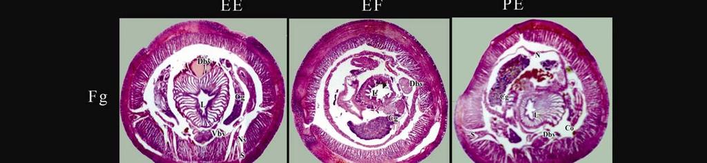
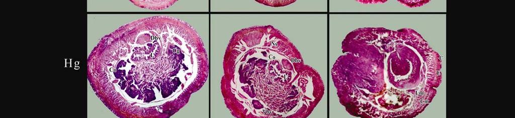
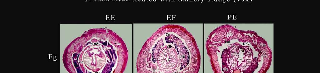
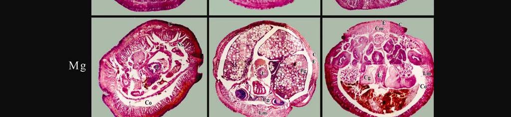
4 Histological studies of foregut, midgut and hindgut regions of the earthworm s E. eugeniae, E. fetida and P. excavatus treated with tannery sludge Foregut Histology of the tested fore gut reveals that the cuticle is untouched with the damage. Not much spoilage in the area of circular muscles but in some part of the longitudinal muscle can see the signs of injury. Detachment of peritoneum from longitudinal muscles can also be seen in some of the areas. The lumen of the intestine is not much harmed and the epithelial lining. The ventral nerve cord is damaged to a smaller extend. The dorsal and ventral blood vessel ruined largely. In brief, the 45% of the distillery sludge does not affect the tissues of earthworm much. Midgut In mid gut region of the cuticle and the epidermis are intact. The circular and longitudinal muscles are also unaffected. The peritoneum shows the signs of disconnect at several places. The typhlosole and the nephridiopore are clearly visible. The chlorachogen cells crumble to a considerable extent. The ventral nerve cord is not much damaged where as the dorsal blood vessel and ventral blood vessel have shown the signs of damage. The nephridia are feebly visible. Generally, the tissue of the test individuals were not much affected in this concentrations. Hindgut The cuticle of the earthworm hind gut region; under it, the epidermis is faintly visible. Light destruction is visible in some parts of circular muscles. However, in the longitudinal muscles more damage and fissures are seen in few areas. The peritoneum covering the intestine is injured largely. The intestinal epithelial lining, the lumen of the intestine are also damaged. Greater degree of injury to the dorsal and ventral blood vessel is observed. Ventral nerve cord is damaged beyond recognition. In short, the acute concentration of distillery sludge has a telling effect on the histology of the earthworms E. eugeniae E. fetida and P. excavatus. Page 131
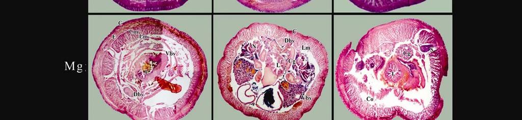
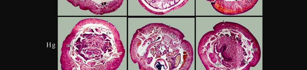
5 Histological studies of foregut, midgut and hindgut regions of the earthworm s E. eugeniae, E. fetida and P. excavatus treated with distillery sludge Foregut The cuticle is well distinct except breaks in one or two places. The epidermis is lightly damaged. The circular and longitudinal muscles are well formed and show no marks of damage. The ventral nerve cord and the ventral blood vessels are very much unaffected. Apart from regular cells, there is a large bright yellow body is de serving to be noticed which could be an eleocyte. Normally the eleocyte are filled with lipid droplets. The peritoneum is damaged in one or two places. The intestine wall and the epithelial lining of the intestine are damaged in several places. The nephridiopore is visible distinctly. The 45% distillery sludge has slightly affected the histology of earthworm. Midgut The cuticle is damaged in some areas. The epidermis has eroded completely in few areas. The circular muscle and longitudinal muscle are well developed in several places. The peritoneum is damaged in most part of the section. The ventral nerve cord and ventral blood vessels are visible clearly. The cholargogen cells are spread throughout the coelom. Accumulations of some foreign bodies are also seen in chloragogen cells. The intestine wall is severely damaged. In general, the effect of 56% concentration of distillery waste is much more visible than in other concentrations. Hindgut The cuticle is slightly damaged in several areas. The epidermis is also damaged largely. Even the circular muscles have shown the signs of damage in several areas. The longitudinal muscle has shown the signs of crakes in major parts of the regions. There is no sings of peritoneum wall at all. The intestinal epithelium is injured beyond recognition. The chlorocogen cells have damaged. Sings of accumulation of foreign bodies and vacuolization of coelom are observed. The ventral nerve cord is faintly visible, where as the dorsal and ventral blood vessel and the nephridia are seen faintly. In short, the clitellar region of 70% distillery sludge exposed earthworms is severally affected. Coelom of earthworms, especially in the hinder segments, are massed aggregations of these phagocytic cells and contain encysted nematodes, worn out Page 132
6 setate, protozoan cysts and large amount of pigment granules form choragogen cells. In the coelomic fluid rounded cells called eleocytes present. These cells are bright yellow colored and always filled with minute lipoid droplets. The feasibility of using earthworms for industrial waste management is dependent on the fundamental knowledge of basic parameters governing the survival and growth of earthworm species. The present set of investigation is aimed in this direction and the results are encouraging. Researchers reported that the passage of organics through the earthworm s gut significantly alters the physical structure of the material. Large particle are broken down into numerous smaller particles, with a resultant enormous increase in surface area. Soil pollution bio-indicators are essential to establish environmental standards. Earthworms and hypogenous micro arthropods were chosen to test ecological risks derived from soil amendment with Tannery and distillery sludge. The objective of culturing earthworms in tannery and distillery sludge revealed that the earthworm could live in the tannery and distillery waste. Investigation by other researchers in other waste products also have yielded encouraging results Gajalakshmi et al., (2001) reported the feasibility of vermi-composing as a viable process for the gainful utilization of industrial sludge in a environmentally clean manner. Pulp and paper industries, which releases waste water to about 330 m 2 /tonne of paper/day. This effluent contains toxic elements which inhibit the growth of the soil micro flora (Sastry et al., 1977; Oblisami and Palanisami, 1991; Sandana, 1995). The chemical immobilization is a relatively inexpensive in situ remediation method that reduce soil contaminant solubility, but the ability of this remediation treatment to reduce heavy metal bioavailability and eco-toxicity to soil invertebrates has not been evaluated Conder et al., (2001). Dominguez, (1997) reported that earthworms can dispose off about 35% to 55% of bio-available heavy metals by accumulating them in their tissues. In the present investigation the Eudrilus eugeniae, E.fetida and P.excavatus was subjected to various concentrations of tannery and distillery sludge. It has been observed that the sludge is toxic to earthworms observed only miner damages. The paradox of the industrial sludge having huge organic load and increased mortality could be solved when the miner damage is assessed histological studies. The histology of earthworm, which is subjected to various concentrations of tannery and distillery, show that increase in the concentration of sludge gradually increases the damages of the tissues. The soft peritoneal membrane lining intestinal epithelium is more affected. The Page 133
7 brown bodies in the coelom in the experimental groups are chloragogen cells. Chloragogen cells are the sites of accumulation of waste products is already been well established. The presences of yellow bodies, elecocytes where the fat droplets are accumulated like adipose tissues in higher organism are also the sites of accumulation of toxicants from the industrial sludge. The results and the discussions of the present investigation reconfirm the belief that there is no such thing as industrial wastes only wasted organics. Earthworm can be used to dispose off all sorts of industrial waste including tannery and distillery waste. The processing of waste by earthworm would help to reduce a major environmental problem, the mounting solid wastes. Page 134
8
9

10
Yara Saddam. Amr Alkhatib. Ihsan
 1 Yara Saddam Amr Alkhatib Ihsan NOTE: Yellow highlighting=correction/addition to the previous version of the sheet. Histology (micro anatomy) :- the study of tissues and how they are arranged into organs.
HOLE S ANATOMY CHAPTER 5, PART II Lecture notes
 HOLE S ANATOMY CHAPTER 5, PART II Lecture notes I. Connective Tissue A. Structure 1. have few cells that are spaced apart and can divide; two categories: a. fixed cells cells that are present in tissue
LABORATORY EXERCISE 4 PHYLUM PLATYHELMINTHES
 GENUS PLANARIA Planaria sp. Lab 4, pg 1 LABORATORY EXERCISE 4 PHYLUM PLATYHELMINTHES With a soft brush, place a live Planaria in a small dish with a few millimeters of pond water. BEHAVIOR. Examine the
Tissues. Tissues - Overview. Bio211 Laboratory 2. Epithelial and Connective Tissues
 Bio211 Laboratory 2 Epithelial and Connective Tissues 1 Tissues Tissues to be examined under the microscope Epithelial Tissue (p. 79 Lab Manual) [TODAY] Connective Tissue (p. 93 Lab Manual) [TODAY] Muscle/Nervous
Study of different tissues Abnormal cells and tissues can be compared to normal tissues to identify disease, such as cancer Being able to know and
 CHAPTER 4 Study of different tissues Abnormal cells and tissues can be compared to normal tissues to identify disease, such as cancer Being able to know and recognize normal tissues under the microscope
Zoology Exercise #10: Phylum Nematoda Lab Guide
 Zoology Exercise #10: Phylum Nematoda Lab Guide All animals with bilateral symmetry, except the acoelomates, have a body cavity. They are either true coelomates (where peritoneum covers both the inner
Tissues and organs PART 1
 Tissues and organs PART 1 Animals and plants are multicellular (made of many cells). Cells become specialised according to their function Tissues: Many cells that perform one or several functions; they
Biology Earthworm Dissection
 Biology 521 - Earthworm Dissection Kingdom Phylum Class Order Genus Species Animalia Annelida Oligochaeta Haplotaxida Lumbricus L. terrestris PRELAB: The earthworm is an excellent organism to study as
NOTES: CH 40 Introduction to Human Anatomy & Physiology
 NOTES: CH 40 Introduction to Human Anatomy & Physiology THE HUMAN BODY Anatomy Physiology (= structures) (= functions or processes) Characteristics of LIFE: 1) Made up of 1 or more CELLS. 2) Obtain and
Tissues. Tissues - Overview. Bio 101 Laboratory 3. Epithelial Tissues and Integument
 Bio 101 Laboratory 3 Epithelial Tissues and Integument 1 Tissues Tissues to be examined under the microscope Epithelial Tissue Integument Connective Tissue **We will be doing muscle and nervous tissues
Digestive System. The group of organs which performs the function of digestion constitute digestive system.
 Digestive System Definition:- The active biological process by which food materials impermeable to the cell membrane is converted into permeable to the cell membrane is called digestion. The group of organs
Tissues 10/21/2016. Epithelial Tissue
 Tissues This is a generalized cell diagram. It shows the anatomy of a cell, but most cells do not actually look like this. Cells can have a wide variety of shapes and sizes, depending on their function.
Anatomy & Histology of The Small intestine
 Anatomy & Histology of The Small intestine Prof. Abdulameer Al-Nuaimi E-mail: a.al-nuaimi@sheffield.ac.uk E. mail: abdulameerh@yahoo.com Jejunum Ileum Histology: Duodenum, jejunum, and ileum
Tissue Outline (chapter 4) Tissues group of cells that perform structural and roles. List the 4 types:
 Tissues group of cells that perform structural and roles. List the 4 types:") Tissue Outline (chapter 4) Tissues group of cells that perform structural and roles. List the 4 types: 1. 2. 3. 4. I. Epithelial Tissue covers all the surfaces, inside & out. Are the major tissues of,
Tissue Outline (chapter 4) Tissues group of cells that perform structural and roles. List the 4 types: 1. 2. 3. 4. I. Epithelial Tissue covers all the surfaces, inside & out. Are the major tissues of,
Lecture Overview. Marieb s Human Anatomy and Physiology. Chapter 4 Tissues: The Living Fabric Epithelial Tissues Lecture 9. Introduction to Tissues
 Marieb s Human Anatomy and Physiology Marieb Hoehn Chapter 4 Tissues: The Living Fabric Epithelial Tissues Lecture 9 Lecture Overview Introduction to Tissues Epithelial Tissues Location General characteristics
Phylum Platyhelminthes
 Phylum Platyhelminthes Class? Dugesia (planaria, non-parasitic flatworm) Class? Liver fluke Class? Tapeworm Phylum Platyhelminthes Class Turbellaria Dugesia (planaria, non-parasitic flatworm) Class Trematoda
Anatomy and Physiology Homework: Chapters 3-4
 Anatomy and Physiology Homework: Chapters 3-4 CHAPTER 3: Cells and Tissues 1. The smallest unit of living tissue is called a. All living organisms are composed of these basic units where all life processes
Lecture Overview. Chapter 4 Epithelial Tissues Lecture 9. Introduction to Tissues. Epithelial Tissues. Glandular Epithelium
 Visual Anatomy & Physiology First Edition Martini & Ober Chapter 4 Lecture 9 Lecture Overview Introduction to Tissues Location General characteristics Functions Classification Glandular Epithelium 2 Where
What is histology? HISTOLOGY
 Introduction to Histology What is histology? HISTOLOGY histo = tissue ogy = study So HISTOLOGY = the study of tissues! What is a TISSUE? Tissues are groups of cells with specialized structural and functional
(b) Stomach s function 1. Dilution of food materials 2. Acidification of food (absorption of dietary Fe in small intestine) 3. Partial chemical digest
 Stomach s function 1. Dilution of food materials 2. Acidification of food (absorption of dietary Fe in small intestine) 3. Partial chemical digest") (1) General features a) Stomach is widened portion of gut-tube: between tubular and spherical; Note arranged of smooth muscle tissue in muscularis externa. 1 (b) Stomach s function 1. Dilution of food
(1) General features a) Stomach is widened portion of gut-tube: between tubular and spherical; Note arranged of smooth muscle tissue in muscularis externa. 1 (b) Stomach s function 1. Dilution of food
KIDSPIRATION by Riedell
 WORM DISSECTION KIDSPIRATION by Riedell CLASSIFICATION Kingdom: Phylum: ANIMALIA Annelida little rings Class: OLIGOCHAETA few bristles SETA (plural: setae) BRISTLES on VENTRAL surface http://www.pgjr.alpine.k12.ut.us/science/whitaker/animal_kingdom/earthworm/earthworm.html
31-2. The Earthworm. . Relate the structure of systems. . Demonstrate dissection technique. . Identifythe major advancesof
 Name Class Date INVESTIGATION 31-2 The Earthworm Introduction The earthworm is a segmented worm. It exhibits more complex structures than any of the more primitive animals that you have studied thus far.
B17 instructions for 227. April 15, 2011
 Microviewer 227: Comparative Digestive Systems Introduction This set is one of a series of lessons examining comparative life function systems. In these sets, you will examine slides of different animals,
General Structure of Digestive Tract
 Dr. Nabil Khouri General Structure of Digestive Tract Common Characteristics: Hollow tube composed of a lumen whose diameter varies. Surrounded by a wall made up of 4 principal layers: Mucosa Epithelial
The Anatomy of the Earthworm
 Carolina Biological Supply Company presents The Anatomy of the Earthworm 2700 York Road Box 187 Burlington, North Carolina 27215 Gladstone, Oregon 97027 Abstract. This program facilitates a study of the
The Gastrointestinal Helminth Infection among Backyard Fowl Population of Selected areas of North 24 Parganas, West Bengal
 Biological Forum An International Journal 8(2): 181-18(216) ISSN (Print): 97-11 ISSN (Online): 2249-29 The Gastrointestinal Helminth Infection among Backyard Fowl Population Selected areas North 24 Parganas,
Tissues. tissue = many cells w/ same structure and function. cell shape aids its function tissue shape aids its function
 Tissues tissue = many cells w/ same structure and function cell shape aids its function tissue shape aids its function Histology = study of tissues 4 types of tissues Epithelial coverings contact openings
DIGESTIVE TRACT ESOPHAGUS
 DIGESTIVE TRACT From the lower esophagus to the lower rectum four fundamental layers comprise the wall of the digestive tube: mucosa, submucosa, muscularis propria (externa), and adventitia or serosa (see
CYTOMORPHOLOGY MODULE 28.1 INTRODUCTION OBJECTIVES 28.2 GENERAL GUIDELINES. Notes
 28 CYTOMORPHOLOGY 28.1 INTRODUCTION Light microscopic examination of stained cells in smears is the method of choice of diagnostic cytology. It allows classification of most normal cells as to type and
STRUCTURAL ORGANSTAION
 STRUCTURAL ORGANSTAION IN ANIMALS ANIMAL TISSUES :- Are Classified in to four types 1.Epithelal Tissues 2.Connective Tissues 3.Muscular Tissues 4.Neural Tissues Vikasana - CET 2012 1.Epithelial Tissues
THE TISSUE LEVEL OF ORGANIZATION PART I: EPITHELIAL TISSUE
 THE TISSUE LEVEL OF ORGANIZATION PART I: EPITHELIAL TISSUE 4 Main Tissue Types Epithelium Covers surfaces, lines cavities, forms glands Connective Tissue Support and protects body Muscular Tissue Movement
Small intestine. Small intestine
 General features Tubular organ longest part; 5-6 m most of chemical digestion absorption of nutrients reabsorption of H2O occurs. Two structural features; maximize the lumenal surface area villi microvilli
Science 8 - Cells & Cell Organization Notes
 Science 8 - Cells & Cell Organization Notes 1.1 - Characteristics of Living Things Composed of cells Reproduce, grow & repair themselves Require energy o Plants usually from sun o Animals from plants or
Tissues Chapter 5...Tissue - a group or mass of similar cells working together to perform certain common functions
 Tissues Chapter 5...Tissue - a group or mass of similar cells working together to perform certain common functions There are 4 major types of tissue Epithelial Connective Muscle Nervous 1. Epithelial Tissue
B. Incorrect! The ectoderm does not produce the dermis. C. Incorrect! The dermis is derived from the mesoderm.
 Human Anatomy - Problem Drill 04: The Integumentary System Question No. 1 of 10 Instructions: (1) Read the problem and answer choices carefully, (2) Work the problems on paper as 1. From the inner cell
Prepared By Student. Dania Abed Al-majeed. Rahma Raad Hanna. Balqees Mohammed Aasim. Dania Hisham. Rasha Rafiee
 Prepared By Student Rahma Raad Hanna Balqees Mohammed Aasim Dania Hisham Dania Abed Al-majeed Rasha Rafiee Epithelia Epithelia can be derived from ectoderm, mesoderm or endoderm -ectoderm gives rise to
ABCD rule. apocrine glands. arrector pili. ceruminous glands. contact dermatitis
 ABCD rule assessing moles: asymmetric, broder irregularity, color, diameter (larger than 6mm) apocrine glands arrector pili sweat glands in the pubic and underarm areas that secrete thicker sweat, that
Exercise 6. Procedure
 Exercise 6 Procedure Growing of root tips Select a few medium-sized onion bulbs. Carefully remove the dry roots present. Grow root tips by placing the bulbs on glass tubes (of about 3 4 cm. diameter) filled
Urinary System Laboratory
 Urinary System Laboratory 1 Adrenal gland Organs of The Urinary System Renal artery and vein Kidney Ureter Urinary bladder Figure 26.1 2 Urethra Functions of the urinary system organs: Urethra expels urine
Epithelium tissue system
 Epithelium tissue system Histology : is the study of the microscopic anatomy (microanatomy) of cells and tissues of plants and animals. It is commonly performed by examining cells and tissues under a light
A adipose cells. B capillary. C epithelium
 EPITHELIA Objective The objective of this class is to observe how different epithelia vary in terms of cell shape, size and number of cell layers enabling them to be well adapted for functions in different
Body Tissues Pearson Education, Inc.
 Body Tissues Tissues Groups of cells with similar structure and function Four primary types: Epithelial tissue (epithelium).1 Connective tissue.2 Muscle tissue.3 Nervous tissue.4 Epithelial Tissues Locations:
Lab Animal Tissue. LEARNING OBJECTIVES: To understand the relationship between the structure and function of different animal tissues
 Name: Bio A.P. PURPOSE: HYPOTHESIS: NONE Lab Animal Tissue BACKGROUND: In animals, groups of closely related cells specialized to perform the same function are called tissues. There are four general classes
Skin and Body Membranes Body Membranes Function of body membranes Cover body surfaces Line body cavities Form protective sheets around organs
 Skin and Body Membranes Body Membranes Function of body membranes Cover body surfaces Line body cavities Form protective sheets around organs Classification of Body Membranes Epithelial membranes Cutaneous
Urinary system. Urinary system
 Distal convoluted tubule (DCT) Highly coiled, ~ 5 mm in length Last part of the nephron. Wall; simple cuboidal epithelium Less metabolically active than the PCT no brush border light eosinophilic cytoplasm
Dr. Heba Kalbouneh. Dr. Heba Kalbouneh. Dr. Heba Kalbouneh
 Dr. Heba Kalbouneh Dr. Heba Kalbouneh Dr. Heba Kalbouneh Tissue: is a group of cells that serve the same function, they are surrounded by extra cellular matrix. The 4 basic types of tissue: 1. epithelial
Tissue: The Living Fabric: Part A
 PowerPoint Lecture Slides prepared by Janice Meeking, Mount Royal College C H A P T E R 4 Tissue: The Living Fabric: Part A Tissues Groups of cells similar in structure and function Types of tissues Epithelial
Cell and Tissue Types. Epithelial, Connective, Muscle, Nerve
 Cell and Tissue Types Epithelial, Connective, Muscle, Nerve Objectives Explain the major stages of the cell cycle and cellular division (mitosis). Describe specific events occurring in each of the phases
Figure Nutrition: omnivore, herbivore, carnivore
 Figure 41.1 Nutrition: omnivore, herbivore, carnivore Essential Nutrients: Amino acids Fatty acids Vitamins Minerals Figure 41.2 Complete vs incomplete Omnivore vs herbivore (vegetarian) Table 41.1 Table
Bio & 241 A&P Unit 1 / Lecture 3
 Bio & 241 A&P Unit 1 / Lecture 3 Tissues All body tissues arise from three fundamental embryonic tissues. Endoderm: forms epithelial tissues lining internal organs such as the GI tract Mesoderm: connective
BI 121 LAB. WEEK 2: Tissues (continued); Integumentary System
; Integumentary System") BI 121 LAB 2-1 WEEK 2: Tissues (continued); Integumentary System This week you will 1) Review the four major tissue types 2) Review the characteristics of epithelial tissues. 3) Learn the major characteristics
BI 121 LAB 2-1 WEEK 2: Tissues (continued); Integumentary System This week you will 1) Review the four major tissue types 2) Review the characteristics of epithelial tissues. 3) Learn the major characteristics
Outline. Bio 105: Tissues Laboratory. Organization of the Human Body. Tissue - Epithelium. Tissues 3/2/ Copyright 2009 Pearson Education, Inc
 Outline Bio 105: Tissues Laboratory Laboratory 5 Reading: Chapter 4 I. Cell to cell contact II. Body Cavities III. Membranes IV. Homeostasis V. Integumentary System I. Includes skin, hair and nails 1 2
Class XI Chapter 7 Structural Organisation in Animals Biology
 Question 1: in one word or one line. (i) Give the common name of Periplaneta americana. (ii) How many spermathecae are found in earthworm? (iii) What is the position of ovaries in the cockroach? (iv) How
Class XI Chapter 7 Structural Organisation in Animals Biology
 Question 1: in one word or one line. (i) Give the common name of Periplaneta americana. (ii) How many spermathecae are found in earthworm? (iii) What is the position of ovaries in the cockroach? (iv) How
Lower Secondary Science Blood Circulatory System Notes / Advanced Notes
 Lower Secondary Science Blood Circulatory System Notes / Advanced Notes Double Circulation in Mammals In mammals, there is a double circulation (i.e. blood passes through the heart twice in one complete
Excretion and Water Balance
 Excretion and Water Balance In the body, water is found in three areas, or compartments: Plasma, the liquid portion of the blood without the blood cells, makes up about 7 percent of body fluid. The intercellular
HISTOLOGY. Simple squamal lungs
 HISTOLOGY Lab Objectives: Students should be able to... 1. Visually identify each class of tissue and examples within each class 2. Indicate the location (in the human body and/or organ) and function of
A Single Neuron from the Brain
 Nervous Tissue A Single Neuron from the Brain Dendrites Cell Body Axon Nerve cells, called neurons, transmit signals throughout our bodies. These signals tell our bodies what to do. Dendrites transmit
Lysosomes. Gr: lysis solution, soma body. Membrane bounded vesicles. Usually round ovoid or irregular electron dense bodies m.
 Lysosomes Gr: lysis solution, soma body Membrane bounded vesicles Usually round ovoid or irregular electron dense bodies 0.05 0.5 m. Lysosomes No. varies from a few to several hundred per cell, in different
Sphincters heartburn diaphragm The Stomach gastric glands pepsin, chyme The Small Intestine 1-Digestion Is Completed in the Small Intestine duodenum
 Sphincters are muscles that encircle tubes and act as valves. The tubes close when the sphincters contract and they open when the sphincters relax. When food or saliva is swallowed, the sphincter relaxes
BIO Lab 18: Dissection of the Earthworm
 The Earthworm Harken to me, you that know what is just, my people who have My law in their heart: Fear not the reproach of men and be not afraid of their blasphemies. For the worm shall eat them up as
HISTOLOGY VIRTUAL LABORATORY GASTROINTESTINAL SYSTEM
 HISTOLOGY VIRTUAL LABORATORY GASTROINTESTINAL SYSTEM LIP (Slides GI 1, 2) Identify the outer portion lined by stratified squamous (keratinized) epithelium. Note the hair follicles and sebaceous glands
Alimentary Canal (I)
") Alimentary Canal (I) Esophagus and Stomach (Objectives) By the end of this lecture, the student should be able to discuss the microscopic structure in correlation with the function of the following organs:
Alimentary Canal (I) Esophagus and Stomach (Objectives) By the end of this lecture, the student should be able to discuss the microscopic structure in correlation with the function of the following organs:
Epithelial Tissues. Types of Epithelial Tissues: Lining of Kidney
 Epithelial Tissues Covers the entire body surface and most of the body s inner cavities Outer epidermis (skin) protects from injury and drying out Inner epidermal tissue (on internal surfaces) often serves
Dr Narmeen S. Ahmad. Lab 1
 Dr Narmeen S. Ahmad Lab 1 1 Tissues are groups of cells with a common structure (form) and function (job). There are (4) types of tissue: 1. Epithelial 2. Connective 3. Muscle 4. Nervous 2 Epithelial cells
ANATOMY & PHYSIOLOGY ONLINE COURSE - SESSION 13 THE DIGESTIVE SYSTEM
 ANATOMY & PHYSIOLOGY ONLINE COURSE - SESSION 13 THE DIGESTIVE SYSTEM The digestive system also known as the alimentary canal or gastrointestinal tract consists of a series of hollow organs joined in a
They cells can not function death.
 Jenna Hellack Jan 2001 Tissues What do you think happens when the cells use up their food and oxygen before there is time to replenish it? They cells can not function death. Blood Cell Cancer cell Plant
Tissues. tissue = many cells w/ same structure and function. cell shape aids function tissue shape aids function. Histology = study of tissues
 Tissues tissue = many cells w/ same structure and function cell shape aids function tissue shape aids function Histology = study of tissues 4 types of tissues Epithelial coverings contact openings Connective
Histology 101! !! Name:! Block: Identify and describe the functions of major tissue types including their subclasses and varieties!
 Histology 101 Identify and describe the functions of major tissue types including their subclasses and varieties Name: Block: "1 Introduction to Tissues Histology Notes Tissue (living fabric) : groups
Organisation. AQA Biology topic 2
 Organisation AQA Biology topic 2 2.1 Principles of Organisation Cells, tissues, organs and systems Basically, all living things are made up of cells A group of CELLS makes up a TISSUE A group of TISSUES
Body Tissues. Cells are specialized for particular functions Tissues - groups of cells with similar structure. and function Four primary tissue types:
 Chapter 3 Tissues Body Tissues Cells are specialized for particular functions Tissues - groups of cells with similar structure and function Four primary tissue types: Epithelium Connective tissue Nervous
Ch 7 Nutrition in humans
 Ch 7 Nutrition in humans Think about (Ch 7, p.2) 1. The stomach churns food into smaller pieces physically. The stomach wall secretes proteases to chemically digest proteins. It also releases hydrochloric
The Human Body: An Overview of Anatomy. Anatomy. Physiology. Anatomy - Study of internal and external body structures
 C H A P T E R 1 The Human Body: An Orientation An Overview of Anatomy Anatomy The study of the structure of the human body Physiology The study of body function Anatomy - Study of internal and external
PowerPoint Lecture Slide Presentation by Patty Bostwick-Taylor, Florence-Darlington Technical College Skin and Body Membranes
 PowerPoint Lecture Slide Presentation by Patty Bostwick-Taylor, Florence-Darlington Technical College Skin and Body Membranes 4 Body Membranes Function of body membranes Cover body surfaces Line body cavities
Toxicology. Toxicity. Human Health Concerns. Health Effects of Hazardous Materials
 Human Health Concerns Health Effects of Hazardous Materials Toxicology Study of the nature, effects, and detection of poisons in organisms Humans are obvious focal point Other species and ecosystem function
MCAT Biology Problem Drill 20: The Digestive System
 MCAT Biology Problem Drill 20: The Digestive System Question No. 1 of 10 Question 1. During the oral phase of swallowing,. Question #01 A. Initially, the food bolus is moved to the back of the tongue and
Internal Morphology. 1.Cut the legs and wings (if present) off your specimen. 5.Use forceps to pull skeleton apart, exposing internal systems.
 off your specimen. 5.Use forceps to pull skeleton apart, exposing internal systems.") Internal Morphology Insect Dissections Often the best approach to understanding internal morphology is by way of a dissection. For this reason, the entire chapter should be treated as a laboratory activity.
Internal Morphology Insect Dissections Often the best approach to understanding internal morphology is by way of a dissection. For this reason, the entire chapter should be treated as a laboratory activity.
Epithelial Tissue lining, covering, glandular tissue > Function protect, absorption, filtration, secretion, excretion
 Chapter 4: TISSUES IX. Tissues Intro Epithelial Tissue lining, covering, glandular tissue > Function protect, absorption, filtration, secretion, excretion Connective Tissue most widespread tissue type
Tissues. Connective Tissue. Bio 101 Laboratory 4. Connective Tissues
 Bio 101 Laboratory 4 Connective Tissues 1 Tissues Tissues to be examined under the microscope Connective Tissue Refer to your Marieb s Lab Manual for pictures of tissues/guidance Lab Guide (handout) for
Nervous system (blue) Insect Internal Systems and Physiology. Decentralized nervous system. Synapse gap 8/22/2012
 Insect Internal Systems and Physiology. Decentralized nervous system. Synapse gap 8/22/2012") Nervous system (blue) Insect Internal Systems and Physiology Decentralized nervous system Brain (left) several lobes Ventral nerve cord Ganglia in many segments masses of nerve cells Synapse gap Synapse
Nervous system (blue) Insect Internal Systems and Physiology Decentralized nervous system Brain (left) several lobes Ventral nerve cord Ganglia in many segments masses of nerve cells Synapse gap Synapse
Why are cells shaped the way they are?
 Why are cells shaped the way they are? # 1 Cheek Cells These cells were gently scraped from the inner surface of a person s cheek, and placed on a microscope slide. The cheek lining cells are thin and
The Digestive System. Chapter 25
 The Digestive System Chapter 25 Introduction Structure of the digestive system A tube that extends from mouth to anus Accessory organs are attached Functions include Ingestion Movement Digestion Absorption
An overview of the digestive system. mouth pharynx esophagus stomach small intestine large intestine rectum anus
 An overview of the digestive system mouth pharynx esophagus stomach small intestine large intestine rectum anus Why GIT? What are the main steps in the digestive process? Ingestion intake of food via the
Histology Notes -Part 1: Epithelial Tissues
 Introduction Group of cells w/ similar structure & function = TISSUE Four Basic Tissue Types 1. Epithelial-covers 2. Connective-supports 3. Muscular*-produces movement (will discuss in the muscular system
Unit I Problem 9 Histology: Basic Tissues of The Body
 Unit I Problem 9 Histology: Basic Tissues of The Body - What is the difference between cytology and histology? Cytology: it is the study of the structure and functions of cells and their contents. Histology:
Lesson 9A Tissues in Animals
 Lesson 9A Tissues in Animals Levels of Organization in the Human Body Similar types of cells Different types of tissues Different organs Many organ systems cell tissue organ organ system organism Levels
Histology Urinary system
 Histology Urinary system Urinary system Composed of two kidneys, two ureters, the urinary bladder, and the urethra, the urinary system plays a critical role in: 1- Blood filtration,(filtration of cellular
Chapter 4 The Integumentary System and Body Membranes. HAP Susan Chabot Lemon Bay High School
 Chapter 4 The Integumentary System and Body Membranes HAP Susan Chabot Lemon Bay High School Classification of Body Membranes Epithelial Membranes Cutaneous Membranes = The Skin Mucous Membranes Serous
Lab 1 ANIMAL TISSUES
 Lab 1 ANIMAL TISSUES Levels of Organization Animals are multicellular heterotrophs whose cells lack cell walls. Most animals exhibit a hierarchical level of organization: Cells are organized into tissues
Histology of the myocardium and blood vessels. Prof. Abdulameer Al-Nuaimi
 Histology of the myocardium and blood vessels Prof. Abdulameer Al-Nuaimi E-mail: a.al-nuaimi@sheffield.ac.uk E-mail: abdulameerh@yahoo.com Histology of blood vessels The walls of arteries and veins are
Cell Theory Vocabulary Flashcards
 Mr. Powner Biology Cell Theory Vocabulary Flashcards Instructions: Cut out the flashcards from the following pages. The following word list is the vocabulary for studying cell theory. Write each word on
Tissues. Group of cells that are similar in structure and function. 4 primary types. Epithelium (covering) Connective (support) Nervous(control)
 Connective (support) Nervous(control)") Tissues Tissues Group of cells that are similar in structure and function 4 primary types Epithelium (covering) Connective (support) Nervous(control) Epithelial tissue (epithelium) Lining, covering, and
Tissues Tissues Group of cells that are similar in structure and function 4 primary types Epithelium (covering) Connective (support) Nervous(control) Epithelial tissue (epithelium) Lining, covering, and
ULTRASTRUCTURAL CHANGES IN THE INFECTIVE LARVAE OF NIPPOSTRONGYLUS BRASILIENSIS IN THE SKIN OF IMMUNE MICE
 ULTRASTRUCTURAL CHANGES IN THE INFECTIVE LARVAE OF NIPPOSTRONGYLUS BRASILIENSIS IN THE SKIN OF IMMUNE MICE by D. L. Lee ABSTRACT Infective stage larvae of Nippostrongylus brasiliensis are immobilized within
Urinary Anatomy. Lab 40. Kidneys. Nephrons. Renal Corpuscle
 Urinary Anatomy Lab 40. Urinary Anatomy and Kidney Dissection Kidneys: filters blood, produces urine Ureters: convey urine to bladder Bladder: holding tank Urethra: carries urine to the outside for elimination
Morphological characters of the pheretimoid earthworms in North America north of Mexico
 Morphological characters of the pheretimoid earthworms in North America north of Mexico This document is modified from the following paper: Chang, C.-H., Snyder, B., Szlavecz K. (2016) Asian pheretimoid
1. Three Main Functions. Chapter 19: 2. Two Groups of digestive organs. 2. Two Groups of digestive organs 6/1/2015. The Wall of the Digestive Tract
 1. Three Main Functions Chapter 19: General Structure and Function of the Digestive System Digestion-breakdown of food into small particles for transport to blood Absorption- into bloodstream to take to
Lec #2 histology. Bronchioles:
 Lec #2 histology. Last lecture we talked about the upper respiratory tract histology, this one is about the lower part histology. We will discuss the histology of: -bronchioles -respiratory bronchioles
Ingestion Digestion- Absorption- Elimination
 DIGESTIVE SYSTEM 1 FUNCTIONS Organization GI tract==mouth anus Accessory organs Salivary glands, liver, pancreas, gallbladder Major Functions: Ingestion-mouth, teeth, tongue Digestion- chemical and mechanical
Animal Nutrition. Chapter 41. Biology Eighth Edition Neil Campbell and Jane Reece. PowerPoint Lecture Presentations for
 Chapter 41 Animal Nutrition PowerPoint Lecture Presentations for Biology Eighth Edition Neil Campbell and Jane Reece Lectures by Chris Romero, updated by Erin Barley with contributions from Joan Sharp
LESSON ASSIGNMENT. The Human Integumentary and Fascial Systems. After completing this lesson, you should be able to:
 LESSON ASSIGNMENT LESSON 3 The Human Integumentary and Fascial Systems. TEXT ASSIGNMENT Paragraphs 3-1 through 3-14. LESSON OBJECTIVES After completing this lesson, you should be able to: 3-1. Define integumentary