CT Imaging at the Point-of-Care
|
|
|
- Delilah Sherman
- 5 years ago
- Views:
Transcription


1 ENGLISH


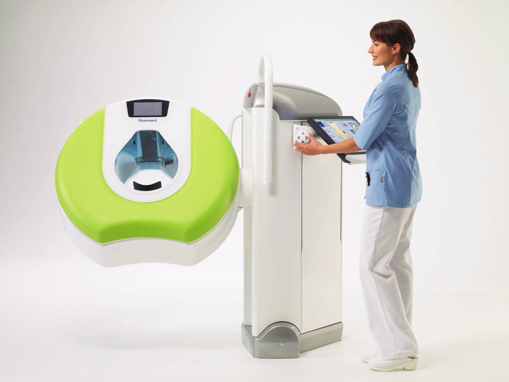
2 True Dedication The new Planmed Verity Extremity CT Scanner revolutionizes extremity CT imaging. The compact unit brings 3D imaging at emergency departments, orthopedic clinics or trauma centers for fast diagnoses at the Point-of-Care. Superior image quality serves radiologists, orthopedists, and extremity specialists all alike. With surprisingly low radiation dose of only one tenth of that of conventional CT, Planmed Verity helps to find subtle extremity fractures at the first visit to the clinic. 2 3


 or")
 is required.")


 technology to")
 images of the")


3 CT Imaging at the Point-of-Care Need for dedicated orthopedic imaging The most commonly missed fractures are within the extremities. Initial diagnosis is typically based on plain 2D radiographs obtained on the day of the patient s visit to the orthopedic clinic. Very often, however, more detailed assessment with Computed Tomography (CT) or Magnetic Resonance Imaging (MRI) is required. Unfortunately these imaging modalities may not be readily available at the time of the visit and important diagnostic information will be missed when it is most valuable for the patient s care. Introducing Planmed Verity Extremity CT Scanner Planmed Verity utilizes CBCT (Cone Beam Computed Tomography) technology to provide high resolution volumetric (3D) images of the extremities at a particularly low dose. The unit is designed for extremity CT imaging at the Point-of-Care in emergency departments, orthopedic clinics and trauma centers. Typical users are radiologists, orthopedists, and extremity specialists such as hand surgeons and podiatrists. Less missed fractures with Planmed Verity Overlapping structures seriously limit the visibility of the subtle fracture line in 2D radiographs. In the worst case, this leads to patient suffering for months and repeated radiographs. Not only has the healing process been delayed or completely stopped but the patient has been exposed to unnecessary radiation dose. Planmed Verity Extremity CT Scanner is designed to find subtle and occult fractures at the first visit to the imaging facility. The system features specialized functions and tools for extremity imaging that provide optimal imaging technique for different targets and purposes. 4 5


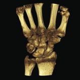

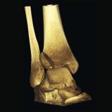
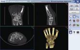
4 Detailed diagnostics with 3D imaging High image quality with low dose CBCT technology with flat panel detector enables the use of significantly lower radiation dose compared to extremity imaging with conventional MDCT. For the Planmed Verity Extremity CT Scanner, dose level is similar to repeated plain radiographs that are often needed for optimal visualization of subtle fractures. On the other hand, the exposure from Planmed Verity examination is comparable to one chest X-ray examination, or less than one week of background radiation. Even though Planmed Verity Extremity CT Scanner utilizes remarkably low level of radiation, the image quality is equal or even superior to the expensive MDCT units. Depending on the imaging protocol, isotropic resolution of up to 0.2 mm (optional 0.1 mm high resolution) is available. Volumetric imaging with multi-planar reconstruction (MPR) and surface rendering provide optimal visualization, without structure overlap. Optimal diagnostics and treatment planning With Planmed Verity, the technologist can achieve perfect quality without retakes. Also the radiologist can rely on the volumetric image dataset and focus on diagnostic work. This will significantly help in decision making process and reduce oversight errors. The 3D visualization capabilities of Planmed Verity provide the physicians with multiple possibilities for diagnosis and planning of the care and possible surgical intervention. MPR and surface renderings offer multiple choices for image display and excellent metal artefact removal algorithm ensures visibility of even the finest details of complex metal implants. 6 7


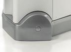


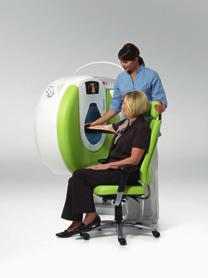
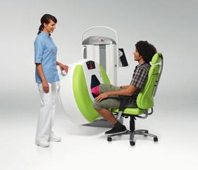
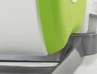
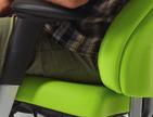
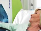
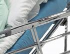
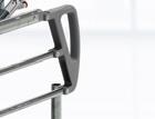
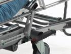
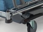
5 Adaptable CT Extremity imaging using MDCT can be challenging. Very often, patients have limited mobility and can not be positioned easily on the examination table. Furthermore, awkward posture is needed when imaging e.g. elbow to avoid unnecessary X-ray exposure of sensitive organs. Adaptable gantry with soft surface Planmed Verity Extremity CT Scanner provides motorized gantry with adjustable height and tilt for the best possible extremity positioning. Dedicated, carbon fiber positioning trays ensure that the target is always perfectly positioned in the field of view (FOV). During the imaging, the patient can lean on a comfortable, soft gantry that reduces discomfort and thereby motion artefacts. Planmed Verity can always be positioned in a way that is the most convenient for the patient. The versatile positioning also enables imaging for instance directly on a hospital bed making the imaging event fast and simple for both the technologist and the patient. 8 9


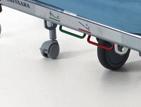


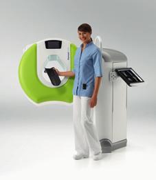
6 Easy siting The possibility to move a dedicated extremity CT scanner is a truly unforeseen feature. In the optional mobile configuration, Planmed Verity can be sited to virtually any X-ray room for example right next to the existing plain radiography Bucky table. If the room space is of concern, the compact Planmed Verity can be moved to a storage position when not in use. It will only take a moment to reactivate the unit when needed. When installing the unit, no extensive room preparation or external cooling systems are required. The stand-alone unit plugs in to a regular power outlet and connects to the information system through a standard Ethernet connection. The mobile Planmed Verity is an excellent choice for example for facilities with two adjacent X-ray rooms as the unit can be switched between rooms when general X-ray system is being serviced


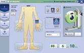
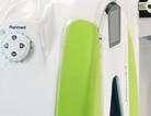
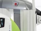
7 Verity Manager Software Dedicated positioning system Specially designed positioning trays and optional positioning camera ensure that the target is always in the center of the field of view. The carbon fiber trays also help in achieving correct anatomical orientation for fracture visualization, and provide optimal image quality for CBCT imaging. Ingenious control interface Planmed Verity is equipped with ingenious, single-handed control system for gantry and tray movements. The user can easily and precisely operate the device while positioning the patient. Optimal target visibility With extended, TearDrop -shaped bore, Planmed Verity offers excellent target visibility and access, and less anxiety and claustrophobia for the patient. The imaging volume is indicated with red and green lasers. Optionally, video camera and multifunctional info screen on the gantry can be used for positioning. Touch-screen optimized workflow The user is guided through the imaging procedure by an intuitive, touch screen optimized user interface. User configurable pre-set imaging programs guarantee fluent workflow and easy operation. Multiple, touch-screen optimized software tools are available for image processing, image stack definition, surface rendering, and more. The Verity touch-screen has adjustable height and tilt for optimal working ergonomics. Connectivity Communication to hospital imaging network is easy with the Verity Manager software. The software has built in worklist management and image transfer protocols for communication with Hospital Information Systems (HIS), Radiology Information System (RIS) and Picture Archiving and Communication System (PACS). Verity Manager is compliant with Digital Imaging and Communications in Medicine (DICOM)


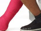
8 Advanced Extremity CT Weight-bearing CT One of the most intriguing new applications of Planmed Verity Extremity CT Scanner is weight-bearing option in which the patient stands inside the gantry during image acquisition. Weight-bearing imaging of the ankle or knee shows the anatomy under natural load. With this new imaging application many of the previously unanswered questions can be addressed. Soft-surface gantry Correct patient positioning is the key to successful imaging. For patients with fractures it may be difficult to remain still during imaging. The soft gantry surface is one of the features that help the patient to find and maintain comfortable position. Nice addition to this element is the possibility to select a color that speaks the right language. The strong, vivid color can also help the patient to relax during image acquisition


9 Enhanced digital imaging Fast 3D image reconstruction With Planmed Verity Extremity CT Scanner 3D reconstruction is readily available at the unit within couple of minutes after image acquisition. During the reconstruction process, proprietary image processing algorithms fine-tune the image for optimal presentation of clinically relevant data, which is then available for DICOM transfer to PACS and radiologist workstation. Extended volume It is also possible to extend the imaging volume if needed. This optionally available, so called stitching algorithm automatically combines two adjacent images into one volume. This is useful for visualizing unusually long fractures or other targets requiring extended imaging volume. Artefact removal Planmed Verity s advanced artefact removal algorithm has been developed to provide optimal image presentation even in challenging post-operative imaging studies. The unit can display detailed bone structure all the way to the very vicinity of the metal-bone interface, which is invaluable for fracture and joint surface diagnostics. Future applications For Planmed Verity and CBCT technology, we can find many new, intriguing uses. For instance, bone healing process can be visualized in a novel way without removing the cast. It is also possible to use contrast media for arthrography or other specialized imaging technique. These are just some examples of many possible future applications for the Planmed Verity Extremity CT Scanner. With the low dose and easy accessibility combined to the high quality clinical images, Planmed Verity will revolutionize orthopedic extremity imaging practices


10 Planmed Verity Lilac Planmed Verity Dark Blue Planmed Verity Mint Planmed Verity Sahara Yellow Planmed Verity Lime World of Planmed Verity 18 19


11 Planmed Oy develops, manufactures and markets advanced imaging equipment and accessories for mammography and orthopedic imaging. Planmed s extensive mammography product range covers digital and analog units, stereotactic biopsy devices, and breast positioning systems for an early detection of breast cancer. Within orthopedic 3D imaging Planmed offers low dose extremity CT imaging for quicker, easier and more accurate diagnosis at the Point-of-Care. Planmed Oy Asentajankatu Helsinki Finland tel fax sales@planmed.com Planmed Verity is pending FDA market clearance. Images may contain optional items not included in the standard delivery. Rights for changes reserved /0211/en
ENGLISH. Digital Mammography
 ENGLISH Digital Mammography Medical imaging excellence Planmed is strongly committed to improving the early detection of breast cancer. Our mission is to provide top quality imaging systems for mammographic
Dedicated to Mammography
 ENGLISH True Dedication As the number of women diagnosed with breast cancer increases every year, more women are surviving cancer and are able to carry on living their everyday lives. Thanks to screening
Dedicated to Digital Mammography
 ENGLISH True Dedication Although the number of breast cancers diagnosed increases each year more women than ever before are surviving. Higher survival rates are closely linked to earlier detection and
Dental Line. 3D digital panoramic system. radiology ahead
 Dental Line 3D digital panoramic system radiology ahead new generation 3D digital panoramic unit 3D imaging s value available for anyone Following the incredible success of the innovative digital panoramic
Planmeca ProMax 3D s Planmeca ProMax 3D ENGLISH
 Planmeca ProMax 3D s Planmeca ProMax 3D ENGLISH Genuine all-in-one unit Planmeca ProMax 3D s and Planmeca ProMax 3D units are designed to obtain complete information on patient anatomy in the minutest
WITH. The Next Step in Office MRI
 WITH The Next Step in Office MRI Introducing S-scan the Next Step in Office MRI Based on extensive customer feedback and years of engineering, Esaote has designed the S-scan with exp Technology, an optimized
JiangSu Magspin Instrument Co., Ltd
 Page 1 of 5 Magspin ARMOUS - 0.25T Dedicated Open Extremity MRI System The Beginning of a New Era in Extremity MR Imaging Superior Sitting Position MR Imaging ARMOUS is the long awaited Dedicated Open
OnTarget. OnTime. OnBudget. On-Site Extremity CT Exams Deliver Clinical Excellence and Superb Productivity.
 OnSight 3D Extremity System OnTarget. OnTime. OnBudget. On-Site Extremity CT Exams Deliver Clinical Excellence and Superb Productivity. SMART IMAGING SOLUTIONS A Great Source of Diagnostic Insight Can
The value of weight-bearing functional CT scans
 The value of weight-bearing functional scans In musculoskeletal medicine, advanced imaging like computed axial tomography () scanning, has become invaluable to the evaluation and management of patients
For true visualisation
 ENGLISH For true visualisation Planmeca ProModel is a patient-specific physical model for high-end maxillofacial operations and dental surgery. By reproducing the anatomy of the patient in real-size, Planmeca
Profound understanding of anatomy
 ENGLISH Profound understanding of anatomy The unique Planmeca ProMax 3D product family offers equipment for all maxillofacial imaging. All volume sizes from the smallest special cases to whole head images
The Key to Confidence
 WITH The Key to Confidence The Key to Confidence More detail, better accuracy, greater confidence The G-scan Brio is a revolutionary MRI approach for all musculoskeletal applications, which allows you
CT Scanning Protocol For V2R Guided Surgery Solutions
 CT Scanning Protocol For V2R Guided Surgery Solutions 2 V2R CT Scanning Protocol \\ Contents Contents General requirements... 3 V2R Dual Scan Protocol... 5 V2R Single Scan Protocol... 8 Overview... 10
Varian Acuity BrachyTherapy Suite One Room Integrated Image-Guided Brachytherapy
 Varian Acuity BrachyTherapy Suite One Room Integrated Image-Guided Brachytherapy The Acuity BrachyTherapy Suite Integrating Imaging, Planning, and Treatment in a Single Room Each component draws on the
X-ray (Radiography) - Bone
 - Bone") Scan for mobile link. X-ray (Radiography) - Bone Bone x-ray uses a very small dose of ionizing radiation to produce pictures of any bone in the body. It is commonly used to diagnose fractured bones or
Scan for mobile link. X-ray (Radiography) - Bone Bone x-ray uses a very small dose of ionizing radiation to produce pictures of any bone in the body. It is commonly used to diagnose fractured bones or
Digital mammography imaging from Carestream Health solutions for great workflow, productivity, and patient care.
 Digital Mammography Imaging on KODAK CR Systems Digital mammography imaging from Carestream Health solutions for great workflow, productivity, and patient care. Commercial distribution of the CR Mammography
How-To Evaluate a Veterinary Digital Radiography System A SPECIAL REPORT
 How-To Evaluate a Veterinary Digital Radiography System A SPECIAL REPORT INTRODUCTION The more information you have, the easier decisions are to make. Experiencing a live hands-on demonstration will take
Profound understanding of anatomy
 ENGLISH Profound understanding of anatomy The unique Planmeca ProMax 3D product family offers equipment for all maxillofacial imaging. All volumes sizes from the smallest special cases to whole head images
3Shape X1 Scanning redefined
 3Shape X1 Scanning redefined Why choose the X1 Give your patients a great experience No head fixation and sleek design create a comfortable scanning experience for your patient High image quality low dose
Breast positioning system for full field digital mammography and digital breast tomosynthesis system
 Breast positioning system for full field digital mammography and digital breast tomosynthesis system Mari Varjonen* a, Martti Pamilo b, Pirjo Hokka b, Riina Hokkanen a, Pekka Strömmer a a Planmed Oy Asentajankatu
ANNOUNCING THE NEW STONY BROOK UNIVERSITY OUTPATIENT IMAGING CENTER
 ANNOUNCING THE NEW STONY BROOK UNIVERSITY OUTPATIENT IMAGING CENTER PROVIDING THE MOST ADVANCED DIAGNOSTICS FOR THE HIGHEST QUALITY OF CARE Call Our Dedicated Line: (631) 638-2121 When you need the benefit
3Shape X1 Scanning redefined
 3Shape X1 Scanning redefined Why choose the X1 Give your patients a great experience No head fixation and sleek design create a comfortable scanning experience for your patient High image quality low dose
Head to new heights with your imaging SCANORA 3D
 SCANORA 3D Head to new heights with your imaging Benefits at a glance The solution for dentomaxillofacial and ENT imaging Easy Patient seated for added stability during exposure. Clear, self-explinatory
CT SCAN PROTOCOL. Shoulder
 CT SCAN PROTOCOL Shoulder Purpose and Summary CT images made with this protocol are used to provide the orthopedic surgeon with a detailed 3D anatomical reconstruction of the patient s scapula and proximal
Superior Performance. Lower Dose.* 1,2. World s first and only. 3D breast biopsy. Breast Biopsy Guidance System. Affirm
 World s first and only 3D breast biopsy Superior Performance. Lower Dose.* 1,2 TM Affirm Breast Biopsy Guidance System For Stereotactic and Tomosynthesis Interventional Procedures Breast and Skeletal Health
HEALTHCARE AI DEVELOPMENT CYCLE
 Dr. Keith Dreyer Chief Science Officer, ACR Data Science Institute ACR Board of Chancellors, Chairman Informatics Commission Chief Data Science Officer, MGH, BWH, Partners Healthcare Associate Professor
Powered by. Dedicated MRI
 Powered by Dedicated MRI Provides the latest software and hardware upgrade configuration powered by exp technology: boosting productivity, increasing image quality, and adding new acquisition techniques.
2D AND 3D/2D WALL-MOUNTED PANORAMIC UNITS
 2D AND 3D/2D WALL-MOUNTED PANORAMIC UNITS KEEP YOUR CLINIC ONE STEP AHEAD! Wall-mounted concept: zero foot print 62kg - the lightest unit on the market Face to face positioning High Definition The fruit
Planmeca ProMax 3D s Planmeca ProMax 3D ENGLISH
 Planmeca ProMax 3D s Planmeca ProMax 3D ENGLISH Learn more: Planmeca Imaging for ipad Genuine all-in-one unit Planmeca ProMax 3D s and Planmeca ProMax 3D units are designed to obtain complete information
IMAGE-GUIDED RADIATION THERAPY
 IMAGE-GUIDED RADIATION THERAPY Your Single Source Oncology Solutions Provider Plan. Target. Treat. At Best NOMOS, we design products and solutions that help medical professionals treat a variety of cancers.
Full ultrasound breast volumes. Faster scans. Streamlined workflow. ACUSON S2000 Automated Breast Volume Scanner. Answers for life.
 Full ultrasound breast volumes. Faster scans. Streamlined workflow. ACUSON S2000 Automated Breast Volume Scanner Answers for life. 1 ACQUIRE An automated whole breast solution. Reduced acquisition time.
Excellence by experience Analog Mammography. MAMMOMAT 1000 and MAMMOMAT 3000 Nova. Answers for life.
 Excellence by experience Analog Mammography MAMMOMAT 1000 and MAMMOMAT 3000 Nova Answers for life. Excellence by experience 5000 sold systems. More than 30 years experience. Clear results. Breast cancer
DIAGNOSTIC IMAGING. OPTIMIZED.
 ABOUT LED DENTAL SEE THE DIFFERENCE Using our years of business insight and clinical experience as a foundation, LED Dental takes the uncertainty out of your imaging purchase decision. We offer our clients
Delivering Top Performance. Outstanding Value. Exceptional Reliability. ACUSON X150 Ultrasound System. Answers for life.
 Delivering Top Performance. Outstanding Value. Exceptional Reliability. ACUSON X150 Ultrasound System Answers for life. Gynecology Imaging Ovarian Follicles Abdominal Imaging Renal Vasculature, Color Doppler
Medical Diagnostic Imaging
 Medical Diagnostic Imaging Laboratories Medical Diagnostic Imaging Lab Name Location Person in Charge Programs Served Courses Served Patient Care and Management (2) Introduction to MDI Radiographic Technique
2
 1 2 3 4 5 6 7 8 9 10 11 12 13 Cine loop of tomosynthesis slice images through the chest. 14 Standard PA chest radiograph (left) and single slice from the tomosynthesis image dataset (right) of a patient
Cone-beam CT for extremity imaging
 Cone-beam CT for extremity imaging Poster No.: C-0297 Congress: ECR 2011 Type: Scientific Paper Authors: K. Mattila, J. A. Kankare, M. Kortesniemi, J. Salo, N. C. 1 2 1 1 2 2 1 2 2 Lindfors, J. T. Mattila,
CURRENTLY FDA APPROVED ARE FULL FIELD DIGITAL MAMMOGRAPHY SYSTEMS AND FILM SCREEN STILL BEING USED AT SOME INSTITUTIONS
 ABBY DUROJAYE,M.D CURRENTLY FDA APPROVED ARE FULL FIELD DIGITAL MAMMOGRAPHY SYSTEMS AND FILM SCREEN STILL BEING USED AT SOME INSTITUTIONS BOTH HAVE BEEN SHOWN TO BE EFFECTIVE TOOLS EARLY DETECTION OF BREAST
vertaplan the spine surgeon s software vertaplan System for successful reconstruction of the individual sagittal balance
 the spine surgeon s software System for successful reconstruction of the individual sagittal balance What do you think of patient-specific reconstruction of the spine geometry? Optimum surgical outcome
Lunar Prodigy Advance
 GE Medical Systems Lunar Prodigy Advance Direct-Digital Densitometry imagination at work Your practice needs to move fast, yet you want peace of mind. A partnership is a journey - expertise, support and
User Guide for Dental and Maxillofacial Cone Beam Computed Tomography (CBCT)
") User Guide for Dental and Maxillofacial Cone Beam Computed Tomography (CBCT) Poster No.: C-0756 Congress: ECR 2014 Type: Educational Exhibit Authors: J. Ukkonen, J. Asp; Helsinki/FI Keywords: Education
User Guide for Dental and Maxillofacial Cone Beam Computed Tomography (CBCT) Poster No.: C-0756 Congress: ECR 2014 Type: Educational Exhibit Authors: J. Ukkonen, J. Asp; Helsinki/FI Keywords: Education
Profound understanding of anatomy
 ENGLISH Profound understanding of anatomy Planmeca ProMax 3D, the intelligent and multipurpose X-ray unit, is designed to obtain complete information on patient anatomy in the minutest detail. The unit
ADVANCED 3D IMAGING. CEFLA s.c. Via Selice Provinciale 23/a Imola Italy t newtom.
 CEFLA s.c. Via Selice Provinciale 23/a 40026 Imola Italy t. +39 045 8202727 045 583500 info@newtom.it newtom.it 05/2018 NVGEGB181S00 According to the standards in force, in extra-eu areas the availability
THE WAIT IS OVER CS D. 3D imaging is now available for everyone
 THE WAIT IS OVER CS 8100 3D 3D imaging is now available for everyone COMPLEXITY IS NO LONGER THE STANDARD NOW THERE ARE MANY REASONS TO MOVE TO 2D/3D IMAGING Now it s possible to experience nothing but
Breast Tomosynthesis. What is breast tomosynthesis?
 Scan for mobile link. Breast Tomosynthesis Breast tomosynthesis is an advanced form of mammography, a specific type of breast imaging that uses low-dose x-rays to detect cancer early when it is most treatable.
Reliable versatility. Philips HD5 ultrasound system
 Reliable versatility Philips HD5 ultrasound system Affordability and features in one Designed to perform as you need it Every day, your patients come to you for high-quality care. Now, there s an ultrasound
ENGLISH. Planmeca Compact a
 ENGLISH Planmeca Compact a Affordable approach Planmeca Compact a is the ideal choice for any dentist looking for an affordable high-performing dental unit with pneumatically driven instrument system.
PAN CEPH 3D CONE BEAM
 PAN CEPH 3D CONE BEAM 2D - 3D panoramic units PANORAMIC CEPHALOMETRIC 3D CONE BEAM IMAGING I-MAX TOUCH Tactile & naturally intuitive panoramic imaging Discover the simplicity and efficiency this unit can
Computed tomography. Department of Radiology, University Medical School, Szeged
 Computed tomography Department of Radiology, University Medical School, Szeged voxel +1-4 +2 +5 +3 +1 0-2 pixel -2 0 +1-4 -6 +5 +2 +1 Department of Radiology, University Medical School, Szeged
X X X. GXS-700 Direct USB Digital Intraoral Sensors. Buy a Sensor Combo and a Digital Pan Unit, Receive $600 Off! GO.BENCO benco.
 8 0 0. G O. B E N C O b e n c o. c o m GS-700 Direct USB Digital Intraoral Sensors Designed to make migrating from film, or upgrading an existing digital system, easier than ever High quality image capture
Introducing the future of DXA. Powerful images. Clear answers. Horizon DXA System
 Introducing the future of DXA Powerful images. Clear answers. Horizon DXA System Hologic turns ideas into innovation. Again. Hologic cares about you and your patients about keeping their bones healthy,
Integra. Limit uncertainty by simplifying operation and maximizing performance with NXT Digital Architecture
 Integra CUSA NXT Ultrasonic Tissue Ablation System Limit uncertainty by simplifying operation and maximizing performance with NXT Digital Architecture Integra CUSA NXT Ultrasonic Tissue Ablation System
NRT - for more than 25 years providing medical x-ray solutions. Diagnostic excellence... nrt celex brochure 05/03/03 10:25 Side 1
 nrt celex brochure 05/03/03 10:25 Side 1 NRT - for more than 25 years providing medical x-ray solutions NRT - Nordisk Røntgen Teknik A/S Birkegaardsvej 16 DK-8361 Hasselager Denmark Phone +45 86 28 35
INFORMATION for PATIENTS
 INFORMATION for PATIENTS What is MRI? Magnetic Resonance Imaging uses a computer, magnetic fields and radio waves to generate images of the body. It can be used for virtually all parts of the body, generating
CS 9300 Family. The power of flexibility
 CS 9300 Family The power of flexibility The new CS 9300 digital imaging system from Carestream Dental take the guesswork out of examinations The all-in-one CS 9300 is the most versatile multimodality imaging
Policy Library Clinical Advantages of Digital Breast Tomosynthesis in Symptomatic Patients
 Policy Library Clinical Advantages of Digital Breast Tomosynthesis in Symptomatic Patients Version: 1 Approved by: Faculty of Clinical Radiology Council Date of approval: Click and type: day month and
Look differently. Invenia ABUS. Automated Breast Ultrasound
 Look differently. Invenia ABUS Automated Breast Ultrasound InveniaTM ABUS from GE Healthcare offers a view beyond mammography, with breast screening technology that looks differently. 40 % The unseen risk.
True Low Dose. Exact time to display image on screen may vary upon computer and network configuration.
 RAYSCAN ALPHA PLUS True Low Dose Cone Beam CT Industry Leading Resolution High resolution images provide all the clinical information needed while keeping radiation exposure low. Endodontics - Smallest
Case Studies. Any other questions? medicom.us
 Case Studies Radiology Group Uses Medicom to Replace Cumbersome Cloud to Efficiently Send Imaging Studies to Referring Physicians Location: Florida, United States Business Challenge: Referring physicians
ESWL Based on Experience + Innovation
 Dreifachfokus DSR- Ortung Dreifachfokus DSR Localization Triple-focus Double-layer Piezo Technology Automatic Patient Positioning ESWL Based on Experience + Innovation Richard Wolf GmbH and ELvation Medical
3D Cone beam CT & Digital Radiography Dedicated to Otorhinolaryngology
 3D Cone beam CT & Digital Radiography Dedicated to Otorhinolaryngology Multi-functional imaging solution3 RAYSCAN m is an unique 2-in-1 imaging solution, combining Cone Beam CT and Digital Radiography,
5G XL - R EN ENGLISH
 5G XL - R15.0 - EN ENGLISH Sede legale ed amministrativa - Headquarters QR srl - Via Selice Provinciale, 23/a - 40026 Imola - Bo (Italy) Stabilimento - Plant Via Fermi, 40-37136 Verona (Italy) Tel. +39
WOMEN S HEALTH SOLUTIONS. The power and promise of breast tomosynthesis is here. Selenia Dimensions system with Acquisition Workstation 8000
 WOMEN S HEALTH SOLUTIONS The power and promise of breast tomosynthesis is here Selenia Dimensions system with Acquisition Workstation 8000 3D mammography: A new dimension in early breast cancer detection
B R E A S T I M A G I N G S O L U T I O N S. Selenia Dimensions A Revolution in Breast Imaging
 B R E A S T I M A G I N G S O L U T I O N S Selenia Dimensions A Revolution in Breast Imaging The promise of breast tomosynthesis is here Hologic has been at the forefront of the industry s transformation
Captus 3000 Thyroid Uptake And Well System The Ultimate... User Friendly... Intuitive
 Captus 3000 Thyroid Uptake And Well System The Ultimate... User Friendly... Intuitive Performing Thyroid Uptake measurements has never been easier. The Captus 3000 combines a software program with the
dysect FAQ CT Technologist FAQs
 401 Washington Avenue Suite 1010 Towson, Maryland 21204 Tel +1 (410) 583.0680 Fax +1 (410) 583.0696 info@dejarnette.com dysect FAQ CT Technologist FAQs What will be different with the DICOM modality worklist
ACR MRI Accreditation: Medical Physicist Role in the Application Process
 ACR MRI Accreditation: Medical Physicist Role in the Application Process Donna M. Reeve, MS, DABR, DABMP Department of Imaging Physics University of Texas M.D. Anderson Cancer Center Educational Objectives
Radiologic Imaging Magnetic Resonance Imaging (MRI)
") Radiologic Imaging X-ray has always been the golden rule in diagnosing and treating podiatric patients. Unfortunately, for some patients the diagnosis is not as evident. That is when we need to utilize
Radiologic Imaging X-ray has always been the golden rule in diagnosing and treating podiatric patients. Unfortunately, for some patients the diagnosis is not as evident. That is when we need to utilize
COMPUTED TOMOGRAPHY COURSE
 CT Radiography RAD 421 4 th year semester 2 Course Lecture Tutorial Practical Credit hours CT Radiography 2-1 2 Course Description The course explores the basic physical and technical principles of CT
X-ray (Radiography) - Chest
 - Chest") Scan for mobile link. X-ray (Radiography) - Chest Chest x-ray uses a very small dose of ionizing radiation to produce pictures of the inside of the chest. It is used to evaluate the lungs, heart and chest
Scan for mobile link. X-ray (Radiography) - Chest Chest x-ray uses a very small dose of ionizing radiation to produce pictures of the inside of the chest. It is used to evaluate the lungs, heart and chest
Improving Methods for Breast Cancer Detection and Diagnosis. The National Cancer Institute (NCI) is funding numerous research projects to improve
 is funding numerous research projects to improve") CANCER FACTS N a t i o n a l C a n c e r I n s t i t u t e N a t i o n a l I n s t i t u t e s o f H e a l t h D e p a r t m e n t o f H e a l t h a n d H u m a n S e r v i c e s Improving Methods for
CANCER FACTS N a t i o n a l C a n c e r I n s t i t u t e N a t i o n a l I n s t i t u t e s o f H e a l t h D e p a r t m e n t o f H e a l t h a n d H u m a n S e r v i c e s Improving Methods for
Background Information
 Background Information Erlangen, November 26, 2017 RSNA 2017 in Chicago: South Building, Hall A, Booth 1937 Artificial intelligence: Transforming data into knowledge for better care Inspired by neural
FEE RULES RADIATION ONCOLOGY FEE SCHEDULE CONTENTS
 Tel: +27-21-9494060 Fax: +27-21-9494112 E-mail: leon.gouws@cancercare.co.za FEE RULES RADIATION ONCOLOGY FEE SCHEDULE CONTENTS 1. EXTERNAL BEAM RADIATION... 2 2. PLANNING OF TREATMENT... 2 3. DELIVERY
created by high-voltage devices Examples include medical and dental x-rays, light, microwaves and nuclear energy
 What is radiation? Radiation is energy emitted from a source, that travels through space and can penetrate matter. Listed below are two types that we are exposed to and contribute to our overall radiation
Universal Power Procedures Table
 230 Universal Power Procedures Table 230 Universal Procedures Table The Right Thing to Do For Your Practice, Your Staff and Your Patients As the practice of medicine changes, more procedures are being
PARTNER MEDIZINISCHE BILDGEBUNG
 RADIOLOGICAL PRACTICE 9 PARTNER MEDIZINISCHE BILDGEBUNG Radiological Practice Calw Leonberg OUR PHILOSOPHY To us patient care and modern radiological imaging go hand in hand. The patient entrusted to us
A Navigation System for Minimally Invasive CT-Guided Interventions
 A Navigation System for Minimally Invasive CT-Guided Interventions Markus Nagel 1,GerdSchmidt 2, Ralf Petzold 3, and Willi A. Kalender 1 1 Institute of Medical Physics, University of Erlangen-Nürnberg,
NewTom 5G XL EXTRA.VISION
 CEFLA s.c. Via Selice Provinciale 23/a 40026 Imola Italy t. +39 045 8202727 045 583500 info@newtom.it newtom.it 06/2018 N5GXGB181S00 According to the standards in force, in extra-eu areas the availability
Appendix A: Introduction to Imaging Modalities for Which Data Were Collected in the 2017 Imaging Inventory
 Appendix A: Introduction to Imaging Modalities for Which Data Were Collected in the 207 Imaging Inventory Computed Tomography Computed tomography (CT) employs X-rays as a source of ionizing radiation,
Bringing the power of imaging to your patient. Portable full body 32-slice CT scanner
 Bringing the power of imaging to your patient Portable full body 32-slice CT scanner Point-of-care CT imaging Your multi-departmental imaging solution Orthopedic surgery Arthroplasty Musculoskeletal disorders
Imaging Choices in Occult Hip Fracture
 Introduction Imaging Choices in Occult Hip Fracture Jesse Cannon, MD; Salvatore Silvestri, MD; Mark Munro, MD J Emerg Med. 2009;32(3):144-152 Reporter PGY 宋兆家 Supervisor VS 侯勝文 990220 High dependence on
Low Dose Excellent Image Quality Rapid Reconstruction
 Low Dose Excellent Image Quality Rapid Reconstruction Efficient 3 in 1 Dental X-ray System CBCT > Precise 3-D Anatomical structures - Accurate diagnosis for doctors - Safe implant for patients > Significant
Dental Cone Beam CT. What is Dental Cone Beam CT?
 Scan for mobile link. Dental Cone Beam CT Dental cone beam computed tomography (CT) is a special type of x-ray equipment used when regular dental or facial x-rays are not sufficient. Your doctor may use
The power and promise of breast tomosynthesis is here. Selenia Dimensions system with Acquisition Workstation 5000
 WOMEN S HEALTH BREAST SOLUTIONS HEALTH The power and promise of breast tomosynthesis is here Selenia Dimensions system with Acquisition Workstation 5000 3D mammography: A new dimension in early breast
Nuclear Medicine and PET. D. J. McMahon rev cewood
 Nuclear Medicine and PET D. J. McMahon 150504 rev cewood 2018-02-15 Key Points Nuclear Medicine and PET: Imaging: Understand how Nuc Med & PET differ from Radiography & CT by the source of radiation. Be
RAD. Experiences Using RADspeed Pro EDGE for Tomosynthesis. 2. Background to Adoption. 1. Profile of Imamura General Hospital
 RAD Experiences Using RADspeed Pro EDGE for Tomosynthesis Takayuki Baba R.T. Department of Diagnostic Imaging, Imamura General Hospital Takayuki Baba 1. Profile of Imamura General Hospital 2. Background
Breast Tomosynthesis An additional screening tool in the fight against breast cancer
 What to Expect Breast Tomosynthesis An additional screening tool in the fight against breast cancer Every woman over 40 should be examined for breast cancer once a year. American Cancer Society What to
WHAT TO EXPECT. Breast Tomosynthesis An additional screening tool in the fight against breast cancer HOLOGIC. The Women's Health Company
 WHAT TO EXPECT Breast Tomosynthesis An additional screening tool in the fight against breast cancer HOLOGIC The Women's Health Company ...,. Screening for breast cancer Doctors and scientists agree that
ACR MRI Accreditation Program. ACR MRI Accreditation Program Update. Educational Objectives. ACR accreditation. History. New Modular Program
 ACR MRI Accreditation Program Update Donna M. Reeve, MS, DABR, DABMP Department of Imaging Physics University of Texas M.D. Anderson Cancer Center Educational Objectives Present requirements of the new
VisuMax from ZEISS Defining the pulse rate in refractive surgery
 VisuMax from ZEISS Defining the pulse rate in refractive surgery Remarkable precision and detail Defining new trends in modern corneal surgery As a ground-breaking, high-performance femtosecond laser
BRAINLAB ELEMENTS RADIOTHERAPY PLANNING
 BRAINLAB ELEMENTS RADIOTHERAPY PLANNING BUILD REACH AND RICHNESS BRAINLAB ELEMENTS Medical technology just got a whole lot easier and innovation is within reach with pre-planning software apps by Brainlab.
Breast Health and Imaging Glossary
 Contact: Lorna Vaughan HerSpace Breast Imaging & Biopsy Associates 300 State Route 35 South W. Long Branch, NJ 07764 732-571-9100, ext. 104 lorna@breast-imaging.com Breast Health and Imaging Glossary Women
PINPOINTING RADIATION THERAPY WITH THE PRECISION OF MR.
 GE Healthcare PINPOINTING RADIATION THERAPY WITH THE PRECISION OF MR. MR Radiation Oncology Suite MAXIMIZE YOUR PRECISION. HELP MINIMIZE PATIENT COMPLICATIONS. Our goal in MR radiation oncology is to
Introducing Wollongong Diagnostics Corrimal
 Introducing Wollongong Diagnostics Corrimal 432-434 PRINCES HIGHWAY, CORRIMAL NSW 2518 www.wollongongdiagnostics.com.au WELCOME TO SERVICES MR MSK Neurology Prostate Body Imaging MULTISLICE CT CT angiography
Revised: 8/05; 9/08; 9/09; 8/11; 8/12; 1/13 Reviewed: 3/10
 DCH RADIOGRAPHY PROGRAM CURRICULUM 17 Hours of Pre-requisites are required before entering into the professional phase of the Radiography Program. Pre-requisites English Comp I Intermediate College Algebra
SRT-100 Skin cancer TReaTmenT Simplified
 SRT-100 Skin cancer treatment simplified More Patients, Few Choices Cases of skin cancer have been on the rise for decades, and are dramatically escalating as the population ages. According to the Centers
Scans in Neurofibromatosis
 Scans in Neurofibromatosis A scan creates an image or picture of internal organs of the body such as bone or soft tissue. Scans are used by doctors to help to identify the cause of your symptoms. Your
Echo in a Heartbeat. Article from the customer magazine Medical Solutions, April
 Echo in a Heartbeat Article from the customer magazine Medical Solutions, April 2009 www.siemens.com/healthcare-magazine Echo in a Heartbeat Recently, Siemens introduced a new echocardiography system that
Intuitive. Intelligent. Innovative. General Imaging
 Intuitive. Intelligent. Innovative. General Imaging CLARITY CONFIDENCE EASE OF USE 2 The perfect fit Aplio i700 helps you provide better quality of care in the shortest possible time. Combining superior
BRAINLAB ORTHOPEDICS SOFTWARE SOLUTIONS TO IMPROVE PATIENT CARE
 BRAINLAB ORTHOPEDICS SOFTWARE SOLUTIONS TO IMPROVE PATIENT CARE BUZZ Buzz, the digital O.R. from Brainlab, is the clever central multi-touch information hub that routes, displays, interacts, streams, records,