Osteochondral Lesions of the Talus A Unique Surgical Approach. Mark J. Mendeszoon, DPM, FACFAS, FACFAOM
|
|
|
- Lee Bond
- 5 years ago
- Views:
Transcription
1 Osteochondral Lesions of the Talus A Unique Surgical Approach Mark J. Mendeszoon, DPM, FACFAS, FACFAOM
2 Introduction Osteochondral lesions of the talar dome can cause significant functional impairment and a decreased quality of life. Defined as a separation of articular cartilage from the talar dome, with varying amounts of subchondral bone. These lesions can be chronic in nature, as seen in Osteochondritis Dissecans (OCD).
3 Introduction In 1888, Francis Konig described osteochondritis dissecans as a subchondral inflammatory process of the knee resulting in a loose cartilaginous fragments. In 1922, Kappis described the same process in the talus (5).

4 Osteochondral lesions of the talus (OLTs) occur in 70% of sprains & fractures of the ankle 98% of lateral lesions involve trauma 70% of medial lesions involve trauma Conservative treatment successful in less than 45% MRI is modality of choice for visualization OLTs Hannon, C.P. et al. Osteochondral Lesions of the Talus: Aspects of Current Management. The Bone and Joint Journal. February 2014, Vol 96B, pg
5 Etiology Trauma is often a causative factor (3) Occur in 2-6% of all ankle sprains Estimated to be accompanied by concurrent ligamentous injuries 28-45% of the time (2). High incidence following ankle fractures May occur without a history of Trauma Attributed to difference in mechanical properties between articulating TTJ surface. Tibial cartilage may be stiffer resulting in microtrauma, leading to an OLT Idiopathic Osteonecrosis Associated with ETOH, Endocrine, Steroids, Genetics, ect.
6 Incidence Talar osteochondral injuries represents 1% of all talar fractures and 4% of all osteochondral lesions (2, 4) More commonly seen in males (2). Average age affected between 20 and 30 years old 10% of these lesions occur bilaterally (3).
7 Incidence True incidence of OLT s may be underreported due to missed or delayed diagnosis. OLT s in patients with unexplained chronic ankle pain has been reported as high as 81%.
8 Classification The Berndt Hardy Classification is most commonly used in describing the severity of OLTs. 1959, an extensive review, including staging criteria was performed by Berndt and Harty (1). Using cadaver studies, they postulated that lateral lesions were the result of dorsiflexion and inversion, while plantarflexion and inversion lead to medial lesions.
9 Canale & Belding retrospective 35-year follow-up review that concluded that some stage III lesions and all stage IV lesions require surgical intervention (1, 2). Updates by Anderson et. al resulted in two subclasses added to stage III injuries.
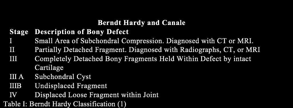
10 Classification
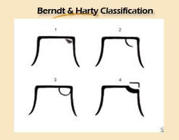
11 Classification
12 Types of Lesions Reported that 57% occur posteromedially and 43% occur anterolaterally (4). Lateral lesions are located in the middle third of the talar dome and are shallow and wafershaped. Medial lesions are typically located in the posterior third of the talar dome and are deeper and cup shaped (2).
13 Presentation Most often present with a chief complaint of a sprained ankle. Often report a history of trauma, recurrent sprains or chronic instability(4). Pain increased with WB Common Symptoms include pain, swelling, weakness, and decreased range of motion, ankle joint stiffness.
14 Presentation Physical Exam Findings Non specific: Patients often have pain on palpation of the anterolateral or posteromedial aspects of the ankle joint, along with pain with dorsiflexion and inversion. Note: With ankle sprains, pain and swelling should subside within a few months with conservative treatment.
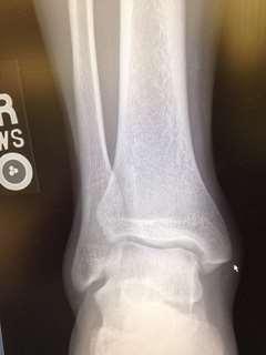
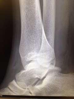
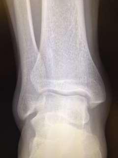
15 Plain X-rays: Radiographic Exam Anterior Posterior Lateral Mortise Plantarflexed mortise may help better visualize posterior medial lesions Dorsiflexed mortise may help better visualize anterior medial and lateral lesions ***Because patients often present with a chief complaint of ankle pain without radiographic evdience of acute fracture (i.e Stage I compression fractures) these lesions are often misdiagnosed ***

16 Plain X-ray
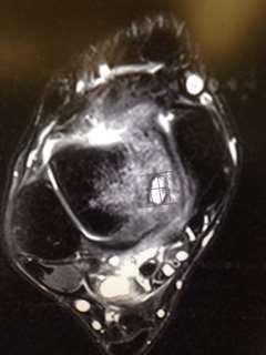
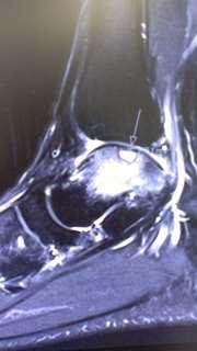
17 Radiographic Exam MRI: *Occult OLT s Cartilaginous surfaces Surrounding Bony Edema Fragment stability Other soft tissue injuries

18 MRI: Stage I

19 MRI: Stage IV
20 Differential Diagnosis Bony and soft tissue impingement Lateral ankle instability Ankle and/or subtalar joint arthritis Tendinitis RSD Tarsal coalitions Synovitis
21 Important Points Contained Lesions UnContained Lesions 150 mm2 21
22 Size Really Does Matter Chuck-Paiwong et al: Good-excellent results in 100% under 15mm 31/32 patients over 15mm had poor result 73 patients Choi et al: 80% with lesions over 15mm had poor outcome 25 patients 22
23 Treatments Various: Non operative vs. Operative Tol et al systemic review (7) Summarized 65 study groups in 52 studies Systematically screened Electronic databases from January 1966 to December 2006 Non operative treatment 25-40% success rate All stages involved OATS, BMS and ACI scored success rates of 87, 85 and 76%, respectively. Stage III and IV Bone marrow stimulation (BMS) was identified as the best treatment option.
24 Treatment Symptomatic, Non-displaced lesions are often treated conservatively NWB in short leg cast; crutches Rest ICE NSAIDs Physical therapy 3-6 months non-operative treatment
25 Treatment Surgical intervention is often reserved symptomatic lesions that have failed conservative therapy or displaced, stage III or IV lesions; smaller lesions <1.5 cm Excision and Curettage : Arthroscopic or Open; remove fragment BMS: Drilling or microfracturing: Disrupts intra osseous vessels Growth Factors Angiogenesis Bone Marrown Cells Fibrocartilage
26 Larger Lesions Treatment Fresh Osteochondral Allograft Mosaicplasty with Autogenous Graft Lesions 1-4 cm^2 6.5, 4.5, 3.5 cylindrical plugs autogenous graft derived from ipsilateral knee Medial upper part of the medical femoral condyle is primary harvest site. Goal is to reproduce the mechanical, structural and biochemical properties of the original hyaline articular cartilage which has become damaged
27 27
28 Treatment Osteochondral Autologus Transfer system (OATs) Similar Concept as Mosaicplasty Complete osteochondral plug is removed from site of the lesion 6-10 mm osteochondral plugs are transferred from ipsilateral knee to deficit; never leaves harvest tube
29 Treatment Autologous Chondrocyte Transplantation (ACT) (9) Osteochondral slices (10x 3mm) from ipsilateral knee sterile tub lab Eznymatic break down cartilage, isolation chondrocytes, which are then cultivated in culture medium 2 weeks Cultured cells are injected under tibial periosteal flap (8)
30 Microfracture Indicated for lesions up to 15mm in diameter Multiple holes created at 3-4mm intervals Stimulate mesenchymal stem cells (MSCs) and growth factors Results in fibrin clot & eventually fibrocartilaginous repair Fibrocartilage mostly Type I collagen Softer & more easily damaged than hyaline 2. Polat, G. et al. Long-Term Results of Microfracture in the Treatment of Talus Osteochondral Lesions. European Society of Sports Traumatology. February 2016 Vol 24, pg
31 Subchondral Drilling vs Microfracture Heat necrosis is main concern of drilling May cause bone necrosis, pain, edema, or stress fracture Microfracture avoids heat necrosis, but can create loose body particles If not removed, may cause locking & cartilage damage Particles may block access channels to bone marrow, impeding healing 3. Choi, J.I. and Lee, Keun-Bae. Comparison of Clinical Outcomes Between Arthroscopic Subchondral Drilling and Microfracture for Osteochondral Lesions of the Talus. Knee Surgical Sports Traumatology Arthroscopy. January pg 31


32 Autologous Osteochondral Transplant (Mosaicplasty / OATS) Cylindrical osteochondral grafts harvested from NWB portion of ipsilateral knee Indicated for lesions over 15mm in diameter May result in cystic formation due to incongruence with surrounding cartilage Zengerink et al: 87% good-excellent results 243 patients Hannon, C.P. et al. Osteochondral Lesions of the Talus: Aspects of Current Management. The Bone and Joint Journal. February 2014, Vol 96B, pg
33 Drilling vs Microfracture cont. Choi et al cont: Drilling Microfracture Patients 40 (28M,12F) 50 (40M,10F) Pre-op AOFAS Post-op AOFAS Mean f/u 38.1 months 38.5 Mean lesion size 1.0cm cm 2 Results: Excellent 30 (75%) 34 (68%) Good 5 (12.5%) 10 (20%) Fair 5 (12.5%) 6 (12%) 33


34 Drilling vs Microfracture cont. Subchondral Drilling Microfracture 3-4 mm apart -Adequate bleeding must be verified upon releasing tourniquet 34
35 Surgical Technique Local Ipsilateral Allograft Less Morbidity One Surgical Incision Decreased Surgical Time
36 36
37 37
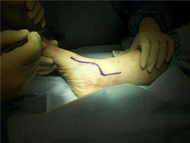
38 Incision

39 Medial Malleolar Osteotomy Preparation 39

40 Medial Osteotomy Creation 40

41 Finalizing Medial Osteotomy 41

42 Medial Osteotomy Take Down 42

43 Medial Talar Dome Lesion Exposure 43

44 Talar Dome Lesion Exposure 44

45 Medial Talar Dome Defect 45

46 Medial Talar Dome Lesion Excision 46

47 Talus Dome Core Decompression 47

48 Inferior Talus Harvest Site 48

49 Harvest Site

50 Harvested Plug

51 Insertion Osteochondral Plug
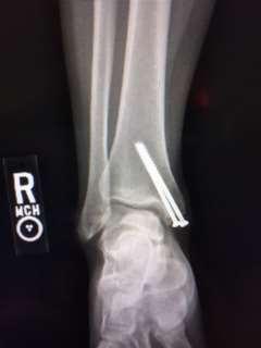
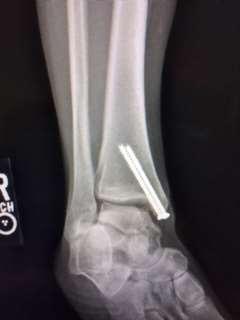
52 Reposition Medial Malleolus 52
53 Reinforcement of Deltoid Ligament 53
54 54
55 Potential Complications Post-operative pain, infection, nerve and arterial compromise, hypertrophic scar formation, RSD, DVT, PE, non-union, delayed union, amputation, and death. Failure of graft, Non-union of the osteotomy site
56 Post Operative Course NWB 4-6 weeks splint/ cast Walking Boot 4-6 weeks 4 weeks PT week 8 Shoe weeks Minimize narcotics. Selective on NSAIDS. Anticoagulation 4 weeks. MVI, Vit. D 2000U, Vit. C 1000 mg
. Philadelphia, PA: Mosby. 3. Cuttica, Daniel J., W. B. Smith, Christopher F. Hyer, Terrence M. Philbin, and Gregory C. Bertlet.")
57 References 1. Hansen, S. T. (2000). Functional Reconstruction of the Foot and Ankle (pp ). Philadelphia, PA: Lippincott Williams & Wilkins. 2. Coughlin, M. J., Mann, R. A., & Saltzman, C. L. (2007). Surgery and the Foot And Ankle (8th ed., Vol. II, pp ). Philadelphia, PA: Mosby. 3. Cuttica, Daniel J., W. B. Smith, Christopher F. Hyer, Terrence M. Philbin, and Gregory C. Bertlet. "Osteochondral Lesions of the Talus: Predictors of Clinical Outcome." Foot and Ankle International (2011): Print 4. Dragoni, Massimiliano, Davide E. Bonasia, and Annunziato A. Amendola. "Osteochondral Talar Allograft for Large Osteochondral Defects: Technique Tip." Foot and Ankle International 32.9 Sept. (2011): Print. 5. Raikin, Thomas M. Massive Osteochondral Defects of the Talus. Foot and Ankle Clinics Vol. 9. Philadelphia: Elsevier, Print. 6. Hangody, Laszlo. "The mosaicplasty technique for osteochonral lesions of the talus." Foot and Ankle Clinics 8 (2003): Print. 7. Tol, Johannes, Peter A. Strujis, and Maartje Zengerink. "Treatment of osteochondral lesions of the talus: a systematic review." Knee Surgery, Sports Traumatology, Arthroscopy (2009). Web. 1 Dec < 8. Alan S Banks, Michael S Downey, Dennis E Martin, Stephen J Miller. McGlamry s Comprehensive Textbook of Foot and Ankle Surgery. Lippincott Williams & Wilkins Pp Peterson, L, Mats Brittberg, and Anders Lindahl. "Autologous chondrocyte transplantation." Foot and Ankle Clinics 8.2 (2003): Print.

58 Thank you 58
June 2013 Case Study. Author: T. Walker Robinson, MD, MPH, Nationwide Children s Hospital
 June 2013 Case Study Author: T. Walker Robinson, MD, MPH, Nationwide Children s Hospital Chief Complaint: Right ankle pain HPI: A 10 year old female dancer presents to the clinic with a five day history
OCD: Beyond Microfracture. Disclosures. OCD Talus: My Approach 2/23/2018
 OCD: Beyond Microfracture Gregory C Berlet MD, FRCS(C), FAOA Orthopedic Foot and Ankle Center Columbus Ohio Disclosures Consultant/Speaker Bureau/Royalties/ Stock: Wright Medical, Stryker, ZimmerBiomet,
NORTHERN OHIO FOUNDATION
 NORTHERN OHIO FOOT & ANKLE FOUNDATION! The!Northern!Ohio!Foot!and!Ankle!Journal!!!!!!!!!!!!!!!Official!Publication!of!the!NOFA!Foundation!! Osteochondral Lesions of the Talus: Literature Review by Neil
Ankle Arthroscopy PAULO ROCKETT, M.D. Porto Alegre Brazil
 Ankle Arthroscopy PAULO ROCKETT, M.D. Porto Alegre Brazil Ankle sprains are among the most common injuries in sports and at work. Between 20 and 40% of patients treated with conservative therapy may have
Arthroscopy Of the Ankle.
 Arthroscopy Of the Ankle www.fisiokinesiterapia.biz Ankle Arthroscopy Anatomy Patient setup Portal placement Procedures Complications Anatomy Portals Anterior Anteromedial Anterolateral Anterocentral Posterior
Autologous Osteochondral Transplantation for Osteochondral Lesions of the Talus: Functional and T2 MRI Outcomes at Mid to Long-term follow-up
 Autologous Osteochondral Transplantation for Osteochondral Lesions of the Talus: Functional and T2 MRI Outcomes at Mid to Long-term follow-up September 2014 Seán Flynn, Keir Ross, Charles P. Hannon, Hunter
Ankle Arthroscopy.
 Ankle Arthroscopy Key words: Ankle pain, ankle arthroscopy, ankle sprain, ankle stiffness, day case surgery, articular cartilage, chondral injury, chondral defect, anti-inflammatory medication Our understanding
Sequalae of Ankle Sprains: Peri Articular Fractures of the Ankle in Sports Medicine.
 Sequalae of Ankle Sprains: Peri Articular Fractures of the Ankle in Sports Medicine www.fisiokinesiterapia.biz Chronic Ankle Pain The most common cause of chronic pain following an ankle sprain is a missed
Osteochondral Lesions: Medial Versus Lateral, Persistent Pain, Cartilage Restoration Options and Indications
 Osteochondral Lesions: Medial Versus Lateral, Persistent Pain, Cartilage Restoration Options and Indications AnnunziatoAmendola, MD a,b, *, Ludovico Panarella, MD, PhD c,d KEYWORDS Talus Osteochondral
POSITION STATEMENT The Use of Osteochondral Transplantation for the Treatment of Osteochondral Lesions of the Talus
 Position Statement POSITION STATEMENT The Use of Osteochondral Transplantation for the Treatment of Osteochondral Lesions of the Talus The American Orthopaedic Foot & Ankle Society (AOFAS) endorses the
Autologous Chondrocyte Implantation. Gerard Hardisty FRACS
 Autologous Chondrocyte Implantation Gerard Hardisty FRACS Disclosure Orthopaedic Surgeons Strong as an OX and half as bright Orthopaedic Innovation Arthroscopy Joint replacement Trauma management MIS Early
Osteochondritis Dissecans of the Knee. M Lucas Murnaghan MD, MEd, FRCSC
 Osteochondritis Dissecans of the Knee M Lucas Murnaghan MD, MEd, FRCSC Outline 1. Clinical Presentation 2. Investigations 3. Classification 4. Non-operative Treatment 5. Operative Treatment 6. Treatment
Peggers Super Summaries: Foot Injuries
 Lisfranc Injury ANATOMY Roman arch with recessed 2 nd MT base AP medial side of intermediate cuneiform to 2 nd MT base Oblique medial side of lateral cuneiform with 3 rd MT base and 4 th with medial boarder
First Metatarsal Head Osteochondral Defect Treatment with Particulated Juvenile Cartilage Allograft Transplantation
 First Metatarsal Head Osteochondral Defect Treatment with Particulated Juvenile Cartilage Allograft Transplantation Bryan Van Dyke, DO Gregory C. Berlet, MD Justin L. Daigre, MD Christopher F. Hyer, DPM,
Case Report. Byung Ill Lee, MD and Byoung Min Kim, MD Department of Orthopedic Surgery, Soonchunhyang University Hospital, Seoul, Korea
 Case Report Knee Surg Relat Res 2015;27(4):263-268 http://dx.doi.org/10.5792/ksrr.2015.27.4.263 pissn 2234-0726 eissn 2234-2451 Knee Surgery & Related Research Concomitant Osteochondral utograft Transplantation
Osteochondral lesions were first described by Konig
 155 Osteochondral Lesions of the Talar Dome Alexander Finger, M.D., and Steven C. Sheskier, M.D. Osteochondral lesions were first described by Konig in 1888. 1 He theorized that spontaneous necrosis was
Cartilage Repair Options
 Imaging of Cartilage Repair Carl S. Winalski, MD Imaging Institute Department of Biomedical Engineering Cleveland Clinic Cartilage Repair Options Direct repair Marrow stimulation Autologous transplantation
Outline. Ankle/Foot Anatomy Ankle Sprains Ottawa Ankle Rules DDx: The Sprain That Wasn t
 Ankle Injuries Outline Ankle/Foot Anatomy Ankle Sprains Ottawa Ankle Rules DDx: The Sprain That Wasn t Anatomy: Ankle Mortise Bony Anatomy Lateral Ligament Complex Medial Ligament Complex Ankle Sprains
Welcome to the: Orthopaedic Opinion Online Website The website for the answer to all your Orthopaedic Questions
 Welcome to the: Orthopaedic Opinion Online Website The website for the answer to all your Orthopaedic Questions Orthopaedic Opinion Online is a website designed to provide information to patients who have
UvA-DARE (Digital Academic Repository) Treatment of osteochondral defects of the talus van Bergen, C.J.A. Link to publication
 Treatment of osteochondral defects of the talus van Bergen, C.J.A. Link to publication") UvA-DARE (Digital Academic Repository) Treatment of osteochondral defects of the talus van Bergen, C.J.A. Link to publication Citation for published version (APA): van Bergen, C. J. A. (2014). Treatment
UvA-DARE (Digital Academic Repository) Treatment of osteochondral defects of the talus van Bergen, C.J.A. Link to publication Citation for published version (APA): van Bergen, C. J. A. (2014). Treatment
Posterior Ankle Impingement: Don t Get Pinched
 Posterior Ankle Impingement: Don t Get Pinched 11 th Annual Sports Medicine Continuing Education Conference Gregory P Witkowski, MD Orthopaedic Trauma and Foot/Ankle Surgery Disclosures I have nothing
PATIENT GUIDE TO CARTILAGE INJURIES
 Lucas Wymore, MD Sports Medicine 23000 Moakley Street Suite 102 Leonardtown MD 20650 Office Phone: 301-475-5555 Office Fax: 301-475- 5914 Email: lwymore@somdortho.com PATIENT GUIDE TO CARTILAGE INJURIES
OSTEOCHONDRAL ALLOGRAFTS AND AUTOGRAFTS IN THE TREATMENT OF FOCAL ARTICULAR CARTILAGE LESIONS
 Status Active Medical and Behavioral Health Policy Section: Surgery Policy Number: IV-115 Effective Date: 10/22/2014 Blue Cross and Blue Shield of Minnesota medical policies do not imply that members should
Ankle Injuries. Ankle Sprain. Range of Motion. The most likely diagnosis is lateral ligament sprain. Dorsiflexion Plantarflexion Inversion
 Ankle Injuries Dr Peter Brukner, OAM Sports Physician Associate Professor Centre for Sports Medicine Research & Education The University of Melbourne Adjunct Professor School of Human Movement Studies
OSTEOCHONDRAL ALLOGRAFT RECONSTRUCTION FOR MASSIVE BONE DEFECT
 OSTEOCHONDRAL ALLOGRAFT RECONSTRUCTION FOR MASSIVE BONE DEFECT Angelo J. Colosimo, MD -Head Orthopaedic Surgeon University of Cincinnati Athletics -Director of Sports Medicine University of Cincinnati
Ankle Pain After a Sprain.
 Ankle Pain After a Sprain www.fisiokinesiterapia.biz Anterior Drawer Stress Test Talar Tilt Talar Tilt (CFL) Difficult to isolate from subtalar ROM Slight plantar flexion (dorsi = relative subtalar isolation)
Review relevant anatomy of the foot and ankle. Learn the approach to examining the foot and ankle
 Objectives Review relevant anatomy of the foot and ankle Learn the approach to examining the foot and ankle Learn the basics of diagnosis and treatment of ankle sprains Overview of other common causes
TREATMENT OF CARTILAGE LESIONS
 TREATMENT OF CARTILAGE LESIONS Angelo J. Colosimo, MD -Head Orthopaedic Surgeon University of Cincinnati Athletics -Director of Sports Medicine University of Cincinnati Medical Center -Associate Professor
Greetings From SCOI. Richard D. Ferkel, M.D
 Greetings From SCOI Richard D. Ferkel, M.D OLT In the Athlete Operative Treatment and Return to Play The Following relationships exist: Royalties and stock options Smith and Nephew Consulting income
Knee Preservation and Articular Cartilage Restoration
 Knee Preservation and Articular Cartilage Restoration With Special Thanks to Aaron Krych, MD and Riley Willims, MD Zak Knutson, MD Articular Cartilage Layer of tissue covering the bone which are part of
A Patient s Guide to Osteochondritis Dissecans of the Talus. Foot and Ankle Center of Massachusetts, P.C.
 A Patient s Guide to Osteochondritis Dissecans of the Talus Welcome to Foot and Ankle Center of Massachusetts, where we believe in accelerating your learning curve with educational materials that are clearly
Ankle Ligament Injury: Don t Worry- It s Only a Sprain Wes Jackson MD Orthopaedic Foot & Ankle
 Ankle Ligament Injury: Don t Worry- It s Only a Sprain Wes Jackson MD Orthopaedic Foot & Ankle Outline I. Epidemiology II. Classification and Types of Sprains III. Anatomy IV. Clinical Assessment and Imaging
Posttraumatic subchondral bone contusions and fractures of the talotibial joint: Occurrence of kissing lesions
 KISSING CONTUSIONS CHAPTER 7 Posttraumatic subchondral bone contusions and fractures of the talotibial joint: Occurrence of kissing lesions Elizabeth S. Sijbrandij 1, Ad P.G. van Gils 1, Jan Willem K.
Osteochondritis Dissecans
 Osteochondritis Dissecans Introduction Osteochondritis dissecans (OCD) is a problem that affects the knee, mostly at the end of the big bone of the thigh (the femur). A joint surface damaged by OCD doesn't
The prevalence of osteochondritis
 Brief Communication Communication abrégée OSTEOCHONDRITIS DISSECANS OF THE TALAR DOME TREATED WITH AN OSTEOCHONDRAL AUTOGRAFT Edward Chang, BSc(PT);* Eric Lenczner, MD The prevalence of osteochondritis
Persistent ankle pain after inversion lesions: what the radiologist must look for
 Persistent ankle pain after inversion lesions: what the radiologist must look for Poster No.: P-0118 Congress: ESSR 2016 Type: Authors: Keywords: DOI: Educational Poster R. Leao, L. C. Zattar-Ramos, E.
Rakesh Patel, MD 4/9/09
 Rakesh Patel, MD 4/9/09 Chondral Injuries Very common Present in 63-66% patients undergoing arthroscopy 11-19% full-thickness lesions Up to 79% patients with ACL deficient knee have some form of chondral
Disclosures. Syndesmosis Injury. Syndesmosis Ligaments. Objectives. Mark M. Casillas, M.D.
 Disclosures Syndesmosis Injury No relevant disclosures Mark M. Casillas, M.D. 1 Objectives Syndesmosis Ligaments Understand the syndesmosis anatomy and function Classify syndesmosis injuries Describe treatment
High Ankle Sprains: Diagnosis & Treatment
 High Ankle Sprains: Diagnosis & Treatment Mark J. Mendeszoon, DPM, FACFAS, FACFAOM Precision Orthopaedic Specialties University Regional Hospitals Advanced Foot & Ankle Fellowship- Director It Is Only
Sports Injuries of the Ankle and Ankle Arthritis. Mr Amit Amin Consultant Foot and Ankle Surgeon Parkside Hospital
 Sports Injuries of the Ankle and Ankle Arthritis Mr Amit Amin Consultant Foot and Ankle Surgeon Parkside Hospital Impingement Painful mechanical limitation of full ankle movement secondary to osseous
Anterior Impingement
 Anterior Impingement Ziali Sivardeen BMedSci, (MRCS), AFRCS, FRCS (Tr & Orth) Consultant Trauma and Orthopaedic Surgeon (Shoulder, Knee and Sports Injuries) Aims Causes of Anterior Ankle Pain Ankle Impingement
V E R I TAS MGH 1811 MGH 1811 V E R I TAS. *Gerber JP. Persistent disability with ankle sprains. Foot Ankle Int 19: , 1998.
 MGH 1811 Management of Ankle Instability Richard J. de Asla, M.D. V E R I TAS MGH 1811 I have no potential conflicts with this presentation. V E R I TAS It s just a sprain Lateral Ankle Sprains Most common
UvA-DARE (Digital Academic Repository) Treatment of osteochondral defects of the talus van Bergen, C.J.A. Link to publication
 Treatment of osteochondral defects of the talus van Bergen, C.J.A. Link to publication") UvA-DARE (Digital Academic Repository) Treatment of osteochondral defects of the talus van Bergen, C.J.A. Link to publication Citation for published version (APA): van Bergen, C. J. A. (2014). Treatment
UvA-DARE (Digital Academic Repository) Treatment of osteochondral defects of the talus van Bergen, C.J.A. Link to publication Citation for published version (APA): van Bergen, C. J. A. (2014). Treatment
Articular Cartilage Surgical Restoration Options
 Articular Cartilage Surgical Restoration Options Randy Schwartzberg, M.D. Assistant Professor - UCF College of Medicine Rationale Our bodies do not make articular/hyaline cartilage. gics injections to
Comparison of Outcomes of Osteochondral Transplantation with Autografts and Allografts for Osteochondral Lesions of the Talus
 Comparison of Outcomes of Osteochondral Transplantation with Autografts and Allografts for Osteochondral Lesions of the Talus Yoshiharu Shimozono, MD, Eoghan T Hurley, MB, BCh, Timothy W Deyer, MD, John
Craig S. Radnay, M.D. 1/27/2016. Access to the Talus for Treatment of Osteochondral Lesions. Epidemiology of OLT. Treatment of OLT
 Access to the Talus for Treatment of Osteochondral Lesions Craig S. Radnay, MD, MPH ISK Institute for Orthopaedics and Sports Medicine NYU/Hospital for Joint Diseases Tampa, FL January 23, 2016 Epidemiology
Clinical evaluation where no obvious fracture a. Squeeze test
 7:43 am The Syndesmotic Injury: From Subtle to Severe Robert B. Anderson, MD Chief, Foot and Ankle Carolinas Medical Center OrthoCarolina (Charlotte, North Carolina) 7:30-8:25 am Symposium 1: Management
OATS for the Foot and Ankle Surgical Technique
 OATS for the Foot and Ankle Surgical Technique Osteochondral Autograft Transfer System Scientific Support for Small Joint OATS Outcome of Osteochondral Autograft Transplantation for Type-V Cystic Osteochondral
17/10/2017. Foot and Ankle
 17/10/2017 Alicia M. Yochum RN, DC, DACBR, RMSK Foot and Ankle Plantar Fasciitis Hallux Valgus Deformity Achilles Tendinosis Posterior Tibialis Tendon tendinopathy Stress Fracture Ligamentous tearing Turf
Arthroscopic Treatment of Osteochondral Talar Defects
 Arthroscopic Treatment of Osteochondral Talar Defects Christiaan J.A. van Bergen, MD, Ruben Zwiers, MSc, and C. Niek van Dijk, MD, PhD Based on an original article: J Bone Joint Surg Am. 2013 Mar 20;95(6):519-25.
ANKLE OSTEOCHONDRAL ALLOGRAFTS
 ANKLE OSTEOCHONDRAL ALLOGRAFTS UNIVERSITY OF CALIFORNIA, SAN DIEGO CHOLL KIM, M.D., Ph.D. AMIR JAMALI, M.D. F. RICHARD CONVERY, M.D. WILLIAM BUGBEE, M.D. ANKLE OSTEOCHONDRAL ALLOGRAFTS BACKGROUND - ORGAN
Ankle Sprains and Their Imitators
 Ankle Sprains and Their Imitators Mark Halstead, MD Dr. Mark Halstead is the Associate Professor of the Departments of Orthopedics and Pediatrics at Washington University School of Medicine; Director of
A Patient s Guide to Osteochondritis Dissecans of the Knee
 A Patient s Guide to Osteochondritis Dissecans of the Knee 2350 Royal Boulevard Suite 200 Elgin, IL 60123 Phone: 847.931.5300 Fax: 847.931.9072 DISCLAIMER: The information in this booklet is compiled from
UvA-DARE (Digital Academic Repository) Treatment of osteochondral defects of the talus van Bergen, C.J.A. Link to publication
 Treatment of osteochondral defects of the talus van Bergen, C.J.A. Link to publication") UvA-DARE (Digital Academic Repository) Treatment of osteochondral defects of the talus van Bergen, C.J.A. Link to publication Citation for published version (APA): van Bergen, C. J. A. (2014). Treatment
UvA-DARE (Digital Academic Repository) Treatment of osteochondral defects of the talus van Bergen, C.J.A. Link to publication Citation for published version (APA): van Bergen, C. J. A. (2014). Treatment
Triplane fracture of the ankle is an uncommon injury
 A Case Report & Literature Review Intramalleolar Triplane Fracture With Osteochondral Talar Defect Wendy L. Heusch, DO, and Henry W. Albers, MD Triplane fracture of the ankle is an uncommon injury that
Ankle Sprain. 43 Thames Street, St Albans, Christchurch 8013 Phone: (03) Website: philip-bayliss.com
 Website: philip-bayliss.com") 43 Thames Street, St Albans, Christchurch 8013 Phone: (03) 356 1353 Website: philip-bayliss.com Ankle Sprain An Ankle sprain is one of the most common musculoskeletal injuries. Patients typically describe
43 Thames Street, St Albans, Christchurch 8013 Phone: (03) 356 1353 Website: philip-bayliss.com Ankle Sprain An Ankle sprain is one of the most common musculoskeletal injuries. Patients typically describe
Achilles Tendonitis and Tears
 Achilles Tendonitis and Tears The Achilles tendon is an important structure for normal ankle motion and normal function, even for daily activities such as walking. Achilles tendonitis can occur in patients
emoryhealthcare.org/ortho
 COMMON SOCCER INJURIES Oluseun A. Olufade, MD Assistant Professor, Department of Orthopedics and PM&R 1/7/18 GOALS Discuss top soccer injuries and treatment strategies Simplify hip and groin injuries in
Avascular Necrosis of the Foot. Dr. Hema Choudur MD, FRCPC Associate Professor. Dept. of Radiology. McMaster University, Hamilton, Canada.
 Avascular Necrosis of the Foot Dr. Hema Choudur MD, FRCPC Associate Professor. Dept. of Radiology. McMaster University, Hamilton, Canada. Avascular Necrosis: Pathophysiology Ischemia to the bone from oxygen
3/13/2018. Cartilage Cases. Case. Physical exam
 Cartilage Cases Aaron J. Krych, MD Professor, Orthopedic Surgery Sports Medicine Fellowship Director Sports Medicine Research Fellowship Director Mayo Clinic 2014 MFMER slide-1 Case 19 yo F division I
BIOMECHANICS OF ANKLE FRACTURES
 BIOMECHANICS OF ANKLE FRACTURES William R Reinus, MD MBA FACR Significance of Ankle Fractures Most common weight-bearing Fx 70% of all Fxs Incidence is increasing Bimodal distribution Men 15-24 Women over
Ankle Arthritis and Ankle Replacement
 Ankle Arthritis and Ankle Replacement Ryan DeBlis, MD Disclosures I have no disclosures. 1 Diagnosis Ankle arthritis Majority (70%) of patients are post-traumatic (ie, after ankle fracture) Primary arthritis
Mary Lloyd Ireland, M.D. Associate Professor University of Kentucky Dept. of Orthopaedic Surgery and Sports Medicine Lexington, Kentucky
 Common Ankle Injuries: Diagnosis and Treatment Mary Lloyd Ireland, M.D. Associate Professor University of Kentucky Dept. of Orthopaedic Surgery and Sports Medicine Lexington, Kentucky Disclaimer Slide
Talus Fractures: When and Why on Screws and Plates
 Talus Fractures: When and Why on Screws and Plates Frank A. Liporace, MD Associate Professor Director of Orthopaedic Research New York University / Hospital for Joint Diseases, NY, NY Director Orthopaedic
UNDERSTANDING ARTHROSCOPY
 UNDERSTANDING ARTHROSCOPY Diagnosing and Treating Your Joint Problem Looking into a Problem Joint Whether you re taking a step or raising your hand, your joints help you move freely. A worn, torn, or injured
Introduction Knee Anatomy and Function Making the Diagnosis
 Introduction Knee injuries are a very common problem among active individuals. It is important for us to understand how your knee was injured. Most knee injuries are associated with non-contact mechanisms.
over the counter nonsteroidal antiinflammatory drugs with minimal relief.
 CHAPTER I3 FRESFI TAIARALLOGRAFT FORA IARGE POSTERIOR MEDIAL TALUT DOME LE,SION: A Case Study Richard Zirm, DPM INTRODUCTION Osteochondral defects of the talus are relatively rare occurring in approximately
Osteochondritis Dissecans
 P R O C E D U R E 1 4 Ammar Anbari, Adam B. Yanke, and Brian J. Cole ch014-x4397.indd 221 4/11/2008 10:50:21 AM 222 Treatment Options Conservative management Fixation in situ Elevate the OCD lesion, débride
ANTERIOR CRUCIATE LIGAMENT INJURY
 ANTERIOR CRUCIATE LIGAMENT INJURY WHAT IS THE ANTERIOR CRUCIATE LIGAMENT? The anterior cruciate ligament (ACL) is one of four major ligaments that stabilizes the knee joint. A ligament is a tough band
Medical Policy An independent licensee of the Blue Cross Blue Shield Association
 Autografts and Allografts in the Treatment of Focal Page 1 of 30 Medical Policy An independent licensee of the Blue Cross Blue Shield Association Title: Autografts and Allografts in the Treatment of Focal
Post-traumatic osteonecrosis of distal tibia
 Injury Extra (2007) 38, 262 266 www.elsevier.com/locate/inext CASE REPORT Post-traumatic osteonecrosis of distal tibia D. Chakravarty a, *, A. Khanna a,1, A. Kumar b,2 a Department of Orthopaedics, Peterborough
INVISION Total Ankle Replacement System with PROPHECY Preoperative Navigation Revision of a Failed Agility Total Ankle Replacement
 016625 REVISION R INVISION Total Ankle Replacement System with PROPHECY Preoperative Navigation Revision of a Failed Agility Total Ankle Replacement CASE STUDY Patient History The patient was a 65-year-old
Osteochondral Allograft Transplantation and Autograft Transfer System (OATS/mosaicplasty) in the Treatment of Articular
 in the Treatment of Articular") Osteochondral Allograft Transplantation and Autograft Transfer System (OATS/mosaicplasty) in the Treatment of Articular Cartilage Lesions Corporate Medical Policy File name: Osteochondral Allograft Transplantation
Osteochondral Allograft Transplantation and Autograft Transfer System (OATS/mosaicplasty) in the Treatment of Articular Cartilage Lesions Corporate Medical Policy File name: Osteochondral Allograft Transplantation
Particulated Juvenile Articular Cartilage Allograft Transplantation With Bone Marrow Aspirate Concentrate for Treatment of Talus Osteochondral Defects
 SPECIAL FOCUS Particulated Juvenile Articular Cartilage Allograft Transplantation With Bone Marrow Aspirate Concentrate for Treatment of Talus Osteochondral Defects Mark C. Drakos, MD and Conor I. Murphy,
Foot and ankle update
 Foot and ankle update Mr Ian Garnham Consultant Foot and Ankle Surgeon Whipps Cross University Hospital Hallux Rigidus Symptoms first ray and 1st MTP pain and swelling worse with push off or forced dorsiflexion
AAP Boot Camp KNEE AND ANKLE EXAM
 AAP Boot Camp KNEE AND ANKLE EXAM Disclosures I have no relevant financial relationships with the manufacturers of any commercial products and or providers of commercial services discussed in this CME
Life. Uncompromised. The KineSpring Knee Implant System Surgeon Handout
 Life Uncompromised The KineSpring Knee Implant System Surgeon Handout 2 Patient Selection Criteria Patient Selection Criteria Medial compartment degeneration must be confirmed radiographically or arthroscopically
3/21/2011 PCL INJURY WITH OPERATIVE TREATMENT A CASE STUDY PCL PCL MECHANISM OF INJURY PCL PREVALENCE
 PCL PCL INJURY WITH OPERATIVE TREATMENT A CASE STUDY K. Anderson, S. Hjortedal, Y. Jingi, E. Sutcliffe & S. Witschen Washington State University Origin Posterior aspect of tibia Insertion Medial femoral
Barriers Between Injury and Returnto-Work. Lower Extremity. Why the Extreme Variability
 Barriers Between Injury and Returnto-Work in the Lower Extremity Why the Extreme Variability Barriers to Diagnosis Failure or delay in reporting injury Employee expectations: Not really a serious injury
Histologic change of cartilage layer of osteochondritis dissecans before and after fixation in the knee
 1 Histologic change of cartilage layer of osteochondritis dissecans before and after fixation in the knee Mitsuo Ochi, M.D. PhD Professor and chairman Department of Orthopaedic Surgery Graduate School
Ligament lesions of the ankle. Marc C. Attinger
 Ligament lesions of the ankle Marc C. Attinger Anatomy Mechanism of injury Each lig with its function during ROM in dorsiflexion/er ATFL slack, CFL tight in plantarflexion/ir CFL slack, ATFL tight Acute
Rehabilitation Protocol:
 Rehabilitation Protocol: Patellofemoral resurfacing: Osteochondral Autograft Transplantation (OATS), Autologous Chondrocyte Implantation (ACI) and Microfracture Department of Orthopaedic Surgery Lahey
Index. Clin Sports Med 23 (2004) Note: Page numbers of article titles are in boldface type.
 Note: Page numbers of article titles are in boldface type.") Clin Sports Med 23 (2004) 169 173 Index Note: Page numbers of article titles are in boldface type. A Achilles enthesopathy, calcaneal spur with, 133 clinical presentation of, 135 136 definition of, 131
Clin Sports Med 23 (2004) 169 173 Index Note: Page numbers of article titles are in boldface type. A Achilles enthesopathy, calcaneal spur with, 133 clinical presentation of, 135 136 definition of, 131
These FAQs apply to lateral and medial ankle ligament sprains, but not to syndesmosis (high) ankle sprains, which are treated differently.
 ankle sprains, which are treated differently.") ISO 9001:2015 FS 550968 What is an ankle sprain? A sprain is a tear of a ligament. Ligaments are the tough fibrous tissues that connect the bones together across a joint. Ligament tears can vary from very
ISO 9001:2015 FS 550968 What is an ankle sprain? A sprain is a tear of a ligament. Ligaments are the tough fibrous tissues that connect the bones together across a joint. Ligament tears can vary from very
Arthroscopic Management of Osteochondral Lesions of the Talus
 Med. J. Cairo Univ., Vol. 77, No. 3, June: 147-153, 2009 www.medicaljournalofcairouniversity.com Arthroscopic Management of Osteochondral Lesions of the Talus AHMAD KHOLEIF, M.D.*; KAMAL SAMY ABDEL MEGUID,
Impingement Syndromes of the Ankle. Noaman W Siddiqi MD 5/4/2006
 Impingement Syndromes of the Ankle Noaman W Siddiqi MD 5/4/2006 Ankle Impingement Overview Clinical DX Increasingly recognized cause of chronic ankle pain Etiology can be soft tissue or osseous Professional
Basics of Cartilage Restoration Introduction of TruFit
 Basics of Cartilage Restoration Introduction of TruFit Philip A. Davidson, MD Heiden Orthopaedics Park City, Utah USA Smith & Nephew Seminar London, UK October 2008 Cartilage Restoration A wide realm between..
Anterior Knee Pain in Children. Joseph Chorley, MD Associate Professor, Pediatrics Baylor College of Medicine
 Anterior Knee Pain in Children Joseph Chorley, MD Associate Professor, Pediatrics Baylor College of Medicine Goals and Objectives To learn how to care for patients with chronic knee pain To be able to
Clinical Commissioning Policy: Autologous chrondrocyte implantation for osteochondral lesions of the talus
 Clinical Commissioning Policy: Autologous chrondrocyte implantation for osteochondral lesions of the talus Reference: NHS England: 16012/P NHS England INFORMATION READER BOX Directorate Medical Operations
Foot and Ankle Complaints.
 Foot and Ankle Complaints www.fisiokinesiterapia.biz INTRODUCTION Anatomy and Function Foot Ankle Common complaints Common diagnoses FOOT AND ANKLE ANATOMY 26 bones and 2 sesamoids Forefoot Metatarsals
UvA-DARE (Digital Academic Repository) Treatment of osteochondral defects of the talus van Bergen, C.J.A. Link to publication
 Treatment of osteochondral defects of the talus van Bergen, C.J.A. Link to publication") UvA-DARE (Digital Academic Repository) Treatment of osteochondral defects of the talus van Bergen, C.J.A. Link to publication Citation for published version (APA): van Bergen, C. J. A. (2014). Treatment
UvA-DARE (Digital Academic Repository) Treatment of osteochondral defects of the talus van Bergen, C.J.A. Link to publication Citation for published version (APA): van Bergen, C. J. A. (2014). Treatment
2017 SAFSA CONGRESS PROGRAMME
 2017 SAFSA CONGRESS PROGRAMME THURSDAY, MAY 25 07h45 07h55: WELCOME & INTRODUCTIONS Forefoot I: Hallux Valgus and Lesser Toes (08h00-10h00 Lectures) 08h00 08h30: Surgical Management of Hallux Valgus Rippstein,
Osteochondral Grafting
 Last Review Date: January 19, 2017 Number: MG.MM.SU.68 Medical Guideline Disclaimer Property of EmblemHealth. All rights reserved. The treating physician or primary care provider must submit to EmblemHealth
Osteochondral transplantation of autologous graft for the treatment of osteochondral lesions of talus: 5- to 7-year follow-up
 DOI 10.1007/s00167-014-3389-3 ANKLE Osteochondral transplantation of autologous graft for the treatment of osteochondral lesions of talus: 5- to 7-year follow-up Dimitrios Georgiannos Ilias Bisbinas Athanasios
Case Report Combined Posterior and Anterior Ankle Arthroscopy
 Case Reports in Orthopedics Volume 2012, Article ID 693124, 4 pages doi:10.1155/2012/693124 Case Report Combined Posterior and Anterior Ankle Arthroscopy Peter E. Scholten 1, 2 andc.niekvandijk 2 1 Department
UvA-DARE (Digital Academic Repository) Treatment of osteochondral defects of the talus van Bergen, C.J.A. Link to publication
 Treatment of osteochondral defects of the talus van Bergen, C.J.A. Link to publication") UvA-DARE (Digital Academic Repository) Treatment of osteochondral defects of the talus van Bergen, C.J.A. Link to publication Citation for published version (APA): van Bergen, C. J. A. (2014). Treatment
UvA-DARE (Digital Academic Repository) Treatment of osteochondral defects of the talus van Bergen, C.J.A. Link to publication Citation for published version (APA): van Bergen, C. J. A. (2014). Treatment
AUTOLOGOUS CHONDROCYTE IMPLANTATION FOR FOCAL ARTICULAR CARTILAGE LESIONS
 CARTILAGE LESIONS Non-Discrimination Statement and Multi-Language Interpreter Services information are located at the end of this document. Coverage for services, procedures, medical devices and drugs
AOFAS 2016 Annual Convention July 20-23, 2016 Toronto, Canada
 ANKLE ARTHROSCOPY FOR OSTEOCHONDRAL LESIONS OF THE TALUS: THE EFFECT OF LIMITED ANKLE RANGE OF MOTION ON ANTERIOR AND POSTERIOR ARTHROSCOPIC ACCESSIBILITY Phinit Phisitkul, Craig Akoh, Kevin Dibbern, Vinay
.org. Ankle Fractures (Broken Ankle) Anatomy
 Anatomy") Ankle Fractures (Broken Ankle) Page ( 1 ) A broken ankle is also known as an ankle fracture. This means that one or more of the bones that make up the ankle joint are broken. A fractured ankle can range
Ankle Fractures (Broken Ankle) Page ( 1 ) A broken ankle is also known as an ankle fracture. This means that one or more of the bones that make up the ankle joint are broken. A fractured ankle can range