Surgical fracture treatment, fixation types. dr. Varga Attila
|
|
|
- Benjamin Banks
- 5 years ago
- Views:
Transcription
1 Surgical fracture treatment, fixation types III. External Skeletal Fixation dr. Varga Attila
2 History of External Fixation Already used at the end of the 19th century Gained popularity during/after II WW Widespread use delayed (missing standards) Widely used at present (biological osteosynthesis)
3 Advantages Minimally invasive Robust mechanical environment Effective in infected areas as well Dynamization at a later stage of healing Highly customizable (PMMA/APEF)
4 Disadvantages Outside of weight bearing axis Pin tracts are infection risks Some systems are not flexible in use (K-E) Somewhat limited range of orthopedic situations Can injure patient/owner
5 Indications TIBIA RADIUS & ULNA HUMERUS & FEMUR (with restrictions) Shearing injury, Gunshot trauma Some mandibular fractures Very small patients Mature patients/ Immature patients (growth plate!!)


6 Tibia & Radius/Ulna ESF most applicable to shaft fractures highly comminuted diaphyseal fracures
 Bigger mobility of soft tissues (ex femoropatellar lig, quadriceps etc)")
7 Humerus & Femur Less appropriate than R & U Only unilateral frames Thick soft tissue coverage (pin tract infection) Bigger mobility of soft tissues (ex femoropatellar lig, quadriceps etc)
8 Shearing injuries ESF provides easy access to the wound thus facilitates daily management Rigid fixation a major advantage in the case of articular injuries


9 Shearing injuries

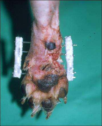
10 Shearing injuries

11 Shearing injuries

12 Shearing injuries


13 Gunshot trauma


14 Gunshot trauma


15 Gunshot trauma


16 Gunshot trauma

17 Mandibular fractures


18

19 Debridement

20 Fixateur externe


21 Very small & Immature patients Very small Minimally invasive Preserving soft tissue & blood supply Yorkshire radius, foot, carpus/tarsus tarsus,, MC/MT Immature MUST NOT bridge active growth plate Limb elongating operations
22 Bridging a joint Only mature animals!! When extra place is required to insert pins Temporary fixation!! Keep joint in functional angle!! Supporting a weak internal fixation


23 Limb correction osteotomy
24 Elements of an ESF system Fixation pins Fixation column (connecting column/rod rod)
25 The different systems K-E system Securos, Imex-SK Synthes-Aesculap PMMA/APEF Manuflex
26 Kirschner-Ehmer system Elements Disadvantages no potential for using smaller/larger larger pins difficulty to use positive profile pins frequent failure/loosening loosening of clamps overcomplex frames to protect weak frame components Straight connecting bar makes ideal pin placement difficult
27 Securos system/ / IMEX-SK system Elements Advantages to K-E system Better strength and versatility Positive profile pins


28 Securos system


29 IMEX-SK system

30 IMEX-SK system


31 Elements PMMA/APEF

32 PMMA/APEF Advantages ease of use no preop planning/preassembly preassembly minimal risk of pin/bar loosening or failure FREEDOM of placing pins pin location not subservient to connecting bar position Easily combined with other types
33 PMMA/APEF Disadvantages Single use Postop adjustment not easy Dynamising not easy K-E


34 Elements Manuflex
35 Classification of frames Unilateral (type I) type Ia type Ib type I tie-in in config (IM pin) Bilateral (type II) Multiplanar (type III) -ex.. a unilateral/type type Ia added to a bilateral Circular/Ilizarov Strength & stiffness: type type Ia < type Ib < type I tie- in < type II < type III)

 type")

36 Classification of frames Unilateral (type I) type Ia type Ib type I tie-in in config (IM pin)


37 Classification of frames Bilateral (type II) Multiplanar (type III) -ex.. a unilateral/type type Ia added to a bilateral Circular/Ilizarov
38 Pin Types Smooth pins (K-wire, Steinmann pins) Cheap but minimal resistance to pullout Inserted in an angle (70 degrees) Positive profile threaded pins More expensive Increased pin-bone interface ( pin-loosening loosening) No need for angulation Preserves core diameter Less prone to pin breakage
 Full")

39 Pin Types Negative threaded pins Bigger pin-bone interface Prone to pin breakage (not recommended) Full pins Better strength & stability Harder to find safe soft tissue corridors Demands more careful alignment Half pins
40 Pin Insertion MOST IMPORTANT: correct limb alignment M-L alignment and torsion!!(valgus valgus/varus, abnormal stresses on adjacent joints) Some CRAN-CAUD CAUD malalignment can be tolerated (elbow/carpus,stifle/tarsustarsus can accomodate)
41 Pin Insertion Releasing soft tissues (prevent windup) Pre-drilling pin holes(decrease heat necrosis) Pins should be min 0,5-1 1 cm from joint surface and min 2 cm from fracture line (fissures!) Two cortices engaged Begin with prox/dist ones alignment
42 Pin Insertion -controlling heat- Adverse reactions: local thermal bone injury+excessive local stress Pin-bone interface affected necrosis necrosis fibrous connective tissue micromovement of pin bone resorption pin pin-loosening Trocar points of pins dont facilitate egress of bone shards impaction impaction HEAT >50rpm thermal necrosis minimized
43 Pin Insertion Larger pins greater pin-bone interface (don t exceed 30%) More pins increase strength (max 3-44 per segment) Stiffer pins more even load sharing Softer pins exerts more stress on near cortex stress resorption
44 Pin Insertion Summary At least 3 pins per fragment Near/far from fracture site Largest possible pins (dont exceed 30%) Stiff pins Positive profile threaded pins If using smooth pins 70 degrees!
45 Bone healing Factors affecting bone healing Age & general health Location & type of fracture Surrounding soft tissue damage Obtained degree of fragment apposition Mechanical environment provided by ESF Primary bony union Rigid internal fixation + close contact of fragments
46 Bone healing Secondary bone healing Fracture gaps + some interfragmentary motion ESF is good example as interfragmentay compr. usually is not applied (ESF preserves blood supply, important for 2ndary union)


47 Primary and secondary bony union
48 Bone healing TRAUMA Soft tissue and bone damage HEMATOME
49 Bone healing FIBRIN STABILIZATION HEMATOME CYTOKINES MESENCHYMAL CELL PROLIFERATION VASCULARISATION
50 Differentiation of mesenchymal cells Small amount of interfragmentary strain Osteoblasts And high O 2 environment More strain And lower O 2 environment Chondroblasts Large amount of strain And low O 2 environment Fibroblasts
51 Callus Formation Outside of callus has best mechanical & biological environment most resistant to bending forces (less strain) periosteal blood supply maintains high O2 environment Fracture ends under high strain + low O2 tension (more chondro/fibroblasts fibroblasts) Outer edge of callus gives most info on secondary callus formation!
52 Callus Formation Limited amount of strain helps bony union (max 2% of fracture area) Excess strain leads to chondro/fibroblast formation + low O2 env NONUNION! Axial forces promote bending bending/twisting forces hinder bony union
53 Postop Xray AAAA=alignment alignment, apposition, apparatus, activity Are there adequate number of pins/segments segments? Pin size correct? Are pins properly centered, safe distance away from skin/fracture lines? Does frame config match fracture type/patient patient size?
54 Postop Xray Amount of external callus is inversely proportional to the rigidity of the frame Osteomyelitis: rough/irregular callus margins Pin-loosening loosening: 1mm radiolucent area around pin
55 Postop Xray Resorption/lack of bridging callus indicates delayed/non non-union reexploration bone grafting new new fixation Generalized loss of bone density indicates stress protection staged dynamization
56 Postop Xray Ideally fractures fill up with bone density material after 6-8 weeks Rechecks at 7-10 days,, 6-86 weeks then every 3-4 weeks 7-10 days recheck Partially weight bearing Some edema Mild pain on pin site
 Xrays (evaluate AAAAs) decision more time, removal, intervention or")
57 Postop Xray 6-8 weeks recheck Usually healed by this time Weight bearing Pain free pin sites (otherwise loosening) Xrays (evaluate AAAAs) decision more time, removal, intervention or dynamization
58 ESF can be removed Animal is weight bearing Sufficient bony callus bridging the fracture gap (density adjacent bone) If severe lameness but good callus find reason (often loose pin!)
59 More time needed Weight bearing but callus not completely bridged Bridging callus of insufficient radiodensity (too stiff frame?)
60 Complications Soft tissue impalement: muscles, nerves (can be severe, wind-up ) tendons, vessels ESF faliure: frame faliure,, pin breakage, pin loosening Infection: osteomyelitis, sequestration, major/minor pin tract infections
61 Pin breakage and loosening Pin breakage: negative threaded pin?, reassess frame strength/rigidity rigidity Pin pullout: smooth pins inserted parallel Pramature pin loosening: very common complication, often poor placement! If good bone union process remove loose pin(s) = dynamization If frame is not stabile anymore review ESF
62 Pin loosening Pin tract drainage Instability Infection
 Sequestrum result of thermal necrosis (poor technique, calcaneus!")
63 Osteomyelitis and sequestrum ESF is a very effective method to manage ongoing osteomyelitis coming from other type of fixation method (internal!) Sequestrum result of thermal necrosis (poor technique, calcaneus!) Remove pin and drill out tract, drain freely

64 Major pin tract infection Common compl. causes premature pin loosening Bacterial colonization of pin-skin interface Pain Purulent discharge Pin loosening
65 Minor pin tract infection To some degree its normal (where thick soft tissue) Bacterial contamination of the pin-skin surface Limited granulation tissue formation Light serous discharge Lack of pain No pin loosening

66 Thank You!

67 Practical training
.org. Tibia (Shinbone) Shaft Fractures. Anatomy. Types of Tibial Shaft Fractures
 Shaft Fractures. Anatomy. Types of Tibial Shaft Fractures") Tibia (Shinbone) Shaft Fractures Page ( 1 ) The tibia, or shinbone, is the most common fractured long bone in your body. The long bones include the femur, humerus, tibia, and fibula. A tibial shaft fracture
Tibia (Shinbone) Shaft Fractures Page ( 1 ) The tibia, or shinbone, is the most common fractured long bone in your body. The long bones include the femur, humerus, tibia, and fibula. A tibial shaft fracture
External Skeletal Fixation (ESF)
") External Skeletal Fixation (ESF) Technique for fracture repair in animals Introduction External Skeletal Fixation is a versatile and effective technique for fracture repair in animals, rigidly stabilizing
External Skeletal Fixation (ESF) Technique for fracture repair in animals Introduction External Skeletal Fixation is a versatile and effective technique for fracture repair in animals, rigidly stabilizing
Biomechanics of Fractures and Fixation
 Biomechanics of Fractures and Fixation Theodore Toan Le, MD Original Author: Gary E. Benedetti, MD; March 2004 New Author: Theodore Toan Le, MD; Revised October 09 Basic Biomechanics Material Properties
Monolateral External Fixation System for Trauma and Orthopaedics
 MEFiSTO Monolateral External Fixation System for Trauma and Orthopaedics Surgical Technique Original Instruments and Implants of the Association for the Study of Internal Fixation AO/ASIF MEFiSTO Table
Types of Plates 1. New Dynamic Compression Plate: Diaphyseal fracture: Radius, Ulna, Humerus, Rarely tibia
 Types of Plates 1. New Dynamic Compression Plate: DCP Diaphyseal fracture: Radius, Ulna, Humerus, Rarely tibia 1. Undercut adjacent to the holes low contact: less stress shield 2. Undercut at the undersurface
pediatric orthopedic implants fixator
 pediatric orthopedic implants DDDAF fixator DAF HUMERUS FIXATOR It is a single plan external fixation tool and used for the treatment of the fractures of humerus, radius and ulna for adults. It is also
ISPUB.COM. Z Ali, L Khurshid, S Vakil, A Anjum, S Dhar OBJECTIVE METHOD PATIENTS
 ISPUB.COM The Internet Journal of Orthopedic Surgery Volume 22 Number 1 Prevalence Of Pin Tract Infection And Role Of Combined Saline And Povidone Iodine With Combined Spirit (Isopropyl Alcohol 70% V/V)
Zimmer Small Fragment Universal Locking System. Surgical Technique
 Zimmer Small Fragment Universal Locking System Surgical Technique Zimmer Small Fragment Universal Locking System 1 Zimmer Small Fragment Universal Locking System Surgical Technique Table of Contents Introduction
Knee spanning solutions
 Knee spanning solutions System features Indications Intended to be used on adults or pediatric patients as required for fracture fixation (open or closed); post-traumatic joint contracture which has resulted
EXTERNAL FIXATION SYSTEM
 EXTERNAL FIXATION SYSTEM The Apex Pin Fixation System is a one step procedure reducing insertion time and reducing insertion temperature. There are three types of fixation pins: Self-drilling / Self-tapping
MEFiSTO. Monolateral External Fixation System for Trauma and Orthopaedics.
 MEFiSTO. Monolateral External Fixation System for Trauma and Orthopaedics. Surgical Technique This publication is not intended for distribution in the USA. Instruments and implants approved by the AO Foundation.
BRIDGE PLATING OF COMMINUTED SHAFT OF FEMUR FRACTURES
 BRIDGE PLATING OF COMMINUTED SHAFT OF FEMUR FRACTURES Mohammad Abul kalam, Pradeep Kumar, Mohammad Afzal Hussain and Iqbal Ahmad Abstract A prospective study of forty comminuted femoral shaft fractures,
QUICK REFERENCE GUIDE. The PreFix Fixator (92000 Series) ALWAYS INNOVATING
 ALWAYS INNOVATING") 21 The PreFix Fixator (92000 Series) ALWAYS INNOVATING INTRODUCTION The PreFix fixator is designed to provide temporary external fixation. This may be needed when local facilities or the condition of the
21 The PreFix Fixator (92000 Series) ALWAYS INNOVATING INTRODUCTION The PreFix fixator is designed to provide temporary external fixation. This may be needed when local facilities or the condition of the
Clinical. Solutions. Synthes Solutions. Foot and Ankle.
 Clinical Solutions Foot and Ankle. Foot and Ankle. Fractures of the tibial shaft Fractures of the distal fibula Fractures of the distal tibia Fractures and osteotomies of the calcaneus Arthrodesis Fractures,
Elbow Hinge Fixator. Guided Flexion/Extension for Unstable Elbow Fractures.
 Elbow Hinge Fixator. Guided Flexion/Extension for Unstable Elbow Fractures. Surgical Technique MR Safe Radiolucent Table of Contents System Description 3 Indications and Contraindications 4 Fixation Components
Small Fragment Plating System. Securing optimal fixation through locked and compression plating technology
 Small Fragment Plating System Securing optimal fixation through locked and compression plating technology Contents Design Rationale Introduction Interfragmentary Fixation Insertion of a 3.5 mm Cortical
Small Fragment Plating System
 Small Fragment Plating System Securing optimal fixation through locked and compression plating technology SURGICAL TECHNIQUE RECOVERY FUNCTION SURVIVORSHIP DePuy believes in an approach to trauma surgery
Surgical Technique. Distal Humerus Locking Plate
 Surgical Technique Distal Humerus Locking Plate PERI-LOC Locked Plating System Distal Humerus Locking Plate Surgical Technique Table of Contents Introduction...2 Indications...3 Plate Features...3 Patient
Small External Fixator Nonspanning Wrist Frame. For the treatment of wrist fractures.
 Small External Fixator Nonspanning Wrist Frame. For the treatment of wrist fractures. Technique Guide Part of the Small External Fixation System Small External Fixator Nonspanning Wrist Frame When to use
Humerus shaft - Reduction & Fixation - Compression plate - AO Surgery Reference. Compression plating
 Humerus shaft 12-A3 ORIF 1. Principles Compression plating Authors Compression plate Compression plating provides fixation with absolute stability for two-part fracture patterns, where the bone fragments
LCP Medial Distal Tibia Plate, without Tab. The Low Profile Anatomic Fixation System with Angular Stability and Optimal Screw Orientation.
 LCP Medial Distal Tibia Plate, without Tab. The Low Profile Anatomic Fixation System with Angular Stability and Optimal Screw Orientation. Technique Guide LCP Small Fragment System Table of Contents Introduction
Technique Guide. DCP and LC-DCP Systems. Dynamic Compression Plates (DCP) and Dynamic Compression Plates with Limited Bone Contact (LC-DCP).
 and Dynamic Compression Plates with Limited Bone Contact (LC-DCP).") Technique Guide DCP and LC-DCP Systems. Dynamic Compression Plates (DCP) and Dynamic Compression Plates with Limited Bone Contact (LC-DCP). Table of Contents Introduction DCP and LC-DCP Systems 2 Indications
Technique Guide DCP and LC-DCP Systems. Dynamic Compression Plates (DCP) and Dynamic Compression Plates with Limited Bone Contact (LC-DCP). Table of Contents Introduction DCP and LC-DCP Systems 2 Indications
Medical Devices HUMERUS. WRIStX
 Medical Devices DDDAF HUMERUS WRIStX DAF HUMERUS FIXATOR It is a single plan external fixation tool and used for the treatment of the fractures of humerus, radius and ulna for adults. It is also used at
Fracture fixation. Types. Mechanical considerations. Biomechanics of fracture fixation. External fixation. Internal fixation
 Fracture fixation Biomechanics of fracture fixation Types External fixation Mechanical considerations Internal fixation Mechanical considerations in treatment of 1. In the external fixation: fracture When
Mini External Fixator
 Stabilize the Phalanges and Metacarpals Mini External Fixator Surgical Technique Table of Contents Introduction Mini External Fixator 2 Indications 4 Surgical Technique Technique Overview 5 Product Information
TIPMED EXTERNAL FIXATION SYSTEMS
 TIPMED EXTERNAL FIXATION SYSTEMS ANATOMICAL LOCATIONS FOR EXTERNAL FIXATION SYSTEMS Humeral Dynamic Axial Fixator Elbow Fixator Pelvic Dynamic Axial Fixator Pennig Wrist Fixator Hand Fixator Finger Fixator
7/23/2018 DESCRIBING THE FRACTURE. Pattern Open vs closed Location BASIC PRINCIPLES OF FRACTURE MANAGEMENT. Anjan R. Shah MD July 21, 2018.
 BASIC PRINCIPLES OF FRACTURE MANAGEMENT Anjan R. Shah MD July 21, 2018 DESCRIBING THE FRACTURE Pattern Open vs closed Location POLL OPEN HOW WOULD YOU DESCRIBE THIS FRACTURE PATTERN? 1 Spiral 2 Transverse
Flexible Fragment Fixation. Surgical Technique
 Flexible Fragment Fixation Surgical Technique 2 F 3 Flexible Fragment Fixation The F 3 Fragment Plating System offers low profile, yet strong fixation in a locked plating construct that can be contoured
Types of fracture healing and association with fixation
 Types of fracture healing and association with fixation MR ANTHONY J THAYAPARAN SPECIALIST REGISTRAR I N TRAUMA AND ORTHOPAEDICS LONDON NORTH WEST HOSPITALS What are we covering? Types of Fracture Healing
Wrist Fixation System
 Wrist Fixation System Anatomy / Fracture Implant EXTRA & SIMPLE ARTICULAR Volar Radius Volar Fixed Angle Plate Volar Bearing Plate Radial Peg Plate Volar Hook Plate Volar Buttress Pin Volar Shear Plate
Ethan M. Braunstein, M.D. 1, Steven A. Goldstein, Ph.D. 2, Janet Ku, M.S. 2, Patrick Smith, M.D. 2, and Larry S. Matthews, M.D. 2
 Skeletal Radiol (1986) 15:27-31 Skeletal Radiology Computed tomography and plain radiography in experimental fracture healing Ethan M. Braunstein, M.D. 1, Steven A. Goldstein, Ph.D. 2, Janet Ku, M.S. 2,
Mandible External Fixator II. Provides treatment for fractures of the maxillofacial area.
 Mandible External Fixator II. Provides treatment for fractures of the maxillofacial area. Lightweight single-phase system Adjustable throughout application Rigid construct using three basic components
Surgical Care at the District Hospital. EMERGENCY & ESSENTIAL SURGICAL CARE
 Surgical Care at the District Hospital 1 18 Orthopedic Trauma Key Points 2 18.1 Upper Extremity Injuries Clavicle Fractures Diagnose fractures from the history and by physical examination Treat with a
Operative technique for external fixation. Thomas Beck Allgemeine und Unfallchirurgie Spitalzentrum Oberwallis
 Operative technique for external fixation Thomas Beck Allgemeine und Unfallchirurgie Spitalzentrum Oberwallis Operative technique of external fixation Basic principles Operative technique & Introduction
The Flower Medial Column Fusion Plate
 The Flower Medial Column Fusion Plate PROCEDURE GUIDE www.flowerortho.com The Flower Foot & Ankle Application NC FUSION PLATE 2-HOLE COMPRESSION PLATE TMT FUSION PLATE LAPIDUS FUSION PLATE COMPRESSION
Locked Plating: Biomechanics and Biology
 Techniques in Orthopaedics 22(4):E1 E6 2007 Lippincott Williams & Wilkins, Inc. Locked Plating: Biomechanics and Biology Kyle F. Dickson, M.D., M.B.A., John W. Munz, M.D. Summary: Since the early ideas
LISS DF and LISS PLT. Less Invasive Stabilization Systems for Distal Femur and Proximal Lateral Tibia.
 LISS DF and LISS PLT. Less Invasive Stabilization Systems for Distal Femur and Proximal Lateral Tibia. LISS DF and LISS PLT. Less Invasive Stabilization Systems for Distal Femur and Proximal Lateral Tibia.
Mandible External Fixator II. Provides treatment for fractures of the maxillofacial area.
 Mandible External Fixator II. Provides treatment for fractures of the maxillofacial area. Technique Guide This publication is not intended for distribution in the USA. Instruments and implants approved
Technique Guide. The Distraction Osteogenesis Ring System. Nonarticular tibia frame.
 Technique Guide The Distraction Osteogenesis Ring System. Nonarticular tibia frame. Table of Contents Introduction The Distraction Osteogenesis Ring System 2 AO Principles 4 Indications 5 Surgical Technique
A locking plate system that expands a surgeon s options in trauma surgery. Zimmer NCB Plating System
 A locking plate system that expands a surgeon s options in trauma surgery Zimmer NCB Plating System The Power of Choice The power of having true intraoperative options is at your fingertips. Using standard
Orthopedics in Motion Tristan Hartzell, MD January 27, 2016
 Orthopedics in Motion 2016 Tristan Hartzell, MD January 27, 2016 Humerus fractures Proximal Shaft Distal Objectives 1) Understand the anatomy 2) Epidemiology and mechanisms of injury 3) Types of fractures
Hoffmann II Compact External Fixation System
 Trauma Hoffmann II Compact External Fixation System Modular System for Upper Extremity Foot Introduction In 1938, Raoul Hoffmann, a surgeon from Geneva, Switzerland, designed a revolutionary External Fixation
MEFiSTO. Monolateral External Fixation System for Trauma and Orthopaedics.
 MEFiSTO. Monolateral External Fixation System for Trauma and Orthopaedics. Surgical Technique This publication is not intended for distribution in the USA. Instruments and implants approved by the AO Foundation.
Primary internal fixation of fractures of both bones forearm by intramedullary nailing
 Original article 21 Primary internal fixation of fractures of both bones forearm by intramedullary nailing Nepal Medical College and Teaching Hospital, Kathmandu, Nepal Correspondenc to: Dr R P Singh,
Surgical Technique. Calcaneal Locking Plate
 Surgical Technique Calcaneal Locking Plate PERI-LOC Locked Plating System Calcaneal Locking Plate Surgical TechniqueCatalog Infor Table of Contents Introduction...2 Indications...3 Plate Features...3 Patient
Vasu Pai FRACS, MCh, MS, Nat Board Ortho Surgeon Gisborne
 Vasu Pai FRACS, MCh, MS, Nat Board Ortho Surgeon Gisborne FRACTURE MANAGEMENT I Simple closed fracture : Complete or Incomplete Stable or unstable II Open fracture III Multiple fracture IV Polytrauma Fractures
Tibial deformity correction by Ilizarov method
 International Journal of Research in Orthopaedics http://www.ijoro.org Case Report DOI: http://dx.doi.org/10.18203/issn.2455-4510.intjresorthop20180422 Tibial deformity correction by Ilizarov method Robert
Technique Guide. Compact 2.0 LOCK Mandible. The locking system for the mandible.
 Technique Guide Compact 2.0 LOCK Mandible. The locking system for the mandible. Table of Contents Introduction Compact 2.0 LOCK Mandible 2 AO Principles 4 Indications and Contraindications 5 Surgical
Open reduction; plate fixation 1 Principles
 Executive Editor: Peter Trafton Authors: Martin Jaeger, Frankie Leung, Wilson Li Proximal humerus 11-A2 Open reduction, plate fixation Search search... Shortcuts All Preparations All Approaches All Reductions
2.7 mm/3.5 mm Variable Angle LCP Elbow System DJ9257-B 1
 2.7 mm/3.5 mm Variable Angle LCP Elbow System DJ9257-B 1 System overview Simply complete: A comprehensive system, consisting of five (5) distal humerus plates and three (3) types of olecranon plates Implant
Midfoot - Reduction & Fixation - ORIF - screw fixation - AO Surgery Reference. ORIF - screw fixation
 Midfoot - TMT (Lisfranc) injury 1. Diagnosis ORIF - screw fixation Authors Mechanism of the injury Tarso-metatarsal (Lisfranc) injuries may be caused by direct or indirect forces. Direct forces include
QUICK REFERENCE GUIDE. The XCaliber Meta-Diaphyseal Fixator
 17 The XCaliber Meta-Diaphyseal Fixator GENERAL POINTS The XCaliber Fixator is made of radiolucent material for unobstructed X-ray visualization. The metallic bolts and the cam and bush of each ball-joint,
Hand Fracture System with Small Bone External Fixation System. Product Overview
 Hand Fracture System with Small Bone External Fixation System Product Overview Acumed Hand Fracture System The Acumed Hand Fracture System is designed to provide both standard and fracture-specific fixation
Surgical Technique. Cannulated Angled Blade Plate 3.5 and 4.5, 90
 Surgical Technique Cannulated Angled Blade Plate 3.5 and 4.5, 90 Cannulated Angled Blade Plate 3.5 and 4.5, 90 Table of contents Indications/Contraindications 2 Implants 3 Surgical technique 5 Implant
Technique Guide. LCP Proximal Femoral Hook Plate 4.5/5.0. Part of the LCP Periarticular Plating System.
 Technique Guide LCP Proximal Femoral Hook Plate 4.5/5.0. Part of the LCP Periarticular Plating System. Table of Contents Introduction Features and Benefits 2 AO ASIF Principles 4 Indications 5 Surgical
Part 2: Selection of Fixation Technique & External Coaptation
 A Practitioner s Guide to Fracture Management Peer Reviewed A Practitioner s Guide to Fracture Management Part 2: Selection of Fixation Technique & External Coaptation Meredith Kapler, DVM North Carolina
Surgical Technique. Anterolateral and Medial Distal Tibia Locking Plates
 Surgical Technique Anterolateral and Medial Distal Tibia Locking Plates PERI-LOC Periarticular Locked Plating System Anterolateral and Medial Distal Tibia Locking Plates Surgical Technique Contents Product
Technique Guide. Small Fragment Locking Compression Plate (LCP) System. Stainless Steel and Titanium.
 System. Stainless Steel and Titanium.") Technique Guide Small Fragment Locking Compression Plate (LCP) System. Stainless Steel and Titanium. Table of Contents Introduction Small Fragment Locking Compression Plate (LCP) System 2 AO Principles
Technique Guide Small Fragment Locking Compression Plate (LCP) System. Stainless Steel and Titanium. Table of Contents Introduction Small Fragment Locking Compression Plate (LCP) System 2 AO Principles
Technique Guide. Small Fragment Locking Compression Plate (LCP) System. Stainless steel and titanium.
 System. Stainless steel and titanium.") Technique Guide Small Fragment Locking Compression Plate (LCP) System. Stainless steel and titanium. Table of Contents Introduction Small Fragment Locking Compression Plate (LCP) System 2 AO Principles
Technique Guide Small Fragment Locking Compression Plate (LCP) System. Stainless steel and titanium. Table of Contents Introduction Small Fragment Locking Compression Plate (LCP) System 2 AO Principles
Simply Complete. VA-LCP Elbow Plating System.
 Simply Complete. VA-LCP Elbow Plating System. Building on success Founded upon the established principles of Synthes LCP technology and the first generation elbow plates, the Variable Angle LCP Elbow Plating
Orthopedic Nursing, Part 2. External Fixation. Nursing Best Practice Guidelines
 Orthopedic Nursing, Part 2 External Fixation Nursing Best Practice Guidelines Reasons for External Fixation Device Placement.. a technique of fracture immobilization a series of transfixing pins are inserted
Surgical Technique. Distal Radius and Foot
 Surgical Technique Distal Radius and Foot JET-X BAR Unilateral Fixator Distal Radius and Foot Surgical Technique Contents Design Features...2 Distal Radius Surgical Technique Indications...10 Surgical
LCP Anterolateral Distal Tibia Plate 3.5. The low profile anatomic fixation system with optimal plate placement and angular stability.
 LCP Anterolateral Distal Tibia Plate 3.5. The low profile anatomic fixation system with optimal plate placement and angular stability. Technique Guide LCP Small Fragment System Table of Contents Introduction
Zimmer Small Fragment Universal Locking System
 Zimmer Small Fragment Universal Locking System Surgical Technique Flexibility and compatibility in one system Zimmer Small Fragment Universal Locking System Zimmer Small Fragment Universal Locking System
Technique Guide. 3.5 mm LCP Olecranon Plates. Part of the Synthes locking compression plate (LCP) system.
 system.") Technique Guide 3.5 mm LCP Olecranon Plates. Part of the Synthes locking compression plate (LCP) system. Table of Contents Introduction 3.5 mm LCP Olecranon Plates 2 AO Principles 3 Indications 3 Clinical
Technique Guide 3.5 mm LCP Olecranon Plates. Part of the Synthes locking compression plate (LCP) system. Table of Contents Introduction 3.5 mm LCP Olecranon Plates 2 AO Principles 3 Indications 3 Clinical
Patient Guide. Intramedullary Skeletal Kinetic Distractor For Tibial and Femoral Lengthening
 Patient Guide Intramedullary Skeletal Kinetic Distractor For Tibial and Femoral Lengthening Introduction You have decided to have a limb lengthening operation. The surgery you have chosen uses a device
Low Profile Neuro Plating System. Surgical Technique
 Low Profile Neuro Plating System Surgical Technique TABLE OF CONTENTS INTRODUCTION Low Profile Neuro Plating System 2 SURGICAL TECHNIQUE Technique 5 PRODUCT INFORMATION Low Profile Neuro Plates 10 Low
Small External Fixator Wrist Spanning Frame. For the treatment of wrist fractures.
 Small External Fixator Wrist Spanning Frame. For the treatment of wrist fractures. Technique Guide Part of the Small External Fixation System Small External Fixator Wrist Spanning Frame When to use The
Technique Guide. 2.4 mm Variable Angle LCP Distal Radius System. For fragment-specific fracture fixation with variable angle locking technology.
 Technique Guide 2.4 mm Variable Angle LCP Distal Radius System. For fragment-specific fracture fixation with variable angle locking technology. Table of Contents Introduction 2.4 mm Variable Angle LCP
Choice, Simplicity, Transition. TransFx External Fixation System Small and Mini Surgical Technique
 Choice, Simplicity, Transition TransFx External Fixation System Small and Mini Surgical Technique TransFx External Fixation System Small and Mini Surgical Technique 1 Surgical Technique For TransFx External
MatrixNEURO. The next generation cranial plating system.
 MatrixNEURO. The next generation cranial plating system. Technique Guide CMF Matrix This publication is not intended for distribution in the USA. Instruments and implants approved by the AO Foundation
LCP Anterolateral Distal Tibia Plate 3.5. The low profile anatomic fixation system with optimal plate placement and angular stability.
 LCP Anterolateral Distal Tibia Plate 3.5. The low profile anatomic fixation system with optimal plate placement and angular stability. Technique Guide LCP Small Fragment System Table of Contents Introduction
A Tale of Four Traumas. Understanding how bones break
 A Tale of Four Traumas Understanding how bones break Bone Break Classifications First Classification Based on whether the break is open to the environment or not. CLOSED- not an open wound. Broken but
LCP Distal Tibia Plate
 Surgical Technique LCP Locking Compression Plate Original Instruments and Implants of the Association for the Study of Internal Fixation AO/ASIF Table of contents Indications 3 Implants/Instruments 5 Surgical
Tibial Shaft Fractures
 Tibial Shaft Fractures Mr Krishna Vemulapalli Consultant Orthopaedics Surgeon Queens & King George Hospitals Queens Hospital 14/03/2018 Google Maps Map data 2018 Google 10 km Orthopaedics Department Covers
NEW INSTRUMENTS FOR INTERNAL FIXATION OF FRACTURES USING MINIMALLY INVASIVE TECHNIQUES
 NEW INSTRUMENTS FOR INTERNAL FIXATION OF FRACTURES USING MINIMALLY INVASIVE TECHNIQUES Dr.eng. Comşa Stanca, sing. Gheorghiu Doina, eng. Ciobota Dan National Institute of Research & Development for fine
Technique Guide. 6.5 mm Midfoot Fusion Bolt. For intramedullary fixation of the medial column of the foot.
 Technique Guide 6.5 mm Midfoot Fusion Bolt. For intramedullary fixation of the medial column of the foot. Table of Contents Introduction 6.5 mm Midfoot Fusion Bolt 2 AO Principles 4 Indications 5 Surgical
EXTERNAL FIXATION SYSTEM
 EXTERNAL FIXATION SYSTEM 2 3 1 4 5 1. DJD II Body 2. Humeral Guide 3. Pin Insertion Guides 4. Hoffmann II Compact Instruments 5. Hoffmann II Compact Components and Apex Pins 2 Overview The DJD II is a
Knee Surgical Technique
 Knee Surgical Technique COMPASS Universal Hinge by Jimmy Tucker, M.D. Orthopaedic Surgeon Director, Arkansas Sports Medicine, P.A. Little Rock, Arkansas Table of contents Design features 3 Indications
Surgical Technique. 3.5mm and 4.5mm Lateral Proximal Tibia Locking Plates
 Surgical Technique 3.5mm and 4.5mm Lateral Proximal Tibia Locking Plates PERI-LOC Periarticular Locked Plating System 3.5mm and 4.5mm Lateral Proximal Tibia Locking Plate Surgical Technique Contents Product
The Flower Four Corner Fusion Plate
 The Flower Four Corner Fusion Plate PROCEDURE GUIDE www.flowerortho.com The Flower Upper Extremity Application PROXIMAL HUMERUS PLATE SMALL BONE PLATES FOUR CORNER FUSION PLATE ANATOMIC DISTAL RADIUS PLATE
LCP Medial Proximal Tibial Plate 4.5/5.0. Part of the Synthes LCP periarticular plating system.
 LCP Medial Proximal Tibial Plate 4.5/5.0. Part of the Synthes LCP periarticular plating system. Technique Guide This publication is not intended for distribution in the USA. Instruments and implants approved
Surgical Technique. Targeter Systems Overview
 Surgical Technique Targeter Systems Overview PERI-LOC Locked Plating System Targeter Systems Overview Table of contents Product overview... 2 Introduction... 2 Indications... 2 Design features and benefits...
LCP Distal Humerus Plates
 The anatomic fixation system for the distal humerus with angular stability Surgical technique LCP Locking Compression Plate Contents Indications and contraindications 2 Implants 3 Instruments 5 Preparation
Mini External Fixator.
 Mini External Fixator. Assembly and Surgical Technique This publication is not intended for distribution in the USA. Instruments and implants approved by the AO Foundation. Image intensifier control Warning
Fractures of the tibia shaft treated with locked intramedullary nail Retrospective clinical and radiographic assesment
 ARS Medica Tomitana - 2013; 4(75): 197-201 DOI: 10.2478/arsm-2013-0035 Șerban Al., Botnaru V., Turcu R., Obadă B., Anderlik St. Fractures of the tibia shaft treated with locked intramedullary nail Retrospective
LCP Metaphyseal Plate for distal medial tibia. Anatomically precontoured metaphyseal plate.
 Indications The LCP Metaphyseal Plate for distal medial tibia is a preshaped plate that allows optimal treatment of juxta-articular fractures of the distal tibia extending into the shaft area. This plate
Foot and Ankle Technique Guide Proximal Inter-Phalangeal (PIP) Fusion
 Fusion") Surgical Technique Foot and Ankle Technique Guide Proximal Inter-Phalangeal (PIP) Fusion Prepared in consultation with: Phinit Phisitkul, MD Department of Orthopedics and Rehabilitation University of Iowa
Surgical Technique Foot and Ankle Technique Guide Proximal Inter-Phalangeal (PIP) Fusion Prepared in consultation with: Phinit Phisitkul, MD Department of Orthopedics and Rehabilitation University of Iowa
Thoracolumbar Spine Locking Plate (TSLP) System. A low-profile plating system for anterior stabilization of the thoracic and lumbar spine.
 System. A low-profile plating system for anterior stabilization of the thoracic and lumbar spine.") Thoracolumbar Spine Locking Plate (TSLP) System. A low-profile plating system for anterior stabilization of the thoracic and lumbar spine. Technique Guide Instruments and implants approved by the AO Foundation
Thoracolumbar Spine Locking Plate (TSLP) System. A low-profile plating system for anterior stabilization of the thoracic and lumbar spine. Technique Guide Instruments and implants approved by the AO Foundation
PROXIMAL TIBIAL PLATE
 SURGICAL NÁSTROJE TECHNIQUE PRO ARTROSKOPII PROXIMAL INSTRUMENTS TIBIAL FOR PLATE ARTHROSCOPY Proximal Tibial Plate Description of medical device The Proximal Tibial Plate is used in epyphyseal and metaphyseal
Fracture and Dislocation of Metacarpal Bones, Metacarpophalangeal Joints, Phalanges, and Interphalangeal Joints ( 1-Jan-1985 )
") In: Textbook of Small Animal Orthopaedics, C. D. Newton and D. M. Nunamaker (Eds.) Publisher: International Veterinary Information Service (www.ivis.org), Ithaca, New York, USA. Fracture and Dislocation
In: Textbook of Small Animal Orthopaedics, C. D. Newton and D. M. Nunamaker (Eds.) Publisher: International Veterinary Information Service (www.ivis.org), Ithaca, New York, USA. Fracture and Dislocation
A Patient s Guide to Adult Forearm Fractures
 A Patient s Guide to Adult Forearm Fractures Orthopedic and Sports Medicine 825 South 8th Street, #550 Minneapolis, MN 55404 Phone: 612-333-5000 Fax: 612-333-6922 1 DISCLAIMER: The information in this
External Fixator Brochure
 External Fixator Brochure Response Ortho is a global orthopaedic trauma solutions manufacturer offering premium products created under its founding principles of innovation, excellence by design and functional
Elbow Plating System
 Elbow Plating System Elbow Plating System Acumed is a global leader of innovative orthopaedic and medical solutions. We are dedicated to developing products, service methods and approaches that improve
Technique Guide. TomoFix Osteotomy System. A comprehensive plating system for stable fixation of osteotomies around the knee.
 Technique Guide TomoFix Osteotomy System. A comprehensive plating system for stable fixation of osteotomies around the knee. Table of Contents Introduction TomoFix Osteotomy System 2 AO Principles 4 Indications
3.5 mm Locking Attachment Plate
 For Treatment of Periprosthetic Fractures 3.5 mm Locking Attachment Plate Surgical Technique Table of Contents Introduction 3.5 mm Locking Attachment Plate 2 Indications 4 Surgical Technique Preparation
Humeral SuturePlate. Surgical Technique
 Humeral SuturePlate Surgical Technique The humeral SuturePlate is an anatomically designed, low profile, titanium polyaxial locking plate and screw system. Multiple chamfered suture eyelets along the margin
AO / Synthes Proximal Posterior Medial Tibia Plate. Tibial Plateau Fx. Osteosynthesis
 Tibial Plateau Fx. Osteosynthesis Articular Fractures Osteosynthesis Characteristics of the LCP system LCP 3.5 system LCP 4.5 system Many different traditional plates (small, large) Lag screws Rafting