Anterior and Medial compartments of the thigh. Dr. Heba Kalbouneh Associate Professor of Anatomy and Histology
|
|
|
- Kimberly Hunt
- 5 years ago
- Views:
Transcription


1 Anterior and Medial compartments of the thigh Dr. Heba Kalbouneh Associate Professor of Anatomy and Histology
2 Terms Related to Movements Movement Flexion Extension Abduction Adduction Medial (internal) rotation Lateral (external) rotation Elevation Depression Protraction Retraction Opposition Definition A movement that decreases the angle between two body parts A movement that increases the angle between two body parts A movement away from the midline A movement towards the midline A rotational movement towards the midline A rotational movement away from the midline A movement in a superior direction A movement in an inferior direction Anterior movement of the jaw Posterior movement of the jaw The movement that involves grasping of the thumb and fingers
3 Movement Supination Pronation Dorsiflexion Plantarflexion Definition A rotational movement where the forearm and palm are turned outwards A rotational movement where the forearm and palm are turned inwards Movement at the ankle, so that the foot points more superiorly Movement at the ankle, so that the foot points inferiorly Inversion Eversion Circumduction The movement of the sole towards the median plane so that the sole faces in a medial direction The movement of the sole away from the median plane so that the sole faces in a lateral direction A conical movement of a body part. Circumduction is a combination of flexion, extension, adduction and abduction.

4 Plantarflexion of foot The foot points inferiorly

5 Dorsiflexion of foot The foot points more superiorly

6 Inversion of foot The movement of the sole towards the median plane so that the sole faces in a medial direction

7 Eversion of foot The movement of the sole away from the median plane so that the sole faces in a lateral direction
 with deep veins Deep veins are deeper in the body and accompany the principle")
8 Superficial veins are those closer to the surface of the body, and have no corresponding arteries. They return blood from skin and subcutaneous structures by anastomosing (inter-connecting) with deep veins Deep veins are deeper in the body and accompany the principle arteries and usually have the same name Superficial veins can be seen under the skin
 saphenous vein Drains the lateral end of the dorsal venous arch. At the knee.")
9 Superficial veins of lower limb Large veins embedded in the subcutaneous (superficial) fascia are: Great (long) saphenous vein Drains the medial end of the dorsal venous arch. Small (short) saphenous vein Drains the lateral end of the dorsal venous arch. At the knee. The great saphenous vein s surface marking is classically one hand's breadth posterior to the medial border of the patella or just posterior to the medial condyle of the femur.
 Small (short) saphenous vein Passes up the back of the leg to penetrate the deep fascia and join the popliteal vein in the popliteal fossa")
10 Superficial veins of lower limb Great (long) saphenous vein Passes up the medial side of the leg, knee, and thigh Ascends in company with the Saphenous nerve Passes through the Saphenous opening Joins the femoral vein (in front of medial malleolus) Small (short) saphenous vein Passes up the back of the leg to penetrate the deep fascia and join the popliteal vein in the popliteal fossa Ascends in company with the Sural nerve (behind lateral malleolus)
11 The normal flow of blood in the lower limbs is from skin and subcutaneous tissues to the superficial veins, which drain by perforating veins to the deep veins, which in turn drain into the iliac veins and inferior vena cava Perforating vein Deep vein Superficial vein

12 Fascia lata: Is a strong fibrous sheet that surrounds the whole of the thigh like a tight trouser. Thick on its lateral side to form the iliotibial tract Saphenous opening is an oval opening in the upper part of the fascia lata of the thigh. Its center lies 1.5 inch below and lateral to the pubic tubercle and is about 3 cm long and 1.5 cm wide The saphenous opening is filled with loose connective tissue called the cribriform fascia

 attached above to the iliac")


13 Iliotibial tract Is a strong wide band (thickening of the deep fascia (fascia lata)) on the lateral side of the thigh) attached above to the iliac tubercle and below to the lateral condyle of tibia. Gluteus maximus Tensor fascia latae Receives the insertion of tensor fascia latae and gluteus maximus muscles

14 Deep fascia of the thigh (fascia lata) Fascia lata is connected to the linea aspera by three intermuscular septa; 1-Medial intermuscular septum 2-Lateral intermuscular septum 3-Posterior intermuscular septum
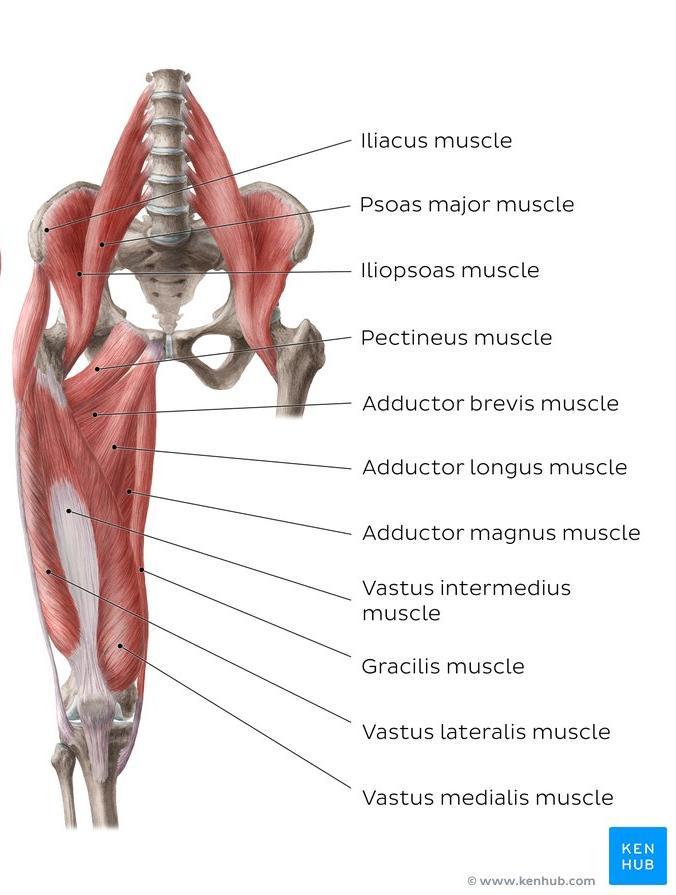
15 Anterior Fascial Compartments of the Thigh Contents of the Anterior Fascial Compartment of the Thigh 1-Muscles: Sartorius, iliacus, psoas, and quadriceps femoris, pectineus 2-Blood supply: Femoral artery 3-Nerve supply: Femoral nerve Note: Not all the contents of the anterior compartment have the same function. For example illopsoas is the main flexor of the thigh at the hip joint while quadriceps femoris is the main extensor of the leg at the knee joint
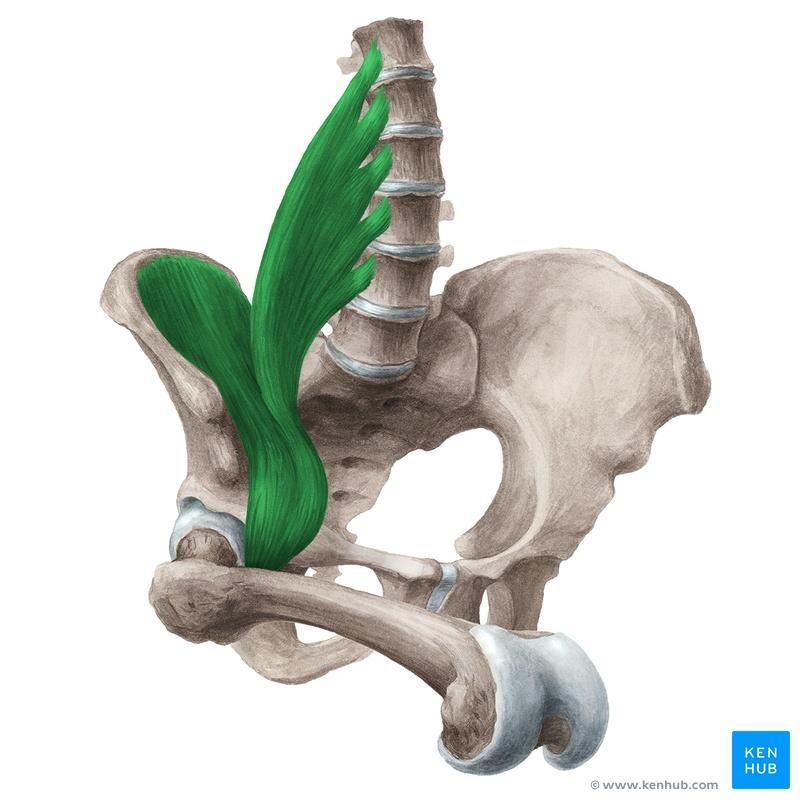
16 Iliacus Origin: Iliac fossa Insertion: with psoas into lesser trochanter Nerve supply: Femoral nerve Action: Flexes thigh on trunk; if thigh is fixed, it flexes the trunk on the thigh as in sitting up from lying down Lesser trochanter
17 Psoas Origin: Transverse processes, bodies, and intervertebral discs of the 12th thoracic and lumbar vertebrae Insertion: With iliacus into lesser trochanter Nerve supply: Lumbar plexus Action: Flexes thigh on trunk; if thigh is fixed, it flexes the trunk on the thigh as in sitting up from lying down
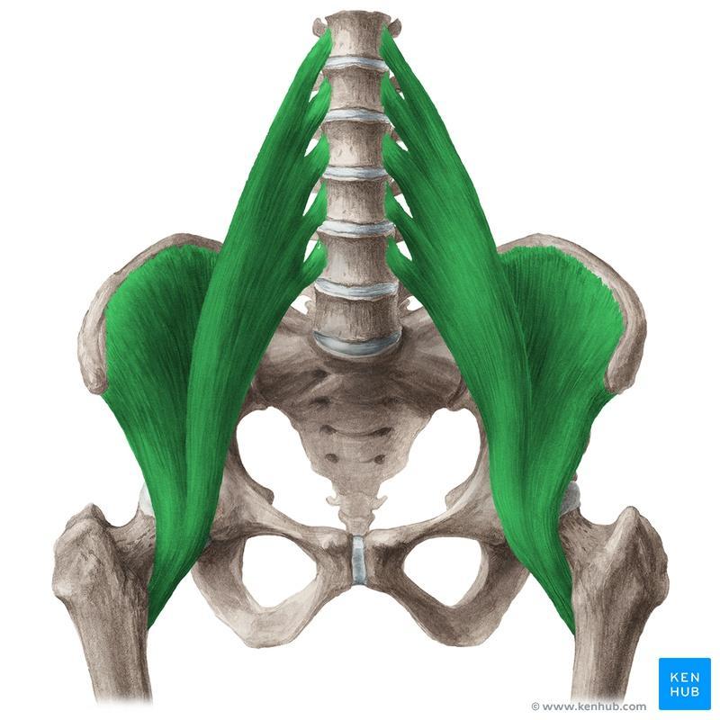
18 Iliopsoas
19
20 Quadriceps femoris
21 Rectus femoris Origin: Straight head: anterior inferior iliac spine (AIIS) Reflected head: ilium above acetabulum Insertion: Quadriceps tendon into patella, then via ligamentum patellae into tibial tuberosity Nerve supply: Femoral nerve Action: Extension of leg at knee joint; flexes thigh at hip joint


22 Vastus lateralis Vastus= Great or Wide Origin: Shaft of femur Insertion: Quadriceps tendon into patella, then via ligamentum patellae into tibial tuberosity Nerve supply: Femoral nerve Action: Extension of leg at knee joint
23 Vastus medialis Origin: Shaft of femur Insertion: Quadriceps tendon into patella, then via ligamentum patellae into tibial tuberosity Nerve supply: Femoral nerve Action: Extension of leg at knee joint; stabilizes patella
24 Vastus intermedius Origin: Shaft of femur Insertion: Quadriceps tendon into patella, then via ligamentum patellae into tibial tuberosity Nerve supply: Femoral nerve Action: Extension of leg at knee joint; articularis genu retracts synovial membrane

25 Sartorius Sartor = Tailor Origin: Anterior superior iliac spine (ASIS) Insertion: Upper medial surface of shaft of tibia (SGS) Nerve supply: Femoral nerve Action: Flexes, abducts, laterally rotates thigh at hip joint; flexes and medially rotates leg at knee joint Helps rotate the leg to the cross-legged position This name was chosen in reference to the cross-legged position in which tailors once sat
26


27 The upper medial surface of shaft of tibia is called SGS area for the attachments of 3 muscles: The combined tendons of insertion are termed pes anserinus (goose foot) Sartorius Gracilis Semitendinosus
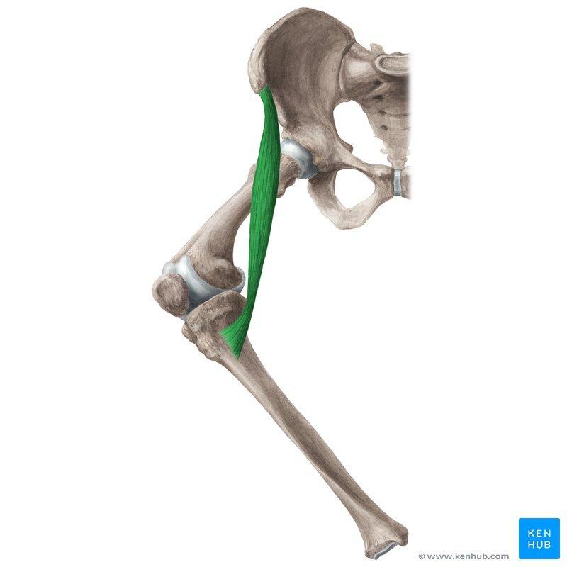
28 Pectineus Origin: Pectineal line of pubis Insertion: Pectineal line of femur Nerve supply: Femoral nerve Action: Flexes and adducts thigh at hip joint

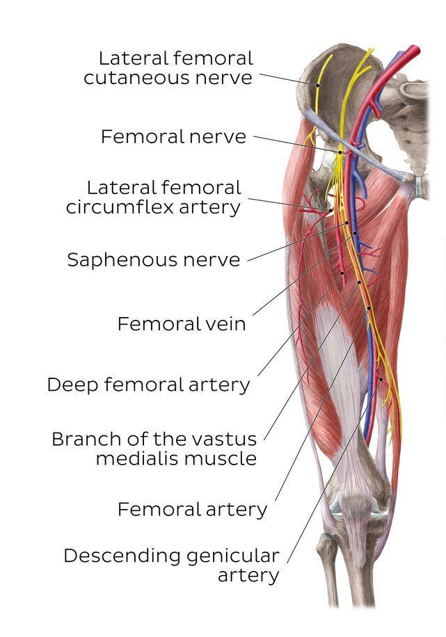
29 Psoas Femoral nerve Is the largest branch of the lumbar plexus (L2, 3, and 4). It emerges from the lateral border of the psoas muscle Leaves the abdomen by passing through the gap behind the inguinal ligament Enters the thigh lateral to the femoral artery and the femoral sheath Innervates all muscles in the anterior compartment of the thigh Innervates skin over the anterior and medial aspects of the thigh, the medial aspect of the knee, leg and the foot (as far as the ball of the big toe) Femoral vein Femoral artery
30 Note that the femoral nerve does not enter the thigh within the femoral sheath Cutaneous branches 1- Medial cutaneous nerve of the thigh 2- Intermediate cutaneous nerve of the thigh 3- Saphenous nerve L2 L3 Muscular branches Iliacus Pectineus Sartorius Quadriceps femoris L4 Articular branches Hip joint Knee joint
31 Femoral nerve L2 L3 L4 Iliacus Inguinal ligament Pectineus Sartorius Quadriceps femoris
 Lateral")
32 Femoral branch of genitofemoral nerve Ilio-inguinal nerve Lateral cutaneous nerve of the thigh Obturator nerve Femoral nerve (Intermediate and medial cutaneous nerves of thigh) Lateral Medial


33 Femoral nerve (Intermediate and medial cutaneous nerves of thigh) Femoral nerve Saphenous nerve THE SAPHENOUS NERVE Runs downward and medially. It emerges between the tendons of sartorius and gracilis It then runs down in company with the great saphenous vein. It passes in front of the medial malleolus Femoral nerve (Saphenous nerve) Supplies the medial side of the knee, leg and foot as far as the ball of the big toe


34 Saphenous nerve The saphenous nerve accompanies the femoral artery through the adductor canal, but does not pass through the adductor hiatus with the femoral artery. Rather, the saphenous nerve penetrates the deep fascia and appears between the sartorius and gracilis muscles on the medial side of the knee, and continues down the medial side of the leg to the foot, and supplies skin on the medial side of the knee, leg, and foot.
35 Medial Fascial Compartment of the Thigh Can you think of a bone that can be suitable to provide an origin for an adductor muscle of the hip joint? The Pubic bone Would you be able to think of a bone that can be a good insertion FOR the adductor muscles? The femur
36 Medial fascial compartment of the Thigh Contents of the Medial Fascial Compartment of the Thigh 1- Muscles: Pectineus, Adductor Longus, Adductor Brevis, Adductor Magnus, Gracilis Obturator externus 2-Nerve supply: Obturator nerve 3-Blood supply: Profunda femoris artery and obturator artery In the practical session Remember that the adductor muscles are arranged in three layers in similar way to that of the pages of the book The first layer (page) contains: pectineus and adductor longus The second layer contains: adductor brevis only The third layer contains: adductor magnus only
37 Action of the adductor muscles as a group 1- Adduct the thigh, although adduction of the thigh is not important in the mechanism of walking and standing 2-Because their origin is in front of the hip joint (in a plane that is in front of the hip joint) they can flex the thigh at the hip joint 3- They assist in medial rotation of the thigh
38 Gracilis Origin: Body, inferior ramus of pubis, ramus of ischium Insertion: Upper part of shaft of tibia on medial surface (SGS) Nerve supply: Obturator nerve Action: Adducts thigh at hip joint; flexes leg at knee joint
39 Adductor longus Origin: Body of pubis, inferior to pubic tubercle Insertion: Posterior surface of shaft of femur (linea aspera) Nerve supply: Obturator nerve Action: Adducts thigh at hip joint
40 Adductor brevis Origin: Body and inferior ramus of pubis Insertion: Posterior surface of shaft of femur (linea aspera) Nerve supply: Obturator nerve Action: Adducts thigh at hip joint
41 Adductor magnus Origin: Adductor part: Inferior ramus of pubis, ramus of ischium Hamstring part: ischial tuberosity Insertion: Adductor part: Posterior surface of shaft of femur Hamstring part: adductor tubercle of femur Nerve supply: Adductor part: obturator nerve Hamstring part: sciatic nerve Action: Adducts thigh at hip joint; hamstring portion extends thigh at hip joint

42 Obturator nerve L2 L3 Arises from the lumbar plexus (L2, 3, and 4) anterior divisions Emerges on the medial border of the psoas muscle Enters the thigh by passing through the obturator canal It divides into anterior and posterior divisions Supplies all muscles in the medial compartment except the hamstring part of adductor magnus and pectineus Supplies obturator externus Supplies the skin on the medial side of upper thigh L4 Femoral nerve (Intermediate and medial cutaneous nerves of thigh)
43 L2 L3 L4 Psoas Femoral nerve Obturator nerve


44 Descends on the anterior surface of adductor brevis The anterior division Muscular branches to 3 muscles : Gracilis, Adductor brevis, Adductor longus Articular branches to the hip joint Cutaneous branches to the skin on the medial side of the thigh. Descends on the posterior surface of adductor brevis The posterior division Muscular branches to 2 muscles : Obturator externus Adductor part of the adductor magnus Articular branches to the knee joint Referred pain Is the pain perceived at a location other than the site of the painful stimulus.

45 Hilton s law states that the nerves crossing a joint supply 1-The muscles acting on it 2-The skin over the joint 3-The joint itself For example, The hip receives fibres from the femoral, sciatic and obturator nerves. It is important to note that these nerves also supply the knee joint and, for this reason, it is not uncommon for a patient, particularly a child, to complain bitterly of pain in the knee and for the cause of the mischief, the diseased hip, to be overlooked


46 Articularis Genu Origin: Anterior surface of distal part of the shaft of the femur Insertion: Proximal part of the suprapatellar bursa (an extension of the synovial cavity of the knee joint) It is often part of the vastus intermedius. This muscle lies deep to the vastus intermedius Pulls the bursa away from the knee joint during extension Articularis: joint Genu: knee

47 Superficial fascia of the thigh Contents: 1- Cutaneous nerves 2- Superficial arteries 3- Superficial veins 4- Superficial inguinal lymph nodes
48 Genitofemoral nerve Skin of the thigh Ilioinguinal nerve Femoral nerve (Intermediate and medial cutaneous nerves of thigh)
49 3 2 1 Superficial arteries: 1- Superficial external pudendal artery: passes medial to external genitalia 2- Superficial epigastric artery: passes upward toward the umbilicus 3- Superficial circumflex iliac artery: passes laterally toward the iliac crest Femoral triangle All these are branches of femoral artery Lateral Medial
50 3 2 1 Superficial veins: Great saphenous vein and its tributaries The superficial veins that correspond to the superficial branches of the femoral artery drain into the great saphenous vein Femoral triangle Lateral Medial

51 Superficial inguinal lymph nodes Lies below the inguinal ligament Divided into two groups; horizontal and vertical. A-The horizontal group lies below and parallel to the inguinal ligament. B-The vertical group lies along the terminal part of Saphenous vein. Lymph nodes cannot be palpated or seen unless they are enlarged
) 4- Anterior abdominal wall")
52 Remember that if the patient presented to you with an enlarged superficial inguinal lymph nodes you should ask about and check the below mentioned areas Both groups (horizontal and vertical) receive superficial lymph vessels from: 1- All the lower limb 2- Gluteal region 3- Perineum (Lower half of anal canal, Distal urethra, Genitalia of both sexes (but not the testes)) 4- Anterior abdominal wall below umbilicus The efferent lymph vessels from the superficial inguinal nodes pass through the saphenous opening in the deep fascia and join the deep inguinal nodes
53 Great saphenous vein The horizontal group of lymph nodes

54 Great saphenous vein Saphenous opening transmits the great saphenous vein, some superficial branches of the femoral artery, and lymph vessels
55
56
ANATYOMY OF The thigh
 ANATYOMY OF The thigh 1- Lateral cutaneous nerve of the thigh Ι) Skin of the thigh Anterior view 2- Femoral branch of the genitofemoral nerve 5- Intermediate cutaneous nerve of the thigh 1, 2 and 3 are
ANATYOMY OF The thigh
 ANATYOMY OF The thigh 1- Lateral cutaneous nerve of the thigh Ι) Skin of the thigh Anterior view 2- Femoral branch of the genitofemoral nerve 5- Intermediate cutaneous nerve of the thigh 1, 2 and 3 are
ANATYOMY OF The thigh
 ANATYOMY OF The thigh 1- Lateral cutaneous nerve of the thigh Ι) Skin of the thigh Anterior view 2- Femoral branch of the genitofemoral nerve 1, 2 and 3 are From the lumber plexus 5- Intermediate cutaneous
rotation of the hip Flexion of the knee Iliac fossa of iliac Lesser trochanter Femoral nerve Flexion of the thigh at the hip shaft of tibia
 Anatomy of the lower limb Anterior & medial compartments of the thigh Dr. Hayder The fascia lata encloses the entire thigh like a sleeve/stocking. Three intramuscular fascial septa (lateral, medial, and
The thigh. Prof. Oluwadiya KS
 The thigh Prof. Oluwadiya KS www.oluwadiya.com The Thigh: Boundaries The thigh is the region of the lower limb that is approximately between the hip and knee joints Anteriorly, it is separated from the
Baraa Ayed حسام أبو عوض. Ahmad Salman. 1 P a g e
 4 Baraa Ayed حسام أبو عوض Ahmad Salman 1 P a g e Today we are going to cover these concepts: Iliotibial tract Anterior compartment of the thigh and the hip Medial compartment of the thigh Femoral triangle
Lecture 08 THIGH MUSCLES ANTERIOR COMPARTMENT. Dr Farooq Khan Aurakzai. Dated:
 Lecture 08 THIGH MUSCLES ANTERIOR COMPARTMENT BY Dr Farooq Khan Aurakzai Dated: 11.02.2017 INTRODUCTION to the thigh Muscles. The musculature of the thigh can be split into three sections by intermuscular
Identify the muscles associated with the medial compartment of the thigh. Identify the attachment points of the medial thigh muscles.
 L 8 A B O R A T O R Y Thigh MEDIAL THIGH Identify the muscles associated with the medial compartment of the thigh. Identify the attachment points of the medial thigh muscles. Identify the actions of these
The Hip (Iliofemoral) Joint. Presented by: Rob, Rachel, Alina and Lisa
 Joint. Presented by: Rob, Rachel, Alina and Lisa") The Hip (Iliofemoral) Joint Presented by: Rob, Rachel, Alina and Lisa Surface Anatomy: Posterior Surface Anatomy: Anterior Bones: Os Coxae Consists of 3 Portions: Ilium Ischium Pubis Bones: Pubis Portion
The Hip (Iliofemoral) Joint Presented by: Rob, Rachel, Alina and Lisa Surface Anatomy: Posterior Surface Anatomy: Anterior Bones: Os Coxae Consists of 3 Portions: Ilium Ischium Pubis Bones: Pubis Portion
lower limb Anterior Compartment: lecture 3 The deep fascia ( fascia lata) divides the thigh into 3 compartments:
 divides the thigh into 3 compartments:") lower limb lecture 3 The deep fascia ( fascia lata) divides the thigh into 3 compartments: 1. Anterior Extensor compartment 2. Medial Adductor compartment 3. Posterior Flexor compartment Anterior Compartment:
lower limb lecture 3 The deep fascia ( fascia lata) divides the thigh into 3 compartments: 1. Anterior Extensor compartment 2. Medial Adductor compartment 3. Posterior Flexor compartment Anterior Compartment:
Lower limb summary. Anterior compartment of the thigh. Done By: Laith Qashou. Doctor_2016
 Lower limb summary Done By: Laith Qashou Doctor_2016 Anterior compartment of the thigh Sartorius Anterior superior iliac spine Upper medial surface of shaft of tibia 1. Flexes, abducts, laterally rotates
Muscles of the Thigh. 6.1 Identify, describe the attachments of and deduce the actions of the muscles of the thigh: Anterior group
 Muscles of the Thigh 6.1 Identify, describe the attachments of and deduce the actions of the muscles of the thigh: Anterior group Sartorius: This is a long strap like muscle with flattened tendons at each
Femoral Triangle and Adductor Canal. Dr. Heba Kalbouneh Associate Professor of Anatomy and Histology
 Femoral Triangle and Adductor Canal Dr. Heba Kalbouneh Associate Professor of Anatomy and Histology Femoral Triangle and Adductor Canal Femoral triangle Is a triangular depressed area located in the upper
Muscles of Lesson Five. Muscular Nomenclature and Kinesiology - Two. Muscles of Lesson Five, cont. Chapter 16
 Chapter 16 Muscular Nomenclature and Kinesiology - Two Lessons 5-6 Muscles of Lesson Five Iliopsoas (psoas major, iliacus) Hip outward rotators (piriformis, gemellus superior, gemellus inferior, obturator
Muscles of the lower extremities. Dr. Nabil khouri MD, MSc, Ph.D
 Muscles of the lower extremities Dr. Nabil khouri MD, MSc, Ph.D Posterior leg Popliteal fossa Boundaries Biceps femoris (superior-lateral) Semitendinosis and semimembranosis (superior-medial) Gastrocnemius
Human Anatomy Biology 351
 Human Anatomy Biology 351 Lower Limb Please place your name on the back of the last page of this exam. You must answer all questions on this exam. Because statistics demonstrate that, on average, between
HUMAN BODY COURSE LOWER LIMB NERVES AND VESSELS
 HUMAN BODY COURSE LOWER LIMB NERVES AND VESSELS October 22, 2010 D. LOWER LIMB MUSCLES 2. Lower limb compartments ANTERIOR THIGH COMPARTMENT General lfunction: Hip flexion, knee extension, other motions
lesser trochanter of femur lesser trochanter of femur iliotibial tract (connective tissue) medial surface of proximal tibia
 medial surface of proximal tibia") LOWER LIMB MUSCLES OF THE APPENDICULAR SKELETON The muscles that act on the lower limb fall into three groups: those that move the thigh, those that move the lower leg, and those that move the ankle, foot,
LOWER LIMB MUSCLES OF THE APPENDICULAR SKELETON The muscles that act on the lower limb fall into three groups: those that move the thigh, those that move the lower leg, and those that move the ankle, foot,
Lumbar Plexus. Ventral rami L1 L4 Supplies: Major nerves.. Abdominal wall External genitalia Anteromedial thigh
 Lower Limb Nerves Lectures Objectives Describe the structure and relationships of the plexuses of the lower limb. Describe the course, relationships and structures supplied for the major nerves of the
DISSECTION SCHEDULE. Session I - Hip (Front) & Thigh (Superficial)
 & Thigh (Superficial)") DISSECTION SCHEDULE Session I - Hip (Front) & Thigh (Superficial) Surface anatomy Inguinal region Gluteal region Thigh Leg Foot bones Hip bone Femur Superficial fascia Great saphenous vein Superficial
DISSECTION SCHEDULE Session I - Hip (Front) & Thigh (Superficial) Surface anatomy Inguinal region Gluteal region Thigh Leg Foot bones Hip bone Femur Superficial fascia Great saphenous vein Superficial
The Muscular System. Chapter 10 Part D. PowerPoint Lecture Slides prepared by Karen Dunbar Kareiva Ivy Tech Community College
 Chapter 10 Part D The Muscular System Annie Leibovitz/Contact Press Images PowerPoint Lecture Slides prepared by Karen Dunbar Kareiva Ivy Tech Community College Table 10.14: Muscles Crossing the Hip and
Muscles of the Hip 1. Tensor Fasciae Latae O: iliac crest I: lateral femoral condyle Action: abducts the thigh Nerve: gluteal nerve
 Muscles of the Hip 1. Tensor Fasciae Latae O: iliac crest I: lateral femoral condyle Action: abducts the thigh Nerve: gluteal nerve 2. Gluteus Maximus O: ilium I: femur Action: abduct the thigh Nerve:
Lectures of Human Anatomy
 Lectures of Human Anatomy Lower Limb Gluteal Region and Hip Joint By DR. ABDEL-MONEM AWAD HEGAZY M.B. with honor 1983, Dipl."Gynecology and Obstetrics "1989, Master "Anatomy and Embryology" 1994, M.D.
Gluteal region DR. GITANJALI KHORWAL
 Gluteal region DR. GITANJALI KHORWAL Gluteal region The transitional area between the trunk and the lower extremity. The gluteal region includes the rounded, posterior buttocks and the laterally placed
Human Anatomy Biology 255
 Human Anatomy Biology 255 Exam #4 Please place your name and I.D. number on the back of the last page of this exam. You must answer all questions on this exam. Because statistics demonstrate that, on average,
Lower Limb Nerves. Clinical Anatomy
 Lower Limb Nerves Clinical Anatomy Lumbar Plexus Ventral rami L1 L4 Supplies: Abdominal wall External genitalia Anteromedial thigh Major nerves.. Lumbar Plexus Nerves relation to psoas m. : Obturator n.
musculoskeletal system anatomy nerves of the lower limb 1 done by: dina sawadha & mohammad abukabeer
 musculoskeletal system anatomy nerves of the lower limb 1 done by: dina sawadha & mohammad abukabeer What is the importance of plexuses? plexuses provides us the advantage of a phenomenon called convergence
Main Menu. Joint and Pelvic Girdle click here. The Power is in Your Hands
 1 Hip Joint and Pelvic Girdle click here Main Menu K.6 http://www.handsonlineeducation.com/classes//k6entry.htm[3/23/18, 2:01:12 PM] Hip Joint (acetabular femoral) Relatively stable due to : Bony architecture
Femoral Artery. Its entrance to the thigh Position Midway between ASIS and pubic symphysis
 Lower Limb Vessels Lecture Objectives Describe the major arteries of the lower limb. Describe the deep and superficial veins of the lower limb. Describe the topographical relationships of the arteries
The psoas minor is medial to the psoas major. The iliacus is a fan-shaped muscle that when contracted helps bring the swinging leg forward in walking
 1 p.177 2 3 The psoas minor is medial to the psoas major. The iliacus is a fan-shaped muscle that when contracted helps bring the swinging leg forward in walking and running. The iliopsoas and adductor
MUSCULOSKELETAL LOWER LIMB
 MUSCULOSKELETAL LOWER LIMB Spinal Cord Lumbar and Sacral Regions Spinal cord Dorsal root ganglion Conus medullaris Cauda equina Dorsal root ganglion of the fifth lumbar nerve End of subarachnoid space
Anatomy & Physiology. Muscles of the Lower Limbs.
 Anatomy & Physiology Muscles of the Lower Limbs http://www.ishapeup.com/musclecharts.html Muscles of the Lower Limbs Among the strongest muscles in the body. Because pelvic girdle is composed of heavy,
Anatomage Table Instructors Guide- Lower Limb
 The Lower Limb Anatomage Table Instructors Guide- Lower Limb Table of Contents Lower Limb 1- The Skeletal System...3 1: Hip Bone...3 2: Hip Joint and Femur...4 3: Patella and Knee Joint...7 4: Tibia, Fibula,
Mohammad Ashraf. Abdulrahman Al-Hanbali. Ahmad Salman. 1 P a g e
 - 7 Mohammad Ashraf Abdulrahman Al-Hanbali Ahmad Salman 1 P a g e Structures under the cover of Gluteus Maximus: 1-Bones: Ileum, Femur (Head, greater trochanter and gluteal tuberosity), Ischium (ischial
Human Anatomy Biology 351
 Human Anatomy Biology 351 Lower Limb Please place your name on the back of the last page of this exam. You must answer all questions on this exam. Because statistics demonstrate that, on average, between
The Hay is in the Barn
 Anatomy 1 Practical 1 Review Made by Forrest Allen (nerd) Edited by TJ Williamson (not nerd) The Hay is in the Barn 2019 Thunderbringers Too much to handle https://www.youtube.com/watch?v=glii-kaza d8
THIEME. Anterior and Medial Compartments of the Thigh
 CHAPTER Anterior and Medial Compartments of the Thigh Learning Objectives 2 At the end of the dissection, you should be able to identify the following: Cutaneous nerves innervating the skin of the anterior
1-Muscles: 2-Blood supply: Branches of the profunda femoris artery. 3-Nerve supply: Sciatic nerve
 1-Muscles: B i c e p s f e m o r i s S e m i t e n d i n o s u s S e m i m e m b r a n o s u s a small part of the adductor magnus (h a m s t r i n g p a r t o r i s c h i a l p a r t ) 2-Blood supply:
Contents of the Posterior Fascial Compartment of the Thigh
 Contents of the Posterior Fascial Compartment of the Thigh 1-Muscles: B i c e p s f e m o r i s S e m i t e n d i n o s u s S e m i m e m b r a n o s u s a small part of the adductor magnus (h a m s t
Regional Anaesthesia
 Regional Anaesthesia Lower limb anatomy and blocks Hip and Knee Joint Hip Joint: Nerve supply Lumbar plexus Femoral nerve through the nerve to the Rectus Femoris Ant division of the Obturator nerve The
The University Of Jordan Faculty Of Medicine THE LOWER LIMB. Dr.Ahmed Salman Assistant Prof. of Anatomy. The University Of Jordan
 The University Of Jordan Faculty Of Medicine THE LOWER LIMB Dr.Ahmed Salman Assistant Prof. of Anatomy. The University Of Jordan Gluteal Region Cutaneous nerve supply of (Gluteal region) 1. Lateral cutaneous
The Lower Limb II. Anatomy RHS 241 Lecture 3 Dr. Einas Al-Eisa
 The Lower Limb II Anatomy RHS 241 Lecture 3 Dr. Einas Al-Eisa Tibia The larger & medial bone of the leg Functions: Attachment of muscles Transfer of weight from femur to skeleton of the foot Articulations
Leg. Dr. Heba Kalbouneh Associate Professor of Anatomy and Histology
 Leg Dr. Heba Kalbouneh Associate Professor of Anatomy and Histology Skin of the Leg Cutaneous Nerves Medially: The saphenous nerve, a branch of the femoral nerve supplies the skin on the medial surface
The Hip Joint. Shenequia Howard David Rivera
 The Hip Joint Shenequia Howard David Rivera Topics Of Discussion Movement Bony Anatomy Ligamentous Anatomy Muscular Anatomy Origin/Insertion/Action/Innervation Common Injuries MOVEMENT Flexion Extension
Where should you palpate the pulse of different arteries in the lower limb?
 Where should you palpate the pulse of different arteries in the lower limb? The femoral artery In the femoral triangle, its pulse is easily felt just inferior to the inguinal ligament midway between the
MUSCLES OF THE LOWER LIMBS
 MUSCLES OF THE LOWER LIMBS Naming, location and general function Dr. Nabil khouri ROLES THAT SHOULD NOT BE FORGOTTEN Most anterior compartment muscles of the hip and thigh Flexor of the femur at the hip
Lesson 24. A & P Hip
 Lesson 24 A & P Hip 1 Aims of the Session This session will allow candidates to have an understanding of the bony prominences and soft tissues of the hip 2 Learning Outcomes By the end of the lesson the
Adductor canal (Subsartorial) or Hunter s canal
 or Hunter s canal") Adductor canal (Subsartorial) or Hunter s canal John Hunter described the exposure and ligation of the femoral artery in this canal for aneurysm of the popliteal artery; this method has the advantage that
Adductor canal (Subsartorial) or Hunter s canal John Hunter described the exposure and ligation of the femoral artery in this canal for aneurysm of the popliteal artery; this method has the advantage that
5.1 Identify, describe the attachments of and deduce the actions of the muscles of the thigh:
 5.1 Identify, describe the attachments of and deduce the actions of the muscles of the thigh: Anterior group Proximal attachment Distal attachment Sartorius ASIS» Upper part of shaft tibia (middle surface)»
Hip joint and pelvic girdle. Lower Extremity. Pelvic Girdle 6/5/2017
 Hip joint and pelvic girdle Lower Extremity The relationship between the pelvic girdle and hip is similar to that between the shoulder girdle and shoulder joint. The lower limbs are attached to the axial
Acland's DVD Atlas of Human Anatomy. Transcript for Volume Robert D Acland
 Acland's DVD Atlas of Human Anatomy Transcript for Volume 2 2007 Robert D Acland This free downloadable pdf file is to be used for individual study only. It is not to be reproduced in any form without
Scapula Spine Lateral edge of clavicle. Medial border Scapula. Medial border of Scapula, between superior angle and root of spine. Scapula.
 Muscle attachments and actions answer sheet Muscle Origins insertions Movements Joints crossed Trapezius Base of skull Spinous process of C7 Thoracic Spine Lateral edge of clavicle Elevation Retraction
Posterior compartment of the thigh. Dr. Heba Kalbouneh Associate Professor of Anatomy and Histology
 Posterior compartment of the thigh Dr. Heba Kalbouneh Associate Professor of Anatomy and Histology Posterior compartment of the thigh 1-Muscles: Biceps femoris Semitendinosus Semimembranosus Adductor magnus
First practical session. Bones of the gluteal region
 First practical session 2017 Bones of the gluteal region The Hip bone The hip bone is made of: 1 The ilium: superior in position 2 The ischium:postero-inferior in position 3 The pubis: antero-inferior
ANATOMY TEAM GLUTEAL REGION & BACK OF THIGH
 ANATOMY TEAM GLUTEAL REGION & BACK OF THIGH OBJECTIVES By the end of this lecture, the student should be able to identify and discuss: Contents of gluteal region: Groups of Glutei muscles and small muscles
Sports Medicine 15. Unit I: Anatomy. The knee, Thigh, Hip and Groin. Part 4 Anatomies of the Lower Limbs
 Sports Medicine 15 Unit I: Anatomy Part 4 Anatomies of the Lower Limbs The knee, Thigh, Hip and Groin Anatomy of the lower limbs In Part 3 of this section we focused upon 11 of the 12 extrinsic muscles
Topic 7: Hip and pelvis. Parts of the hip. Parts of the femur
 Topic 7: Hip and pelvis Parts of the hip Parts of the femur Classifying the hip joint Ball and socket Synovial Multiaxial Movements of the hip: Abduction/adduction Flexion/extension Medial/lateral rotation
musculoskeletal system anatomy nerves of the lower limb 2 done by: Dina sawadha & mohammad abukabeer
 musculoskeletal system anatomy nerves of the lower limb 2 done by: Dina sawadha & mohammad abukabeer #Sacral plexus : emerges from the ventral rami of the spinal segments L4 - S4 and provides motor and
Year 2004 Paper one: Questions supplied by Megan
 QUESTION 47 A 58yo man is noted to have a right foot drop three days following a right total hip replacement. On examination there is weakness of right ankle dorsiflexion and toe extension (grade 4/5).
CHAPTER 8: THE BIOMECHANICS OF THE HUMAN LOWER EXTREMITY
 CHAPTER 8: THE BIOMECHANICS OF THE HUMAN LOWER EXTREMITY _ 1. The hip joint is the articulation between the and the. A. femur, acetabulum B. femur, spine C. femur, tibia _ 2. Which of the following is
Muscles of the Gluteal Region
 Muscles of the Gluteal Region 1 Some of the most powerful in the body Extend the thigh during forceful extension Stabilize the iliotibial band and thoracolumbar fascia Related to shoulders and arms because
Muscles of Gluteal Region
 1 The Gluteal Region In the gluteal region the skin is tough with many layers underneath. Directly under it is the superficial fascia followed by the deep fascia then the muscles and the bones of the thigh.
Bones of Lower Limb. Dr. Heba Kalbouneh Associate Professor of Anatomy and Histology
 Bones of Lower Limb Dr. Heba Kalbouneh Associate Professor of Anatomy and Histology Bones of the lower limb Hip Bone Made up of 3 bones: 1) Ilium (flat), superior in position 2) Ischium (L), postero-inferior
5 Testing the Muscles of the Lower Extremity
 C H A P T E R 5 Testing the Muscles of the Lower Extremity Hip Flexion Hip Flexion, Abduction, and External Rotation with Knee Flexion Hip Extension Hip Abduction Hip Abduction from Flexed Position Hip
Figure 1 - Hip and Pelvis
 Hip Figure 1 - Hip and Pelvis The terms hip and pelvis are frequently used interchangeably, but strictly speaking, the pelvis is a girdle of bones and the hip is a joint. The pelvis consists of The sacrum
Myology of the Knee. PTA 105 Kinesiology
 Myology of the Knee PTA 105 Kinesiology Objectives Describe the planes of motion and axes of rotation of the knee joint Visualize the origins and insertions of the muscles about the knee List the innervations
LOWER LIMB. As we know the bony part of the body is divided into Axial and Appendicular (upper and lower Limbs)
") LOWER LIMB As we know the bony part of the body is divided into Axial and Appendicular (upper and lower Limbs) Bones of the Lower limb: 1-Pelvic Girdle: composed of: 1. Right hip bone : is formed by 3
LOWER LIMB As we know the bony part of the body is divided into Axial and Appendicular (upper and lower Limbs) Bones of the Lower limb: 1-Pelvic Girdle: composed of: 1. Right hip bone : is formed by 3
Bony Anatomy. Femur. Femoral Head Femoral Neck Greater Trochanter Lesser Trochanter Intertrochanteric Crest Intertrochanteric Line Gluteal Tuberosity
 Hip Anatomy Bony Anatomy Femur Femoral Head Femoral Neck Greater Trochanter Lesser Trochanter Intertrochanteric Crest Intertrochanteric Line Gluteal Tuberosity Bony Anatomy Pelvic Girdle Acetabulum 3 bones
Lower Limb Dr. Robin Paudel
 Lower Limb n What is a limb? n Skeleton n Joints n Pelvis or limb girdle n Hip/Hip Muscles n Lumber and sacral plexus getting spinal nerves out onto limb n Muscles anterior and posterior compartments n
The Lower Limb. Anatomy RHS 241 Lecture 2 Dr. Einas Al-Eisa
 The Lower Limb Anatomy RHS 241 Lecture 2 Dr. Einas Al-Eisa The bony pelvis Protective osseofibrous ring for the pelvic viscera Transfer of forces to: acetabulum & head of femur (when standing) ischial
VENOUS DRAINAGE OF THE LOWER LIMB
 Anatomy of the lower limb Superficial veins & nerve injuries Dr. Hayder VENOUS DRAINAGE OF THE LOWER LIMB The venous drainage of the lower limb is of huge clinical & surgical importance. Since the venous
Venous drainage of the lower limb
 Venous drainage of the lower limb INTRODUCTION It is of immense clinical and surgical importance. The venous blood against gravity. FACTORS HELPING THE VENOUS DRAINAGE OF THE LOWER LIMB The contraction
Ali Yaghi. Omar Eyad. Ahmad Salman. 1 P a g e
 5 Ali Yaghi Omar Eyad Ahmad Salman 1 P a g e **There are two types of groin hernia; the femoral hernia and the inguinal hernia. But how can we differentiate between the inguinal hernia and the femoral
The front of the thigh. Dr.Amjad shatarat
 The front of the thigh Femoral triangle (Scarpa s triangle) Is a triangular depressed area located in the upper part of the medial aspect of the thigh immediately below the inguinal ligament. Superiorly:
The Knee. Clarification of Terms. Osteology of the Knee 7/28/2013. The knee consists of: The tibiofemoral joint Patellofemoral joint
 The Knee Clarification of Terms The knee consists of: The tibiofemoral joint Patellofemoral joint Mansfield, p273 Osteology of the Knee Distal Femur Proximal tibia and fibula Patella 1 Osteology of the
Anatomy MCQs Week 13
 Anatomy MCQs Week 13 1. Posterior to the medial malleolus of the ankle: The neurovascular bundle lies between Tibialis Posterior and Flexor Digitorum Longus The tendon of Tibialis Posterior inserts into
To describe he knee joint, ligaments, structure & To list the main features of other lower limb joints
 To describe he knee joint, ligaments, structure & neurovascular supply To demonstrate the ankle joint anatomy To list the main features of other lower limb joints To list main groups of lymph nodes in
Applied anatomy of the hip and buttock
 CHAPTER CONTENTS The hip joint e9 Capsule and ligaments e9 s e0 Flexor muscles................... e0 Extensor muscles.................. e Abductor muscles.................. e Adductor muscles..................
THE LOWER LIMB NERVES VESSELS
 THE LOWER LIMB NERVES VESSELS LOWER LIMB: FEMORAL TRIANGLE FEMORAL TRIANGLE LOWER LIMB: FEMORAL TRIANGLE FEMORAL TRIANGLE is a triangular landmark useful in dissection and in understanding relationships
Muscle Testing of Knee Extensors. Yasser Moh. Aneis, PhD, MSc., PT. Lecturer of Physical Therapy Basic Sciences Department
 Muscle Testing of Knee Extensors Yasser Moh. Aneis, PhD, MSc., PT. Lecturer of Physical Therapy Basic Sciences Department Muscle Testing of Knee Extensors othe Primary muscle Quadriceps Femoris -Rectus
Abdominal muscles. Subinguinal hiatus and ingiunal canal. Femoral and adductor canals. Neurovascular system of the lower limb. Sándor Katz M.D.,Ph.D.
 Abdominal muscles. Subinguinal hiatus and ingiunal canal. Femoral and adductor canals. Neurovascular system of the lower limb. Sándor Katz M.D.,Ph.D. External oblique muscle Origin: outer surface of the
Location Terms. Anterior and posterior. Proximal and Distal The term proximal (Latin proximus; nearest) describes where the appendage joins the body.
 describes where the appendage joins the body.") HUMAN ANAT OMY Location Terms Anterior and posterior In human anatomical usage, anterior refers to the front of the individual. Similarly, posterior refers to the back of the subject. In standard anatomical
HUMAN ANAT OMY Location Terms Anterior and posterior In human anatomical usage, anterior refers to the front of the individual. Similarly, posterior refers to the back of the subject. In standard anatomical
Bio 113 Anatomy and Physiology The Muscles. Muscles of the Head and Neck. Masseter. Orbicularis occuli. Orbicularis oris. Sternocleidomastoid
 Bio 113 Anatomy and Physiology The Muscles Muscles of the Head and Neck Masseter Orbicularis occuli Orbicularis oris Sternocleidomastoid Temporalis BIO 113 Fall 2011 Muscles Page 1 of 5 Muscles of the
Organization of the Lower Limb
 Organization of the Lower Limb Limb Development Lower limb develops in an aterolateral position at the level of the L2 to S3 trunk segments Great toe positioned cephalic direction with the soles of the
Anatomy of the lower limb
 Anatomy of the lower limb 1. Bones of the lower limb Pelvis Hip bone/coxal bone Acetabulum o Acetabular margin o Acetabular fossa o Acetabular notch o Lunate surface Ischiopubic ramus Obturator foramen
Body Organizations Flashcards
 1. What are the two main regions of the body? 2. What three structures are in the Axial Region? 1. Axial Region (Goes down midline of the body) 2. Appendicular Region (limbs) 3. Axial Region (Goes down
Copyright 2003 Pearson Education, Inc. publishing as Benjamin Cummings. Dr. Nabil Khouri MD, MSc, Ph.D
 Dr. Nabil Khouri MD, MSc, Ph.D Pelvic Girdle (Hip) Organization of the Lower Limb It is divided into: The Gluteal region The thigh The knee The leg The ankle The foot The thigh and the leg have compartments
this makes sense, however this is lower order thinking and does not solve the lower leg
 Functional Knee Valgus in a Barbell Squat 1 One of the most common lower leg dysfunction we see in athletes, particularly general population is functional knee valgus, or better referred to as the knees
BONES JOINTS MUSCLES OF THE LOWER LIMB
 BONES JOINTS MUSCLES OF THE LOWER LIMB LOWER LIMB: BONES LOWER LIMB GLUTEAL REGION consists of 6 major segments: FEMORAL REGION (THIGH) KNEE REGION LEG REGION TALOCRURAL REGION (ANKLE) FOOT REGION LOWER
In-Depth Foundations: Anatomy Terms to Know
 Be familiar with / able to identify and define all the following parts. The Spine Cranium Vertebrae Cervical, Thoracic, Lumbar Sacrum Coccyx Bones of Upper Body Cranium Mastoid process; Occipital condyle,
Balanced Body Movement Principles
 Balanced Body Movement Principles How the Body Works and How to Train it. Module 3: Lower Body Strength and Power Developing Strength, Endurance and Power The lower body is our primary source of strength,
LAB Notes#1. Ahmad Ar'ar. Eslam
 LAB Notes#1 Ahmad Ar'ar Eslam 1 P a g e Anatomy lab Notes Lower limb bones :- Pelvic girdle: It's the connection between the axial skeleton and the lower limb; it's made up of one bone called the HIP BONE
Cadaver Muscular System Practice Practical
 Cadaver Muscular System Practice Practical Station 1 Station 1 1. Specific structure 1. Rectus sheath 2. Red line 2. Linea alba Station 2 Station 2 3. Red muscle 1. Rectus abdominis 4. Red muscle actions
Joints of the lower limb
 Joints of the lower limb 1-Type: Hip joint Synovial ball-and-socket joint 2-Articular surfaces: a- head of femur b- lunate surface of acetabulum Which is deepened by the fibrocartilaginous labrum acetabulare
The posterior abdominal wall. Prof. Oluwadiya KS
 The posterior abdominal wall Prof. Oluwadiya KS www.oluwadiya.sitesled.com Posterior Abdominal Wall Lumbar vertebrae and discs. Muscles opsoas, quadratus lumborum, iliacus, transverse, abdominal wall
It is formed by fusion of 3 bones: I. Ilium (superior bone). II. Pubis (antero-inferior bone). III. Ischium (postero-inferior bone).
. II. Pubis (antero-inferior bone). III. Ischium (postero-inferior bone).") It is formed by fusion of 3 bones: I. Ilium (superior bone). II. Pubis (antero-inferior bone). III. Ischium (postero-inferior bone). Pubis Acetabulum Ana (242 ) The three constituent of bones of the hip
It is formed by fusion of 3 bones: I. Ilium (superior bone). II. Pubis (antero-inferior bone). III. Ischium (postero-inferior bone). Pubis Acetabulum Ana (242 ) The three constituent of bones of the hip
Organization of the Lower Limb Audrone Biknevicius, Ph.D. Dept. Biomedical Sciences, OU HCOM at Dublin Clinical Anatomy Immersion 2014
 Organization of the Lower Limb Audrone Biknevicius, Ph.D. Dept. Biomedical Sciences, OU HCOM at Dublin Clinical Anatomy Immersion 2014 www.thestudio1.co.za LIMB FUNCTION choco-locate.com blog.coolibar.com
Lecture 09. Popliteal Fossa. BY Dr Farooq Khan Aurakzai
 Lecture 09 Popliteal Fossa BY Dr Farooq Khan Aurakzai Dated: 14.02.2018 What is popliteus? Introduction Anything relating to, or near the part of the leg behind the knee. From New Latin popliteus the muscle
Peripheral Nervous System: Lower Body
 Peripheral Nervous System: Lower Body MSTN121 - Neurophysiology Session 11 Department of Myotherapy Lumbar Plexus Iliohypogastric nerve (T12-L1) Motor: Transverse abdominis and internal obliques Sensory:
RN(EC) ENC(C) GNC(C) MN ACNP *** MECHANISM OF INJURY.. MOST IMPORTANT ***
 ENC(C) GNC(C) MN ACNP *** MECHANISM OF INJURY.. MOST IMPORTANT ***") HISTORY *** MECHANISM OF INJURY.. MOST IMPORTANT *** Age of patient - Certain conditions are more prevalent in particular age groups (Hip pain in children may refer to the knee from Legg-Calve-Perthes
HISTORY *** MECHANISM OF INJURY.. MOST IMPORTANT *** Age of patient - Certain conditions are more prevalent in particular age groups (Hip pain in children may refer to the knee from Legg-Calve-Perthes