warwick.ac.uk/lib-publications
|
|
|
- Nickolas Richardson
- 5 years ago
- Views:
Transcription
1 A Thesis Submitted for the Degree of PhD at the University of Warwick Permanent WRAP URL: Copyright and reuse: This thesis is made available online and is protected by original copyright. Please scroll down to view the document itself. Please refer to the repository record for this item for information to help you to cite it. Our policy information is available from the repository home page. For more information, please contact the WRAP Team at: warwick.ac.uk/lib-publications
2 THE ROLE OF IMAGING IN ADVANCING THE UNDERSTANDING OF THE PATHOGENESIS, DIAGNOSIS AND STAGING OF CENTRAL CHONDROID BONE TUMOURS By Hassan Douis A Thesis by Publication Submitted in Partial Fulfilment of the Requirements for the Degree of Doctor of Philosophy (PhD) Warwick Medical School, University of Warwick Submitted November
3 Table of Contents List of Tables and Illustrated Material... 4 Acknowledgements... 6 Dedication... 6 Declaration of Authorship... 7 Abstract Abbreviations Foreword Chapter 1: Introduction Primary bone tumours Chondrogenic tumours Enchondroma Epidemiology of enchondromas Location and clinical presentation of enchondromas Histology of enchondromas Pathogenesis of enchondromas Imaging of enchondromas Management of enchondromas Chondrosarcoma Epidemiology of chondrosarcomas Location and clinical presentation of chondrosarcomas Histology of chondrosarcomas Pathogenesis of chondrosarcomas Imaging of chondrosarcomas Staging of chondrosarcomas Management of chondrosarcomas Enchondroma versus low-grade chondrosarcoma Imaging Radiography Computed Tomography Skeletal Scintigraphy Positron emission tomography Magnetic Resonance Imaging
4 Anatomical Magnetic Resonance Imaging Dynamic Contrast-Enhanced Magnetic Resonance Imaging Diffusion-Weighted Magnetic Resonance Imaging (DWI) Chapter 2: The imaging of cartilaginous bone tumours. I. Benign lesions Chapter 3: The imaging of cartilaginous bone tumours. II. Chondrosarcoma Chapter 4: Can MR imaging challenge the commonly accepted theory of the pathogenesis of solitary enchondroma of long bone? Chapter 5: Is there a role for diffusion-weighted MRI (DWI) in the diagnosis of central cartilage tumors? Chapter 6: MRI differentiation of low-grade from high-grade appendicular chondrosarcoma Chapter 7: Is bone scintigraphy necessary in the initial surgical staging of chondrosarcoma of bone? Chapter 8: Discussion Summary Future research Bibliography Appendix 1: What are the differentiating clinical and MRI-features of enchondromas from low-grade chondrosarcomas? Appendix 2: List of publications by candidate
5 List of Tables and Illustrated Material All histopathology and imaging figures were obtained from the Royal Orthopaedic Hospital, Birmingham, UK in accordance with clinical governance guidelines at the Royal Orthopaedic Hospital. Figures and tables Page Number Table 1 WHO classification of bone tumours...20 Figure 1 Histopathology of enchondroma Figure 2: Radiograph of enchondroma of the proximal tibia Figure 3: Radiograph of enchondroma of the hand Figure 4: Computed Tomography of enchondroma of the femur Figure 5: MRI of enchondroma of the distal femur Figure 6: MRI of enchondroma of the distal femur Figure 7: MRI and bone scintigraphy of enchondroma of the left distal femur.. 34 Figure 8: Bone scintigraphy of enchondroma of the left distal femur Figure 9: Macroscopic pathology of chondrosarcoma of the humerus Figure 10: Histopathology of a grade 1 chondrosarcoma...41 Figure 11: Histopathology of a cartilaginous lesion of unknown malignant potential (CLUMP)
6 Figure 12: Histopathology of a grade 2 chondrosarcoma 44 Figure 13: Histopathology of a grade 3 chondrosarcoma...45 Figure 14: Radiograph of low-grade chondrosarcoma of distal femur Figure 15: Radiograph of a high-grade chondrosarcoma (grade 2 chondrosarcoma) of the femur Figure 16: Radiograph of a high-grade chondrosarcoma of the proximal femur...51 Figure 17: Radiograph of a high-grade chondrosarcoma of the proximal femur...52 Figure 18: Radiograph of a low-grade chondrosarcoma of the left proximal humerus..53 Figure 19: Computed Tomography of a chondrosarcoma of the tibia Figure 20: MRI of a chondrosarcoma of the proximal humerus.56 Figure 21: MRI of a chondrosarcoma of the proximal tibia Figure 22: MRI of a chondrosarcoma of the proximal femur...58 Figure 23: MRI of a grade 1 chondrosarcoma of the proximal tibia. 59 Figure 24: Eccentric enchondroma of the humeral head Figure 25: Grade 1 chondrosarcoma of the left proximal humerus Figure 26: Chondrosarcoma of the proximal femur.65 Figure 27 Illustrations by Rudolf Virchow
7 Acknowledgements Completion of this doctoral research would not have been possible without the support and guidance of numerous people. I particularly would like to express my appreciation and gratitude to my principal supervisor, Professor Charles Hutchinson, whose support and guidance have been invaluable. I also like to express my gratitude to my previous colleagues Dr Asif Saifuddin and Professor Robert Grimer whose guidance and support over the years has helped me to develop my research interest. There are several other people whose support is much appreciated, and I hope that this sentence gives due reference of my gratitude to them. Finally, I would like to thank the patients, without them none of this research would have been possible. To them I owe a lifelong debt of gratitude. Dedication I would like to dedicate this work to my beloved wife Mariam Jafri and my two sons Yusuf and Idris Douis who despite the little time I devoted to them are always on my mind and in my heart. 6
8 Declaration of Authorship This thesis is submitted to the University of Warwick in support of my application for the degree of Doctor of Philosophy. It has been composed by myself and has not been submitted in any previous application for any degree. The work presented (including data generated and data analysis) was carried out by the author. Signed: Date: (Hassan Douis) 7
9 8
10 9
11 10
12 Abstract Central chondroid bone tumours are one of the most common primary bone tumours. Benign central chondroid tumours are termed enchondromas and its malignant counterpart are called chondrosarcomas. Enchondromas are frequently observed on routine imaging. Similarly, chondrosarcomas are the second most common primary bone tumour after osteosarcoma. Imaging is crucial in the diagnosis of central chondroid tumours and in the differentiation of enchondromas from chondrosarcomas. Furthermore, imaging plays a vital role in the staging of chondrosarcomas. In this thesis, the published scientific literature on the role of imaging in the diagnosis of benign chondroid tumours and chondrosarcomas and the role of imaging in the staging of chondrosarcomas is reviewed and summarised. Furthermore, the contribution of the authors published work is highlighted in the thesis. The first two articles are review articles which discuss the clinical and imaging features of benign and malignant chondrogenic tumours and the significance of imaging in the diagnosis of these tumours. The third article is an original article which investigates the theory of the pathogenesis of enchondromas. It is widely believed that enchondromas arise from cartilage islands which are displaced from the growth plate during the process of skeletal maturation. However, this theory is unproven, and the origin of this theory was forgotten prior to the authors study. Based on the incidental prevalence of enchondromas of the knee in the adult population of 2.9%, the study assesses the prevalence of cartilage islands/enchondromas in skeletally immature patients. In this 11
13 study, no cartilage islands/enchondromas in skeletally immature patients were identified. The study therefore shows the rarity of enchondromas in skeletally immature individuals which is in contrast to the adult population. Furthermore, in view of the absence of cartilage islands in this study, the study raises doubts about the validity of the unproven theory. Lastly, the very origin of this theory is rediscovered in this thesis which has been forgotten in modern medicine. The fourth article is an original article which evaluates the role of diffusion-weighted MRI (DWI) in the diagnosis of central cartilage tumours. Prior to the authors study the role of DWI in the diagnosis of central cartilage tumours was uncertain. The authors study demonstrates that DWI cannot be used to differentiate between enchondromas and chondrosarcomas and that DWI does not aid in the distinction of low-grade chondroid tumours from high-grade chondrosarcomas. This is a finding which was not known prior to the study. The fifth article is an original article which assesses the utility of conventional MRI in the differentiation of low-grade from high-grade chondrosarcomas of long bone. Prior to the authors study the role of conventional MRI in the differentiation of lowgrade from high-grade chondrosarcomas of long bone was unknown. The authors study shows that bone expansion, active periostitis, soft tissue mass and tumour length can be used to differentiate high-grade from low-grade chondral lesions of long bone on conventional MRI. Furthermore, the presence of these four MRI features shows a diagnostic accuracy of 95.6%. These findings were not known prior to the study and have significantly furthered the knowledge about the role of conventional MRI in the grading of chondrosarcoma of long bone. 12
14 The sixth article is an original article which evaluates the role of bone scintigraphy and Computed Tomography of the chest in the staging of chondrosarcoma of bone. Whilst guidelines regarding the staging of bone sarcomas state that bone scintigraphy should be performed to assess for the presence of skeletal metastases and that Computed Tomography (CT) of the chest should be performed to evaluate for possible pulmonary metastases, there has been no research on the utility of bone scintigraphy in chondrosarcoma of bone and on the role of CT-chest in the staging of chondrosarcomas. Furthermore, the prevalence of skeletal and pulmonary metastases of chondrosarcoma at presentation was unknown prior to this study. The authors study demonstrated no skeletal metastases on bone scintigraphy in chondrosarcoma of bone at presentation. In contrast, pulmonary metastases were observed in approximately 5% of all patients with chondrosarcoma at presentation on CT-chest. The finding therefore demonstrates the rarity of skeletal metastases in chondrosarcoma of bone at presentation which is in contrast to osteosarcoma and Ewing sarcoma. The study therefore concludes that there is little role for skeletal scintigraphy in the surgical staging of chondrosarcoma. In contrast, the study shows that there is a role for CT-chest in the staging of chondrosarcoma. These above described findings are important new findings and represent a significant contribution to the knowledge base regarding metastatic behaviour of chondrosarcomas at presentation and regarding the staging of chondrosarcoma of bone. 13
15 In summary, the authors publications have significantly enhanced and furthered the understanding of the pathogenesis of enchondromas, the role of functional MRI in the differentiation of enchondromas from chondrosarcomas, the utility of MRI in the grading of chondrosarcomas and the role of skeletal scintigraphy in the staging of chondrosarcomas. 14
16 Abbreviations ADC = Apparent diffusion coefficient AP radiograph = Antero-posterior radiograph CLUMP = cartilaginous lesion of unknown malignant potential CT = Computed Tomography DCE-MRI = Dynamic contrast-enhanced Magnetic Resonance Imaging DWI =Diffusion-weighted Imaging FDG-PET/CT = Fluorodeoxyglucose-Positron Emission Tomography/Computed Tomography IDH = Isocitratedehydrogenase MRI = Magnetic Resonance Imaging PDWI = Proton density weighted Imaging SLICED group = Skeletal Lesions Interobserver Correlation among Expert Diagnosticians Study Group SUV = Standardized uptake value T1WI = T1-weighted Imaging T2WI = T2-weighted Imaging WHO = World Health Organization 2-HG = 2-Hydroxyglutarate 99m-Tc MDP bone scintigraphy = 99 metastable-technetium Methyldiphosphonate 15
17 Foreword Primary bone tumours are a heterogeneous group of tumours, which originate in bone or from bone-derived cells. Whilst benign primary bone tumours are relatively common, malignant primary bone tumours (bone sarcomas) represent a rare disease entity. Primary bone tumours are classified according to the World Health Organization Classification of Tumours dependent on their histological composition. Chondrogenic tumours represent a subgroup of bone tumours, which demonstrate cartilaginous differentiation and are amongst the most common primary bone tumours. Enchondroma is a benign bone tumour of hyaline cartilage which represents the second most common benign bone tumour after osteochondroma whilst its malignant counterpart, the chondrosarcoma is a malignant cartilaginous matrix-producing neoplasm which represents the second most common sarcoma of bone after osteosarcoma. Imaging plays a pivotal role in the detection, diagnosis and staging of bone tumours in general and in chondrogenic bone tumours in particular. This is highlighted by the fact that unlike in carcinomas where histopathological evaluation is the gold standard in the diagnosis of tumours, the diagnosis of bone tumours is reliant on a consensus between histopathology and imaging. This is of particular importance in chondrogenic tumours where the differentiation of benign chondrogenic tumours (enchondromas) from malignant chondrogenic tumours (chondrosarcomas) is one of the most challenging diagnoses in orthopaedic oncology and is based on a consensus between clinical findings, imaging and histopathology. Imaging, may it be Radiography, Computed Tomography, Magnetic Resonance Imaging, Skeletal Scintigraphy and more recently Positron Emission Tomography has 16
18 revolutionized oncology including the care of patients with sarcoma as it facilitates early detection, diagnosis and staging of cancers thereby significantly contributing to the marked improvement in survival of oncology patients which has been observed over the last 40 years. Although there is a plethora of evidence supporting the role of imaging in the diagnosis, grading and staging in a wide variety of cancers, the evidence in chondrosarcomas has been very limited. In particular, there has been a paucity of evidence to support the use of functional imaging techniques in the diagnosis of chondrosarcomas. This thesis discusses four original studies and two review articles published by the author of this thesis which evaluate; 1) the significance of imaging in benign and malignant chondrogenic tumours in general 2) the contribution of MR Imaging in understanding the pathogenesis of enchondromas 3) the role of functional MRI in the diagnosis of chondrosarcomas 4) the significance of conventional MRI in the grading of chondrosarcomas 5) the utility of skeletal scintigraphy in the staging of chondrosarcomas in particular All original articles and review articles have been published in peer-reviewed journals and all original articles represent significant contributions to the knowledge regarding the pathogenesis of enchondromas, the diagnosis, grading and staging of chondrosarcomas. Furthermore, the hereby presented original articles about the role 17
19 of imaging in the diagnosis, grading and staging of chondrosarcomas have resulted in significant changes in patient management. Lastly, the authors work, which is being presented for this thesis, has been quoted in multiple national and international conferences and has been cited in multiple peer-reviewed publications and books. The publications form part of the research portfolio of the applicant and are closely related in that they are all imaging research studies on primary chondrogenic bone tumours. The introduction aims to provide both an overview of the topic and a summary of the publications presented describing the role of imaging in the diagnosis, grading and staging of chondrogenic bone tumours prior to the authors published work. Furthermore, the limitations of imaging in the diagnosis and in the work-up of patients with chondrogenic tumours are being discussed. Subsequently, the presented work of the author demonstrates the significance and contribution of the authors published work in understanding the pathogenesis of enchondromas, the role of imaging in the diagnosis, grading and staging of chondrosarcomas. In the conclusion, the author discusses the potential future role of other advanced imaging techniques in the diagnosis of enchondromas and chondrosarcomas and in the potential role of treatment response in chondrosarcomas. 18
20 Chapter 1: Introduction 1.1. Primary bone tumours Primary bone tumours are divided into benign primary bone tumours, intermediate and malignant primary bone tumours. Whilst benign primary bone tumours are relatively common, intermediate and malignant primary bone tumours are rare disease entities. The true incidence of benign primary bone tumours is unknown because many benign bone neoplasms are clinically indolent and are frequently only depicted as incidental findings on imaging performed for unrelated causes. In contrast, primary malignant bone neoplasms (sarcomas) are rare and account for approximately 0.2% of all neoplasms with an annual incidence of approximately 0.8 per population. 1-3 According to the World Health Organization, primary bone tumours are classified dependent on histopathology. Hence, we differentiate chondrogenic, osteogenic, fibrogenic, fibrohistiocytic, haematopoietic, osteoclastic giant cell rich, notochordal, vascular, myogenic, lipogenic tumours, tumours of undefined neoplastic nature and miscellaneous tumours. The various histological subtypes of primary bone tumours are then further classified dependent on their biological behaviour as benign, intermediate and malignant (Table 1). 4 19
21 Table 1: WHO classification of bone tumours (adapted from World Health Organization Classification of Tumours of Soft Tissue and Bone, 2013) CHONDROGENIC TUMOURS Benign Osteochondroma Chondroma Enchondroma Periosteal chondroma Osteochondromyxoma Subungeal exostosis Bizarre parosteal osteochondromatous proliferation Synovial chondromatosis Intermediate (locally aggressive) FIBROGENIC TUMOURS Intermediate (locally aggressive) Desmoplastic fibroma of bone Malignant Fibrosarcoma of bone FIBROHISTIOCYTIC TUMOURS Benign fibrous histiocytoma/non-ossifying fibroma HAEMATOPOIETIC NEOPLASMS Malignant Plasma cell myeloma Solitary plasmacytoma of bone Chondromyxoid fibroma Atypical cartilaginous tumour/chondrosarcoma grade 1 Intermediate (rarely metastasizing) Chondroblastoma Malignant Chondrosarcoma grade 2 and 3 Dedifferentiated chondrosarcoma Mesenchymal chondrosarcoma Clear cell chondrosarcoma OSTEOGENIC TUMOURS Benign Osteoma Osteoid osteoma Intermediate (locally aggressive) Osteoblastoma Malignant Low-grade central osteosarcoma Conventional osteosarcoma Chondroblastic osteosarcoma Fibroblastic osteosarcoma Osteoblastic osteosarcoma Telangiectatic osteosarcoma Small cell osteosarcoma Secondary osteosarcoma Primary non-hodgkin lymphoma of bone GIANT CELL RICH TUMOURS Benign Giant cell lesion of small bones Intermediate (locally aggressive, rarely metastasizing) Giant cell tumour of bone Malignant Malignancy in giant cell tumours of bone NOTOCHORDAL TUMOURS Benign Benign notochordal tumour Malignant Chordoma VASCULAR TUMOURS Benign Haemangioma Intermediate (locally aggressive, rarely metastasizing) Epithelioid haemangioma Malignant Epithelioid haemangioendothelioma Angiosarcoma TUMOURS OF UNDEFINED NEOPLASTIC NATURE Benign Simple bone cyst Parosteal osteosarcoma Fibrous dysplasia Periosteal osteosarcoma Osteofibrous dysplasia High-grade surface osteosarcoma Chondromesenchymal hamartoma MYOGENIC Rosai-Dorfman disease Benign Intermediate (locally aggressive) Leiomyoma of bone Aneurysmal bone cyst Malignant Langerhans cell histicytosis Leiomyosarcoma of bone Monostotic LIPOGENIC Polyostotic Benign Erdheim-Chester disease Lipoma of bone MISCELLANEOUS TUMOURS Malignant Ewing sarcoma Liposarcoma of bone Adamantinoma Undifferentiated high-grade pleomorphic sarcoma 20
22 Benign bone tumours are characterized by a limited capacity of local recurrence whilst intermediate tumours often recur and are associated with an infiltrative and locally destructive growth pattern but do not metastasize or rarely metastasize. In contrast, malignant bone tumours (termed bone sarcomas) in addition to demonstrating a locally destructive growth and recurrence also have a significant risk to metastasize which ranges from approximately 20% to 100%. 1,4 Chondrosarcomas, osteosarcomas, leiomyosarcomas and fibrosarcomas of bone are further graded based on their histopathological features such as relative proportion of cells to matrix, nuclear atypia of tumour cells, irregularity of nuclear contour, enlargement and hyperchromasia of nuclei, mitotic figures and necrosis. The two histopathological grading systems which are most widely adopted are the two-tier system which divides malignant tumours as low-grade and high-grade and the three-tier system which divides malignant bone tumours into grade 1, grade 2 and grade 3 malignant bone tumours Chondrogenic tumours Chondrogenic bone tumours are tumours which consist of cartilage and they represent the second largest group of bone tumours after osteogenic bone tumours. Benign chondrogenic bone tumours include osteochondroma, enchondroma, periosteal chondroma, chondromyxoid fibroma, osteochondromyxoma, bizarre parosteal osteochondromatous proliferation, synovial chondromatosis and the chondroblastoma. In contrast, malignant chondrogenic tumours include chondrosarcomas (grade 1-3), periosteal chondrosarcoma, dedifferentiated 21
23 chondrosarcoma, mesenchymal chondrosarcoma and clear cell chondrosarcoma (see Table 1). In the following sections, I will focus on one of the most common benign cartilage tumours, the enchondroma and its malignant counterpart, the central chondrosarcoma Enchondroma Enchondroma is a benign hyaline cartilage neoplasm which arises in the medullary cavity Epidemiology of enchondromas Enchondromas are relatively common and account for approximately 10-25% of all surgically removed benign bone tumours. 5 The true incidence is significantly higher because many enchondromas are asymptomatic and are frequently only discovered incidentally. This has been highlighted in two previous MRI-studies which discovered that the incidental prevalence of enchondromas on routine MRI-examinations of the knee is 2.9% 6 and the incidental prevalence of enchondromas on routine MRIexaminations of the shoulder is 2.1% 7. In contrast, enchondromatosis, which is a group of skeletal dysplasias characterized by multiple enchondromas and which includes Ollier s disease and Maffucci syndrome are rare disease entities. 8 An indepth discussion of enchondromatosis is beyond the remit of this thesis. I will therefore focus on solitary enchondromas in this thesis unless particularly stated. 22
24 Enchondromas demonstrate a wide age distribution which ranges from 5 years to 80 years although the average age at time of diagnosis is 40 years. 9, Location and clinical presentation of enchondromas The most common location of enchondromas are the long tubular bones of the hand (40-65% of all enchondromas) followed by the major long bones (approximately 25% of all enchondromas) with the femur being the most common location within the long bones followed by the humerus and tibia. Furthermore, 7% of all enchondromas are located within the feet. In contrast, enchondromas are rare in the flat bones such as the pelvis, spine, ribs, scapula and sternum. 9 Whilst enchondromas in the hands and feet may present clinically with palpable swelling or a pathological fracture, enchondromas of the long tubular bones are often asymptomatic and are therefore frequently detected incidentally. 6,7,9, Histology of enchondromas On histopathology, enchondromas are hypocellular, avascular hyaline tumours, which demonstrate abundant hyaline cartilage without cellular atypia (Figure 1). Endosteal erosion may be present in some cases however an important differentiating feature from chondrosarcomas is the lack of entrapment of the host bone by tumour cells. In contrast to enchondromas of long bone, enchondromas of the hands and feet can be more cellular and may demonstrate cellular atypia
. 1.2.1.4 Pathogenesis of enchondromas The pathogenesis of enchondromas is poorly understood.")
25 Figure 1: Figure 1: Histopathology of enchondroma: Histology shows well-differentiated, hypocellular, avascular hyaline cartilage without cellular atypia and no host bone entrapment. (Image courtesy of Dr F. Puls, Department of Pathology, Royal Orthopaedic Hospital, Birmingham, UK) Pathogenesis of enchondromas The pathogenesis of enchondromas is poorly understood. Although it is widely believed that enchondromas arise from cartilage remnants, which have been displaced from the growth plate during the process of skeletal maturation, this widely held belief is unproven and the origin of this theory is unknown in modern medicine. 12 In chapter 4 of this thesis, I investigate the origin of this theory unravelling unexpected and surprising findings into the very origin of this theory and 24
26 critically appraise the origin of this theory. Furthermore, we investigate the theory using MRI Imaging of enchondromas Imaging is crucial in the identification and diagnosis of enchondromas. Enchondromas of long bones are usually located centrally or eccentrically in a metadiaphyseal location whilst epiphyseal involvement is only observed in 2-5% of all enchondromas of long bones. On radiography, enchondromas usually appear as well-defined lytic lesions within the medullary cavity which demonstrate a geographic pattern of bone destruction. 9 Approximately 95% of all enchondromas in the long bones demonstrate some degree of matrix calcification which is typically referred to as ring-and-arc or popcorn calcification. 13 Enchondromas are usually less than 5cm in maximum length, demonstrate only minimal endosteal scalloping and minimal cortical thickening (Figure 2). 10 Periosteal reaction, bone expansion and cortical destruction are not features of central intramedullary enchondromas of long bones and if present are suspicious for the development of a chondrosarcoma 13,14. In contrast to enchondromas of long bones, enchondromas in the hands and feet frequently demonstrate extensive endosteal scalloping, bony expansion and cortical thinning (Figure 3). These findings should therefore not be used to raise the suspicion for the development of a chondrosarcoma in the hands and feet. Furthermore, it may be difficult to demonstrate a calcified matrix in enchondromas of the hand unlike in enchondromas of the long bones. In the absence of trauma however, cortical disruption and periosteal reaction are unusual in enchondromas of the hands and 25

27 feet and therefore raise the suspicion of malignant transformation into a chondrosarcoma. 10,13 Figure 2: Figure 2: Radiograph of enchondroma of the proximal tibia: Anteroposterior radiograph of the knee demonstrates a 4.3cm in the maximum cranio-caudal dimension measuring enchondroma within the proximal tibia which shows chondroid matrix calcification (arrow) but no endosteal scalloping, cortical destruction or periosteal reaction. 26
. The imaging findings are in keeping with an enchondroma. 27")
28 Figure 3: Figure 3: Radiograph of enchondroma of the hand: Anteroposterior radiograph of the little finger demonstrates an expansile radiolucent lesion of the middle phalanx which shows marked endosteal scalloping, cortical thinning and bony expansion with subtle foci of matrix calcification within it (arrow). The imaging findings are in keeping with an enchondroma. 27

29 Computed Tomography is useful in demonstrating radiographically occult matrix calcification in enchondromas (Figure 4) and is the imaging modality of choice in the assessment of the degree of endosteal scalloping if present. 10,13 Figure 4: Figure 4: Computed Tomography of enchondroma of the femur. Sagittal CT of an enchondroma of the distal femur demonstrates extensive chondroid matrix calcification (arrow) but no endosteal scalloping 28
30 On MRI, enchondromas of long bones typically demonstrate a well-defined lobular contour, with the lesion appearing as intermediate signal intensity on the T1- weighted images and of increased signal intensity on the T2-weighted images in keeping with hyaline cartilage (Figure 5a,b). Small foci of high signal intensity on the T1-weighted images may be observed within the lesion (Figure 5a) and are thought to represent engulfed normal yellow marrow.. On MRI, matrix calcification if present appears as punctate or curvilinear foci of signal void both on T1-weighted images and fluid-sensitive sequences (Figure 6a,b). 10,13 Thin septa which are of low-signal intensity on the T2-weighted images, are often seen between lobules of cartilage and demonstrate fibrovascular septa which enhance after gadolinium administration (Figure 6c). 29

 which represent foci of engulfed normal")
31 Figure 5: Figure 5a: Figure 5b: Figure 5: MRI of enchondroma of the distal femur: Coronal T1W SE MRI (a) and coronal T2W FS MRI (b) of an enchondroma of the distal femur shows a well-defined lobulated lesion which is of low to intermediate signal intensity on the T1-weighted images (a, small arrow) and of increased signal intensity on the T2 fat-suppressed images (b, small arrow). There are small foci of high signal intensity on the T1-weighted images, (a, large arrow) which represent foci of engulfed normal yellow marrow. 30


32 Figure 6: Figure 6a: Figure 6b: 31
 and on the T2 fat-suppressed images (b, arrow) in keeping with matrix calcification.")
33 Figure 6c: Figure 6: MRI of enchondroma of the distal femur. Coronal T1W SE MRI (a) and coronal T2W FS MRI (b) of an enchondroma of the distal femur demonstrates a 4.9cm chondroid lesion which shows foci of signal void both on the T1-weighted image (a, arrow) and on the T2 fat-suppressed images (b, arrow) in keeping with matrix calcification. Gadolinium-enhanced fat-suppressed T1-weighted MRI examination of the same lesion shows avid peripheral and septal enhancement (c, arrow) as well as nodular enhancement. 32
34 On 99 m -Technitium methylene diphosphonate (MDP) bone scintigraphy, enchondromas usually demonstrate mild to moderate homogeneous radio-isotope uptake which is equal or less than the radio-isotope uptake observed within the anterior iliac crest (Figure 7a,b). However approximately 30% of all enchondromas demonstrate avid heterogeneous radio-isotope uptake on 99m-Technitium methylene diphosphonate (MDP) bone scintigraphy which is greater than the uptake observed in the anterior iliac crest (Figure 8). 9,10,13,15 Hence, this finding cannot be reliably utilized to differentiate enchondromas from low-grade chondrosarcomas. 33
 demonstrates a 4.")
 bone scintigraphy, the lesion demonstrates only mild homogeneous")
35 Figure 7: Figure 7a: Figure 7b: Figure 7: MRI and bone scintigraphy of enchondroma of the left distal femur. Coronal T1W SE MRI of the left femur (a) demonstrates a 4.5cm in the maximum cranio-caudal dimension measuring enchondroma in the distal femur. On 99m-Technitium methylene diphosphonate (MDP) bone scintigraphy, the lesion demonstrates only mild homogeneous radio-isotope uptake (b, black arrow) which is less than the radio-isotope uptake observed within the anterior iliac crest. 34
 bone scintigraphy of the enchondroma of the distal femur seen in figure 6")
36 Figure 8: Figure 8: Bone scintigraphy of enchondroma of the left distal femur. 99m-Technitium methylene diphosphonate (MDP) bone scintigraphy of the enchondroma of the distal femur seen in figure 6 demonstrates avid radio-isotope uptake (black arrow). 35
37 On fluoro-deoxyglucose-positron emission tomography (FDG-PET), enchondromas demonstrate mildly increased mean standardized uptake value. However, there is considerable overlap in the SUVmax values between enchondromas and low-grade chondrosarcomas resulting in a poor specificity when SUVmax values fall between 2 and 4.5 which is observed in approximately 46% of all benign and malignant chondroid lesions. 16, Management of enchondromas Unlike chondrosarcomas which always require surgical intervention unless surgery is contraindicated, the management of enchondromas is dependent on patient symptoms. Asymptomatic enchondromas do not require treatment. In contrast, symptomatic enchondromas can be safely treated with curettage Chondrosarcoma Epidemiology of chondrosarcomas Chondrosarcomas are the second most common malignant primary bone tumour after osteosarcoma accounting for approximately 20% of all malignant bone tumours and for 3.5% of all biopsied primary bone tumours. 14 According to the WHO Classification of bone tumours, chondrosarcomas are defined as a locally aggressive or malignant group of cartilaginous matrix-producing neoplasms with diverse morphological features and clinical behaviour. 19 Chondrosarcomas are divided into 36
38 conventional chondrosarcomas, dedifferentiated chondrosarcomas, mesenchymal chondrosarcomas and clear cell chondrosarcomas. 4 Conventional chondrosarcomas are further divided dependent on location and origin into primary central chondrosarcomas, secondary central chondrosarcomas, secondary peripheral chondrosarcomas and periosteal chondrosarcomas. 19 Primary central chondrosarcomas are chondrosarcomas, which arise centrally in bone without a benign precursor whilst secondary central chondrosarcomas are central chondrosarcomas, which arise in a pre-existing enchondroma. In contrast, secondary peripheral chondrosarcomas are chondrosarcomas which arise from an osteochondroma whilst periosteal chondrosarcomas originate from the periosteum and hence occur on the surface of bone. 19 Although the above described classification reveals a significant variety of chondrosarcomas, primary central chondrosarcoma is the most common subtype of chondrosarcoma accounting for approximately 85% of all chondrosarcomas. In this thesis, I will therefore focus on primary central chondrosarcoma and will refer to it as chondrosarcoma unless otherwise stated Location and clinical presentation of chondrosarcomas Most patients diagnosed with chondrosarcoma are in their 6 th decade of life. Although the lesion can arise in any bone which is derived from endosteal ossification, the most common sites in the skeleton are the pelvis, followed by the proximal femur, proximal humerus, distal femur and ribs. 19 In contrast, 37
39 chondrosarcomas of the hands and feet are rare. 20 Similarly, chondrosarcoma in the spine and the craniofacial bones are a rare occurrence. 21 The most frequent clinical presentations are pain and local swelling which are usually present for months prior to diagnosis. 15,19 Pathological fracture is observed in 3-17% of all chondrosarcomas Histology of chondrosarcomas On macroscopy, chondrosarcomas have a translucent, lobular blue-gray or white surface which corresponds to hyaline cartilage. Yellow-white foci are frequently identified within the lesion and represent areas of mineralization. Cortical erosion and destruction, bone expansion and an associated soft tissue mass may be seen (Figure 9). 19 On histology, the tumour is composed of irregularly shaped lobules of hyaline cartilage which vary in size and shape. 19,22 The chondrocytes demonstrate atypia, show enlarged hyperchromatic nuclei and frequently demonstrate myxoid change. The presence of host bone entrapment (= permeation of cortical and/or medullary bone) is diagnostic of a chondrosarcoma (Figure 10). In cases where permeation is not identified but cellular atypia is present, the diagnosis of a low-grade chondrosarcoma cannot be reliably made however a low-grade chondrosarcoma cannot be excluded. The classification of these low-grade chondroid tumours remains controversial. Whilst the latest WHO classification of bone tumours does not mention these lesions as a separate disease entity, they invariably are being classified in some centres as enchondromas, in others as grade 1 chondrosarcomas despite the 38
40 lack of a permeative growth pattern whilst other institutions classify them as atypical enchondromas, borderline cartilage tumours, grade 0 chondrosarcomas or as cartilaginous tumours of unknown malignant potential (CLUMPs) (Figure 11). 19,23-25 The histological criteria for the diagnosis of a chondrosarcoma of the phalanges are different and reliant on the presence of cortical destruction, soft tissue extension or the presence of mitosis. 19,20 39
.")
41 Figure 9: Figure 9: Macroscopic pathology of a chondrosarcoma of the humerus. Surgical specimen of a coronally sectioned humerus demonstrates lobular white or blue-grey foci of hyaline cartilage within the medullary cavity of the humerus (long, white arrow). Yellow-white foci (green arrow), which are seen within the lesion represent areas of mineralisation. The lesion results in bone expansion, cortical remodelling (curved white arrow) and a soft tissue mass (small, thick arrow). (Image courtesy of Dr S. Vaiyapuri, Department of Pathology, Royal Orthopaedic Hospital, Birmingham, UK). 40
. (Image courtesy of Dr F. Puls, Department of Pathology, Royal Orthopaedic Hospital, Birmingham, UK).")
42 Figure 10: Figure 10: Histopathology of a grade 1 chondrosarcoma: The specimen demonstrates the hallmark of a grade 1 chondrosarcoma: the presence of host bone entrapment or permeation (black arrows). (Image courtesy of Dr F. Puls, Department of Pathology, Royal Orthopaedic Hospital, Birmingham, UK). 41
.")

43 Figure 11: Figure 11: Histopathology of a cartilaginous lesion of unknown malignant potential (CLUMP). The chondrocytes demonstrate atypia and myxoid change. There is however no host bone entrapment. (Image courtesy of Dr S. Vaiyapuri, Department of Pathology, Royal Orthopaedic Hospital, Birmingham, UK). 42
44 Conventional intramedullary chondrosarcomas are graded on a scale from I to III. The grading is based on nuclear size, hyperchromasia, cellularity and mitosis. Grade I chondrosarcomas are moderately cellular and contain hyperchromatic nuclei (Figure 10). Grade II chondrosarcomas are more cellular demonstrating more nuclear atypia, hyperchromasia and nuclear size (Figure 12). Grade III chondrosarcomas show increased cellularity, are more pleomorphic, demonstrate increased mitoses and show spindle shape at the periphery of the cartilage lobules (Figure 13). 19 Grading of chondrosarcomas is important because the grade of the lesion correlates with prognosis and overall survival. 19,26-30 This fact has been highlighted in multiple studies, most recently in a large retrospective study which demonstrated that the 10- year survival of grade I chondrosarcomas was 95%, the 10-year survival for grade 2 chondrosarcomas was 86% whilst the 10-year survival for grade 3 chondrosarcomas was 55%. 31 A large study of chondrosarcomas identified that 61% of all chondrosarcomas were grade I chondrosarcomas, 36% were grade II chondrosarcomas whilst only 3% were grade III chondrosarcomas
.")
45 Figure 12: Figure 12: Histopathology of a grade 2 chondrosarcoma: The chondrocytes demonstrate more nuclear atypia and hyperchromasia. (Image courtesy of Dr F. Puls, Department of Pathology, Royal Orthopaedic Hospital, Birmingham, UK). 44

46 Figure 13: Figure 13: Histopathology of a grade 3 chondrosarcoma: The tumour cells are more pleomorphic and increasingly spindle shaped. (Image courtesy of Dr F. Puls, Department of Pathology, Royal Orthopaedic Hospital, Birmingham, UK). 45
47 Pathogenesis of chondrosarcomas The pathogenesis of chondrosarcomas remains poorly understood. However, a previous study has demonstrated that somatic mutations in isocitrate dehydrogenase (IDH) 1 and 2 are frequent events in central chondrosarcomas, central and periosteal chondromas occurring in at least 56% of these tumours. In contrast, osteochondromas, peripheral chondrosarcomas and mesenchymal tumours other than central chondrosarcomas, central and periosteal chondromas do not show this mutation. 33 Mutations in IDH 1 and 2 result in production of the oncometabolite 2-Hydroxyglutarate (2-HG) which in turn drives tumour progression. The exact mechanism by which accumulation of 2-HG may lead to tumourigenesis remains uncertain. However, increasing evidence suggests a possible epigenetic mechanism. The above described findings therefore suggest a causal role of IDH1 and 2 mutations in tumourigenesis of central chondrosarcomas. 33 In the future, this discovery may therefore potentially result in the development of drugs which target and effectively treat chondrosarcomas showing IDH 1 and 2 mutations Imaging of chondrosarcomas Imaging is crucial in the diagnosis of central chondrosarcomas. On radiography, chondrosarcomas typically demonstrate as mixed lytic and sclerotic lesions with a variable degree of chondroid matrix mineralization which is seen in 60-78% of all chondrosarcomas (Figure 14). 14,15,19,21 46
 within the")
48 Figure 14: Figure 14: Radiograph of low-grade chondrosarcoma of distal femur. Anteroposterior radiograph of the distal femur demonstrates a well-demarcated, geographical pattern of bone destruction (large arrow) within the distal femur with extensive chondroid matrix calcification (small arrow). 47
49 The amount of calcification is variable, however high-grade chondrosarcomas demonstrate less chondroid matrix mineralization than low-grade chondrosarcomas. Low-grade chondrosarcomas frequently show a geographical pattern of bone destruction (Figure 14) whilst a moth-eaten, permeative pattern of bone destruction favours a high-grade chondrosarcoma or a dedifferentiated chondrosarcoma (Figure 15). 10,14. Erosion of the cortex is termed endosteal scalloping. Endosteal scalloping of more than two thirds of the depth of the cortex is a hallmark of chondrosarcoma reflecting its increased biological activity (Figure 15). 48
 and bone expansion.")
50 Figure 15: Figure 15: Radiograph of a high-grade chondrosarcoma (grade 2 chondrosarcoma) of the femur. Lateral radiograph of the femur shows a moth-eaten, permeative pattern of bone destruction within the mid-diaphysis with marked endosteal scalloping (large arrow) and bone expansion. A relatively small focus of chondroid-matrix calcification (small arrow) is observed within the lesion. 49
51 Continued growth of the lesion may result in cortical remodeling, cortical thickening, cortical destruction, periosteal reaction, bony expansion (Figure 16) and the development of a soft tissue mass (Figure 17). 14,15,21 Within the long bones, a central chondroid lesion which is larger than 5cm is regarded as suspicious for a chondrosarcoma (Figure 18). 10,15 Computed Tomography is the imaging modality of choice in the assessment of endosteal scalloping of chondroid tumours (Figure 19). Furthermore, Computed Tomography is particularly useful in the identification of occult matrix mineralization which is present in 90%-94% of all chondrosarcomas on CT. 10,14,15 50
 which demonstrates cortical remodelling,")
52 Figure 16: Figure 16: Radiograph of a high-grade chondrosarcoma of the proximal femur. Anteroposterior radiograph of the proximal femur shows a mixed lytic, sclerotic lesion (large arrow) which demonstrates cortical remodelling, cortical thickening and bony expansion (small arrow). 51

53 Figure 17: Figure 17: Radiograph of a high-grade chondrosarcoma of the proximal femur. Anteroposterior radiograph of the proximal femur shows an ill-defined lesion within the right proximal femur which demonstrates extensive chondroid matrix calcification and which results in cortical destruction and a pathological fracture of the greater trochanter. There is extension of chondroid tissue into the surrounding soft tissue in keeping with an associated large soft tissue mass (arrow). 52
 which was a histologically")
54 Figure 18: Figure 18: Radiograph of a low-grade chondrosarcoma of the left proximal humerus. Anteroposterior radiograph of the left proximal humerus shows an 10cm in the maximum cranio-caudal dimension measuring chondroid lesion in the left proximal humerus (arrow) which was a histologically confirmed grade 1 chondrosarcoma. Note the absence of other aggressive features such as bone expansion, cortical destruction or periosteal reaction. 53
 endosteal")
55 Figure 19: Figure 19: Computed Tomography of a chondrosarcoma of the tibia. Axial CT of the tibia exquisitely demonstrates extensive (more than 2/3) endosteal scalloping (arrow). 54
56 Magnetic Resonance Imaging (MRI) most accurately depicts the intraosseous tumour extent, the presence and extent of a soft tissue mass. On MRI, chondrosarcomas typically demonstrate a lobulated appearance. The lesion is of low to intermediate signal intensity on the T1-weighted images, frequently demonstrates punctate foci of signal void due to matrix mineralization and may demonstrate foci of high signal intensity on the T1-weighted images which are due to entrapped areas of yellow marrow. On the fluid-sensitive sequences, the tumour demonstrates very high signal intensity due to the high water content within the chondrocytes (Figure 20a,b). The high signal intensity lobules of cartilage cells are frequently separated by septa, which are hypointense on the fluid-sensitive sequences and demonstrate enhancement after gadolinium administration (Figure 21a,b). Endosteal scalloping, soft tissue extension, cortical changes and periosteal reaction are well appreciated on MRI (Figure 22a,b). Whilst perilesional bone marrow-like oedema is uncommon, its presence favours the diagnosis of a chondrosarcoma (Figure 23a,b). 14,34 Accurate preoperative grading of chondrosarcomas is important because the treatment of low-grade chondrosarcomas (grade 1 chondrosarcomas) significantly differs from that of high-grade chondrosarcomas (grade 2 and grade 3 chondrosarcomas) in many centres. In many bone tumour centres, low-grade chondrosarcomas are treated with curettage whilst high-grade chondrosarcomas are treated with en-bloc excision or amputation
 and coronal T2W FS MRI (b) of the humerus shows an 8.")
, of high signal intensity on the T2 fat-suppressed images (b)")
57 Figure 20 Figure 20a: Figure 20b: Figure 20: MRI of a chondrosarcoma of the proximal humerus. Coronal T1W SE MRI (a) and coronal T2W FS MRI (b) of the humerus shows an 8.9cm in the maximum cranio-caudal dimension measuring lesion in the proximal humeral diaphysis which is of low to intermediate signal intensity on the T1-weighted images (a), of high signal intensity on the T2 fat-suppressed images (b) and which demonstrates foci of signal void (arrow) due to matrix mineralisation. 56

 which demonstrates avid")
58 Figure 21 Figure 21a: Figure 21b: Figure 21: MRI of a chondrosarcoma of the proximal tibia. Coronal T1W SE MRI (a) and gadolinium-enhanced T1W MRI (b) of the tibia shows a chondrosarcoma of the proximal tibia (a) which demonstrates avid septal enhancement after gadolinium administration (b, arrow). 57
 and axial T2W FS MRI of the left femur shows a 17.")
, marked endosteal scalloping (b, small arrow), cortical thickening and periosteal")
59 Figure 22 Figure 22a: Figure 22b: Figure 22: MRI of a chondrosarcoma of the proximal femur. Coronal T2W FS MRI (a) and axial T2W FS MRI of the left femur shows a 17.1cm in the maximum cranio-caudal dimension measuring chondroid lesion with bony expansion (a, arrow), marked endosteal scalloping (b, small arrow), cortical thickening and periosteal reaction (b, large arrow). 58
 and axial (b) PD FS MRI-study of the knee shows an only 2.")
,")
60 Figure 23: Figure 23a: Figure 23b: Figure 23: MRI of a grade 1 chondrosarcoma of the proximal tibia. Sagittal (a) and axial (b) PD FS MRI-study of the knee shows an only 2.1cm chondroid lesion within the proximal tibial epiphysis. Despite the small size of the lesion, there is extensive peritumoural bone marrowlike oedema (long arrow), soft tissue oedema and cortical destruction (short arrow). Biopsy confirmed a grade 1 chondrosarcoma. 59
61 Although biopsy is frequently performed prior to surgery, only a small area of the tumour is sampled which may result in erroneous down-grading of the tumour. Unpublished data from the Royal National Orthopaedic Hospital, Stanmore performed by the author of this PhD has shown that CT-guided biopsy of chondrosarcomas results in erroneous down-grading of the tumour in approximately 10% of all cases. More worryingly, a recent study published by Roitman et al., demonstrated that concordance between preoperative CT-guided bone biopsy and the final pathological grading in chondrosarcomas was 83% in long bones and only 36% in the pelvis. 44 This significant discrepancy between preoperative histological grading and final histological grading in chondrosarcomas may result in inadequate treatment of a high-grade chondrosarcoma. Although a few studies have attempted to evaluate the role of MRI in the differentiation of low-grade from high-grade chondrosarcomas, the results of these studies were hampered by small sample size and by the fact that these studies only evaluated very few MRI-criteria associated with chondrosarcomas leading to conflicting results Therefore, there remained uncertainty about the role of MRI in the differentiation of low-grade chondrosarcomas from high-grade chondrosarcomas. However, the clinical significance of accurate preoperative grading of chondrosarcomas can not be overemphasized as erroneous downgrading of the tumour due to sampling errors on biopsy may lead to curettage of high-grade chondrosarcomas which is inadequate and hence may result in further surgery. In chapter 6 of this thesis, I therefore present a study which forms part of this PhD- 60
62 thesis evaluating the utility of MRI in the differentiation of low-grade from high-grade chondrosarcomas. Fluorodeoxyglucose-positron emission tomography (FDG-PET) is widely used in oncological imaging in the detection, diagnosis, staging, treatment response assessment and evaluation of recurrence of tumours due to its ability to detect glucose uptake in cells with high metabolic activity. 49 PET/CT has therefore been utilized in an attempt to differentiate benign cartilage tumours from malignant cartilage tumours and in the grading of chondrosarcomas. Whilst there is conflicting evidence about the role of PET/CT in the differentiation of benign from malignant cartilage tumours (Figures 24,25), PET/CT can be used to differentiate benign and low-grade malignant cartilage tumours from high-grade malignant cartilage tumours as it has been demonstrated that high-grade chondrosarcomas have a higher SUV than low-grade chondroid tumours and that the SUV increases with higher histological grade of the tumour. 16,50 However, this differentiation can be reliably made on MRI as demonstrated by our group 51 and hence the role of PET/CT in the diagnosis and characterization of chondrosarcomas is of doubtful clinical significance especially when one considers that MRI forms part of the routine practice in the locoregional staging of chondrosarcomas whilst PET/CT is not usually used in the staging of chondrosarcomas. Furthermore, in contrast to MRI, PET/CT is associated with a radiation burden. Hence, PET/CT plays no significant role in the diagnostic workup of patients with chondrosarcoma. 61


63 Figure 24: Figure 24a: Figure 24b: Figure 24: Eccentric enchondroma of the humeral head. Axial T2W FS MRI-study of the left humerus (a) shows imaging features in keeping with an eccentric enchondroma of the 62

 shows a 19cm in the maximum craniocaudal dimension measuring grade 1 chondrosarcoma of the left proximal humerus.")
64 humeral head (a, arrow). On FDG-PET/CT (b), the lesion demonstrates increased FDG-uptake with an SUVmax of 3.6. The lesion was a histologically confirmed enchondroma. Figure 25: Figure 25a: Figure 25b: Figure 25: Grade 1 chondrosarcoma of the left proximal humerus. Coronal CT (a) shows a 19cm in the maximum craniocaudal dimension measuring grade 1 chondrosarcoma of the left proximal humerus. On FDG-PET/CT (b), the lesion demonstrates mild increased FDGuptake with an SUVmax of 3.4. The lesion was a histologically confirmed grade 1 chondrosarcoma. 63
65 On 99m-Technitium methylene diphosphonate (MDP) bone scintigraphy, 82% of all chondrosarcomas demonstrate marked radio-isotope uptake which is greater than the uptake of the anterior iliac crest (Figure 26a,b) whilst avid isotope uptake is only observed in 21% of all enchondromas. 15 Similarly, a heterogeneous radionuclide uptake pattern is observed in 63% of chondrosarcomas but only in 30% of enchondromas. 15 As the above figures however demonstrate, there is significant overlap in the intensity of radio-isotope uptake and the pattern of uptake between enchondromas and chondrosarcomas. Therefore, skeletal scintigraphy cannot be used to differentiate between the two disease entities. 64
 shows a heavily calcified ill-defined lesion (arrow) in the right proximal")
 bone scintigraphy (b), the lesion demonstrates a heterogeneous and avid")
66 Figure 26 Figure 26a: Figure 26b: Figure 26: Chondrosarcoma of the proximal femur. Anteroposterior radiograph of the femur (a) shows a heavily calcified ill-defined lesion (arrow) in the right proximal femur. On 99m- Technitium methylene diphosphonate (MDP) bone scintigraphy (b), the lesion demonstrates a heterogeneous and avid radio-isotope uptake which is greater than the uptake of the anterior iliac crest. The lesion was a histologically confirmed grade 2 chondrosarcoma. 65
67 Staging of chondrosarcomas In contrast, skeletal scintigraphy is recommended and therefore widely used in the staging of chondrosarcomas at time of presentation to exclude/detect skeletal metastases. Similarly, Computed Tomography of the chest is widely recommended in the staging of chondrosarcomas to evaluate the presence of pulmonary metastases An in depth review of the literature however reveals that this recommendation is largely based on evidence acquired from osteosarcomas and Ewing sarcomas where skeletal and pulmonary metastases at presentation are observed in 4%-19% of all patients However, the incidence of skeletal and pulmonary metastases in chondrosarcomas at presentation, the role of skeletal scintigraphy and Computed Tomography of the chest in the staging of chondrosarcomas have not been evaluated previously. In chapter 7, I present a study which forms part of the submitted thesis evaluating the role of bone scintigraphy and Computed Tomography of the chest in the staging of chondrosarcomas showing some intriguing findings Management of chondrosarcomas In contrast to enchondromas which are only treated if found to be symptomatic, chondrosarcomas are always treated with surgery. This is of particular importance in view of the fact that chondrosarcomas are insensitive to radiotherapy or chemotherapy and therefore the only hope for cure at present is surgery. The lack of 66
68 non-surgical treatment options is therefore associated with a poor prognosis in patients with metastatic chondrosarcoma. There has been a change in the surgical treatment technique of low-grade and highgrade chondrosarcomas in recent years. Whilst previously, both low-and high-grade chondrosarcomas were treated with limb-salvage surgery and endoprosthetic reconstruction or amputation, low-grade chondrosarcomas are treated in many centres with intralesional curettage and local adjuvant therapy. In contrast, highgrade chondrosarcomas continue to be treated with endoprosthetic reconstruction or amputation ,63-66 Therefore, accurate preoperative differentiation of lowgrade chondrosarcomas from high-grade chondrosarcomas is crucial Enchondroma versus low-grade chondrosarcoma The differentiation of enchondroma from low-grade chondrosarcoma is one of the most difficult topics in musculoskeletal oncology and is challenging for pathologists, radiologists and clinicians alike. In the following section, the scientific evidence regarding the role and limitation of both histopathology and the various imaging modalities in the differentiation of enchondromas from low-grade chondrosarcomas is discussed in-depth thereby emphasizing the difficulties in the differentiation of the two disease entities. 67
69 Histology The difficulties that pathologists face in the distinction of the two disease entities has been highlighted in two previous studies which evaluted the interobserver variability in the differentiation of enchondromas from low-grade chondrosarcomas and in the grading of chondrosarcomas even by experienced musculoskeletal pathologists. 67,68 The Skeletal Lesions Interobserver Correlation among Expert Diagnosticians (SLICED) Study Group quantified the interobserver reliability in the differentiation of enchondromas from low-grade chondrosarcomas and in the grading of chondrosarcomas among a group of experienced musculoskeletal pathologists and radiologists. Nine musculoskeletal pathologists and eight musculoskeletal radiologists who were experts in the interpretation of chondroid tumours reviewed forty-six cartilaginous tumours of long bone which underwent open biopsy, curettage or excision. Both pathologists and radiologists were asked to classify the chondroid tumours as benign, low-grade malignant or high-grade-malignant using whatever criteria they use in their daily clinical practice. This study demonstrated that the interobserver reliability in the grading of cartilaginous neoplasms in long bones was only moderate for pathologists (kappa value=0.443) and only fair for radiologists (kappa value=0.345) although the inclusion of Magnetic Resonance Imaging resulted in a slightly improved agreement between radiologists (kappa value=0.437). 68 Similarly, Eefting and co-workers evaluated the interobserver variability in the histological diagnosis of enchondromas from grade 1 chondrosarcomas and in the grading of chondrosarcomas. In their study, the authors included 16 chondroid tumours which were evaluated by 18 musculoskeletal pathologists. The authors 68
70 discovered that the distinction between enchondroma and grade 1 chondrosarcoma was most discordant demonstrating only moderate agreement (kappa coefficient=0.54) whilst the differentiation between grade 1 and grade 2 chondrosarcomas demonstrated substantial agreement (kappa coefficient=0.8). 67,69 This discrepancy is of significance particularly when comparing these findings with the interobserver reliability in the grading observed in other tumours. In contrast to this finding for example the interobserver reliability in the histopathological grading of soft tissue sarcomas demonstrates kappa coefficients of 0.68 and ,71 Similarly, histopathological grading of breast carcinoma has shown similar kappa coefficients ranging from 0.69 to The diagnostic dilemma in the differentiation of enchondromas from grade 1 chondrosarcomas has also been highlighted in multiple publications. In these publications central cartilage tumours which on histology are moderately cellular, may show focal myxoid change and mild nuclear atypia however fail to demonstrate a permeative growth have been variably classified as CLUMPs (= cartilaginous lesions of unknown malignant potential), 24 grade 0 chondrosarcomas, atypical enchondromas 25 or as borderline lesions. Whilst the diagnostic uncertainty in the differentiation of enchondromas from grade 1 chondrosarcomas is widely accepted, proven and highlighted in the latest World Health Organization Classification of bone tumours published in 2013, the above described terms for low-grade chondroid tumours which are difficult to classify havenot been adpoted in the most recent WHO Classification of bone tumours. 75 These terms continue to be used in many bone tumour centres highlighting the diagnostic dilemma in the differentiation of the two disease entities. 69
71 The reported interobserver variability and diagnostic uncertainty in the distinction of enchondromas from grade 1 chondrosarcomas is of clinical relevance as the lack of a gold standard in the diagnosis of the two disease entities may result in inappropriate surgical treatment of an enchondroma or in lack of treatment of a grade 1 chondrosarcoma with potentially adverse consequences. 68 Furthermore, there is controversy regarding the biological behaviour and hence treatment approach of these borderline cartilage tumours with some institutions opting for a watch-andwait approach 24,76 whilst other centres decide to perform intralesional curettage for these lesions. 25,51,66 Therefore, in the absence of a permeative growth pattern on histology, consensus between imaging findings, histopathology and clinical findings is most prudent in the distinction of enchondromas from grade 1 chondrosarcomas. 10,14 Hence, in the next section, the scientific evidence of the role and the limitations of the various imaging modalities in the differentiation of enchondromas from low-grade chondrosarcomas is discussed. The differentiation of enchondromas and grade 1 chondrosarcomas is particularly challenging in the long tubular bones such as the femur, tibia and humerus where enchondromas are very commonly observed. Similarly, the long tubular bones are the most common location for chondrosarcomas accounting for approximately 45% of all cases
72 Imaging As stated previously, imaging is vital in the differentiation of enchondromas from grade 1 chondrosarcomas. In the following section, the role of the various imaging modalities in the differentiation of enchondromas from grade 1 chondrosarcomas will be discussed in more detail. The imaging modalities discussed below include: Radiography, Computed Tomography, Skeletal scintigraphy, Positron Emission Tomography and Magnetic Resonance Imaging with a particular emphasis on advanced MRI-techniques which include Diffusion-weighted MRI and Dynamiccontrast enhanced MRI Radiography Geirnaert and co-workers have previously evaluated the role of radiography in the differentiation of enchondromas from grade 1 chondrosarcomas. The authors included 35 enchondromas and 43 central grade 1 chondrosarcomas. The diagnosis of chondroid tumours was based on histology and long-term follow-up. In their study, 51% of all enchondromas were located in the hands and feet whilst only 5% of all grade 1 chondrosarcomas were located in the hands and feet. In contrast, within the axial skeleton, grade 1 chondrosarcomas were statistically more commonly observed than enchondromas whilst there was no statistically significant difference in the number of enchondromas and grade 1 chondrosarcomas identified in the femora and humeri. Furthermore, in their study, grade 1 chondrosarcomas were significantly larger (median size: 5cm) than enchondromas (median size: 2cm) although there was 71
73 significant overlap in size when enchondromas of the hands and feet were excluded. The authors also discovered that only the presence of ill-defined margins and lobulated contours on radiography were statistically significant differentiating features between enchondromas and grade 1 chondrosarcoma. The authors however reported that radiographic features such as endosteal scalloping, cortical thinning, destruction, periosteal reaction and soft tissue extension were not statistically significant differentiating features. The major limitation of this publication is however the study design which did not differentiate between the radiographic appearances of low-grade chondroid tumours of the hands and feet, low-grade chondral tumours of the axial skeleton and low-grade chondroid tumours of the long tubular bones. Furthermore, 51% of enchondromas in this study were located in the hands and feet whilst 35% of grade 1 chondrosarcomas were located in the axial skeleton. The diagnostic challenge in the differentiation of enchondromas and grade 1 chondrosarcomas is however largely one centered around the long tubular bones (such as the femur, humerus, tibia) because both enchondromas and chondrosarcomas are frequently observed in this location particularly as 45% of all chondrosarcomas are diagnosed in the long tubular bones. 10 Therefore, Murphey and co-workers evaluated the role of radiographs in the distinction of enchondromas from chondrosarcomas of the appendicular skeleton. The authors included 92 enchondromas and 95 chondrosarcomas which included 35 grade 1 chondrosarcomas (37% of all chondrosarcomas), 29 grade 2 chondrosarcomas (31% of all chondrosarcomas) and 31 grade 3 chondrosarcomas (33% of all chondrosarcomas). They found that a depth of endosteal scalloping of more than two-third of the cortical thickness as well as the extent of endosteal 72
74 scalloping were statistically significant differentiating features on radiography. Furthermore, increased tumour size, the presence of cortical destruction, cortical thickening, pathological fracture, periosteal reaction and soft tissue extension were statistically significant differentiating features favouring the diagnosis of a chondrosarcoma. A major limitation of this study is that the study cohort included chondrosarcomas in general and did not differentiate between low-grade and highgrade chondrosarcomas in their analysis. It is well established that both the histological and imaging differentiation of enchondromas from high-grade chondrosarcomas does not represent a diagnostic challenge. In contrast, the difficulty lies in the differentiation of enchondromas from low-grade chondrosarcomas. Therefore, the results published by Murphey et al. have to be interpreted in the context of this significant limitation and it cannot be assumed that the described differentiating features are also applicable in the differentiation of enchondromas from low-grade chondrosarcomas of the appendicular skeleton Computed Tomography In the above quoted publication by Murphey et al., the authors also evaluated the role of CT in the differentiation of enchondromas and chondrosarcomas of the appendicular skeleton. In their study, the authors analyzed the CT-examinations of 88 lesions of which 39 were enchondromas and 49 lesions were chondrosarcomas. On CT, the size of the lesion, depth and extent of endosteal scalloping, cortical destruction, the presence of a pathological fracture, periosteal reaction, and soft tissue extension were statistically significant differentiating features. In contrast, 73
75 cortical remodeling, cortical thickening, the presence and extent of matrix calcification as well as the attenuation values of the non-mineralized component on CT were not statistically significant. As stated above, the major limitation of this study is the lack of differentiation of enchondromas from low-grade chondrosarcomas Skeletal Scintigraphy The role of skeletal scintigraphy in the differentiation of enchondromas from chondrosarcomas in the appendicular skeleton has also been assessed by Murphey and co-workers. The authors retrospectively reviewed the skeletal scintigraphs of 67 enchondromas and 51 chondrosarcomas. Isotope uptake in the lesions was graded on a scale of 1-3. Grade 1 was classified as uptake within the lesion less than that of the anterior iliac crest, grade 2 was similar to the uptake in the anterior iliac crest whilst grade 3 was defined as uptake greater than in the anterior iliac crest. Furthermore, the authors assessed if the radio-isotope in the lesions was homogeneous or heterogeneous. The authors found that bone scintigraphy showed greater isotope uptake when compared to the anterior iliac crest in 82% of chondrosarcomas (figure 26) whilst this finding was only observed in 21% of all enchondromas (figure 8). In contrast, 79% of all enchondromas demonstrated equal or lower activity than the anterior iliac crest (figure 7b). Similarly, heterogeneous uptake was observed in 63% of all chondrosarcomas but only in 30% of all enchondromas. This finding was thought to be due to increased biological activity of chondrosarcomas. 15 Although the difference in radio-isotope uptake between enchondromas and chondrosarcomas of the appendicular skeleton was statistically 74
76 significant in this study, the authors as stated above did not differentiate between enchondromas and grade 1 chondrosarcomas and therefore these findings cannot be used to differentiate between the two disease entities Positron emission tomography The potential role of FDG PET in the differentiation of benign cartilaginous tumours and chondrosarcomas has been assessed in three studies. Aoki et al. studied the SUV values in four enchondromas, one osteochondroma and six chondrosarcomas which included one grade 1 chondrosarcoma, four grade 2 chondrosarcomas and one grade 3 chondrosarcoma. They found that the mean SUV in benign cartilage tumours was 0.96 whilst the mean SUV of chondrosarcomas was However, the sample size of the study was very small and included only one grade 1 chondrosarcoma. Therefore, the findings cannot be used to differentiate enchondromas from low-grade chondrosarcomas. 77 Similarly, Feldman et al. also studied the role of PET in 29 chondroid tumours. In this study, the benign chondroid tumour group included 11 enchondromas and 7 osteochondromas whilst the chondrosarcoma-group included 5 grade 1 chondrosarcomas, 2 grade 2 chondrosarcomas, 1 grade 3 chondrosarcoma and 2 dedifferentiated chondrosarcomas. The authors found that a cut-off maximum SUV of 2.0 resulted in a sensitivity of 90.9%, a specificity of 100% and a diagnostic accuracy of 96.6% in the differentiation of benign cartilage tumours from chondrosarcomas. In view of the heterogeneity of benign cartilage tumours which were included in this study and in view of the inclusion of low-grade and high-grade chondrosarcomas, no 75
77 conclusions about the role of PET in the differentiation of enchondromas from lowgrade chondrosarcomas can be made based on this study. 50 In contrast, a study by Lee and co-workers retrospectively assessed PET in 35 cartilaginous tumours which included ten enchondromas, three osteochondromas, twelve grade 1 chondrosarcomas, five grade 2 chondrosarcomas and five grade 3 chondrosarcomas. In this study, the mean SUV of benign chondral tumours was 1.147, the mean SUV of grade 1 chondrosarcomas was whilst the mean SUV of high-grade chondrosarcomas was The authors therefore concluded that although PET could be used to differentiate low-grade chondrosarcomas from highgrade chondrosarcomas, it could not distinguish between benign cartilage tumours and grade 1 chondrosarcomas (Figures 24, 25). Hence, there is no role for PET in the differentiation of enchondromas from low-grade chondrosarcomas Magnetic Resonance Imaging The utility of MRI in the differentiation of enchondromas from chondrosarcomas has been investigated in a few studies. In the following section, the role of anatomical MRI in the differentiation of the two disease entities will be discussed followed by a review of the literature on the role of functional MRI (dynamic-contrast enhanced MRI and diffusion-weighted MRI) in the distinction of enchondromas from low-grade chondrosarcomas. 76
78 Anatomical Magnetic Resonance Imaging Murphey et al. retrospectively analyzed the MR Imaging of 35 enchondromas and 33 chondrosarcomas of the appendicular skeleton. They evaluated lesion size, endosteal scalloping, cortical remodeling, cortical destruction, pathological fracture, periosteal reaction, cortical thickening and soft tissue extension. The group found that the mean length of enchondromas was 5cm whilst the mean length of chondrosarcomas was 8cm. Similarly, the depth and extent of endosteal scalloping was significantly different in enchondromas and chondrosarcomas with endosteal scalloping of more than two-thirds being observed in 67% of chondrosarcomas but only in 11% of enchondromas. Furthermore, cortical remodeling, cortical destruction, soft tissue extension and pathological fracture were statistically significant differentiating features between enchondromas and chondrosarcomas whilst cortical thickening and periosteal reaction could not differentiate between the two disease entities. However, a significant limitation of this study is that the authors did not perform a subgroup analysis to evaluate potential differentiating MRIfeatures between enchondromas and low-grade chondrosarcomas. 15 There have been two studies which evaluated the role of MRI in the differentiation of enchondromas from low-grade chondrosarcomas. Ferrer-Santacreu et al. assessed the role of MRI in the differentiation of enchondromas and low-grade chondrosarcomas of the appendicular skeleton in 82 patients. The authors found no statistically significant differentiating imaging features between enchondromas and low-grade chondrosarcomas
79 Another study by Choi et al. assessed the MRI-features in 16 enchondromas and in 18 low-grade chondrosarcomas. In contrast to Ferrer-Santacreu, the authors found that predominantly intermediate signal intensity on T1WI, a multilobular appearance on contrast-enhanced T1WI, cortical destruction, soft tissue extension, surrounding bone marrow oedema, abnormal soft tissue signal, epiphyseal involvement and location in the flat bone were more commonly observed in low-grade chondrosarcomas than in enchondromas. A significant limitation of this publication is that the authors included chondrosarcomas of the flat bones and enchondromas of the hand in their study. As previously stated, the differentiation of enchondromas from low-grade chondrosarcomas does not represent a diagnostic challenge in these locations. Similarly, the imaging findings of chondroid lesions of the hands and pelvis are very different to the imaging findings of chondroid lesions of the appendicular system. Therefore, the role of conventional MRI in the differentiation of enchondromas from low-grade chondrosarcomas remained uncertain until very recently. 79 A recent study by the author of this thesis specifically evaluated the role of conventional MRI, DCE-MRI and clinical findings in the differentiation of enchondromas from low-grade chondrosarcomas of long bones (see Appendix 1). In this study, we retrospectively analyzed the MRI-findings in 60 central chondroid tumours which included 27 enchondromas, 10 cartilaginous tumours of unknown malignant potential, 15 grade 1 chondrosarcomas and 8 high-grade chondrosarcomas. The subgroup analysis which evaluated the conventional MRIfindings of enchondromas and grade 1 chondrosarcomas, revealed that more than 2/3 endosteal scalloping demonstrated a sensitivity of 71.4% (95% confidence intervals: 41.9%-91.6%) and a specificity of 92.9% (95% confidence interval: 76.5%- 78
80 99.2%), bone expansion showed a sensitivity of 50% (95% confidence interval: 23%- 77%) and a specificity of 89.3% (95% confidence interval: 71.8%-97.7%), cortical destruction showed a sensitivity of 35.7% (95% confidence interval: 12.8%-64.9%) and a specificity of 100% (95% confidence interval: 87.7%-100%) whilst the presence of a soft tissue mass revealed a sensitivity of 40% (95% confidence interval: 16.3%- 67.7%) and a specificity of 100% (95% confidence interval: 87.2%-100%). Of note is that the presence of pain attributed to the lesion resulted in a sensitivity of 60% (95% confidence interval: 32.3%-83.7%) and a specificity of 88.9% (95% confidence interval: 70.8%-97.7%) in the differentiation of enchondromas from grade 1 chondrosarcomas. Subsequent calculation of Odds ratios showed an Odds ratio for pain attributable to the lesion of 12 (95% confidence interval: ), an Odds ratio for more than 2/3 endosteal scalloping of 25 (95% confidence interval: ), and an Odds ratio for bone expansion of 14.3 (95% confidence interval: ) whilst cortical destruction and the presence of a soft tissue mass were diagnostic for a grade 1 chondrosarcoma. Of note is that the 95% confidence intervals for the sensitivity, specificity and the Odds ratios were wide. This finding is most likely due to the small sample size of the study. Despite the wide confidence intervals of the results, the study demonstrated a positive relationship between pain attributable to the lesion, more than 2/3 endosteal scalloping, bone expansion and the diagnosis of a grade 1 chondrosarcoma. Although the study confirmed that cortical destruction and the presence of a soft tissue mass are diagnostic for a grade 1 chondrosarcoma, these findings are not commonly observed in grade 1 chondrosarcomas. However, the study demonstrated that more than 2/3 of endosteal scalloping is the most sensitive conventional MRI sign of grade 1 chondrosarcomas. Similarly, pain attributed to the 79
81 lesion was an important clinical sign of grade 1 chondrosarcomas highlighting the importance of a close collaboration between radiologists, clinicians and pathologists in order to differentiate between enchondromas from grade 1 chondrosarcomas Dynamic Contrast-Enhanced Magnetic Resonance Imaging Dynamic contrast-enhanced MRI (DCE-MRI) is a functional MRI-technique which allows the non-invasive assessment of tumour vascularity. 81 DCE-MRI has previously been utilized in an attempt to differentiate benign bone tumours from malignant bone tumours. 82 More specifically, there have been three studies which attempted to differentiate benign chondroid tumours from chondrosarcomas using DCE- MRI. 83,84 Geirnaerdt and co-workers assessed eight enchondromas, eleven osteochondromas and eighteen chondrosarcomas which included seven grade 1 chondrosarcomas, nine grade 2 chondrosarcomas and two grade 3 chondrosarcomas using DCE-MRI in an attempt to differentiate benign cartilage tumours from chondrosarcomas. The authors evaluated the start of contrast uptake (early versus delayed) and the progression of contrast uptake (exponential versus gradual) within chondroid tumours. They found that early enhancement was observed in 89% of all chondrosarcomas but not in enchondromas whilst delayed enhancement was seen in 11% of chondrosarcomas and in 38% of enchondromas. In contrast, no enhancement was described in 63% of all enchondromas but in no case of a chondrosarcoma. Furthermore, exponential uptake was seen in 61% of all chondrosarcomas but not in enchondromas whilst gradual enhancement was 80
82 observed in 38% of enchondromas and in 39% of chondrosarcomas. The authors therefore concluded that lack of enhancement within a cartilage tumour excluded a chondrosarcoma. Furthermore, they concluded that the differentiation of benign cartilage tumours from chondrosarcomas was possible with a sensitivity of 61%, a specificity of 95%, a positive predictive value of 92% and a negative predictive value of 72%. A significant limitation of this study is that the authors assessed the contrast uptake pattern in enchondromas versus chondrosarcomas in general and did not perform a subgroup analysis evaluating the contrast uptake pattern in enchondromas versus low-grade chondrosarcomas. 84 As stated previously, the distinction between enchondromas and high-grade chondrosarcomas does not represent a diagnostic challenge. 10 A further limitation of this study is the small sample size which most likely would have hampered a subgroup analysis. De Coninck et al. also evaluated the role of DCE in the differentiation of enchondromas from chondrosarcomas. In their retrospective study, the authors included 75 enchondromas and 31 chondrosarcomas which included 18 low-grade chondrosarcomas, 10 intermediate grade chondrosarcomas and 3 high grade chondrosarcomas. The authors found that enhancement within the tumour which was two times more when compared to muscle combined with a 76 degree slope of the uptake curve resulted in a 100% sensitivity and 63.3% specificity in the detection of chondrosarcomas. A significant limitation of this study is the inclusion of low-grade and high-grade chondrosarcomas and the fact that the authors did not differentiate between low-grade chondrosarcomas from high-grade chondrosarcomas in this study. Therefore, the obtained results cannot be utilized in the differentiation of enchondromas from low-grade chondrosarcomas. 83 Hence, the role of DCE-MRI in 81
83 the differentiation of the two disease entities remained unclear until very recently. As stated above, the author of this thesis re-evaluated the role of conventional MRI, DCE-MRI and clinical assessment in the differentiation of enchondromas from lowgrade chondrosarcomas and high-grade chondrosarcomas of long bone (see Appendix 1). In this study, we evaluated the median angle of the curve, the median absolute enhancement of the curve and the relative enhancement of the curve for enchondroma, CLUMPs, grade 1 chondrosarcoma and high-grade chondrosarcomas. Whilst the study demonstrated a statistically significant difference in the median angle of the curve this was due to differences in the angle of the curve between enchondromas versus high-grade chondrosarcomas, due to differences in the angle of the curve between the CLUMP-group and grade 1 chondrosarcomas and due to differences in the group between the CLUMP-group and the high-grade chondrosarcoma group. Similarly, the statistically significant difference in the median absolute enhancement of the various curves was due to differences in the absolute enhancement of enchondromas versus high-grade chondrosarcomas and due to differences between the CLUMP-group and high-grade chondrosarcoma group. In contrast, there was no statistically significant difference in the pairwise comparison between the various chondroid groups. Similarly, there was no statistically significant difference of the various DCE-MRI parameters between enchondromas and grade 1 chondrosarcomas. This study therefore demonstrates that the previously reported potential role of DCE-MRI in the differentiation of enchondromas from chondrosarcomas is most likely due to the inclusion of high-grade chondrosarcomas. In contrast, according to our findings, DCE-MRI cannot be utilized to differentiate enchondromas from low-grade chondrosarcomas
84 Diffusion-Weighted Magnetic Resonance Imaging (DWI) Another functional MRI-technique which has gained increased popularity and is a promising tool in the diagnosis and treatment response assessment in oncological imaging is diffusion-weighted MRI (DWI). DWI exploits the principle of Brownian motion of water molecules. Within biological tissues, the motion of water molecules is restricted by barriers such as large molecules and cell membranes. The degree of diffusion restriction of water molecules in biological tissue is inversely correlated to tissue cellularity and the integrity of cell membranes. Hence, high cellular tissue may demonstrate more restricted diffusion than low cellular tissue. 85,86 DWI has therefore been utilized to differentiate benign skeletal lesions from malignant skeletal lesions. 87,88 As one differentiating feature between enchondromas from low-grade chondrosarcomas on histology is cellularity, DWI appears to be an attractive functional imaging technique in the differentiation of the two disease entities. There has been a paucity of evidence in the literature about the potential role of DWI in the differentiation of enchondromas from low-grade chondrosarcomas. A recent study assessed the utility of DWI in the differentiation of enchondromas from chondrosarcomas. The authors found that the ADC-values between enchondromas, grade 1, grade 2 and grade 3 chondrosarcomas were statistically significant, however the differences were only small and the authors therefore concluded that a larger sample size was required to assess the utility of DWI in the differentiation of enchondromas from grade 1 chondrosarcomas. 89 In the fifth chapter of this thesis, the author therefore presents a study evaluating the role of 83
85 DWI in the differentiation of enchondromas from low-grade chondrosarcomas which showed some interesting findings. Whilst it has become evident from the extensive review of the literature, that imaging plays a role in the diagnosis of central chondroid bone tumours, there remains uncertainty about the role that imaging may play in advancing the understanding of the pathogenesis of enchondromas and in the precise role that imaging may play in the diagnosis, grading and staging of chondrosarcomas of bone. In the following chapters, the authors published work submitted for consideration of a PhD therefore investigates the theory that imaging may aid in the understanding of the pathogenesis of enchondromas and may aid in the diagnosis, grading and staging of chondrosarcomas of bone. The first two chapters are review articles which summarize the role of imaging in the diagnosis of enchondromas and chondrosarcomas. The third article investigates the role that MRI may play in advancing the understanding of the pathogenesis of enchondromas. The fourth article assesses the role of DWI-MRI as a functional MRItechnique in the differentiation of enchondromas from low-grade chondrosarcomas. The fifth article evaluates the utility of conventional MRI in the grading of chondrosarcomas of long bone. Finally, the sixth article examines the role of bone scintigraphy and Computed Tomography of the chest in the staging of chondrosarcomas. The presented thesis therefore investigates the role of imaging in the diagnosis, grading and staging of chondrosarcomas. 84
86 Chapter 8: Discussion 8.1 Summary Enchondromas are the second most common benign cartilage tumour whilst chondrosarcomas are the second most common malignant primary bone tumour after osteosarcoma. Despite this, there has been a paucity of evidence about the role of imaging in the diagnosis, grading and staging of chondrosarcomas. The hereby presented portfolio of peer-reviewed published work submitted for consideration of a PhD thesis significantly enhances the understanding of the pathogenesis of enchondromas and significantly contributes to the knowledge about the role of imaging in the diagnosis, grading and staging of chondrosarcomas. The first two articles, which form part of the thesis are review articles providing an overview of imaging of benign chondroid tumours and chondrosarcomas. In these articles, the role of imaging in the diagnosis of benign chondroid tumours and chondrosarcomas, in the grading and staging of chondrosarcomas is discussed. The third article, which is an original article, critically appraises the commonly held belief about the pathogenesis of enchondromas. Whilst it is widely believed that enchondromas arise from cartilage remnants which have been displaced from the growth plate during the process of skeletal maturation, the origin of this very theory has been forgotten in modern medicine. Furthermore, this theory remains unproven. In the original article entitled Can MR imaging challenge the commonly accepted theory of the pathogenesis of solitary enchondroma of long bone?, we have 149
87 investigated the origin of the above stated theory and have surprisingly discovered that this theory was first postulated by the father of modern pathology Rudolf Virchow in 1863 in his 3-volume book Die krankhaften Geschwülste and later expanded upon by Virchow in the article Über die Entstehung des Enchondroma und seine Beziehungen zu der Ecchondrosis und der Exostosis cartilaginea which he published in Monatsberichte der Königlich Preussischen Akademie der Wissenschaften in Furthermore, we discovered after studying the original manuscripts in German, that Virchow s theory was based on observations he made in patients suffering from rickets (Figure 27). 90,91 Until our publication, both the origin and historic background of this theory were unknown in modern medicine. The rediscovery of the very origin, background and reasoning of this theory in our article is therefore a significant contribution to the history of medicine in general and the understanding of the history of the pathogenesis of enchondromas in particular. We subsequently challenged this theory using MR Imaging in skeletally immature patients. The article which forms part of the thesis, raises doubts about the widely believed but unproven theory that enchondromas arise from displaced cartilage islands during the process of skeletal maturation as we did not identify any macroscopically detectable cartilage islands on MRI. The doubt about the validity of this theory is also supported by the discovery of mutations in the genes encoding Isocitrate dehydrogenase 1 and 2 (IDH1 and IDH2) in enchondromas, periosteal chondromas and central chondrosarcomas therefore highlighting their role in the tumourigenesis of enchondromas. 33 There are limitations to our study. Our investigations and study do not refute the theory that enchondromas arise from 150
88 cartilage remnants which have been displaced from the growth plate during the process of skeletal maturation. The study raises doubts about the validity of this theory for the following reasons: Firstly, Virchow based his theory on observations he made in pathology specimen of children with rickets where he observed proliferations and islands of cartilage extending from the growth plate into the metadiaphysis. He therefore concluded that such foci of cartilage proliferation could have persisted into adulthood and would have subsequently formed into enchondromas. Whilst rickets was endemic in Europe of the 19 th century, its incidence although slightly increasing lately, has dramatically reduced since the 19h century and can therefore not be an important cause for the development of enchondromas. Other causes, which could result in disruption of the growth plate, cannot be ruled out as potential causative agents in the pathogenesis of enchondromas. In particular, trauma in the skeletally immature individual, continues to be frequently observed and remains a possible cause for the development of enchondromas. This is of particular importance when one considers that repetitive injuries as seen in elite athletes or single event insults such as fractures, may lead to injury and disruption of the growth plate and on MRI may manifest themselves as extension of physeal cartilage into the metaphysis Similar extensions of cartilage from the growth plate into the metaphysis were observed in seven MRI-examinations of six patients in our studied cohort (2.9% of all cases). In all seven MRI-studies, the cartilage extensions into the growth plate were in continuity with the growth plate. The aetiology of these cartilage extensions in our study remains unknown due to the retrospective nature of the study. We can 151
89 therefore not exclude that such cartilage extensions may become separated from the growth plate at a later stage during the process of skeletal maturation and could potentially develop into enchondromas. This is of importance especially when one considers that growth plate closure is usually observed at the age of 16 years and the median age of patients in our study was 13 years. 99 However, as stated above, similar cartilage extensions have been described previously and have been attributed to single event or multi event injuries such as previous trauma which either regress after cessation of the repetitive injury or in the case of severe injuries may lead to significant clinically evident growth disturbances. 94,96 Furthermore, we did not identify any enchondromas in our cohort which evaluated 240 MRI-studies of the knee in 209 skeletally immature patients. The absence of enchondromas in skeletally immature patients in our study is however in contrast to the prevalence of incidental enchondromas on MRI-examinations of the knee in adults which has been quoted to be %. 6,100 Our study therefore also highlights the rarity of enchondromas in skeletally immature individuals which is in contrast to adults where enchondromas are frequently observed. We cannot exclude that some of the patients in our study could potentially develop displacement of cartilage islands into the growth plate in the future or may have developed displacement of cartilage islands not depicted on conventional MRI. Hence, the presented study does not refute the above stated theory. Our study findings and the investigation and subsequent rediscovery of the very origin of the theory justifies raising doubts about the validity of the widely believed but unproven theory. Furthermore, the discovery that a majority of enchondromas are associated 152
90 with mutations in the genes IDH1 and IDH2 strongly favours that enchondromas are neoplastic lesions rather than developmental abnormalities

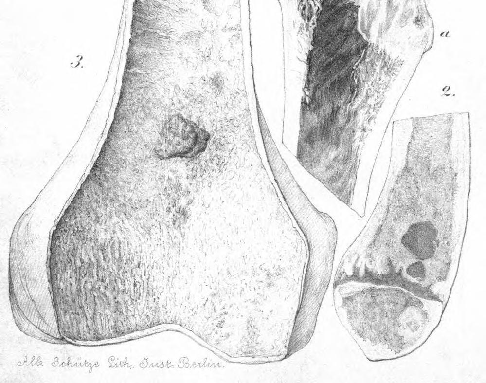
91 Figure 27: 154
92 Figure 27 (Illustrations taken from the article: Über die Entstehung des Enchondroma und seine Beziehungen zu der Ecchondrosis und der Exostosis cartilaginea published in Monatsberichte der Königlich Preussischen Akademie der Wissenschaften in 1875 by R. Virchow) In figure 27.1 Virchow uses an image of pathology specimen in a child with rickets of the distal femur to demonstrate proliferation of cartilage of the growth plate with islands of cartilage which have separated completely and lie backwards within the fully developed spongiosa (translated from German into English by me). In figure 27.2 Virchow shows an image of a pathology specimen in a child with rickets of the distal femur to demonstrate a large and a smaller completely separated island of proliferating cartilage which lies within the spongiosa (translated from German into English by me). In figure 27.3 Virchow demonstrates an image of a pathology specimen of a skeletally mature patient with an enchondroma of the distal femur. In his publication, he states that the location of the enchondroma correlates so precisely with the location of the islands of cartilage observed in the skeletally immature bones (of figure 27.1 and 27.2) that one cannot have any doubt, that such a remnant of cartilage really persisted (into adulthood) (translated from German into English by me). 155
93 The diagnosis and grading of chondrosarcomas represent major challenges for pathologists, clinicians and radiologists alike. Particularly, the differentiation of enchondromas from low-grade chondrosarcomas is one of the most difficult and controversial diagnostic dilemmas in musculoskeletal oncology. Whilst anatomical MRI has been used in attempt to differentiate between the two disease entities, there remains considerable uncertainty in the diagnosis. In the fourth article (original article), we have attempted to use diffusion-weighted MRI (DWI), - a non-invasive functional MRI-technique which indirectly assesses the cellularity of biologic tissues - in an attempt to differentiate enchondromas from chondrosarcomas. A previous retrospective study stated that DWI might be promising in the differentiation of enchondromas from low-grade chondrosarcomas. However, the results of this study were hampered by a small sample size which included only 14 central chondroid tumours. In this study, the authors measured mean ADC-values in three consecutive slices and correlated the mean ADC-values with the histological grading of the chondroid tumours. Whilst the authors found a statistically significant difference in the ADC-values of enchondromas from chondrosarcomas, they concluded that there was only a slight difference in the ADCvalues of enchondromas from chondrosarcomas and therefore a significantly larger patient cohort was required to evaluate the role that DWI may play in the differentiation of enchondromas from chondrosarcomas and low-grade chondrosarcomas in particular. 89 The aim of our study was therefore to investigate the potential role that DWI could play in the differentiation of enchondromas from chondrosarcomas in general and in 156
94 the differentiation of enchondromas from low-grade chondrosarcomas in a larger patient cohort. In the fourth article, which forms part of this PhD-thesis, we therefore re-assessed the role of DWI in the differentiation of enchondromas from chondrosarcomas in a larger patient cohort. We included 52 central chondroid bone tumours of which there 24 enchondromas, 5 cartilaginous lesions of unknown malignant potential, 15 grade 1 chondrosarcomas, three grade 2 chondrosarcomas, two grade 3 chondrosarcomas and three dedifferentiated chondrosarcomas. We found that there was a statistically significant difference in the mean and minimum ADC values in all groups. Post hoc analysis demonstrated that this difference in mean and minimum ADC values was due to the ADC values in the dedifferentiated chondrosarcoma group. In contrast, the mean and minimum ADC-values of lowgrade chondroid tumours and high-grade chondrosarcomas were not statistically significantly different. We therefore concluded that DWI could not differentiate between enchondromas and chondrosarcomas and that it does not aid in the distinction of low-grade chondroid tumours from high-grade chondrosarcomas. This study which forms part of the portfolio of publications submitted for consideration of a PhD therefore significantly advances the understanding about the role of Diffusion-weighted MRI in the differentiation of enchondromas from chondrosarcomas. There are several limitations to our study. Firstly, the retrospective nature of the study and the fact that the study was performed in a bone tumour centre most likely has resulted in a selection bias as many enchondromas observed on routine MRI-examinations are not referred to a 157
95 bone tumour centre where the study was performed. Secondly, whilst 81% of all central cartilage tumours were histologically confirmed, ten enchondromas did not undergo histological confirmation representing 19% of all central cartilaginous tumours and 42 % of all enchondromas in our study. We have applied stringent criteria for designating a lesion as an enchondroma based on widely accepted imaging features. Thirdly, minimum and mean ADC values were measured in one slice. This slice was carefully selected to correspond with the slice on conventional MR imaging, which showed the most aggressive imaging features. Furthermore, there is no standardised technique in the literature which has been widely accepted for analysis of DWI-data. Whilst some authors have analysed DWI-data using a whole-tumour volume approach, analysis of DWI data using the region of interest approach continues to be most widely performed technique similar to the analysis method used in our publication. 87,88, ,111 Although analysis of skeletal lesions using a whole tumour volume approach is likely to be more accurate taking into consideration heterogeneity of ADC-values within skeletal lesions, this technique is very timeconsuming, not widely performed and very difficult to implement in routine clinical practice due to its impracticality and time-consuming nature. Thus, the aim of our data analysis was to investigate an analysis method which could be easily implemented in routine clinical practice and hence if found to be useful could be widely adopted. Whilst there have been no studies which compared the different analysis methods of DWI in bone tumours, there has been a very recent study which evaluated the interobserver variability of three selective region-of-interest 158
96 measurement protocols for apparent diffusion coefficient quantifications in soft tissue tumours and compared them with whole tumour volume ADC measurements. The authors found that whilst all selective and whole tumour volume measurements offered good to excellent interobserver agreement, the selective observer based method of ADC measurement resulted in the closest values to whole tumour volume measurements. The authors therefore concluded that a simplified observer-based manual method of ADC quantification in the evaluation of soft tissue tumours was comparable to whole tumour volume measurements of the minimum and mean ADCs, and required significantly less analysis time. 112 Further research is required to evaluate which analysis method in bone tumours and skeletal lesions are most feasible and accurate in the interpretation of ADC-values in daily clinical practice. This is of importance if DWI is to be implemented as a routine MRI-sequence in daily clinical practice. Furthermore, the inter-observer reliability of measurement of ADC-values has not been tested in this article because both the slice selection and the measurement of ADC-values was performed by one reader only. Therefore, the reliability of the study remains untested. Lastly, we did not calculate 95% confidence intervals for the minimum and mean ADC-values. However, the measured sample mean values in the study may not reflect the population mean values. Therefore, the range of ADC values which contain the true population mean with 95% certainty cannot be quantified based on the study results provided. The fifth article (original article) presented as part of the submitted PhD-thesis represents an original article which evaluates the role of MRI in the grading of 159
97 chondrosarcomas. Whilst a previous publication showed that the presence of a soft tissue mass favoured the diagnosis of a high-grade chondrosarcoma, the utility of this study was hampered by the fact that the authors only assessed a few MRI-features which are associated with chondrosarcomas and by the small sample size of the study. 48 Therefore, there remained uncertainty about the role of other MR Imaging features in the differentiation of high-grade from low-grade appendicular chondrosarcomas. Hence, we evaluated a wide range of MRI-features, which are used in the characterization of chondrosarcomas representing the largest study published on this topic. In this retrospective study, we analysed the MRIexaminations of 179 chondrosarcomas of long bones and divided them into lowgrade chondroid lesions and high-grade chondrosarcomas. We found that on multivariate analysis, bone expansion, active periostitis, the presence of a soft tissue mass, and tumour length are statistically significant differentiating factors between low-grade and high-grade chondroid tumours. On logistic regression analysis, the Odds ratio for bone expansion, active periostitis, soft tissue mass and tumour length were: 8.8 (95% confidence interval ), 52.8 ( ), 21.1 ( ) and 1.4 ( ) for the diagnosis of a high-grade chondrosarcoma. The above stated figures in the study demonstrate wide confidence intervals for all four MRI-features which could be due to the variation of MRI-features in the high-grade chondrosarcoma group although the sample size albeit large in our study could also be a contributing factor. However, it is important to note that the study demonstrated a positive relationship between the above described MRI features and the diagnosis of a highgrade chondrosarcoma. Our study also demonstrated that the presence of the above described four MRI features showed a diagnostic accuracy of 95.6%. We therefore 160
98 concluded that these imaging features accurately predicted the presence of a highgrade chondrosarcoma. Thus, MRI can be used accurately to differentiate low-grade chondroid tumours from high-grade chondrosarcomas of the major long bones which in turn may avoid inadequate surgery. This is of particular clinical importance when one considers that biopsy may result in sampling errors of chondrosarcoma of long bone of up to 17% and hence may result in inappropriate treatment. 44 There are nevertheless limitations to our study. Firstly, due to the retrospective nature of our study, the MRI protocol was not standardised. In view of the rarity of chondrosarcomas, there have been no prospective studies evaluating the role of MRI in the diagnosis and grading of chondrosarcomas. Furthermore, our article continues to be one of the largest studies evaluating the role of MRI in chondrosarcoma hence highlighting challenges faced in the development and conduct of potential prospective studies which would require inclusion of a large patient cohort in order to evaluate MRI-features in the differentiation of low-grade from high-grade chondrosarcomas. Secondly, our study included a higher number of high-grade chondrosarcomas than is usually observed in other series which was most likely due to the tertiary nature of the institution in which the study was conducted. This may therefore have led to a selection bias. Thirdly, the differentiation of enchondromas from low-grade chondrosarcomas demonstrate a low reliability even among subspecialised histopathologists due to the lack of a gold standard in the distinction of the two disease entities. 67,68 The difficulty 161
99 that radiologists, pathologists and clinicians face in the differentiation of enchondromas from low-grade chondrosarcomas was also reflected in our study by the fact that our study included 28 borderline malignant chondroid lesions which were classified as grade 0.5 chondrosarcomas. All chondral tumours at the institution in which the study was performed were reviewed by two musculoskeletal pathologists and the diagnosis of benign versus malignant chondral tumours was made in consensus with radiologists, pathologists and clinicians. Furthermore, the author of this thesis performed an unpublished analysis at the time when the original analysis was performed which excluded the above stated 28 borderline malignant chondroid lesions or CLUMPs. The results of this analysis demonstrated that on multivariate analysis, bone expansion, active periostitis, soft tissue mass and tumour length continued to be statistically significant differentiating factors whilst the other MRI-features continued to be not statistically significant. Therefore, the inclusion of 28 indeterminate chondroid lesions has not resulted in a significant change in the findings of the publication. Lastly, the inter-observer reliability of the above described conventional MRI-findings remains untested as the analysis of the MRI-features was performed in consensus between the two readers and in case of disagreement the opinion of the most senior reviewer was accepted. However, this approach to the data analysis precludes assessment of inter-observer reliability of the above described MRI-features. In conclusion, the above described study identified several MRI features which allow accurate differentiation of low-grade chondroid tumours from high-grade chondrosarcomas of long bones. Therefore, this study significantly enhances the 162
100 understanding and knowledge that MRI plays in the accurate preoperative grading of chondrosarcomas of long bones. Furthermore, the findings of this study may result in a more accurate diagnosis prior to surgery and may significantly improve patient care. 51 The sixth article (original article) included in the submitted PhD-thesis evaluates the role of staging in chondrosarcomas. Prior to this publication, it has been widely assumed that skeletal scintigraphy and Computed Tomography of the chest are mandatory in the initial staging of chondrosarcomas to assess for the presence of skeletal and pulmonary metastases. This management approach however has been based on evidence obtained from other sarcomas such as osteosarcoma and Ewing sarcoma which demonstrate skeletal and pulmonary metastases in 4-19% at initial presentation. The biological behaviour of chondrosarcomas is very different to osteosarcomas and Ewing sarcomas which tend to be more aggressive. Hence, this management approach was not evidence-based. We have therefore investigated the role of whole-body skeletal scintigraphy and Computed Tomography of the chest in the initial surgical staging of chondrosarcomas. In this retrospective study, we evaluated the bone scintigraphy reports and bone scintigraphs in 188 patients with chondrosarcomas. We discovered 195 chondrosarcomas in 188 patients. In our study, there were no patients with skeletal metastases at presentation. However, we discovered 3 patients with multifocal chondrosarcomas, 2 of whom had known Ollier s disease and who would have been adequately imaged on the staging MRI-study. A third patient with multifocal chondrosarcoma was found to have a low-grade central chondrosarcoma 163
101 in the opposite femur. This finding did not alter surgical management as the lesion was observed and remained unchanged. We therefore concluded that there is little role of skeletal scintigraphy in the initial surgical staging of chondrosarcomas. The importance of this publication also lies in demonstrating that unlike in osteosarcoma and Ewing sarcoma, skeletal metastases in chondrosarcomas at presentation are very rare. This is of significance because previous studies have demonstrated that incidental enchondromas are commonly observed on routine MRI-examinations of the knee and shoulder. 6,7 Since publication of our findings, the rarity of skeletal metastases in chondrosarcoma has been corroborated by Gulia et al. who evaluated 69 patients with chondrosarcoma and identified only one patient with skeletal metastases (1.6% of all cases). Of note is, that in their study the single patient with skeletal metastases was also found to have concomitant pulmonary metastases. In contrast, Gulia et al. confirm that the prevalence of pulmonary metastases in chondrosarcoma of bone is significant being observed in 9.8% of all cases (6 patients) in their patient cohort. 113 Similarly, a multicentre retrospective study published by Andreou et al. recently, evaluated the metastatic potential of grade 1 chondrosarcomas. The authors included 225 patients with grade 1 chondrosarcoma and found that 14 patients developed metastases after a median time of 49 months with a range of 4 to 125 months. In their study, only two patients (0.9% of all patients) developed skeletal metastases whilst 12 patients (5.3% of all patients) developed pulmonary metastases. This recent study further supports our findings that skeletal metastases 164
102 are a rare occurrence in chondrosarcoma of bone in contrast to pulmonary metastases. 114 We also assessed the CT-chest reports of all patients with chondrosarcoma at the time of diagnosis. In our study, we identified pulmonary metastases in approximately 5% of chondrosarcomas at initial presentation. In contrast to skeletal scintigraphy, we found therefore evidence supporting the added value of Computed Tomography of the chest in the detection of pulmonary metastases in the initial staging of chondrosarcomas. There are limitations to our study. Firstly, of the 352 patients identified on the database with a chondrosarcoma, 154 did not have bone scintigraphy images or reports available for review. Whilst review of clinical letters in these patients made no reference about the presence of skeletal metastases, we were therefore not in a position to definitely rule out that some of these patients may have presented with occult skeletal metastases. Secondly, of the 195 chondrosarcomas included in the study, only bone scintigrams, which demonstrated increased uptake outside the site of the chondrosarcoma according to the bone scintigraphy report were re-reported by the first author of the article. Whilst review of the clinical notes made no reference to the presence of skeletal metastases, we are again unable to rule out that some of these patients may have had occult skeletal metastases. Furthermore, the inter-observer reliability of the findings of the re-reported bone scintigrams has not been tested because bone scintigraphy studies were re-reported by the author of this thesis only. 165
103 Thirdly, in our patient cohort, three patients were diagnosed with synchronous multifocal chondrosarcomas. Whilst the presence of multifocal synchronous chondrosarcomas could be interpreted as skeletal metastases, two of these patients suffered from Ollier s disease which is characterised by multifocal enchondromas in which malignant transformation is a well-recognised complication therefore strongly favouring that the presence of further chondrosarcomas is due to multiple chondrosarcomas rather than skeletal metastases. In contrast to the other two cases, the third patient with synchronous multifocal chondrosarcomas was not known to suffer from Ollier s disease as there were no further manifestations on other imaging to suggest multiple enchondromas. The existence of multicentric primary chondrosarcomas in the absence of enchondromatosis or hereditary multiple exostosis remains uncertain. 115 The third patient in our cohort was found to have a grade 2 chondrosarcoma of the left pubis, a grade 2 chondrosarcoma of the left proximal femur and a grade 1 chondrosarcoma of the right proximal femur. We therefore cannot exclude that one of the lesions in the left femur or pelvis represented a skip metastasis. The preoperative staging MRI-study accurately depicted both lesions. In contrast, the grade 1 chondrosarcoma of the contralateral right proximal femur was only depicted on the bone scintigraphy study. However, this lesion in view of its lower grade most likely represented a primary chondrosarcoma rather than a skeletal metastasis. Although some clinicians may wish to continue performing skeletal scintigraphy or may even consider whole-body MRI in the initial staging of chondrosarcomas, our findings highlight the rarity of skeletal metastases in this disease entity. Hence, the 166
104 presence of other adjacent chondroid lesions on skeletal scintigraphy or whole-body MRI should not be misinterpreted as skip metastases. This is of particular importance when one considers that incidental enchondromas are identified in approximately 2% of all MRI-scans of the shoulder and in approximately 3% of all MRI-scans of the knee. 6,7 In conclusion, this study, which forms part of the PhD-thesis demonstrates the rarity of skeletal metastases in chondrosarcoma of bone at presentation and highlights the significant prevalence of pulmonary metastases. This study therefore significantly advances the knowledge about the role of skeletal scintigraphy and Computed Tomography of the chest in the initial staging of chondrosarcoma of bone. In summary, the authors published work submitted for consideration of a PhD, investigates the theory that imaging may aid in the understanding of the pathogenesis of enchondromas and may aid in the diagnosis, grading and staging of chondrosarcomas of bone. 167
105 8.2 Future research Whilst the author s work has significantly advanced the understanding about the pathogenesis of enchondromas, the role of imaging in the diagnosis, grading and staging of chondrosarcomas, there remains diagnostic uncertainty in the differentiation of enchondromas from low-grade chondrosarcomas. Although the author s most recent publication demonstrated that the presence of more than 2/3 of endosteal scalloping on conventional MR shows a sensitivity of 71.4% and a specificity of 92.9% in the differentiation of enchondromas from lowgrade chondrosarcomas and hence is the most sensitive feature on conventional MRI, the differentiation of the two disease entities remains challenging. Similarly, diffusion-weighted MRI cannot be used to differentiate between enchondromas and low-grade chondrosarcomas as demonstrated by the author. Furthermore, whilst dynamic-contrast enhanced MRI aids in the detection of high-grade chondrosarcomas, DCE-MRI crucially cannot be utilized to differentiate enchondromas from low-grade chondrosarcomas as published by the author of this thesis very recently (see Appendix 1). 80 Other imaging techniques such as radiography, CT and PET/CT are of limited value in the distinction of the two disease entities. Other functional imaging modalities such as conventional MR-spectroscopy and hyperpolarized MRI have not been evaluated in the diagnosis of enchondromas and low-grade chondrosarcomas. These functional imaging techniques have the potential to allow unique and fascinating insights into the tumour biology, into the molecular and genetic make-up of tumours in general 168
106 and of chondroid tumours in particular. It has been shown relatively recently that a majority of enchondromas and chondrosarcomas have somatic mutations in isocitrate dehydrogenase 1 and 2 (IDH 1 and 2) which produce the oncometabolite 2-hydroxyglutarate. 33 This oncometabolite could therefore potentially be identified and the concentration of this oncometabolite could be quantified non-invasively using conventional MR-spectroscopy or hyperpolarized MRI. Quantification of this oncometabolite could therefore possibly result in a cut-off value which could differentiate enchondromas from low-grade chondrosarcomas more reliably. These imaging techniques may therefore potentially hold the key to unravel the diagnostic uncertainty in the distinction of enchondromas from low-grade chondrosarcomas. Whilst molecular MRI-techniques warrant further research, at present, the differentiation of enchondromas from low-grade chondrosarcomas continues to be challenging. However, a very recent publication by the author of this thesis demonstrated that certain clinical and conventional MRI-features aid in the distinction of the two disease entities. In particular, the presence of more than 2/3 of endosteal scalloping on conventional MRI and pain attributed to the lesion demonstrate a moderate sensitivity in the differentiation of enchondromas from low-grade chondrosarcomas whilst the presence of pain attributed to the lesion and more than 2/3 of endosteal scalloping are highly specific for a grade 1 chondrosarcoma. The authors findings however require validation in a larger prospective patient cohort. Hence, further research is required to evaluate the role of conventional MRI in the differentiation of the two disease entities. 169
107 At present, there remains considerable diagnostic uncertainty in the differentiation of enchondromas from low-grade chondrosarcomas. In particular, the malignant potential of central chondroid lesions which do not demonstrate permeation on histopathology remains unknown. Hence, a major recommendation of this thesis is that future studies should focus on the malignant potential of low-grade central chondroid lesions which are not treated. Therefore, prospective long-term follow-up studies should be performed to assess the malignant potential of such low-grade chondroid lesions and to assess potential predictive imaging biomarkers. Surgery remains the only hope for cure in chondrosarcoma and the presence of skeletal metastases is associated with a bleak outcome. Advances in the molecular understanding of chondrosarcomas are likely to result in the development of nonsurgical therapies such as agents which specifically target mutations in chondrosarcoma cells. Therefore, the above described molecular imaging techniques may in future also allow the development of a personalized, molecular fingerprint of chondrosarcomas which in turn may allow non-invasive monitoring of treatment response in patients with chondrosarcoma beyond the current concept of conventional, anatomical imaging thus potentially resulting in a paradigm shift in imaging. Furthermore, molecular imaging may facilitate the development of new drugs which combine diagnostic and therapeutic capabilities (so-called theranostic drugs). In particular, the efficacy of drugs targeting IDH 1 and 2 mutations in chondrosarcomas could be combined with in-vivo and real-time assessment of treatment response in these patients using the above described functional imaging techniques. 170
108 Therefore, future imaging research in chondrosarcomas should focus on the role of conventional MRI in the differentiation of enchondromas from low-grade chondrosarcomas. However, imaging research should also investigate the potential role that the non-invasive detection and quantification of oncometabolites produced by genetic mutations may play in the diagnosis and treatment response assessment of chondrosarcomas. 171
109 Bibliography 1. Grimer R HP, Vanel D. World Health Organization Classification of Tumours of Soft Tissue and Bone. In: Fletcher C.D.M. BJA, Hogendoorn P.C.W., Mertens F., ed. World Health Organization Classification of Tumours of Soft Tissue and Bone. Lyon: IARCP; 2013: NCI. SEER Cancer statistics review, National Cancer Institute, Bethesda Bethesda: National Cancer Institute Parkin DM WS, Ferlay J. Cancer incidence in five continents. IARCP Press: Lyon World Health Organization Classification of Tumours of Soft Tissue and Bone. In: Fletcher C.D.M. BJA, Hogendoorn P.C.W., Mertens F., ed. World Health Organization Classification of Tumours of Soft Tissue and Bone. Lyon: IARCP; 2013: Unni K. Tumors of the bones and joints. Atlas of Tumor Pathology, Series IV. American Registry of Pathology in collaboration with the Armed Forces Institute of Pathology Walden MJ, Murphey MD, Vidal JA. Incidental enchondromas of the knee. AJR. American journal of roentgenology. 2008;190(6): Hong ED, Carrino JA, Weber KL, Fayad LM. Prevalence of shoulder enchondromas on routine MR imaging. Clin Imaging. 2011;35(5): Bovee JVMG AB. World Health Organization Classification of Tumours of Soft Tissue and Bone. In: Fletcher C.D.M. BJA, Hogendoorn P.C.W., Mertens F., ed. Lyon: IARCPress; 2013: Flemming DJ, Murphey MD. Enchondroma and chondrosarcoma. Seminars in musculoskeletal radiology. 2000;4(1): Logie CI, Walker EA, Forsberg JA, Potter BK, Murphey MD. Chondrosarcoma: A Diagnostic Imager's Guide to Decision Making and Patient Management. Seminars in musculoskeletal radiology. 2013;17(2): Lucas DR VJ. World Health Organization Classification of Tumours of Soft Tissue and Bone. In: Fletcher C.D.M. BJA, Hogendoorn P.C.W., Mertens F., ed. World Health Organization Classification of Tumours of Soft Tissue and Bone. Lyon: IARCP; 2013: Brien EW, Mirra JM, Kerr R. Benign and malignant cartilage tumors of bone and joint: their anatomic and theoretical basis with an emphasis on radiology, pathology and clinical biology. I. The intramedullary cartilage tumors. Skeletal Radiol. 1997;26(6): Douis H, Saifuddin A. The imaging of cartilaginous bone tumours. I. Benign lesions. Skeletal Radiol Douis H, Saifuddin A. The imaging of cartilaginous bone tumours. II. Chondrosarcoma. Skeletal Radiol. 2013;42(5): Murphey MD, Flemming DJ, Boyea SR, Bojescul JA, Sweet DE, Temple HT. Enchondroma versus chondrosarcoma in the appendicular skeleton: differentiating features. Radiographics : a review publication of the Radiological Society of North America, Inc. 1998;18(5): ; quiz Lee FY, Yu J, Chang SS, Fawwaz R, Parisien MV. Diagnostic value and limitations of fluorine-18 fluorodeoxyglucose positron emission tomography for cartilaginous tumors of bone. J Bone Joint Surg Am. 2004;86-A(12):
110 17. Subhawong TK, Winn A, Shemesh SS, Pretell-Mazzini J. F-18 FDG PET differentiation of benign from malignant chondroid neoplasms: a systematic review of the literature. Skeletal Radiol. 2017;46(9): Ryzewicz M, Manaster BJ, Naar E, Lindeque B. Low-grade cartilage tumors: diagnosis and treatment. Orthopedics. 2007;30(1):35-46; quiz Hogendoorn PCW BJ, Nielsen GP. World Health Organization Classification of Tumours of Soft Tissue and Bone. In: Fletcher C.D.M. BJA, Hogendoorn P.C.W., Mertens F., ed. Lyon: IARCPress; 2013: Bovee JV, van der Heul RO, Taminiau AH, Hogendoorn PC. Chondrosarcoma of the phalanx: a locally aggressive lesion with minimal metastatic potential: a report of 35 cases and a review of the literature. Cancer. 1999;86(9): Murphey MD, Walker EA, Wilson AJ, Kransdorf MJ, Temple HT, Gannon FH. From the archives of the AFIP: imaging of primary chondrosarcoma: radiologic-pathologic correlation. Radiographics : a review publication of the Radiological Society of North America, Inc. 2003;23(5): Qasem SA, DeYoung BR. Cartilage-forming tumors. Seminars in diagnostic pathology. 2014;31(1): Crim J, Schmidt R, Layfield L, Hanrahan C, Manaster BJ. Can imaging criteria distinguish enchondroma from grade 1 chondrosarcoma? Eur J Radiol. 2015;84(11): Grimer RJ, Carter SR, Tillman RM, Mangham DC, Abudu A, Fiorenza F. Chondrosarcoma of bone. J Bone Joint Surg Am. 2000;82-A(8): Lee FY, Mankin HJ, Fondren G, et al. Chondrosarcoma of bone: an assessment of outcome. J Bone Joint Surg Am. 1999;81(3): Evans HL, Ayala AG, Romsdahl MM. Prognostic factors in chondrosarcoma of bone: a clinicopathologic analysis with emphasis on histologic grading. Cancer. 1977;40(2): Kreicbergs A, Boquist L, Borssen B, Larsson SE. Prognostic factors in chondrosarcoma: a comparative study of cellular DNA content and clinicopathologic features. Cancer. 1982;50(3): Ozaki T, Hillmann A, Lindner N, Blasius S, Winkelmann W. Metastasis of chondrosarcoma. Journal of cancer research and clinical oncology. 1996;122(10): Sanerkin NG. The diagnosis and grading of chondrosarcoma of bone: a combined cytologic and histologic approach. Cancer. 1980;45(3): Giuffrida AY, Burgueno JE, Koniaris LG, Gutierrez JC, Duncan R, Scully SP. Chondrosarcoma in the United States (1973 to 2003): an analysis of 2890 cases from the SEER database. J Bone Joint Surg Am. 2009;91(5): Angelini A, Guerra G, Mavrogenis AF, Pala E, Picci P, Ruggieri P. Clinical outcome of central conventional chondrosarcoma. Journal of surgical oncology. 2012;106(8): Bjornsson J, McLeod RA, Unni KK, Ilstrup DM, Pritchard DJ. Primary chondrosarcoma of long bones and limb girdles. Cancer. 1998;83(10): Amary MF, Bacsi K, Maggiani F, et al. IDH1 and IDH2 mutations are frequent events in central chondrosarcoma and central and periosteal chondromas but not in other mesenchymal tumours. The Journal of pathology. 2011;224(3): Janzen L, Logan PM, O'Connell JX, Connell DG, Munk PL. Intramedullary chondroid tumors of bone: correlation of abnormal peritumoral marrow and soft-tissue MRI signal with tumor type. Skeletal Radiol. 1997;26(2):
111 35. Brown MT, Gikas PD, Bhamra JS, et al. How safe is curettage of low-grade cartilaginous neoplasms diagnosed by imaging with or without pre-operative needle biopsy? Intralesional curettage and cementation for low-grade chondrosarcoma of long bones: retrospective study and literature review. Bone Joint J. 2014;96-B(8): Campanacci DA, Scoccianti G, Franchi A, et al. Surgical treatment of central grade 1 chondrosarcoma of the appendicular skeleton. Journal of orthopaedics and traumatology : official journal of the Italian Society of Orthopaedics and Traumatology. 2013;14(2): Di Giorgio L, Touloupakis G, Vitullo F, Sodano L, Mastantuono M, Villani C. Intralesional curettage, with phenol and cement as adjuvants, for low-grade intramedullary chondrosarcoma of the long bones. Acta orthopaedica Belgica. 2011;77(5): Funovics PT, Panotopoulos J, Sabeti-Aschraf M, et al. Low-grade chondrosarcoma of bone: experiences from the Vienna Bone and Soft Tissue Tumour Registry. Int Orthop. 2011;35(7): Hickey M, Farrokhyar F, Deheshi B, Turcotte R, Ghert M. A systematic review and meta-analysis of intralesional versus wide resection for intramedullary grade I chondrosarcoma of the extremities. Annals of surgical oncology. 2011;18(6): Kim W, Han I, Kim EJ, Kang S, Kim HS. Outcomes of curettage and anhydrous alcohol adjuvant for low-grade chondrosarcoma of long bone. Surg Oncol. 2015;24(2): Meftah M, Schult P, Henshaw RM. Long-term results of intralesional curettage and cryosurgery for treatment of low-grade chondrosarcoma. J Bone Joint Surg Am. 2013;95(15): Mermerkaya MU, Bekmez S, Karaaslan F, et al. Intralesional curettage and cementation for low-grade chondrosarcoma of long bones: retrospective study and literature review. World journal of surgical oncology. 2014;12: Verdegaal SH, Brouwers HF, van Zwet EW, Hogendoorn PC, Taminiau AH. Lowgrade chondrosarcoma of long bones treated with intralesional curettage followed by application of phenol, ethanol, and bone-grafting. J Bone Joint Surg Am. 2012;94(13): Roitman PD, Farfalli GL, Ayerza MA, Muscolo DL, Milano FE, Aponte-Tinao LA. Is Needle Biopsy Clinically Useful in Preoperative Grading of Central Chondrosarcoma of the Pelvis and Long Bones? Clinical orthopaedics and related research. 2017;475(3): De Beuckeleer LH, De Schepper AM, Ramon F, Somville J. Magnetic resonance imaging of cartilaginous tumors: a retrospective study of 79 patients. Eur J Radiol. 1995;21(1): Geirnaerdt MJ, Bloem JL, Eulderink F, Hogendoorn PC, Taminiau AH. Cartilaginous tumors: correlation of gadolinium-enhanced MR imaging and histopathologic findings. Radiology. 1993;186(3): Varma DG, Ayala AG, Carrasco CH, Guo SQ, Kumar R, Edeiken J. Chondrosarcoma: MR imaging with pathologic correlation. Radiographics : a review publication of the Radiological Society of North America, Inc. 1992;12(4): Yoo HJ, Hong SH, Choi JY, et al. Differentiating high-grade from low-grade chondrosarcoma with MR imaging. European radiology. 2009;19(12): Radiologists TRCoPatRCo. Evidence-based indications for the use of PET-CT in the UK. London: RCP, RCR
112 50. Feldman F, Van Heertum R, Saxena C, Parisien M. 18FDG-PET applications for cartilage neoplasms. Skeletal Radiol. 2005;34(7): Douis H, Singh L, Saifuddin A. MRI differentiation of low-grade from high-grade appendicular chondrosarcoma. European radiology. 2014;24(1): Stacy GS, Mahal RS, Peabody TD. Staging of bone tumors: a review with illustrative examples. AJR. American journal of roentgenology. 2006;186(4): Huang AJ WM, Kattapuram SV, Bredella MA. Surgical staging 2: metastatic disease. Berlin: Springer; Heck RK, Jr., Peabody TD, Simon MA. Staging of primary malignancies of bone. CA: a cancer journal for clinicians. 2006;56(6): RA G. Nuclear medicine. Berlin: Springer; National Guideline C. ACR Appropriateness Criteria primary bone tumors. Accessed 7/12/ Pradhan A, Grimer RJ, Spooner D, et al. Oncological outcomes of patients with Ewing's sarcoma: is there a difference between skeletal and extra-skeletal Ewing's sarcoma? The Journal of bone and joint surgery. British volume. 2011;93(4): Byun BH, Kong CB, Lim I, et al. Comparison of (18)F-FDG PET/CT and (99 m)tc-mdp bone scintigraphy for detection of bone metastasis in osteosarcoma. Skeletal Radiol. 2013;42(12): Paulussen M, Bielack S, Jurgens H, Casali PG, Group EGW. Ewing's sarcoma of the bone: ESMO clinical recommendations for diagnosis, treatment and follow-up. Annals of oncology : official journal of the European Society for Medical Oncology / ESMO. 2009;20 Suppl 4: Bielack S, Carrle D, Casali PG, Group EGW. Osteosarcoma: ESMO clinical recommendations for diagnosis, treatment and follow-up. Annals of oncology : official journal of the European Society for Medical Oncology / ESMO. 2009;20 Suppl 4: Kager L, Zoubek A, Potschger U, et al. Primary metastatic osteosarcoma: presentation and outcome of patients treated on neoadjuvant Cooperative Osteosarcoma Study Group protocols. Journal of clinical oncology : official journal of the American Society of Clinical Oncology. 2003;21(10): Cotterill SJ, Ahrens S, Paulussen M, et al. Prognostic factors in Ewing's tumor of bone: analysis of 975 patients from the European Intergroup Cooperative Ewing's Sarcoma Study Group. Journal of clinical oncology : official journal of the American Society of Clinical Oncology. 2000;18(17): Donati D, Colangeli S, Colangeli M, Di Bella C, Bertoni F. Surgical treatment of grade I central chondrosarcoma. Clinical orthopaedics and related research. 2010;468(2): Mohler DG, Chiu R, McCall DA, Avedian RS. Curettage and cryosurgery for lowgrade cartilage tumors is associated with low recurrence and high function. Clinical orthopaedics and related research. 2010;468(10): Verdegaal SH, Hartigh J, Hogendoorn PC, Brouwers HF, Taminiau AH. Phenol levels during intralesional curettage and local adjuvant treatment of benign and lowgrade malignant bone tumours. Clinical sarcoma research. 2012;2(1): Hanna SA, Whittingham-Jones P, Sewell MD, et al. Outcome of intralesional curettage for low-grade chondrosarcoma of long bones. European journal of surgical oncology : the journal of the European Society of Surgical Oncology and the British Association of Surgical Oncology. 2009;35(12): Eefting D, Schrage YM, Geirnaerdt MJ, et al. Assessment of interobserver variability and histologic parameters to improve reliability in classification and grading of 175
113 central cartilaginous tumors. The American journal of surgical pathology. 2009;33(1): Reliability of histopathologic and radiologic grading of cartilaginous neoplasms in long bones. J Bone Joint Surg Am. 2007;89(10): Landis JR, Koch GG. The measurement of observer agreement for categorical data. Biometrics. 1977;33(1): Coindre JM, Trojani M, Contesso G, et al. Reproducibility of a histopathologic grading system for adult soft tissue sarcoma. Cancer. 1986;58(2): Hasegawa T, Yamamoto S, Nojima T, et al. Validity and reproducibility of histologic diagnosis and grading for adult soft-tissue sarcomas. Human pathology. 2002;33(1): Harvey JM, de Klerk NH, Sterrett GF. Histological grading in breast cancer: interobserver agreement, and relation to other prognostic factors including ploidy. Pathology. 1992;24(2): Robbins P, Pinder S, de Klerk N, et al. Histological grading of breast carcinomas: a study of interobserver agreement. Human pathology. 1995;26(8): Theissig F, Kunze KD, Haroske G, Meyer W. Histological grading of breast cancer. Interobserver, reproducibility and prognostic significance. Pathology, research and practice. 1990;186(6): World Health Organization Classification of Tumours of Soft Tissue and Bone. In: Fletcher C.D.M. BJA, Hogendoorn P.C.W., Mertens F., ed. World Health Organization Classification of Tumours of Soft Tissue and Bone. Lyon: IARCP; 2013: Gaston CL WH, Sumathi V, Douis H, Reddy K, Abudu AT, Carter SR, Jeys LM, Tillman RM, Grimer RJ. Cartilaginous lesions of unknown malignant potential (CLUMP): a different clinical entity from grade 1 chondrosarcomas? Paper presented at: 27th Annual Meeting of the European Musculo-Skeletal Oncology Society (E.M.S.O.S); 21th May - 23rd May 2014, 2014; Vienna, Austria. 77. Aoki J, Watanabe H, Shinozaki T, Tokunaga M, Inoue T, Endo K. FDG-PET in differential diagnosis and grading of chondrosarcomas. Journal of computer assisted tomography. 1999;23(4): Ferrer-Santacreu EM, Ortiz-Cruz EJ, Gonzalez-Lopez JM, Perez Fernandez E. Enchondroma versus Low-Grade Chondrosarcoma in Appendicular Skeleton: Clinical and Radiological Criteria. J Oncol. 2012;2012: Choi BB, Jee WH, Sunwoo HJ, et al. MR differentiation of low-grade chondrosarcoma from enchondroma. Clin Imaging. 2013;37(3): Douis H, Parry M, Vaiyapuri S, Davies AM. What are the differentiating clinical and MRI-features of enchondromas from low-grade chondrosarcomas? European radiology. 2018;28(1): Padhani AR. Dynamic contrast-enhanced MRI in clinical oncology: current status and future directions. Journal of magnetic resonance imaging : JMRI. 2002;16(4): Verstraete KL, De Deene Y, Roels H, Dierick A, Uyttendaele D, Kunnen M. Benign and malignant musculoskeletal lesions: dynamic contrast-enhanced MR imaging-- parametric "first-pass" images depict tissue vascularization and perfusion. Radiology. 1994;192(3): De Coninck T, Jans L, Sys G, et al. Dynamic contrast-enhanced MR imaging for differentiation between enchondroma and chondrosarcoma. European radiology
114 84. Geirnaerdt MJ, Hogendoorn PC, Bloem JL, Taminiau AH, van der Woude HJ. Cartilaginous tumors: fast contrast-enhanced MR imaging. Radiology. 2000;214(2): Koh DM, Collins DJ. Diffusion-weighted MRI in the body: applications and challenges in oncology. AJR. American journal of roentgenology. 2007;188(6): Kransdorf MJ, Bridges MD. Current developments and recent advances in musculoskeletal tumor imaging. Seminars in musculoskeletal radiology. 2013;17(2): Ahlawat S, Khandheria P, Subhawong TK, Fayad LM. Differentiation of benign and malignant skeletal lesions with quantitative diffusion weighted MRI at 3T. Eur J Radiol. 2015;84(6): Hayashida Y, Hirai T, Yakushiji T, et al. Evaluation of diffusion-weighted imaging for the differential diagnosis of poorly contrast-enhanced and T2-prolonged bone masses: Initial experience. Journal of magnetic resonance imaging : JMRI. 2006;23(3): Stratta M, Robba T, Clementi, V, Regis, G, Gallo, A, Piana, R, Linari, A, Faletti, C. DWI in the Differential Diagnosis of Enchondroma and Central Chondrosarcoma. Skeletal Radiol. 2012;41(9): R V. Über die Entstehung des Enchondroma und seine Beziehungen zu der Ecchondrosis und der Exostosis cartilaginea. Monatsberichte der Königlich Preussischen Akademie der Wissenschaften. 1875: R V. Die krankhaften Geschwülste. Berlin: Verlag von August Hirschwald; Chang CY, Shih C, Penn IW, Tiu CM, Chang T, Wu JJ. Wrist injuries in adolescent gymnasts of a Chinese opera school: radiographic survey. Radiology. 1995;195(3): Kleinman PK, Marks SC, Jr., Spevak MR, Belanger PL, Richmond JM. Extension of growth-plate cartilage into the metaphysis: a sign of healing fracture in abused infants. AJR. American journal of roentgenology. 1991;156(4): Laor T, Hartman AL, Jaramillo D. Local physeal widening on MR imaging: an incidental finding suggesting prior metaphyseal insult. Pediatric radiology. 1997;27(8): Laor T, Jaramillo D. MR imaging insights into skeletal maturation: what is normal? Radiology. 2009;250(1): Laor T, Wall EJ, Vu LP. Physeal widening in the knee due to stress injury in child athletes. AJR. American journal of roentgenology. 2006;186(5): Liebling MS, Berdon WE, Ruzal-Shapiro C, Levin TL, Roye D, Jr., Wilkinson R. Gymnast's wrist (pseudorickets growth plate abnormality) in adolescent athletes: findings on plain films and MR imaging. AJR. American journal of roentgenology. 1995;164(1): Shih C, Chang CY, Penn IW, Tiu CM, Chang T, Wu JJ. Chronically stressed wrists in adolescent gymnasts: MR imaging appearance. Radiology. 1995;195(3): Sasaki T, Ishibashi Y, Okamura Y, Toh S, Sasaki T. MRI evaluation of growth plate closure rate and pattern in the normal knee joint. J Knee Surg. 2002;15(2): Stomp W, Reijnierse M, Kloppenburg M, et al. Prevalence of cartilaginous tumours as an incidental finding on MRI of the knee. European radiology Perez-Lopez R, Mateo J, Mossop H, et al. Diffusion-weighted Imaging as a Treatment Response Biomarker for Evaluating Bone Metastases in Prostate Cancer: A Pilot Study. Radiology. 2017;283(1):
115 102. Blackledge MD, Tunariu N, Orton MR, et al. Inter- and Intra-Observer Repeatability of Quantitative Whole-Body, Diffusion-Weighted Imaging (WBDWI) in Metastatic Bone Disease. PloS one. 2016;11(4):e Giles SL, Messiou C, Collins DJ, et al. Whole-body diffusion-weighted MR imaging for assessment of treatment response in myeloma. Radiology. 2014;271(3): Blackledge MD, Collins DJ, Tunariu N, et al. Assessment of treatment response by total tumor volume and global apparent diffusion coefficient using diffusionweighted MRI in patients with metastatic bone disease: a feasibility study. PloS one. 2014;9(4):e Cao J, Xiao L, He B, et al. Diagnostic value of combined diffusion-weighted imaging with dynamic contrast enhancement MRI in differentiating malignant from benign bone lesions. Clinical radiology Cha MJ, Yoon YC. Clinical relevance of the apparent diffusion coefficient value of metastatic bone tumours on diffusion-weighted MRI images: differences according to the types of primary tumour, the affected bones, and clinical factors. Clinical radiology. 2015;70(10): Oh E, Yoon YC, Kim JH, Kim K. Multiparametric approach with diffusion-weighted imaging and dynamic contrast-enhanced MRI: a comparison study for differentiating between benign and malignant bone lesions in adults. Clinical radiology. 2017;72(7): Baunin C, Schmidt G, Baumstarck K, et al. Value of diffusion-weighted images in differentiating mid-course responders to chemotherapy for osteosarcoma compared to the histological response: preliminary results. Skeletal Radiol. 2012;41(9): Douis H, Davies MA, Sian P. The role of diffusion-weighted MRI (DWI) in the differentiation of benign from malignant skeletal lesions of the pelvis. Eur J Radiol. 2016;85(12): Rheinheimer S, Gorlach J, Figiel J, Mahnken AH. Diffusion weighted MRI of osteoid osteomas: Higher ADC values after radiofrequency ablation. Eur J Radiol. 2016;85(7): Subhawong TK, Jacobs MA, Fayad LM. Insights into quantitative diffusion-weighted MRI for musculoskeletal tumor imaging. AJR. American journal of roentgenology. 2014;203(3): Ahlawat S, Khandheria P, Del Grande F, et al. Interobserver variability of selective region-of-interest measurement protocols for quantitative diffusion weighted imaging in soft tissue masses: Comparison with whole tumor volume measurements. Journal of magnetic resonance imaging : JMRI. 2016;43(2): Gulia A, Puri A, Byregowda S. Staging investigations in chondrosarcoma: Is evaluation for skeletal metastases justified? Analysis from an epidemiological study at a tertiary cancer care center and review of literature. South Asian J Cancer. 2016;5(1): Andreou D, Gilg MM, Gosheger G, et al. Metastatic Potential of Grade I Chondrosarcoma of Bone: Results of a Multi-institutional Study. Annals of surgical oncology. 2016;23(1): Damron TA, Sim FH, Unni KK. Multicentric chondrosarcomas. Clinical orthopaedics and related research. 1996(328):


116 Appendix 1: What are the differentiating clinical and MRI-features of enchondromas from low-grade chondrosarcomas? 179
117 180
118 181
119 182
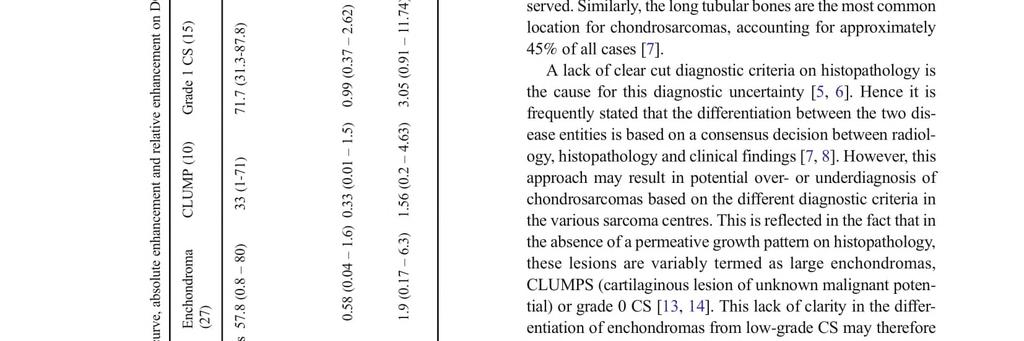
120 183
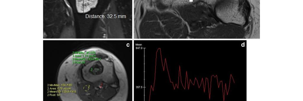
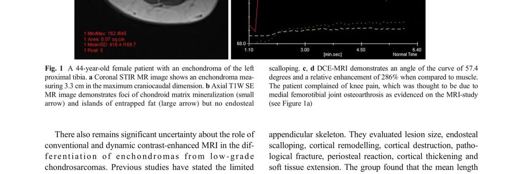
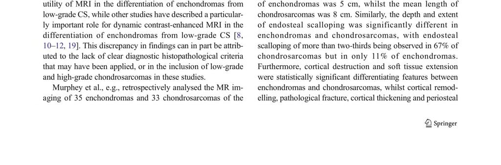
121 184
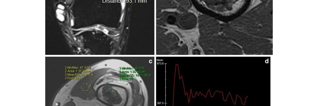
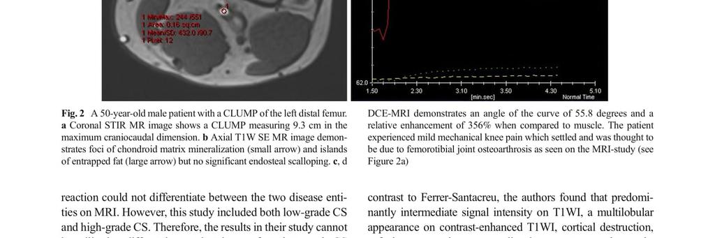
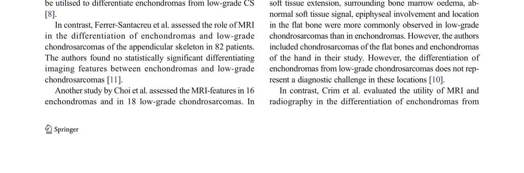
122 185
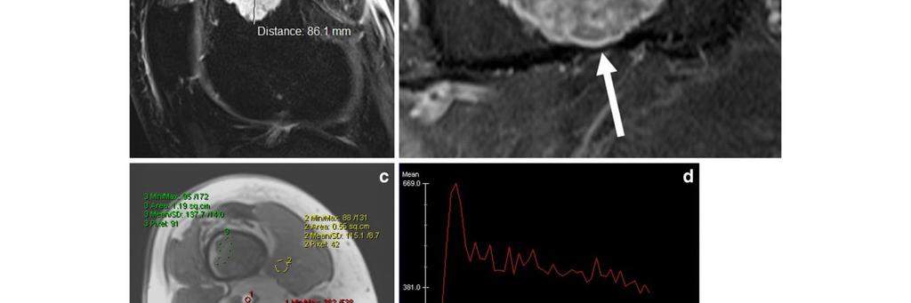
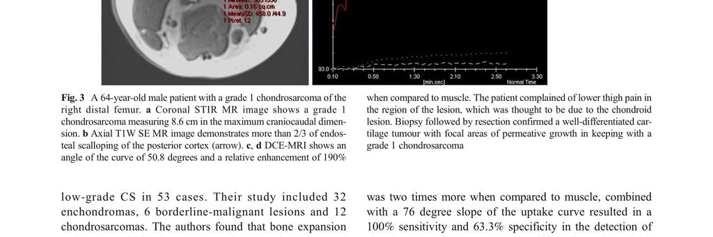
123 186
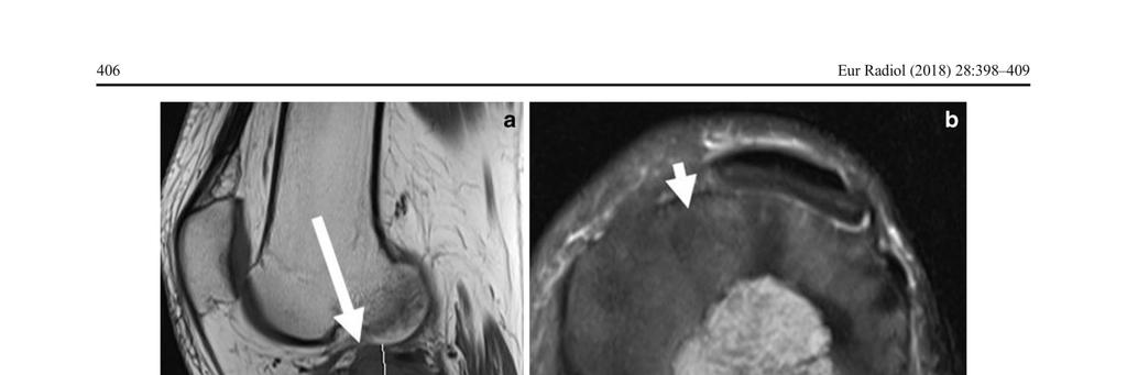
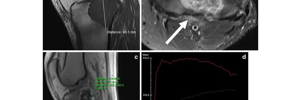
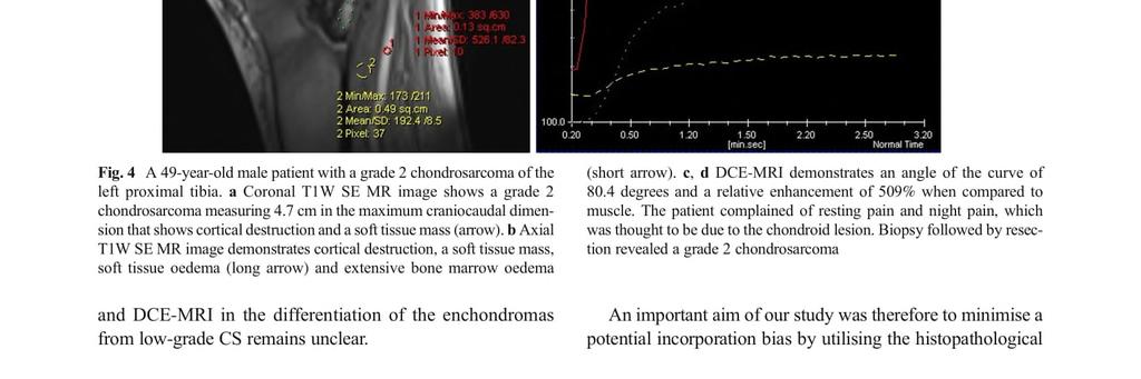
124 187
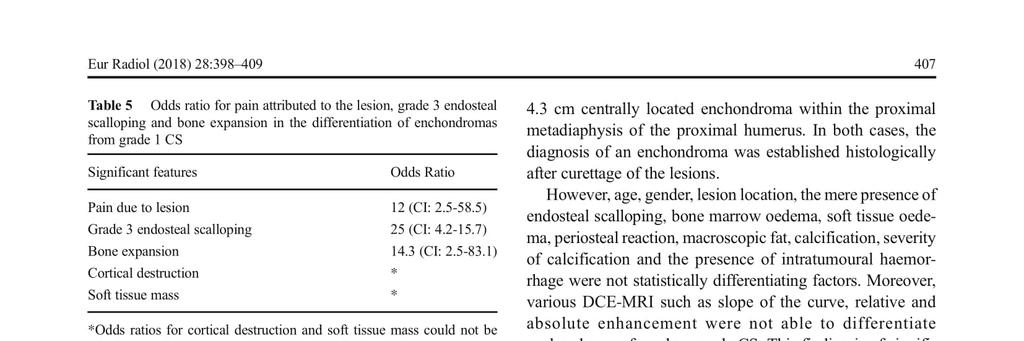
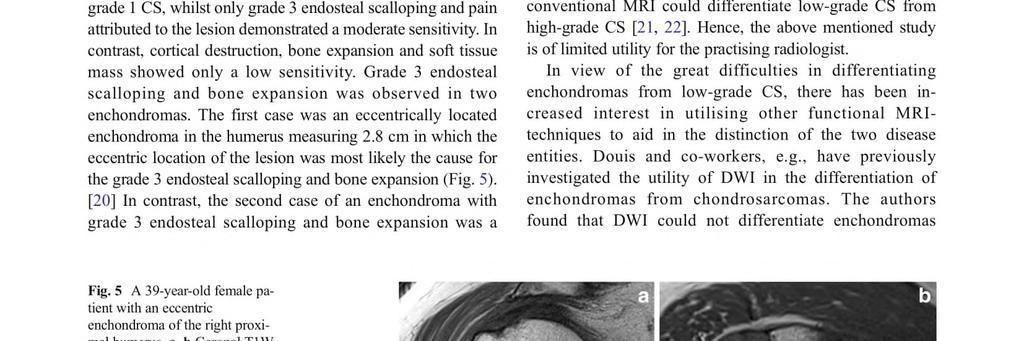
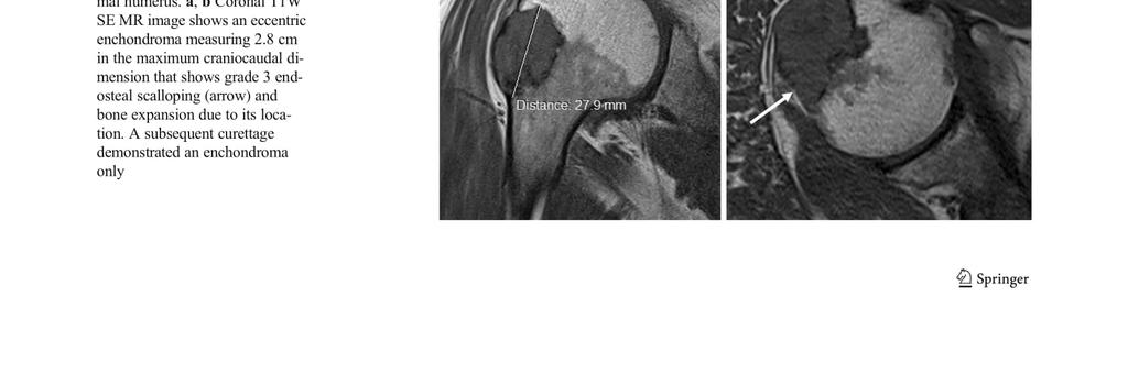
125 188
126 189
127 190
Bone Tumours - a synopsis. Dr Zena Slim SpR in Histopathology QAH 2009
 Bone Tumours - a synopsis Dr Zena Slim SpR in Histopathology QAH 2009 Aims General approach to diagnosis Common entities.and not so common ones. Mini quiz Challenge of bone tumour diagnosis Bone tumours
Typical skeletal location and differential diagnosis of bone tumors.
 Typical skeletal location and differential diagnosis of bone tumors. Poster No.: C-2418 Congress: ECR 2015 Type: Educational Exhibit Authors: M. Barros, L. A. Ferreira, Y. Costa, P. J. V. Coelho, F. Caseiro
Grading of Bone Tumors
 Grading of Bone Tumors Joon Hyuk Choi, M.D. Department of Pathology College of Medicine, Yeungnam University Introduction to grading system of bone tumor used at Mayo Clinic WHO Histologic Classification
Bone Tumors Clues and Cues
 William Herring, M.D. 2002 Bone Tumors Clues and Cues In Slide Show mode, advance the slides by pressing the spacebar All Photos Retain the Copyright of their Authors Clues by Appearance of Lesion Patterns
The Radiology Assistant : Bone tumor - well-defined osteolytic tumors and tumor-like lesions
 Bone tumor - well-defined osteolytic tumors and tumor-like lesions Henk Jan van der Woude and Robin Smithuis Radiology department of the Onze Lieve Vrouwe Gasthuis, Amsterdam and the Rijnland hospital,
USCAP 2014 Common problems in bone and soft tissue pathology: Cartilage tumors
 USCAP 2014 Common problems in bone and soft tissue pathology: Cartilage tumors Andrew Horvai MD PhD Clinical Professor, Pathology UCSF, San Francisco, CA Outline Common intramedullary tumors Enchondroma
Primary bone tumors > metastases from other sites Primary bone tumors widely range -from benign to malignant. Classified according to the normal cell
 Primary bone tumors > metastases from other sites Primary bone tumors widely range -from benign to malignant. Classified according to the normal cell counterpart and line of differentiation. Among the
The Radiology Assistant : Bone tumor - ill defined osteolytic tumors and tumor-like lesions
 Bone tumor - ill defined osteolytic tumors and tumor-like lesions Henk Jan van der Woude and Robin Smithuis Radiology department of the Onze Lieve Vrouwe Gasthuis, Amsterdam and the Rijnland hospital,
Fluid-fluid levels in bone tumors: A pictorial review
 Fluid-fluid levels in bone tumors: A pictorial review Poster No.: C-578 Congress: ECR 2009 Type: Educational Exhibit Topic: Musculoskeletal Authors: L. Figueroa Nasra, C. Martín Hervás, M. Tapia-Viñé,
Introduction to Musculoskeletal Tumors. James C. Wittig, MD Orthopedic Oncologist Sarcoma Surgeon
 Introduction to Musculoskeletal Tumors James C. Wittig, MD Orthopedic Oncologist Sarcoma Surgeon www.tumorsurgery.org Definitions Primary Bone / Soft tissue tumors Mesenchymally derived tumors (Mesodermal)
APMA 2018 Radiology Track Bone Tumors When to say Gulp!
 APMA 2018 Radiology Track Bone Tumors When to say Gulp! DANIEL P. EVANS, DPM, FACFAOM Professor, Department of Podiatric Medicine and Radiology Dr. Wm. Scholl College of Podiatric Medicine Conflict of
MRI XR, CT, NM. Principal Modality (2): Case Report # 2. Date accepted: 15 March 2013
: Case Report # 2. Date accepted: 15 March 2013") Radiological Category: Musculoskeletal Principal Modality (1): Principal Modality (2): MRI XR, CT, NM Case Report # 2 Submitted by: Hannah Safia Elamir, D.O. Faculty reviewer: Naga R. Chinapuvvula, M.D.
Radiological Category: Musculoskeletal Principal Modality (1): Principal Modality (2): MRI XR, CT, NM Case Report # 2 Submitted by: Hannah Safia Elamir, D.O. Faculty reviewer: Naga R. Chinapuvvula, M.D.
Primary bone tumors according to the WHO classification: a review of 13 years with illustrative examples
 Primary bone tumors according to the WHO classification: a review of 13 years with illustrative examples Poster No.: C-1741 Congress: ECR 2015 Type: Educational Exhibit Authors: J. Silva, M. A. Ramírez
Bread and Butter Bone Pathology
 Bread and Butter Bone Pathology NICOLE D. RIDDLE, MD RUFFOLO, HOOPER, AND ASSOC. / UNIVERSITY OF SOUTH FLORIDA Goals: Fundamentals of neoplastic bone pathology Bone Producing Cartilage Producing Miscellaneous
COPYRIGHT 2004 BY THE JOURNAL OF BONE AND JOINT SURGERY, INCORPORATED
 84 COPYRIGHT 2004 BY THE JOURNAL BONE AND JOINT SURGERY, INCORPORATED Radiographic Evaluation of Pathological Bone Lesions: Current Spectrum of Disease and Approach to Diagnosis BY BENJAMIN G. DOMB, MD,
MARK D. MURPHEY MD, FACR. Physician-in-Chief, AIRP. Chief, Musculoskeletal Imaging
 ALPHABET SOUP AND CYSTIC LESIONS OF THE BONE MARK D. MURPHEY MD, FACR Physician-in-Chief, AIRP Chief, Musculoskeletal Imaging ALPHABET SOUP AND CYSTIC LESIONS OF THE BONE Giant cell tumor (GCT) Unicameral
What Do You Need to Know About Bone Pathology? Benjamin L. Hoch M.D. Associate Professor Department of Pathology University of Washington
 What Do You Need to Know About Bone Pathology? Benjamin L. Hoch M.D. Associate Professor Department of Pathology University of Washington What s Do You Need To Know About Bone Pathology? Reactive/pseudosarcomatous
Malignant bone tumors. Incidence Myeloma 45% Osteosarcoma 24% Chondrosarcoma 12% Lyphoma 8% Ewing s Sarcoma 7%
 Malignant bone tumors Incidence Myeloma 45% Osteosarcoma 24% Chondrosarcoma 12% Lyphoma 8% Ewing s Sarcoma 7% Commonest primary bone sarcoma is osteosarcoma X ray Questions to ask 1. Solitary or Multiple
Recognizing Cartilaginous Tumors: Spectrum of Imaging Characteristics with Radiologic-Pathologic correlation.
 Recognizing Cartilaginous Tumors: Spectrum of Imaging Characteristics with Radiologic-Pathologic correlation. Poster No.: C-1451 Congress: ECR 2012 Type: Educational Exhibit Authors: E. Barcina García,
Bone and Joint Part 2. Leslie G Dodd, MD
 Bone and Joint Part 2 Leslie G Dodd, MD Relative rates of cancer Sarcomas are relatively uncommon tumors New cancer cases 2007 All sites 1.4 million prostate 218,890 lung 213,380 breast 180,510 Soft tissue
Malignant Bone Tumours. PathoBasic, Daniel Baumhoer
 Malignant Bone Tumours PathoBasic, 20.03.18 Daniel Baumhoer FNCLCC Grading The differentiation score is defined as the extent to which a tumor resembles adult mesenchymal tissue (score 1), the extent to
Case Report Intramedullary Chondrosarcoma of Proximal Humerus
 Hindawi Publishing Corporation Case Reports in Radiology Volume 2012, Article ID 642062, 7 pages doi:10.1155/2012/642062 Case Report Intramedullary Chondrosarcoma of Proximal Humerus Pratiksha Yadav, Dolly
Disclosures. Giant Cell Rich Tumors of Bone. Outline. The osteoclast. Giant cell rich tumors 5/21/11
 Disclosures Giant Cell Rich Tumors of Bone Andrew Horvai, MD, PhD Associate Clinical Professor, Pathology This lecture discusses "off label" uses of a number of pharmaceutical agents. The speaker is describing
Giant cell tumour of the sternum-two cases
 Giant cell tumour of the sternum-two cases Nishaa.P 1, Raghuram.P 2, Navin patil 3, Jaipal B.R 4 Akkamahadevi patel 5 Assistant Professor ESIC medical college and PGIMSR 1 Professor and HOD, 2 Professor
Imaging Findings Of Bone Tumors: A Pictorial Review
 Imaging Findings Of Bone Tumors: A Pictorial Review Poster No.: C-2511 Congress: ECR 2015 Type: Educational Exhibit Authors: M. Limeme, N. Benzina, A. BelKhiria, H. Zaghouani, S. Majdoub, N. Mallat, H.
04/27/2017. The Spectrum of Cartilaginous Tumors
 The Spectrum of Cartilaginous Tumors L I S A E R C O L A N O, M D M U S C U L O S K E L E T A L O N C O L O G Y D E P A R T M E N T O F O R T H O P A E D I C S A L L E G H E N Y H E A L T H N E T W O R
The Radiology Assistant : Bone tumor A-G
 Bone tumor A-G Bone tumors and tumor-like lesions in alphabethic order Henk Jan van de Woude and Robin Smithuis Radiology department of the Onze Lieve Vrouwe Gasthuis, Amsterdam and the Rijnland hospital,
Recommendations for cross-sectional imaging in cancer management, Second edition
 www.rcr.ac.uk Recommendations for cross-sectional imaging in cancer management, Second edition Musculoskeletal tumours Faculty of Clinical Radiology www.rcr.ac.uk Contents Primary bone tumours 3 Clinical
Bone/Osteoid Producing Lesions
 Chapter 2 Bone/Osteoid Producing Lesions Introduction There are many lesions that are associated with reactive new bone formation; this chapter predominantly covers those in which deposition of osteoid/bone
A Modified Lodwick-Madewell Grading System for the Evaluation of Lytic Bone Lesions
 Musculoskeletal Imaging Original Research Caracciolo et al. Evaluation of Lytic one Lesions Musculoskeletal Imaging Original Research Jamie T. Caracciolo 1 H. Thomas Temple 2 G. Douglas Letson 3 Mark J.
Malignant Bone Tumors - Part I: a brief revision of diagnostic aspects with conventional radiology
 Malignant Bone Tumors - Part I: a brief revision of diagnostic aspects with conventional radiology Poster No.: C-2473 Congress: ECR 2013 Type: Educational Exhibit Authors: I. Candelaria, L. B. Barbosa,
Case 8 Soft tissue swelling
 Case 8 Soft tissue swelling 26-year-old female presented with a swelling on the back of the left knee joint since the last 6 months and chronic pain in the calf and foot since the last 2 months. Pain in
FEGNOMASHIC: from x-ray to MRI
 FEGNOMASHIC: from x-ray to MRI Poster No.: C-2441 Congress: ECR 2015 Type: Educational Exhibit Authors: S. Fouassier, A. L. C. Duarte, C. Ruivo, J. Velez ; Évora/PT, 1 2 1 2 3 1 3 Coimbra/PT, PT Keywords:
Musculoskeletal Sarcomas
 Musculoskeletal Sarcomas Robert C. Orth, M.D., Ph.D. Edward B. Singleton Department of Pediatric Radiology Texas Children s Hospital Page 0 xxx00.#####.ppt 9/23/2012 9:01:18 AM No disclosures Page 1 xxx00.#####.ppt
Bubbly Lesions of Bone
 Residents Section Pattern of the Month w79 08.18.09 Eisenberg Residents Section Pattern of the Month Residents inradiology Ronald L. Eisenberg 1 Eisenberg RL Keywords: bubbly lesions, fegnomashic, skeletal
Bone tumors. RMG: jan
 Bone tumors RMG: jan 217. @Kijohs KIZZA JOHN KIJOHS Diseases arising in bone Lipoma Fibrous cortical defects Non-ossifying fibroma Bone island Benign simple cysts Enchondroma Osteochondroma Osteoid osteoma
Radiologic approach to pediatric lytic bone lesions
 Radiologic approach to pediatric lytic bone lesions Poster No.: C-1177 Congress: ECR 2016 Type: Educational Exhibit Authors: J. L. LERMA GALLARDO, I. de la Pedraja, A. Lancharro 1 1 1 2 1 1 Zapata, J.
Radiography in the Initial Diagnosis of Primary Bone Tumors
 Residents Section Structured Review Costelloe and Madewell Radiography of Primary Bone Tumors Residents Section Structured Review Colleen M. Costelloe 1 John E. Madewell Costelloe CM, Madewell JE Keywords:
ORTHOPAEDIC ONCOLOGY OITE REVIEW COURSE
 ORTHOPAEDIC ONCOLOGY OITE REVIEW COURSE Richard D. Lackman, MD FACS Director, Orthopaedic Oncology Center Cancer Institute Introduction In the evaluation of a patient with a bone tumor, there are several
Review Course «Musculoskeletal Oncology» October 6, 2011 UNIKLINIK BALGRIST. Imaging of Bone and Soft Tissue. Tumors
 Imaging of Bone and Soft Tissue Tumors Approach from a radiologist s point of view Florian Buck Radiology Radio- Radio- Oncologist Oncologist Orthopedist Orthopedist Patient Management Oncologist Oncologist
Multifocal fibrous Dysplasia with enchondroma-like areas: Fibrocartilaginous Dysplasia
 ISPUB.COM The Internet Journal of Pathology Volume 7 Number 2 Multifocal fibrous Dysplasia with enchondroma-like areas: Fibrocartilaginous Dysplasia V Monappa, R Kudva Citation V Monappa, R Kudva. Multifocal
Bone Tumors: Epidemiology, Classification, Pathology
 Bone Tumors: Epidemiology, Classification, Pathology 1 Lars Gunnar Kindblom C O N T E N T S 1.1 Introduction 2 1.2 Epidemiology 2 1.3 Morphologic Diagnosis of Bone Tumors 5 1.4 Types of Bone Tumor Specimens
Chondrosarcoma of 5 th metatarsal Right Foot: An Unusual Presentation and Review of Literature
 IOSR Journal of Dental and Medical Sciences (IOSR-JDMS) e-issn: 2279-0853, p-issn: 2279-0861.Volume 17, Issue 8 Ver. 13 (August. 2018), PP 27-33 www.iosrjournals.org Chondrosarcoma of 5 th metatarsal Right
Chondrosarcoma in Childhood: The Radiologic and Clinical Conundrum
 Chondrosarcoma in Childhood: The Radiologic and Clinical Conundrum Susan M. Mosier 1, Tanvi Patel 1, Karen Strenge 2, Andrew D. Mosier 3* 1. Department of Pediatrics, Ireland Army Community Hospital, Fort
Skeletal metastases are the most common variety of bone tumors and should always be considered in the differential diagnosis, particularly in older
 Dr Brajesh Nandan Skeletal metastases are the most common variety of bone tumors and should always be considered in the differential diagnosis, particularly in older patients. Cancers of the breast, prostate,
* I have no disclosures or any
 Howard Rosenthal, M.D. Associate Professor of Orthopedic Surgery University of Kansas Sarcoma Center I have no disclosures or any conflicts related to the content of this presentation. Objectives 1. Describe
GIANT CELL-RICH OSTEOSARCOMA: A CASE REPORT
 Nagoya J. Med. Sci. 59. 151-157, 1996 CASE REPORTS GIANT CELL-RICH OSTEOSARCOMA: A CASE REPORT KEIJI SATO!, SHIGEKI YAMAMURA!, HISASHI IWATA!, HIDESHI SUGIURA 2, NOBUO NAKASHIMA 3 and TETSURO NAGASAKA
Incidental bone tumors are asymptomatic lesions that are. Incidental Bone Lesions. When to Refer to the Tumor Specialist
 Bulletin of the NYU Hospital for Joint Diseases 2012;70(4):235-40 235 Incidental Bone Lesions When to Refer to the Tumor Specialist LT Suezie Kim, M.D., M.C., U.S.N., Catherine N. Laible, M.D., Leon D.
Department of Radiology, University of Szeged. Imaging of the skeleton
 Imaging of the skeleton Methods of examination: plain x-ray (radiography, densitometry) x-ray with contrast material (fistulography, angiography) ultrasound (b-mode, Doppler, color, duplex) computed tomography
Imaging Evaluation of Malignant Chest Wall Neoplasms 1
 This copy is for personal use only. To order printed copies, contact reprints@rsna.org Imaging Evaluation of Malignant Chest Wall Neoplasms 1 1285 CHEST IMAGING Brett W. Carter, MD Marcelo F. Benveniste,
VALORACIÒN RADIOLÓGICA DE LA LESIÒN ÒSEA SOLITARIA IMAGENOLOGIA MEDICA UNIVERSIDAD HISPANOAMERICANA
 VALORACIÒN RADIOLÓGICA DE LA LESIÒN ÒSEA SOLITARIA IMAGENOLOGIA MEDICA UNIVERSIDAD HISPANOAMERICANA TUMORES ÓSEOS SE PRESENTAN POR RANGOS DE EDAD, PRINCIPALMENTE: MENORES DE 20 AÑOS 20 A 40 AÑOS MAYORES
Immunohistochemistry in Bone and Soft Tissue Tumors. Sahar Rassi Zankoul, MD
 Immunohistochemistry in Bone and Soft Tissue Tumors Sahar Rassi Zankoul, MD Introduction Bone tumors represent a wide variety of tumors of various origins and malignant potentials. These different tumor
Prof. Dr. NAGUI M. ABDELWAHAB,M.D.; MARYSE Y. AWADALLAH, M.D. AYA M. BASSAM, Ms.C.
 Role of Whole-body Diffusion MR in Detection of Metastatic lesions Prof. Dr. NAGUI M. ABDELWAHAB,M.D.; MARYSE Y. AWADALLAH, M.D. AYA M. BASSAM, Ms.C. Cancer is a potentially life-threatening disease,
Primary Tumors of Ribs
 Primary Tumors of Ribs Frank E. Schmidt, M.D., and Max J. Trummer, Capt, MC, USN ABSTRACT An analysis of 50 consecutive patients with primary rib tumors operated on at the U.S. Naval Hospital, San Diego,
Index. Note: Page numbers of article titles are in boldface type.
 Magn Reson Imaging Clin N Am 12 (2004) 185 189 Index Note: Page numbers of article titles are in boldface type. A Acromioclavicular joint, MR imaging findings concerning, 161 Acromion, types of, 77 79
LAC + USC.
 Jeff McDavit,, M.D. LAC + USC mcdavit@usc.edu Clinical History 55 year old male with large, deep, non- tender left thigh mass. Seen at LAC+USC Med Ctr FNA clinic No h/o trauma or radiation Vimentin
Fine Needle Aspiration of Bone Tumours
 Fine Needle Aspiration of Bone Tumours Monographs in Clinical Cytology Vol. 19 Series Editor Svante R. Orell Kent Town Fine Needle Aspiration of Bone Tumours The Clinical, Radiological, Cytological Approach
JMSCR Vol 3 Issue 11 Page November 2015
 www.jmscr.igmpublication.org Impact Factor 3.79 Index Copernicus Value: 5.88 ISSN (e)-2347-176x ISSN (p) 2455-0450 DOI: http://dx.doi.org/10.18535/jmscr/v3i11.36 Diagnostic Dilemmas in Cytodiagnosis of
Monophasic Synovial Carcinoma of knee joint- A Case Report and Review of Literature
 IOSR Journal of Dental and Medical Sciences (IOSR-JDMS) e-issn: 2279-0853, p-issn: 2279-0861.Volume 17, Issue 3 Ver.5 March. (2018), PP 13-17 www.iosrjournals.org Monophasic Synovial Carcinoma of knee
Myositis Ossificans Mimicking Sarcoma, the Importance of Diagnostic Imaging Case Report
 Signature: Pol J Radiol, 2014; 79: 228-232 DOI: 10.12659/PJR.890209 CASE REPORT Received: 2013.12.18 Accepted: 2014.01.27 Published: 2014.07.28 Authors Contribution: A Study Design B Data Collection C
General Approach to Lytic Bone Lesions D. Lee Bennett, MD, MA, Georges Y. El Khoury, MD Appl Radiol. 2004;33(5)
") General Approach to Lytic Bone Lesions D. Lee Bennett, MD, MA, Georges Y. El Khoury, MD Appl Radiol. 2004;33(5) www.medscape.com Abstract and Introduction Abstract When interpreting musculoskeletal radiographs,
General Approach to Lytic Bone Lesions D. Lee Bennett, MD, MA, Georges Y. El Khoury, MD Appl Radiol. 2004;33(5) www.medscape.com Abstract and Introduction Abstract When interpreting musculoskeletal radiographs,
Case 9298 Epithelioid hemangioendothelioma of the femur
 Case 9298 Epithelioid hemangioendothelioma of the femur Manenti G, Antonicoli M, Llubani R, Squillaci E, Dragoni M, Ippolito E, Simonetti G Section: Musculoskeletal System Published: 2011, May. 11 Patient:
A peculiar location of a rare bone tumor: sternal lipoma
 A peculiar location of a rare bone tumor: sternal lipoma Poster No.: P-0033 Congress: ESSR 2016 Type: Authors: Keywords: DOI: Scientific Poster Z. Akkaya, C. Uzun, S. Enon, G. Kocaman, G. Sahin; Ankara/TR
Non-monomelic synchronous primary multicentric chondrosarcoma : A case report
 Acta Orthop. Belg., 2005, 71, 242-248 CASE REPORT Non-monomelic synchronous primary multicentric chondrosarcoma : A case report Yolanda HERNANZ GONZÁLEZ, Javier SALAMANCA, Carlos RESINES ERASUN, Francisco
Paediatric post-traumatic osseous cystic lesion following a distal radial fracture
 Paediatric post-traumatic osseous cystic lesion following a distal radial fracture Joey Chan Yiing Beh 1*, Ehab Shaban Mahmoud Hamouda 1 1. Department of Diagnostic Imaging, KK Women's and Children's Hospital,
An epidemiological survey of tumour or tumour like conditions in the scapula and periscapular region
 SICOT J 206, 2, 34 Ó The Authors, published by EDP Sciences, 206 DOI: 0.05/sicotj/206023 Available online at: www.sicot-j.org RESEARCH OPEN ACCESS An epidemiological survey of tumour or tumour like conditions
Benign Fibrous Histiocytoma with Cystic Change of the Femur: a Case Report
 pissn 2384-1095 eissn 2384-1109 imri 2016;20:264-268 https://doi.org/10.13104/imri.2016.20.4.264 Benign Fibrous Histiocytoma with Cystic Change of the Femur: a Case Report Jung Ah Park, Sung Gyu Moon,
Clinical Study Enchondroma versus Low-Grade Chondrosarcoma in Appendicular Skeleton: Clinical and Radiological Criteria
 Oncology Volume 2012, Article ID 437958, 6 pages doi:10.1155/2012/437958 Clinical Study Enchondroma versus Low-Grade Chondrosarcoma in Appendicular Skeleton: Clinical and Radiological Criteria Eugenio
Calcifying Aponeurotic Fibroma of the Knee: a Case Report with Radiographic and MRI Finding
 pissn 2384-1095 eissn 2384-1109 imri 2017;21:259-263 Calcifying Aponeurotic Fibroma of the Knee: a Case Report with Radiographic and MRI Finding Seung Hyun Lee 1,2, In Sook Lee 1,2, You Seon Song 1,2,
Dr Sneha Shah Tata Memorial Hospital, Mumbai.
 Dr Sneha Shah Tata Memorial Hospital, Mumbai. Topics covered Lymphomas including Burkitts Pediatric solid tumors (non CNS) Musculoskeletal Ewings & osteosarcoma. Neuroblastomas Nasopharyngeal carcinomas
Radiographic features of Ollier s disease two case reports
 Sadiqi et al. BMC Medical Imaging (2017) 17:58 DOI 10.1186/s12880-017-0230-8 CASE REPORT Radiographic features of Ollier s disease two case reports Jamshid Sadiqi 1,3*, Najibullah Rasouly 1, Hidayatullah
A review of Tumoral lesions of the shoulder
 A review of Tumoral lesions of the shoulder Poster No.: P-0109 Congress: ESSR 2013 Type: Scientific Exhibit Authors: M. M. Milán Rodríguez, Á. E. Moreno Puertas, J. M. Giménez, 1 1 1 1 2 1 A. Rubio Fernández,
We are IntechOpen, the world s leading publisher of Open Access books Built by scientists, for scientists. International authors and editors
 We are IntechOpen, the world s leading publisher of Open Access books Built by scientists, for scientists 3,700 108,500 1.7 M Open access books available International authors and editors Downloads Our
Benign Tumors of Bone
 REVIEW ARTICLE Benign Tumors of Bone Subbarao K Padmshri Prof. Dr. Kakarla Subbara, Hyderabad, India. Benign tumors of bone are common while malignant tumors are rare. Benign tumors constitute about 75%
Radiology-Pathology Conference
 July 31, 2009 Radiology-Pathology Conference Daniel T Ginat, M.D., M.S. Sharlin Johnykutty,, M.D. Presentation material is for education purposes only. All rights reserved. 2009 URMC Radiology Page 1 of
Fluid fluid levels in bone tumors and tumoral lesions - Pictorial essay
 Review Fluid fluid levels in bone tumors and tumoral lesions - Pictorial essay Subbarao Kakarla 1,* 1 KIMS Foundation and Research Centre, Minister Road, Secunderabad - 500003, Telangana, India Abstract
Downloaded from by on 11/21/17 from IP address Copyright ARRS. For personal use only; all rights reserved
 Downloaded from www.ajronline.org by 46.3.196.1 on 11/21/17 from IP address 46.3.196.1. opyright RRS. For personal use only; all rights reserved T he scapula is a small bone in which many neoplasms can
A. Nahal MD,* A. Ajlan MD, T. Alcindor MD, and R. Turcotte MD P A T H O L O G Y ABSTRACT 2. CASE DESCRIPTION KEY WORDS 1.
 P A T H O L O G Y Dedifferentiated giant cell tumour of bone in the form of low-grade fibroblastic osteogenic sarcoma: case report of a unique presentation with follow-up A. Nahal MD,* A. Ajlan MD, T.
Chondroblastoma of bone
 Chronology Chondroblastoma of bone LONG-TERM RESULTS AND FUNCTIONAL OUTCOME AFTER INTRALESIONAL CURETTAGE R. Suneja, R. J. Grimer, M. Belthur, L. Jeys, S. R. Carter, R. M. Tillman, A. M. Davies From The
Skeletal System. Chapter 6.1 Human Anatomy & Physiology
 Skeletal System Chapter 6.1 Human Anatomy & Physiology Overview of Skeletal System Bones Joints Skeletal System Cartilage Tendons (bone to muscle) Ligaments (bone to bone) Function of the Skeletal System
Bizarre parosteal osteochondromatous proliferation
 * * Bizarre Parosteal Osteochondromatous Proliferation A Case Report with Literature Review Chi-Fu Kao Yang-Chih Lin Yu-Hung Wu Be-Fong Chen* We report the case of a 12-year-old female with a slowly erythematous
Intraosseous lipoma of the talus
 Intraosseous lipoma of the talus Shigeki Maruyama, M.D. 1, Tetsuji Yamamoto, M.D (!). 2, Masataka Hashimura, M.D. 1, and Nobuhiro Ohsaki, M.D. 1, Toshihiro Akisue, M.D. 2, Shinichi Yoshiya, M.D 2 1 Department
ISSN: DISTRIBUTION OF BONE AND CARTILAGINOUS TUMORS IN PEDIATRIC AGE GROUP IN WESTERN UTTAR-PRADESH: AN EVALUATIVE STUDY
 : 289-295 ISSN: 2277 4998 DISTRIBUTION OF BONE AND CARTILAGINOUS TUMORS IN PEDIATRIC AGE GROUP IN WESTERN UTTAR-PRADESH: AN EVALUATIVE STUDY QADRI S, HASAN M, AKHTAR K * AND SHERWANI RK The Departments
Heterogeneous osteoblastic activity in the right ischium of unclear etiology seen on NaF18-PET/CT
 CASE REPORT Heterogeneous osteoblastic activity in the right ischium of unclear etiology seen on NaF18-PET/CT Aung Zaw Win, Carina Mari Aparici Dept. Radiology, Nuclear Medicine section, San Francisco
Monostotic Paget s Disease: A Case Report
 Chin J Radiol 2002; 27: 117-121 117 CASE REPORT Monostotic Paget s Disease: A Case Report CHI-CHEN HOU 1 CHI WEI LO 2 JINN-MING CHANG 1 CHING-CHERNG TZENG 3 Department of Diagnostic Radiology 1, Orthopedics
The Skeletal System PART A. PowerPoint Lecture Slide Presentation by Patty Bostwick-Taylor, Florence-Darlington Technical College
 PowerPoint Lecture Slide Presentation by Patty Bostwick-Taylor, Florence-Darlington Technical College The Skeletal System 5 PART A The Skeletal System Parts of the skeletal system Bones (skeleton) Joints
History. 33 y/o F with hx of palpable anterior tibial mass x 2 years, only painful with palpation
 History 33 y/o F with hx of palpable anterior tibial mass x 2 years, only painful with palpation Imaging Photo Album Patient also had a smaller lesion 1 cm proximal to this lesion, not seen radiographically.
Essential Dermatopathology. Jinah Kim, MD, PhD Department of Pathology and Dermatology Stanford University Medical Center
 Essential Dermatopathology Jinah Kim, MD, PhD Department of Pathology and Dermatology Stanford University Medical Center OBJECTIVES Review clinical, pathologic and molecular aspects of bone and fat tumors
Key points in the evaluation of focal bone lesions: from plain film to multidetector CT
 Key points in the evaluation of focal bone lesions: from plain film to multidetector CT Poster No.: C-2060 Congress: ECR 2011 Type: Educational Exhibit Authors: I. Rubio Marco, M. Arraiza Sarasa, H. Gómez
Skeletal Radiology. Solitary (unicameral) bone cyst. The fallen fragment sign revisited
 bone cyst. The fallen fragment sign revisited") Skeletal Radiol (1989) 18:261-265 Skeletal Radiology Solitary (unicameral) bone cyst The fallen fragment sign revisited S. Struhl, M.D., C. Edelson, M.D., H. Pritzker, M.D., L.P. Seimon, M.D., and H.D.
Skeletal Radiol (1989) 18:261-265 Skeletal Radiology Solitary (unicameral) bone cyst The fallen fragment sign revisited S. Struhl, M.D., C. Edelson, M.D., H. Pritzker, M.D., L.P. Seimon, M.D., and H.D.
Common Primary Tumors of Bone
 Special Report Common Primary Tumors of Bone Primary bone tumors are a relatively rare occurrence, however, they can have serious deleterious consequences. Many possess the ability to degenerate into malignant
Skeletal System worksheet
 Skeletal System worksheet Name Section A: Intro to Skeletal System The skeletal system performs vital functions that enable us to move through our daily lives. Support - The skeleton provides support and
SMALL ROUND BLUE CELL LESION OF BONE
 DISCLOSURE SMALL ROUND BLUE CELL LESION OF BONE Dr. Alistair Jordan University of South Alabama No financial support or endorsement OBJECTIVES Describe the more common small round cell lesions of bone
Primary Breast Liposarcoma
 Primary Breast Liposarcoma Bhagyam Nagarajan 1*, GayatriAutkar 1, Keyuri Patel 1, Meghal Sanghvi 1 1. Department of Radiology, Wockhardt Hospital, Mumbai, India * Correspondence: Dr Bhagyam Nagarajan,
GIANT CELL TUMOR OF BONE
 GIANT CELL TUMOR OF BONE Definition. First described by Jaffe et al. 1, giant cell tumor of bone is a locally aggressive primary neoplasm of bone that is composed of proliferation of bland looking oval
Chapter 5 The Skeletal System
 Chapter 5 The Skeletal System The Skeletal System Parts of the skeletal system Bones (skeleton) Joints Cartilages Ligaments (bone to bone)(tendon=bone to muscle) Divided into two divisions Axial skeleton:
Unusual location of bone sarcoma in children
 Unusual location of bone sarcoma in children Poster No.: C-1517 Congress: ECR 2014 Type: Educational Exhibit Authors: S. JERBI, A. Khalfalli, G. Abid, O. Bradai, N. chouchane, H. HAMZA; Mahdia/TN Keywords:
University Journal of Surgery and Surgical Specialities
 University Journal of Surgery and Surgical Specialities Volume 1 Issue 1 2015 EXTRA SKELETAL MESENCHYMAL CHONDROSARCOMA :A CASE REPORT Rajaraman R Subbiah S Navin Naushad Kilpaulk Medical College Abstract:
Case Studies in the Skull Base
 Case Studies in the Skull Base Amy C Tsai, MD Neuroradiology Fellow Department of Radiology and Imaging Sciences University of Utah Health Sciences Center Salt Lake City, Utah, USA No disclosures related
The Skeletal System PART A
 5 The Skeletal System PART A PowerPoint Lecture Slide Presentation by Jerry L. Cook, Sam Houston University ESSENTIALS OF HUMAN ANATOMY & PHYSIOLOGY EIGHTH EDITION ELAINE N. MARIEB The Skeletal System