석회성건염 한양의대재활의학교실 이규훈
|
|
|
- Cecil Smith
- 5 years ago
- Views:
Transcription
1 석회성건염 한양의대재활의학교실 이규훈
2 Definition Calcifying tendinitis Acute or chronically painful condition that is caused by inflammation around calcium deposits located in or around the tendons Vascularized, viable soft tissue Cyclic process Within the midsubstance of the tendon Dystrophic calcification
3 Classification Size : small, medium, large Degree and duration : acute, subacute, chronic Radiologic finding : localized, diffuse Clinical : dystrophic, reactive Appear : dry, powdery deposit soft, putty, or toothpaste deposit milky or creamy deposit

4 Sites of calcium deposition
5 Epidemiology Painless shoulder : 2.7% - 20% Painful shoulder : 6.8% in all age 35%-45% in years Common site : supraspinatus tendon (82-90%) Women > men Laterality : Rt. > Lt. Bilaterality : 13-24% Occupation : 주부, 사무직노동자 A rotator cuff tear may coexist : 25% Spontaneous resolution : 9.3% after 3 years 27% after 10 years
6 Pathogenesis Degeneration Hypovascularity Decreased local oxygen tension Metabolic disorder Hereditary
7 Degenerative calcification Degenerative process Focal hyalinization, fibrillate, detach Necrosis Microspherolith, psammoma Aging : decreased vascularity
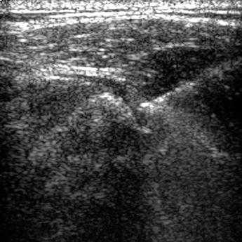
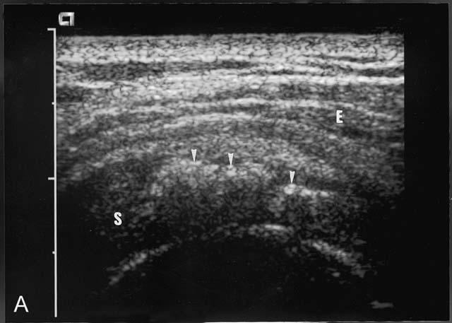
8 Reactive calcification
9
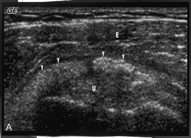
10 Clinical findings Begin wth subacute or chronic symptom Formative phase Pain or tenderness, referred pain Impingement No vascular and no cellular reactions Hard and well defined deposition
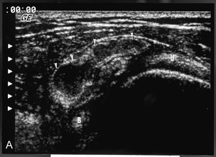
11 Clinical findings Progress to acute symptom Resorptive phase Acute pain Bursitis Vascular and cellular proliferaton -> increase intratendious pressure Fluffy and ill defined deposition

12 Clinical findings
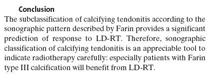
13 Radiologic finidngs Radiograph Confirm and follow up Size : small, medium, large Localize vs diffuse Type A or I : homogeneous calcification and well defined limits Type B or II : heterogeneous calcification and well defined limits Type C or III : heterogeneous calcification and ill defined limits Type D : dystrophic calcification
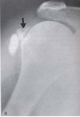
 Plain radiography of a rotator cuff calcification with a well defined periphery. (B) Corresponding axial computed tomography scan.")
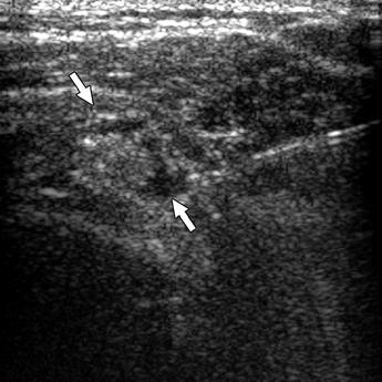
14 Consistency of Rotator Cuff Calcifications: Observations on Plain Radiography, Sonography, Computed Tomography, and at Needle Treatment. FARIN, PEKKA Investigative Radiology. 31(5): , May Figures 1A 1C. (A) Plain radiography of a rotator cuff calcification with a well defined periphery. (B) Corresponding axial computed tomography scan. The calcification is homogeneous with a density of 939 Hounsfield units. (C) Corresponding shows a rotator cuff calcification (arrows) with a clear acoustic shadow (arrowheads). The calcification proved to be hard at ultrasound guided needle treatment. 2
 Plain radiography of a rotator cuff calcification with an ill defined periphery. (B) Corresponding axial computed tomography scan.")
15 Consistency of Rotator Cuff Calcifications: Observations on Plain Radiography, Sonography, Computed Tomography, and at Needle Treatment. FARIN, PEKKA Investigative Radiology. 31(5): , May Figures 2A 2C. (A) Plain radiography of a rotator cuff calcification with an ill defined periphery. (B) Corresponding axial computed tomography scan. The calcification (arrows) is nonhomogeneous with a density of 122 Hounsfield units. (C) Corresponding sonogram shows a rotator cuff calcification (arrows) with no acoustic shadow. The calcification proved to be soft (slurry calcification) at treatment. 3
 Reconstructed sagittal oblique computed tomography scan of a nonhomogeneous calcification (arrows).")
16 Consistency of Rotator Cuff Calcifications: Observations on Plain Radiography, Sonography, Computed Tomography, and at Needle Treatment. FARIN, PEKKA Investigative Radiology. 31(5): , May Figures 3A and 3B. (A) Reconstructed sagittal oblique computed tomography scan of a nonhomogeneous calcification (arrows). The density values from the upper part of the calcification are 120 to 130 Hounsfield units (HU), and the values from the lower part are 400 to 420 HU. (B) Corresponding transverse sonogram shows a hyperechoic mass (arrows) with mixed acoustic shadow. In the center part of the hyperechoic mass is an acoustic shadow (arrowheads). The other parts have no shadow. The upper part of the calcification proved to be soft and the center of the lower part was hard at needle treatment. 4
17 CONCLUSIONS: Ultrasound and CT were reliable in predicting the consistency of rotator-cuff calcifications. 9
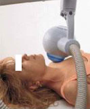
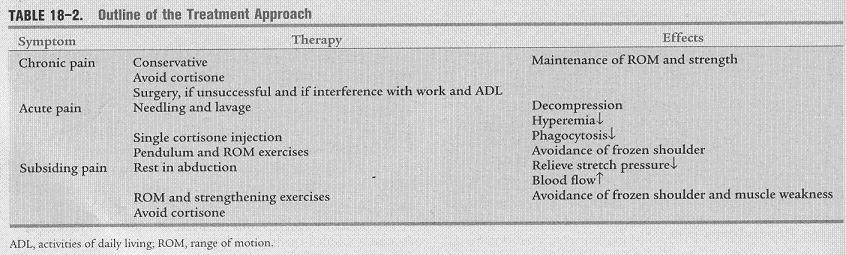
18 Management Non operative NSAIDs Physiotherapy Physical modality Puncture aspiration/needling Radiotherapy Extracorporeal shock wave therapy Surgical treatment
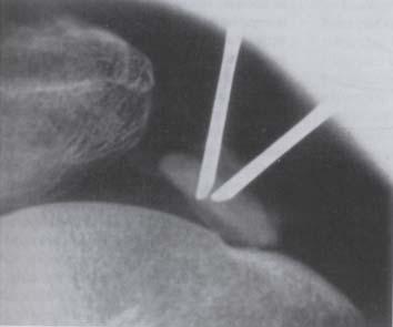
19 Puncture aspiration/needling
20
21
22

23 Table 2. Mean visual analog scale (VAS) scores before and after percutaneous needle puncture treatment (±SD). VAS p With aspiration Without aspiration Time point, weeks (n=41) ( =40) Pretreatment 6.8± ±1.8 NS Posttreatment 1 6.8± ±2.1 NS 2 6.7± ±2.3* S 3 4.9±1.4* 4.7±2.3* NS 6 4.4±1.6* 4.5±1.9* NS ±2.7* 4.3±2.5* NS ±1.9* 3.4±1.6* NS ±2.1* 3.6±2.4* NS *P<0.05 vs. pretreatment. NS=not significant; S=significant (P<0.05).
24
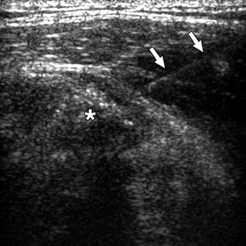
25 Pain : painless : VAS, 0 cm, mild : VAS, 1 4 cm moderate : VAS, 4 8 cm, severe : VAS, 8 10 cm High-resolution ultrasonography (HRUS) arc shaped : an echogenic arc with clear shadowing fragmented or punctate : at least 2 separated echogenic spots or plaques nodular : an echogenic nodule without shadowing cystic : a bold echogenic wall with an anechoic area, weak internal echoes Color Doppler ultrasonography (CDUS) grade 0 : no color flow signal grade 1 : <3 color spots grade 2 : 3 6 color spots grade 3 : >6 color spots

26 Arc shaped Fragmented shaped Nodular shaped Cystic shaped


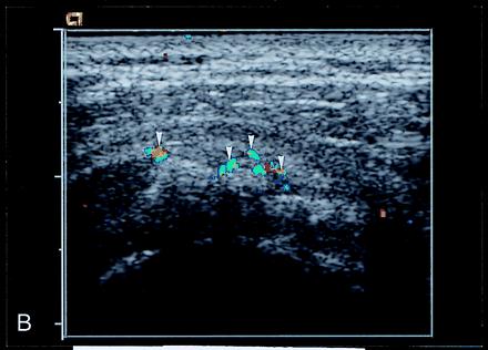
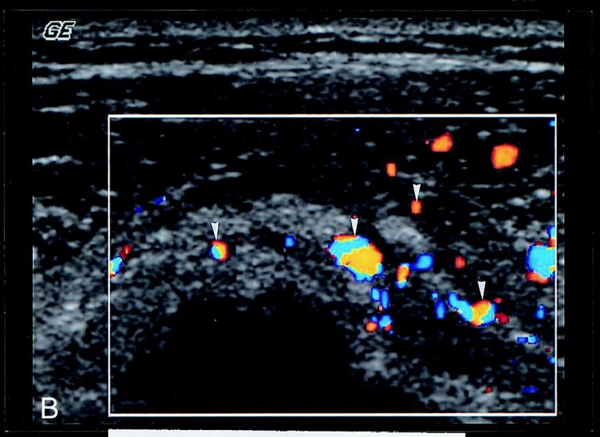
27 Grade 0 Grade 1 Grade 2 Grade 3
28

29 Color Doppler ultrasonography of the rotator cuff is highly correlated to symptoms. Higher-grade CDUS signals usually indicate resorptive status. We recommend a combination of HRUS and CDUS for more accurate prediction of the formative or resorptive status of calcific plaques.
30
31


32 Biceps tendon Greater tuberosity Coronal plane Longitudinal plane
33
Common Applications for Sonography and Guided Intervention: Shoulder
 Common Applications for Sonography and Guided Intervention: Shoulder Jon A. Jacobson, M.D. Professor of Radiology Director, Division of Musculoskeletal Radiology University of Michigan Disclosures: Consultant:
Rotator Cuff and Biceps Pathology
 Rotator Cuff and Biceps Pathology Jon A. Jacobson, M.D. Professor of Radiology Director, Division of Musculoskeletal Radiology University of Michigan Disclosures: Consultant: Bioclinica Advisory Board:
US finding of the shoulder (with live demonstration) 인제의대상계백병원 안재기
 인제의대상계백병원 안재기") US finding of the shoulder (with live demonstration) 인제의대상계백병원 안재기 Shoulder US Biceps tendon & Rotator Cuff Long Head of Biceps Tendon Subscapularis tendon Supraspinatus tendon Infraspinatus tendon Teres
US finding of the shoulder (with live demonstration) 인제의대상계백병원 안재기 Shoulder US Biceps tendon & Rotator Cuff Long Head of Biceps Tendon Subscapularis tendon Supraspinatus tendon Infraspinatus tendon Teres
Ultrasound of Shoulder Pathology and Intervention 서울대학교병원재활의학과 김기원
 Ultrasound of Shoulder Pathology and Intervention 서울대학교병원재활의학과 김기원 Ultrasound for Shoulder Disorder Advantage Dynamic evaluation Immediate clinical correlation + Intervention Weakness Diagnostic accuracy?
US in non-traumatic acute abdomen. Lalita, M.D. Radiologist Department of radiology Faculty of Medicine ChiangMai university
 US in non-traumatic acute abdomen Lalita, M.D. Radiologist Department of radiology Faculty of Medicine ChiangMai university Sagittal Orientation Transverse (Axial) Orientation Coronal Orientation Intercostal
Endometrioma With Calcification Simulating a Dermoid on Sonography
 Case Report Endometrioma With Calcification Simulating a Dermoid on Sonography Kiran A. Jain, MD Several investigators have explored the sonographic diagnostic criteria of endometriomas. Endometriomas
Ultrasound-Guided Calcific Tendinitis Lavage: Application, Technique, and Outcome
 Ultrasound-Guided Calcific Tendinitis Lavage: Application, Technique, and Outcome Andrew Schapiro MD, Humberto Rosas MD, Kenneth Lee MD University of Wisconsin Hospital and Clinics aschapiro@uwhealth.org
CLINICAL PRESENTATION AND RADIOLOGY QUIZ QUESTION
 Donald L. Renfrew, MD Radiology Associates of the Fox Valley, 333 N. Commercial Street, Suite 100, Neenah, WI 54956 9/22/2012 Radiology Quiz of the Week # 91 Page 1 CLINICAL PRESENTATION AND RADIOLOGY
US guided treatment in calcific tendinopathy of body tendons: Techniques and follow-up
 US guided treatment in calcific tendinopathy of body tendons: Techniques and follow-up Poster No.: C-2369 Congress: ECR 2010 Type: Scientific Exhibit Topic: Musculoskeletal - Soft Tissue Authors: T. De
of Thyroid Lesions Comet Tail Crystals
 2 Ultrasound Features of Thyroid Lesions There are many different features indicating a certain benign or malignant tumor type, but many of these are overlapping signs. Combining several features is considered
Ultrasound assessment of most frequent shoulder disorders
 Ultrasound assessment of most frequent shoulder disorders Poster No.: C-2026 Congress: ECR 2014 Type: Educational Exhibit Authors: S. P. Ivanoski; Ohrid/MK Keywords: Trauma, Athletic injuries, Arthritides,
Shear Wave Elastography in diagnostics of supraspinatus tendon.
 Shear Wave Elastography in diagnostics of supraspinatus tendon. Poster No.: C-2168 Congress: ECR 2013 Type: Authors: Keywords: DOI: Scientific Exhibit V. Saltykova; Moscow/RU Musculoskeletal joint, Musculoskeletal
Lipoma Arborescens of Subacromial-subdeltoid Bursa: Ultrasonographic Findings
 C A S E R E P O R T Lipoma Arborescens of Subacromial-subdeltoid Bursa: Ultrasonographic Findings Amelia Bargiela*, Esther Rodriguez, Rafaela Soler The present study describes the ultrasound findings of
Role of imaging in RCC. Ultrasonography. Solid lesion. Cystic RCC. Solid RCC 31/08/60. From Diagnosis to Treatment: the Radiologist Perspective
 Role of imaging in RCC From Diagnosis to Treatment: the Radiologist Perspective Diagnosis Staging Follow up Imaging modalities Limitations and pitfalls Duangkamon Prapruttam, MD Department of Therapeutic
Contents. Basic Ultrasound Principles and Terminology. Ultrasound Nodule Characteristics
 Contents Basic Ultrasound Principles and Terminology Basic Ultrasound Principles... 1 Ultrasound System... 2 Linear Transducer for Superficial Images and Ultrasound-Guided FNA... 3 Scanning Planes... 4
Sonographic Evaluation of Tears of the Gastrocnemius Medial Head ( Tennis Leg )
") Sonographic Evaluation of Tears of the Gastrocnemius Medial Head ( Tennis Leg ) Stefano Bianchi, MD, Carlo Martinoli, MD, Ibrahim Fikry Abdelwahab, MD, Lorenzo E. Derchi, MD, Sandro Damiani, MD Rupture
Sonographic Evaluation of Tears of the Gastrocnemius Medial Head ( Tennis Leg ) Stefano Bianchi, MD, Carlo Martinoli, MD, Ibrahim Fikry Abdelwahab, MD, Lorenzo E. Derchi, MD, Sandro Damiani, MD Rupture
ELENI ANDIPA General Hospital of Athens G. Gennimatas
 ELENI ANDIPA General Hospital of Athens G. Gennimatas Technological advances over the last years have caused a dramatic improvement in ultrasound quality and resolution An established imaging modality
Calcific periarthritis: A love for the shoulder...yet fondness for other joints too! - a multimodality review
 Calcific periarthritis: A love for the shoulder...yet fondness for other joints too! - a multimodality review Poster No.: C-2469 Congress: ECR 2012 Type: Educational Exhibit Authors: S. Basu, W. Bhatti
CLINICAL PRESENTATION AND RADIOLOGY QUIZ QUESTION
 Donald L. Renfrew, MD Radiology Associates of the Fox Valley, 333 N. Commercial Street, Suite 100, Neenah, WI 54956 10/6/2012 Radiology Quiz of the Week # 93 Page 1 CLINICAL PRESENTATION AND RADIOLOGY
Ultrasound Guided Therapeutic Injections in the Treatment of Shoulder Pain: A Multimedia Review
 Ultrasound Guided Therapeutic Injections in the Treatment of Shoulder Pain: A Multimedia Review Poster No.: P-0127 Congress: ESSR 2015 Type: Educational Poster Authors: A. Karsandas, J. Tuckett, R. Sinha,
Ultrasonography of the Neck as an Adjunct to FNA. Nicole Massoll M.D.
 Ultrasonography of the Neck as an Adjunct to FNA Nicole Massoll M.D. Basic Features of Head and Neck Ultrasound and Anatomy Nicole Massoll M.D. University of Arkansas for Medical Sciences, Little Rock
What is Ultrasound? What is Ultrasound? B A. Basic Principles of Ultrasound. Basic Principles of Ultrasound. Basic Principles of Ultrasound
 Introduction to Ultrasound Principles Mani Montazemi, RDMS Baylor College of Medicine Division of Maternal-Fetal Medicine Department of Obstetrics and Gynecology Manager, Maternal Fetal Center Imaging
Ultrasound of Mid and Hindfoot Pathology
 Ultrasound of Mid and Hindfoot Pathology Levon N. Nazarian, M.D. Professor of Radiology Thomas Jefferson University Hospital Disclosures None relevant to this presentation Educational Objective Following
Ultrasonographic Evaluation of Painful Shoulder joint in rural population
 Original article: Ultrasonographic Evaluation of Painful Shoulder joint in rural population Dr. Pankaj Garg*, Dr. V.N. Marathe, Dr. S. G. Gandage, Dr.S.G.Kachewar Department of Radiology, Rural Medical
Poster No.: P-0101 Congress: ESSR Scientific Exhibit. Authors:
 Calcific shoulder tendinitis: outcomes after percutaneous treatment with Ultrasonography-guided needle aspiration of calcific deposits and bursal injection with steroids and local anaesthetics. Award:
Ultrasound Evaluation of Masses
 Ultrasound Evaluation of Masses Jon A. Jacobson, M.D. Professor of Radiology Director, Division of Musculoskeletal Radiology University of Michigan Disclosures: Consultant: Bioclinica Advisory Panel: GE,
Comparative study of high resolusion ultrasonography and magnetic resonance imaging in diagnosing traumatic knee injuries & pathologies
 Original article: Comparative study of high resolusion ultrasonography and magnetic resonance imaging in diagnosing traumatic knee injuries & pathologies Dr. Rakesh Gujjar*, Dr. R. P. Bansal, Dr. Sandeep
Subacute Granulomatous (de Quervain) Thyroiditis
 Thyroiditis") ORIGINL RESERCH Subacute Granulomatous (de Quervain) Thyroiditis Grayscale and Color Doppler Sonographic Characteristics Mary C. Frates, MD, Ellen Marqusee, MD, Carol. enson, MD, Erik K. lexander, MD Received
ORIGINL RESERCH Subacute Granulomatous (de Quervain) Thyroiditis Grayscale and Color Doppler Sonographic Characteristics Mary C. Frates, MD, Ellen Marqusee, MD, Carol. enson, MD, Erik K. lexander, MD Received
Case study # 6 Sharon P
 Patient is a morbidly obese 70 year old female presenting with left shoulder pain after a severe fall. Patient is in moderate to severe pain with extremely limited range of motion due to extensive shoulder
Introduction. Al Kindy Col Med J 2012; Vol. 8 No. 1 P: 63
 Ultrasound of the Rotator Cuff: A Comparison of Ultrasonographic and Physical Examination Finding in Seventy Consecutive Cases *Mohammed S. Al- Iedani, F.I.C.M.S. * Ghadeer H. Majeed, F.I.C.M.S. ** Tamer
Principles of Ultrasound. Cara C. Prideaux, M.D. University of Utah PM&R Sports Medicine Fellow March 14, 2012
 Principles of Ultrasound Cara C. Prideaux, M.D. University of Utah PM&R Sports Medicine Fellow March 14, 2012 None Disclosures Outline Introduction Benefits and Limitations of US Ultrasound (US) Physics
Assessment of crystal deposition diseases with High Resolution Ultrasound
 Assessment of crystal deposition diseases with High Resolution Ultrasound Poster No.: C-2223 Congress: ECR 2010 Type: Educational Exhibit Topic: Musculoskeletal Authors: A. Miquel, P. Bienvenot, C. Pradel,
Case Report Calcific Tendonitis of the Rotator Cuff: An Unusual Case
 Case Reports in Orthopedics Volume 2012, Article ID 806769, 4 pages doi:10.1155/2012/806769 Case Report Calcific Tendonitis of the Rotator Cuff: An Unusual Case Yasuhiro Mitsui, 1 Masafumi Gotoh, 1 Ryo
Soft Tissue Hemangiomas: High-resolution Grayscale and Color Doppler Ultrasonographic Features in 43 Patients
 O R I G I N A L A R T I C L E Soft Tissue Hemangiomas: High-resolution Grayscale and Color Doppler Ultrasonographic Features in 43 Patients Chia-Yu Keng 1,2, Howard Haw-Chang Lan 1,2,3 *, Clayton Chi-Chang
Focused Musculoskeletal Ultrasound
 Focused Musculoskeletal Ultrasound David Lewis Consultant Emergency Medicine Ipswich (Club Doctor, Ipswich Town FC) Advanced Emergency Ultrasound Objectives! General principles! Musculoskeletal anatomy!
EPICONDYLITIS IS AMONG the most common soft-tissue
 738 ORIGINAL ARTICLE Diagnostic Value of Ultrasonography for Clinical Medial Gi-Young Park, MD, PhD, Sung-Moon Lee, MD, Michael Y. Lee, MD, MHA ABSTRACT. Park G-Y, Lee S-M, Lee MY. Diagnostic value of
Ultrasonic Detection of Calcification in Gallstones:
 J Ultrasound Med 3:123-129. Mardl 1984 Ultrasonic Detection of Calcification in Gallstones: "The Reverberation Shadow" Suhas G. Parulekar, MD Observation of reverberations within a gallstone shadow (the
The Essentials Tissue Characterization and Knobology
 The Essentials Tissue Characterization and Knobology Randy E. Moore, DC, RDMS RMSK No relevant financial relationships Ultrasound The New Standard of Care Musculoskeletal sonography has become the standard
Introduction to Radiology
 Introduction - Lecture 1 436 Teams Introduction to Radiology Objectives Introduce the various Medical Imaging Modalities. Understand the basics of image generation. Relate imaging to gross anatomy. Appreciate
WORKPLACE SAFETY AND INSURANCE APPEALS TRIBUNAL DECISION NO. 2718/15
 WORKPLACE SAFETY AND INSURANCE APPEALS TRIBUNAL DECISION NO. 2718/15 BEFORE: S. Netten: Vice-Chair HEARING: December 14, 2015 at Toronto Written DATE OF DECISION: December 23, 2015 NEUTRAL CITATION: 2015
 www.fisiokinesiterapia.biz Shoulder Problems Fractures Instability Impingement Miscellaneous Anatomy Bones Joints / Ligaments Muscles Neurovascular Anatomy Anatomy Supraspinatus Anterior Posterior Anatomy
Killian-Jamieson Diverticulum Revisited: a potential diagnostic pitfall on neck sonography
 Killian-Jamieson Diverticulum Revisited: a potential diagnostic pitfall on neck sonography Poster No.: C-0568 Congress: ECR 2012 Type: Authors: Keywords: DOI: Educational Exhibit C.-Y. Lin; Taipei/CN Diverticula,
DISTINGUISHING BETWEEN ACUTE AND CHRONIC ROTATOR CUFF INJURIES IN WORKERS COMPENSATION PATIENTS
 DISTINGUISHING BETWEEN ACUTE AND CHRONIC ROTATOR CUFF INJURIES IN WORKERS COMPENSATION PATIENTS Lyndon B. Gross M.D. Ph.D. The Orthopedic Center of St. Louis SHOULDER PAIN Third most common musculoskeletal
SHOULDERS MADE DR DR CHRIS MILNE SPORTS PHYSICIAN
 SHOULDERS MADE SHOULDERS MADE SIMPLE SIMPLE Yeah Right DR DR CHRIS MILNE MILNE SPORTS PHYSICIAN SPORTS PHYSICIAN Yeah Right SHOULDER ANATOMY OUTLINE History Examination Investigations MY SHOULDER HURTS!
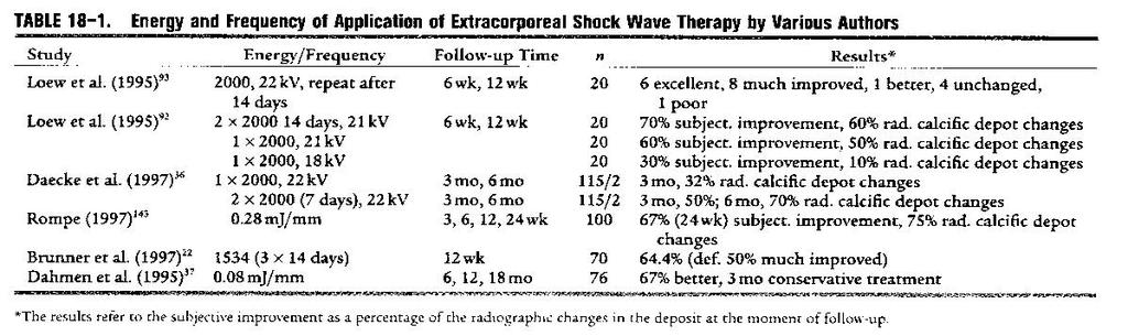
What is the clinical effectiveness of extracorporeal Shock Wave Therapy or barbotage in the management of rotator cuff calcific tendinopathy?
 Specific Question: What is the clinical effectiveness of extracorporeal Shock Wave Therapy or barbotage in the management of rotator cuff calcific tendinopathy? Clinical bottom line There is moderate evidence
Pediatric Ocular Sonography
 Pediatric Ocular Sonography Cicero J Torres A Silva, MD Associate Professor of Radiology 2016 SPR Pediatric Ultrasound Course Yale University School of Medicine None Disclosures Objectives of Presentation
Table of contents. Foreword. Preface. 1 Introduction Historical Perspective 00
 Table of contents Foreword Preface 1 Introduction 00 1.1 Historical Perspective 00 2 Fundamentals of musculoskeletal ultrasound 00 2.1 Frequency and wavelength 00 2.2 Generating ultrasound waves 00 2.3
Ultrasound Physics & Doppler
 Ultrasound Physics & Doppler Endocrine University 2018 Mark Lupo, MD, FACE, ECNU Objectives Review the essential components of ultrasound physics in neck sonography Demonstrate the importance of ultrasound
WELCOME! Introduction to Bedside Ultrasound
 WELCOME! Introduction to Bedside Ultrasound TEACHERS University of California-Irvine School of Medicine Nathan Molina nathan.d.molina@gmail.com Trevor Plescia taplescia90@gmail.com Jack Silva jpsilva42@gmail.com
Pragmatic ultrasound in the diagnosis of soft tissue rheumatic pain. Plamen Todorov
 Pragmatic ultrasound in the diagnosis of soft tissue rheumatic pain Plamen Todorov INTRODUCTION Soft tissue rheumatism: nonsystemic, focal pathological syndromes involving the periarticular structures.
Diagnostic Imaging
 www.fisiokinesiterapia.biz Diagnostic Imaging Diagnostic Imaging is no longer limited to radiography. Major technological advancements have lead to the use of new and improved imaging technologies. The
Sonographic Differences in the Appearance of Acute and Chronic Full-Thickness Rotator Cuff Tears
 Sonographic Differences in the Appearance of Acute and Chronic Full-Thickness Rotator Cuff Tears Sharlene A. Teefey, MD, William D. Middleton, MD, Gregory S. Bauer, MD, Charles F. Hildebolt, DDS, PhD,
Calcific Tendinitis of the Long Head of the Biceps Brachii Distal to the Glenohumeral Joint: Plain Film
 1011 Calcific Tendinitis of the Long Head of the Biceps Brachii Distal to the Glenohumeral Joint: Plain Film Radiographic Findings Amy Beth Goldman1 Calcific tendinitis is a painful condition related to
APPROPRIATE USE GUIDELINES
 APPROPRIATE USE GUIDELINES Appropriateness of Advanced Imaging Procedures (MRI, CT, Bone Scan/PET) in Patients with Shoulder Pain CDI QUALITY INSTITUTE: PROVIDER LED ENTITY (PLE) Compiled by Rob Liddell,
Case Studies. A. Kent Allen, DVM LAMENESS AND IMAGING IN THE SPORT HORSE
 Case Studies A. Kent Allen, DVM Author s address: Virginia Equine Imaging, 2716 Landmark School Road, The Plains, VA 20198; e-mail: vaequine@aol.com. 2007 AAEP. 1. Case Study #1: Medial Collateral Desmitis
Calcifying tendinitis of the rotator cuff with cortical erosion and intraosseus migration: the importance of its recognition.
 Calcifying tendinitis of the rotator cuff with cortical erosion and intraosseus migration: the importance of its recognition. Poster No.: P-0048 Congress: ESSR 2015 Type: Scientific Poster Authors: J.
Chronic Shoulder Disorders
 Chronic Shoulder Disorders Dr. Mustafa Elsingergy Consultant orthopedic surgeon Dallah Hospita Prof. Mamoun Kremli Almaarefa Medical College Contents INTRINSIC Shoulder Pain Due to causes in the shoulder
Shadow because the air
 Thyroid Ultrasound Thyroid US examination needs: 1. high frequency transducer 2. extended patient's neck 3. check all the neck area because the swelling could be in areas other than the thyroid such as
Sonographic Characterization of Carotid Plaque: Detection of Hemorrhage
 311 Sonographic Characterization of Carotid Plaque: Detection of Hemorrhage E. I. Bluth' D.Kai C. R. B. Merritt' M. Sullivan' G. Farr2 N. L. Mills 3 M. Foreman' K. Sloan' M. Schlater' J. Stewart 3 By careful
Complex Fractures and Hip Dislocations
 IMAGING OF HIP PAIN Patients may present with acute (< 2 weeks) or chronic hip pain. Acute pain may be related or not related to an acute traumatic event such as fall or trauma from a motor vehicle accident.
MRI and Sonography of the Shoulder
 Clinical Radiology (1991) 43, 323-327 and of the Shoulder J. HODLER, B. TERRIER*, G. K. yon SCHULTHESS and W. A. FUCHS Departments of Medical Radiology and *Rheumatology, University Hospital, Zurich, Switzerland
MRI of the Shoulder What to look for and how to find it? Dr. Eric Handley Musculoskeletal Radiologist Cherry Creek Imaging
 MRI of the Shoulder What to look for and how to find it? Dr. Eric Handley Musculoskeletal Radiologist Cherry Creek Imaging MRI of the Shoulder Benefits of Ultrasound: * Dynamic * Interactive real time
THYROID NODULES: THE ROLE OF ULTRASOUND
 THYROID NODULES: THE ROLE OF ULTRASOUND NOVEMBER 2017 DR. DEAN DURANT DEFINITION Thyroid nodule: Focal area within the thyroid gland with echogenicity different from surrounding parenchyma. THYROID NODULES
Index. B Backslap technique depth assessment, 82, 83 diaphysis distal trocar, 82 83
 Index A Acromial impingement, 75, 76 Aequalis intramedullary locking avascular necrosis, 95 central humeral head, 78, 80 clinical and functional outcomes, 95, 96 design, 77, 79 perioperative complications,
Dynamic Sonography Evaluation of Shoulder Impingement Syndrome
 Sonography of Shoulder Impingement Syndrome Musculoskeletal Imaging Clinical Observations A C M E D E N T U R I C A L I M A G I N G AJR 2006; 187:216 220 0361 803X/06/1871 216 American Roentgen Ray Society
Case-based discussion:
 Case-based discussion: Pailin Kongmebhol, M.D. Department of Radiology Faculty of Medicine Chiang Mai University There are many guidelines for managing thyroid nodules Two important guidelines: 2015 American
Normal Sonographic Anatomy
 hapter 2:The Liver DUNSTAN ABRAHAM Normal Sonographic Anatomy Homogeneous, echogenic texture (Figure 2-1) Measures approximately 15 cm in length and 10 12.5 cm anterior to posterior; measurement taken
AACE/ACE Advanced Endocrine Neck Ultrasound Training Course 2016
 AACE/ACE Advanced Endocrine Neck Ultrasound Training Course 2016 This 9mm left inferior nodule should remind us all why we re here! There is no absolute number of images required for documentation
Ultrasound (US)-guided Percutaneous Procedures to Treat Inflammatory and Degenerative Diseases of the Upper Limb: How We Do It
-guided Percutaneous Procedures to Treat Inflammatory and Degenerative Diseases of the Upper Limb: How We Do It") Ultrasound (US)-guided Percutaneous Procedures to Treat Inflammatory and Degenerative Diseases of the Upper Limb: How We Do It Poster No.: C-2252 Congress: ECR 2014 Type: Educational Exhibit Authors: G.
Ultrasound (US)-guided Percutaneous Procedures to Treat Inflammatory and Degenerative Diseases of the Upper Limb: How We Do It Poster No.: C-2252 Congress: ECR 2014 Type: Educational Exhibit Authors: G.
Educational Exhibit Authors:
 Renal lymphoma: Patterns of disease with ultrasound (US), colourdoppler ultrasound (CDUS), contrast-enhanced ultrasound (CEUS) and Computed tomography (CT) Poster No.: C-1545 Congress: ECR 2011 Type: Educational
Mr. Duy Thai Orthopaedic Surgeon, Melbourne VIC
 Mr. Duy Thai Orthopaedic Surgeon, Melbourne VIC International Convention of the Vietnamese Physicians, Dentists and Pharmacists of the Free World Melbourne 8 10 August 2014 Conflict of Interest None Subacromial
Contracture of the Deltoid Muscle: Sonographic Evaluation with MRI Correlation
 Sonography and MRI of Deltoid Muscle Contracture Musculoskeletal Imaging Original Research Chung-Cheng Huang 1 Sheung-Fat Ko 1 Jih-Yang Ko 2 Hsuan-Ying Huang 3 Shu-Hang Ng 4 Yung-Liang Wan 4 Min-Chi Chen
CLINICAL PRESENTATION AND RADIOLOGY QUIZ QUESTION
 Donald L. Renfrew, MD Radiology Associates of the Fox Valley, 333 N. Commercial Street, Suite 100, Neenah, WI 54956 12/29/2012 Radiology Quiz of the Week # 105 Page 1 CLINICAL PRESENTATION AND RADIOLOGY
A Practical Approach to Adnexal Masses
 A Practical Approach to Adnexal Masses Darcy J. Wolfman, MD Section Chief of Genitourinary Imaging American Institute for Radiologic Pathology Clinical Associate Johns Hopkins Community Radiology Division
Gallbladder & Pancreas Ultrasonography
 복부초음파 : 담낭과췌장 Gallbladder & Pancreas Ultrasonography 김정훈 Department of Radiology 1 Interaction of sound with matter (1) 반사 (Reflection) (2) 굴절 (Refraction) (3) 흡수 (Absorption) (4) 산란 (Scattering) 음향저항
Kaywan Izadpanah 1*, Martin Jaeger 1, Dirk Maier 1, Norbert P Südkamp 1 and Peter Ogon 2
 Izadpanah et al. BMC Musculoskeletal Disorders 2014, 15:385 RESEARCH ARTICLE Open Access Preoperative planning of calcium deposit removal in calcifying tendinitis of the rotator cuff - possible contribution
CLINICAL PRESENTATION AND RADIOLOGY QUIZ QUESTION
 Donald L. Renfrew, MD Radiology Associates of the Fox Valley, 333 N. Commercial Street, Suite 100, Neenah, WI 54956 11/17/2012 Radiology Quiz of the Week # 99 Page 1 CLINICAL PRESENTATION AND RADIOLOGY
The Shoulder. Systematically scanning the shoulder provides extremely useful diagnostic information. The Shoulder
 1 ! The most ACCESSIBLE to sonographic exam! The most MOBILE and VULNERABLE extremity AND Systematically scanning the shoulder provides extremely useful diagnostic information! The Goal for this section
Calcific tendinitis in atypical location: importance of diagnosis and propose of treatment: Personal experience in 11 patients
 Calcific tendinitis in atypical location: importance of diagnosis and propose of treatment: Personal experience in 11 patients Poster No.: C-2379 Congress: ECR 2010 Type: Topic: Authors: Keywords: DOI:
9/18/18. Welcome- MSK Ultrasound Workshop. Introduction to Musculoskeletal Ultrasound. Acknowledgement of Country. The Workshop.
 Acknowledgement of Country Welcome- MSK Ultrasound Workshop I would like to acknowledge that this meeting is being held on the traditional lands of the Wurundjeri and Boonwurrung people and pay my respect
Case Report Painful Os Peroneum Syndrome: Underdiagnosed Condition in the Lateral Midfoot Pain
 Case Reports in Radiology Volume 2016, Article ID 8739362, 4 pages http://dx.doi.org/10.1155/2016/8739362 Case Report Painful Os Peroneum Syndrome: Underdiagnosed Condition in the Lateral Midfoot Pain
Case Iselin's disease in a Thai boxer.
 Case 13609 Iselin's disease in a Thai boxer. Joris De Win 1, 3, Filip Vanhoenacker 2, 4, Els Goossens3 1: Department of Physical Medicine and Rehabilitation; University Ghent (UGent), Belgium; Email:de_win_joris@hotmail.com
Thyroid Ultrasound Physics and Doppler
 Thyroid Ultrasound Physics and Doppler Advanced AACE-ACE US training course 2017 Dev Abraham MD, MRCP(UK), ECNU, FACE Professor of Medicine, University of Utah No Disclosures Natural Ability to see with
Ultras ono graphic Evaluation of Rotator Cuff Tendons in Patients with Rheumatoid Arthritis
 Med. J. Cairo Univ., Vol. 83, No. 1, June: 395-399, 215 www.medicaljournalofcairouniversity.net Ultras ono graphic Evaluation of Rotator Cuff Tendons in Patients with Rheumatoid Arthritis HALA I. ELGENDY,
Testicular tumors; Ultrasonographic and Pathologic correlation
 Testicular tumors; Ultrasonographic and Pathologic correlation Poster No.: C-0106 Congress: ECR 2014 Type: Educational Exhibit Authors: Y. Kim, S. W. Shin, E. T. Kim, M. Y. Kim ; Kuri City/KR, 1 1 2 1
INTERDISCIPLINARY DISCUSSIONS IN LOCALISED RCC DIAGNOSIS AND SURGICAL STRATEGIES FOR ATYPICAL RENAL CYSTIC LESIONS. Maria Cova
 INTERDISCIPLINARY DISCUSSIONS IN LOCALISED RCC DIAGNOSIS AND SURGICAL STRATEGIES FOR ATYPICAL RENAL CYSTIC LESIONS Maria Cova Radiology Department University of Trieste (IT) Eleventh European International
1 Fundamentals. Basic Definitions and Physics Principles. Fundamentals
 1 To become versed in the language of ultrasonography, it is necessary to review some of the basic principles of physics. The wave physics principles of ordinary (i.e., audible) sound apply to ultrasound
Ultrasound of the Hip: Anatomy, Pathology, and Procedures
 Ultrasound of the Hip: Anatomy, Pathology, and Procedures Jon A. Jacobson, M.D. Professor of Radiology Director, Division of Musculoskeletal Radiology University of Michigan Outline Hip Joint Native hip
The Elbow Scanning Protocol
 The Elbow Scanning Protocol Diagnostic Imaging of the Elbow: Introduction The elbow maybe considered as consisting of four quadrants, anterior, medial, lateral and posterior. Ultrasound would normally
Greater Trochanter: Anatomy and Pathology
 Greater Trochanter: Anatomy and Pathology Jon A. Jacobson, M.D. Professor of Radiology Director, Division of Musculoskeletal Radiology University of Michigan Disclosures: Consultant: Bioclinica Book Royalties:
Sonography of Intramuscular Myxomas
 Article Sonography of Intramuscular Myxomas The Bright Rim and Bright Cap Signs Gandikota Girish, MBBS, FRCS, FRCR, David A. Jamadar, MBBS, FRCS, FRCR, David Landry, MD, Karen Finlay, MD, Jon A. Jacobson,
Educational Exhibit Authors:
 Renal lymphoma: Patterns of disease with ultrasound (US), colourdoppler ultrasound (CDUS), contrast-enhanced ultrasound (CEUS) and Computed tomography (CT) Poster No.: C-1545 Congress: ECR 2011 Type: Educational
Outline. Introduction to imaging modalities of the urinary system. Case base learning of common diseases in urinary tract
 Outline Introduction to imaging modalities of the urinary system Case base learning of common diseases in urinary tract Outline Introduction to imaging modalities of the urinary system Case base learning
Calcific Tendonitis of the Shoulder
 A Patient s Guide to Calcific Tendonitis of the Shoulder 2350 Royal Boulevard Suite 200 Elgin, IL 60123 Phone: 847.931.5300 Fax: 847.931.9072 DISCLAIMER: The information in this booklet is compiled from
Cystic Lymphangioma of the Adrenal Gland: a rare case report
 J Radiol Sci 2013; 38: 59-64 Cystic Lymphangioma of the Adrenal Gland: a rare case report Xiang-Jun Lin Chun-Chao Huang She-Meng Cheng Department of Radiology, Mackay Memorial Hospital and Mackay Medical
Session 2: Ultrasonography for Primary Care Clinicians Learning Objectives
 Session 2: Ultrasonography for Primary Care Clinicians Learning Objectives 1. Assess the main components and functions of a portable ultrasound unit. 2. Identify three clinical applications of portable
Radiology of hepatobiliary diseases
 GI cycle - Lecture 14 436 Teams Radiology of hepatobiliary diseases Objectives 1. To Interpret plan x-ray radiograph of abdomen with common pathologies. 2. To know the common pathologies presentation.
Principal Site Investigator ENHANCE (Evaluation of Thyroid FNA Genomic Signature) study: An IRB approved study with funding to Rochester Regional
 study: An IRB approved study with funding to Rochester Regional") October 20 th 2018 Principal Site Investigator ENHANCE (Evaluation of Thyroid FNA Genomic Signature) study: An IRB approved study with funding to Rochester Regional Health from Veracyte Review ultrasound
October 20 th 2018 Principal Site Investigator ENHANCE (Evaluation of Thyroid FNA Genomic Signature) study: An IRB approved study with funding to Rochester Regional Health from Veracyte Review ultrasound
Outline. Introduction to imaging modalities of the urinary system. Case base learning of common diseases in urinary tract
 Outline Introduction to imaging modalities of the urinary system Case base learning of common diseases in urinary tract Diagnostic Investigations in Urinary System PLAIN KUB EXCRETORY UROGRAPHY RETROGRADE