Bone Tumors Clues and Cues
|
|
|
- Marcia Walsh
- 5 years ago
- Views:
Transcription
1 William Herring, M.D Bone Tumors Clues and Cues In Slide Show mode, advance the slides by pressing the spacebar All Photos Retain the Copyright of their Authors
2 Clues by Appearance of Lesion
3 Patterns of Bone Destruction Geographic Moth-eaten Permeative
4 Geographic Bone Destruction Destructive lesion with sharply defined border Implies a less-aggressive, more slow-growing, benign process Narrow transition zone
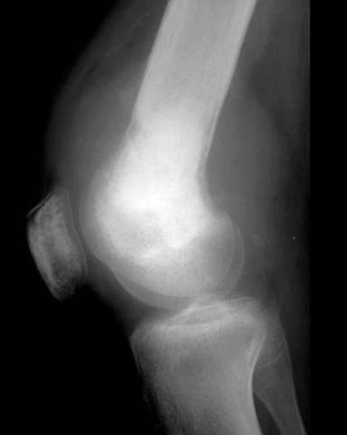
5 Patterns of Bone Destruction Geographic Moth-eaten Permeative Non-ossifying fibroma R 3, 2000
6 Geographic Lesions Examples Non-ossifying fibroma Chondromyxoid fibroma Eosinophilic granuloma
7 Moth-eaten Appearance Areas of destruction with ragged borders Implies more rapid growth Probably a malignancy
8 Patterns of Bone Destruction Geographic Moth-eaten Permeative R 3, 2000 Multiple Myeloma
9 Moth-eaten Appearance Examples Myeloma Metastases Lymphoma Ewing s sarcoma
10 Permeative Pattern Ill-defined lesion with multiple wormholes Spreads through marrow space Wide transition zone Implies an aggressive malignancy Round-cell lesions

11 Patterns of Bone Destruction Geographic Moth-eaten Permeative Leukemia
12 Permeative Pattern Round cell lesions Lymphoma, leukemia Ewing s Sarcoma Myeloma Osteomyelitis Neuroblastoma

13 Patterns of Destruction Geographic Moth-eaten Permeative Less malignant More malignant
14 Periosteal Reactions Benign None Solid More aggressive or malignant Lamellated or onion-skinning Sunburst Codman s triangle
15 Periosteal Reactions Benign None Solid Aggressive/malignant Onion-skinning Sunburst Codman s triangle Non-ossifying fibroma R 3, 2000

16 Periosteal Reactions Benign None Solid Aggressive/malignant Onion-skinning Sunburst Codman s triangle R 3, 2000 Chronic osteomyelitis
17 Periosteal Reactions Benign None Solid Aggressive/malignant Onion-skinning Sunburst Codman s triangle Greenspan, 2000 Ewing s sarcoma
18 Periosteal Reactions Benign None Solid Aggressive/malignant Onion-skinning Sunburst Codman s triangle Greenspan, 2000 Osteosarcoma
19 Periosteal Reactions Benign None Solid Aggressive/malignant Onion-skinning Sunburst Codman s triangle R 3, 2000 Ewing s-codman s triangle

20 Periosteal Reactions Solid Lamellated Sunburst Codman s Less malignant More malignant
21 Tumor Matrix Osteoblastic Fluffy, cotton-like or cloud-like densities Osteosarcoma Cartilaginous Comma-shaped, punctate, annular, popcornlike Enchondroma, chondrosarcoma, chondromyxoid fibroma

22 Tumor matrix Osteoblastic Cartilaginous R 3, 2000 Osteosarcoma

23 Tumor matrix Osteoblastic Cartilaginous Chondrosarcoma R 3, 2000
24 Expansile Lesions of Bone Multiple myeloma Mets Aneurysmal bone cyst Brown tumor Enchondroma Lymphoma Fibrous dysplasia

25 Expansile lesions Multiple myeloma Mets Aneurysmal bone cyst Fibrous dysplasia Brown tumor Enchondroma Lymphoma Multiple Myeloma

26 Expansile lesions Multiple myeloma Mets Aneurysmal bone cyst Fibrous dysplasia Brown tumor Enchondroma Lymphoma Renal Cell Carcinoma
27 Expansile lesions Multiple myeloma Mets Aneurysmal bone cyst Fibrous dysplasia Brown tumor Enchondroma Lymphoma Aneurysmal Bone Cyst

28 Expansile lesions Multiple myeloma Mets Aneurysmal bone cyst Fibrous dysplasia Brown tumor Enchondroma Lymphoma Fibrous Dysplasia
29 Expansile lesions Multiple myeloma Mets Aneurysmal bone cyst Fibrous dysplasia Brown tumor Enchondroma Lymphoma R 3, 2000 Brown Tumor
30 Expansile lesions Multiple myeloma Mets Aneurysmal bone cyst Fibrous dysplasia Brown tumor Enchondroma Lymphoma R 3, 2000 Enchondroma

31 Expansile lesions Multiple myeloma Mets Aneurysmal bone cyst Fibrous dysplasia Brown tumor Enchondroma Lymphoma Lymphoma R 3, 2000
32 Clues by Location of Lesion
33 In the Transverse Plane Central Enchondroma Eccentric GCT, osteosarcoma, chondromyxoid fibroma Cortical Non-ossifying fibroma, osteoid osteoma Parosteal Parosteal osteosarcoma, osteochondroma

34 Osteosarcoma Parosteal sarcoma Osteochondroma Greenspan, Lippincott, 2000 In The Transverse Plane
35 In the Longitudinal Plane Epiphyseal GCT, chondroblastoma Metaphyseal Osteomyelitis, osteo- and chondrosarcoma Diaphyseal Round cell lesions, ABC, enchondroma
36 Tumor Types Characteristic Locations Simple bone cyst Proximal humerus Chondroblastoma Epiphyses Giant Cell tumor Epiphyses

37 Characteristic locations Simple bone cyst Proximal humerus Chondroblastoma Epiphyses Giant Cell tumor Epiphyses R 3, 2000

38 Characteristic locations Simple bone cyst Proximal humerus Chondroblastoma Epiphyses Giant Cell tumor Epiphyses Chondroblastoma R 3, 2000

39 Characteristic locations Simple bone cyst Proximal humerus Chondroblastoma Epiphyses Giant Cell tumor Epiphyses R 3, 2000 Giant Cell Tumor
40 Tumor Types Characteristic Locations Adamantinoma Tibia Chordoma Sacrum, clivus Osteoblastoma Spine, posterior

41 Characteristic locations Adamantinoma Tibia Chordoma Sacrum, clivus Osteoblastoma Spine, posterior R 3, 2000 Adamantinoma
42 Characteristic locations Adamantinoma Tibia Chordoma Sacrum, clivus Osteoblastoma Spine, posterior Chordoma R 3, 2000

43 Characteristic locations Adamantinoma Tibia Chordoma Sacrum, clivus Osteoblastoma Spine, posterior Osteoblastoma R 3, 2000
44 Tumor Types Characteristic Locations Parosteal sarcoma Distal femur Periosteal sarcoma Tibia

45 Characteristic locations Parosteal sarcoma Distal femur Periosteal sarcoma Tibia Parosteal sarcoma

46 Characteristic locations Parosteal sarcoma Distal femur Periosteal sarcoma Tibia R 3, 2000
47 Characteristic Tumors By Body Site
48 Pelvic Lesions Chondrosarcoma Solitary plasmacytoma Chordoma

49 Pelvic lesions Chondrosarcoma Solitary plasmacytoma Chordoma

50 Pelvic lesions Chondrosarcoma Solitary plasmacytoma Chordoma Plasmacytoma
51 Pelvic lesions Chondrosarcoma Solitary plasmacytoma Chordoma R 3, 2000 Chordoma
52 Expansile Rib Lesions Plasmacytoma Metastases Chondrosarcoma Eosinophilic granuloma Neurofibromatosis Fibrous dysplasia
53 Expansile rib lesions Plasmacytoma Metastases Chondrosarcoma Eosinophilic granuloma Neurofibromatosis Fibrous dysplasia

54 Expansile rib lesions Plasmacytoma Metastases Chondrosarcoma Eosinophilic granuloma Neurofibromatosis Fibrous dysplasia Thyroid Carcinoma

55 Expansile rib lesions Plasmacytoma Metastases Chondrosarcoma Eosinophilic granuloma Neurofibromatosis Fibrous dysplasia R 3, 2000

56 Expansile rib lesions Plasmacytoma Metastases Chondrosarcoma Eosinophilic granuloma Neurofibromatosis Fibrous dysplasia R 3, 2000

57 Expansile rib lesions Plasmacytoma Metastases Chondrosarcoma Eosinophilic granuloma Neurofibromatosis Fibrous dysplasia R 3, 2000

58 Expansile rib lesions Plasmacytoma Metastases Chondrosarcoma Eosinophilic granuloma Neurofibromatosis Fibrous dysplasia R 3, 2000
59 Lesions of the Spine Osteoblastoma Expansile, with punctate densities within Chordoma ABC Metastatic disease

60 Spine lesions Osteoblastoma Chordoma ABC Metastatic disease Osteoblastoma R 3, 2000

61 Spine lesions Osteoblastoma Chordoma ABC Metastatic disease Chordoma R 3, 2000
62 Spine lesions Osteoblastoma Chordoma ABC Metastatic disease Aneurysmal bone cyst

63 Spine lesions Osteoblastoma Chordoma ABC Metastatic disease Metastatic Breast Carcinoma R 3, 2000
64 Clues by Density Of Lesion
65 Sclerotic Cortical Lesions Osteoid osteoma Brodie s abscess Stress fracture
66 Sclerotic cortical lesions Osteoid osteoma Brodie s abscess Stress fracture Osteoid Osteoma
67 Sclerotic cortical lesions Osteoid osteoma Brodie s abscess Stress fracture Brodie s abscess R 3, 2000
68 Sclerotic cortical lesions Osteoid osteoma Brodie s abscess Stress fracture Healing Stress Fracture
69 Sclerotic Cortical Lesions Osteoid Osteoma Brodie s abscess Healing Stress Fracture
70 Lytic Lesions in Children Eosinophilic granuloma Neuroblastoma Leukemia

71 Lytic Lesions in Children Eosinophilic granuloma Neuroblastoma Leukemia Eosinophilic granuloma R 3, 2000

72 Lytic Lesions in Children Eosinophilic granuloma Neuroblastoma Leukemia R 3, 2000 Neuroblastoma

73 Lytic Lesions in Children Eosinophilic granuloma Neuroblastoma Leukemia Leukemia R 3, 2000
74 Lytic Lesions in Adults Metastatic lesions Lung Renal Thyroid Multiple myeloma Primary bone tumor

75 Lytic Lesions in Adults Mets Myeloma Primary bone tumor Met from Thyroid Carcinoma

76 Lytic Lesions in Adults Mets Myeloma Primary bone tumor Multiple myeloma

77 Lytic Lesions in Adults Mets Myeloma Primary bone tumor Chondrosarcoma
78 Blastic Lesions in Children Medulloblastoma Lymphoma

79 Blastic Lesions in Children Medulloblastoma Lymphoma Medulloblastoma R 3, 2000
80 Blastic Lesions in Children Medulloblastoma Lymphoma Lymphoma
81 Blastic Lesions in Adults Metastatic disease Breast female Prostate male Lymphoma Paget s disease Etcetera-mastocytosis, fluorosis

82 Blastic Lesions in Adults Mets Lymphoma Paget s Prostate Mets

83 Blastic Lesions in Adults Mets Lymphoma Paget s Breast Mets
84 Blastic Lesions in Adults Mets Lymphoma Paget s Lymphoma
85 Blastic Lesions in Adults Mets Lymphoma Paget s Paget s of Spine
86 Other Clues
87 Benign Lesions Without Sclerotic Boarders Giant Cell tumor Brown tumor Osteolytic phase of Paget s Disease
88 Benign Lesions without Sclerotic Borders Giant cell tumor Brown tumor Osteolytic Paget s R 3, 2000 Giant Cell Tumor
89 Benign Lesions without Sclerotic Borders Giant cell tumor Brown tumor Osteolytic Paget s R 3, 2000 Brown Tumor

90 Benign Lesions without Sclerotic Borders Giant cell tumor Brown tumor Osteolytic Paget s R 3, 2000 Osteolytic Paget s
91 Soft Tissue Extension Usually implies malignancy More likely to form discrete soft tissue mass Benign conditions with soft tissue extension Osteomyelitis Usually infiltration of fat
92 Osteosarcoma R 3, 2000
93 Multiple Lesions More often benign Malignancies with multiple lesions Metastatic disease Multiple myeloma Lymphoma Ewing s sarcoma (rarely) Osteosarcoma (rarely)

94 Multiple lesions Metastatic Multiple myeloma Lymphoma Mets from Ca of Prostate R 3, 2000

95 Multiple lesions Metastatic Multiple myeloma Lymphoma R 3, 2000 Multiple Myeloma
96 Multiple lesions Metastatic Multiple myeloma Lymphoma Lymphoma R 3, 2000

97 Multiple lesions Metastatic Multiple myeloma Lymphoma Osteosarcomatosis R 3, 2000 Osteosarcomatosis

98 Benign vs. Malignant Greenspan, Lippincott, 2000
99 The End
The Radiology Assistant : Bone tumor - well-defined osteolytic tumors and tumor-like lesions
 Bone tumor - well-defined osteolytic tumors and tumor-like lesions Henk Jan van der Woude and Robin Smithuis Radiology department of the Onze Lieve Vrouwe Gasthuis, Amsterdam and the Rijnland hospital,
Typical skeletal location and differential diagnosis of bone tumors.
 Typical skeletal location and differential diagnosis of bone tumors. Poster No.: C-2418 Congress: ECR 2015 Type: Educational Exhibit Authors: M. Barros, L. A. Ferreira, Y. Costa, P. J. V. Coelho, F. Caseiro
Malignant Bone Tumors - Part I: a brief revision of diagnostic aspects with conventional radiology
 Malignant Bone Tumors - Part I: a brief revision of diagnostic aspects with conventional radiology Poster No.: C-2473 Congress: ECR 2013 Type: Educational Exhibit Authors: I. Candelaria, L. B. Barbosa,
The Radiology Assistant : Bone tumor - ill defined osteolytic tumors and tumor-like lesions
 Bone tumor - ill defined osteolytic tumors and tumor-like lesions Henk Jan van der Woude and Robin Smithuis Radiology department of the Onze Lieve Vrouwe Gasthuis, Amsterdam and the Rijnland hospital,
Malignant bone tumors. Incidence Myeloma 45% Osteosarcoma 24% Chondrosarcoma 12% Lyphoma 8% Ewing s Sarcoma 7%
 Malignant bone tumors Incidence Myeloma 45% Osteosarcoma 24% Chondrosarcoma 12% Lyphoma 8% Ewing s Sarcoma 7% Commonest primary bone sarcoma is osteosarcoma X ray Questions to ask 1. Solitary or Multiple
APMA 2018 Radiology Track Bone Tumors When to say Gulp!
 APMA 2018 Radiology Track Bone Tumors When to say Gulp! DANIEL P. EVANS, DPM, FACFAOM Professor, Department of Podiatric Medicine and Radiology Dr. Wm. Scholl College of Podiatric Medicine Conflict of
Bone Tumours - a synopsis. Dr Zena Slim SpR in Histopathology QAH 2009
 Bone Tumours - a synopsis Dr Zena Slim SpR in Histopathology QAH 2009 Aims General approach to diagnosis Common entities.and not so common ones. Mini quiz Challenge of bone tumour diagnosis Bone tumours
Bone tumors. RMG: jan
 Bone tumors RMG: jan 217. @Kijohs KIZZA JOHN KIJOHS Diseases arising in bone Lipoma Fibrous cortical defects Non-ossifying fibroma Bone island Benign simple cysts Enchondroma Osteochondroma Osteoid osteoma
Imaging Findings Of Bone Tumors: A Pictorial Review
 Imaging Findings Of Bone Tumors: A Pictorial Review Poster No.: C-2511 Congress: ECR 2015 Type: Educational Exhibit Authors: M. Limeme, N. Benzina, A. BelKhiria, H. Zaghouani, S. Majdoub, N. Mallat, H.
Grading of Bone Tumors
 Grading of Bone Tumors Joon Hyuk Choi, M.D. Department of Pathology College of Medicine, Yeungnam University Introduction to grading system of bone tumor used at Mayo Clinic WHO Histologic Classification
COPYRIGHT 2004 BY THE JOURNAL OF BONE AND JOINT SURGERY, INCORPORATED
 84 COPYRIGHT 2004 BY THE JOURNAL BONE AND JOINT SURGERY, INCORPORATED Radiographic Evaluation of Pathological Bone Lesions: Current Spectrum of Disease and Approach to Diagnosis BY BENJAMIN G. DOMB, MD,
MARK D. MURPHEY MD, FACR. Physician-in-Chief, AIRP. Chief, Musculoskeletal Imaging
 ALPHABET SOUP AND CYSTIC LESIONS OF THE BONE MARK D. MURPHEY MD, FACR Physician-in-Chief, AIRP Chief, Musculoskeletal Imaging ALPHABET SOUP AND CYSTIC LESIONS OF THE BONE Giant cell tumor (GCT) Unicameral
Primary bone tumors according to the WHO classification: a review of 13 years with illustrative examples
 Primary bone tumors according to the WHO classification: a review of 13 years with illustrative examples Poster No.: C-1741 Congress: ECR 2015 Type: Educational Exhibit Authors: J. Silva, M. A. Ramírez
Bone and Joint Part 2. Leslie G Dodd, MD
 Bone and Joint Part 2 Leslie G Dodd, MD Relative rates of cancer Sarcomas are relatively uncommon tumors New cancer cases 2007 All sites 1.4 million prostate 218,890 lung 213,380 breast 180,510 Soft tissue
Primary bone tumors > metastases from other sites Primary bone tumors widely range -from benign to malignant. Classified according to the normal cell
 Primary bone tumors > metastases from other sites Primary bone tumors widely range -from benign to malignant. Classified according to the normal cell counterpart and line of differentiation. Among the
Introduction to Musculoskeletal Tumors. James C. Wittig, MD Orthopedic Oncologist Sarcoma Surgeon
 Introduction to Musculoskeletal Tumors James C. Wittig, MD Orthopedic Oncologist Sarcoma Surgeon www.tumorsurgery.org Definitions Primary Bone / Soft tissue tumors Mesenchymally derived tumors (Mesodermal)
MRI XR, CT, NM. Principal Modality (2): Case Report # 2. Date accepted: 15 March 2013
: Case Report # 2. Date accepted: 15 March 2013") Radiological Category: Musculoskeletal Principal Modality (1): Principal Modality (2): MRI XR, CT, NM Case Report # 2 Submitted by: Hannah Safia Elamir, D.O. Faculty reviewer: Naga R. Chinapuvvula, M.D.
Radiological Category: Musculoskeletal Principal Modality (1): Principal Modality (2): MRI XR, CT, NM Case Report # 2 Submitted by: Hannah Safia Elamir, D.O. Faculty reviewer: Naga R. Chinapuvvula, M.D.
ORTHOPAEDIC ONCOLOGY OITE REVIEW COURSE
 ORTHOPAEDIC ONCOLOGY OITE REVIEW COURSE Richard D. Lackman, MD FACS Director, Orthopaedic Oncology Center Cancer Institute Introduction In the evaluation of a patient with a bone tumor, there are several
Bubbly Lesions of Bone
 Residents Section Pattern of the Month w79 08.18.09 Eisenberg Residents Section Pattern of the Month Residents inradiology Ronald L. Eisenberg 1 Eisenberg RL Keywords: bubbly lesions, fegnomashic, skeletal
Radiography in the Initial Diagnosis of Primary Bone Tumors
 Residents Section Structured Review Costelloe and Madewell Radiography of Primary Bone Tumors Residents Section Structured Review Colleen M. Costelloe 1 John E. Madewell Costelloe CM, Madewell JE Keywords:
FEGNOMASHIC: from x-ray to MRI
 FEGNOMASHIC: from x-ray to MRI Poster No.: C-2441 Congress: ECR 2015 Type: Educational Exhibit Authors: S. Fouassier, A. L. C. Duarte, C. Ruivo, J. Velez ; Évora/PT, 1 2 1 2 3 1 3 Coimbra/PT, PT Keywords:
Fluid fluid levels in bone tumors and tumoral lesions - Pictorial essay
 Review Fluid fluid levels in bone tumors and tumoral lesions - Pictorial essay Subbarao Kakarla 1,* 1 KIMS Foundation and Research Centre, Minister Road, Secunderabad - 500003, Telangana, India Abstract
Bread and Butter Bone Pathology
 Bread and Butter Bone Pathology NICOLE D. RIDDLE, MD RUFFOLO, HOOPER, AND ASSOC. / UNIVERSITY OF SOUTH FLORIDA Goals: Fundamentals of neoplastic bone pathology Bone Producing Cartilage Producing Miscellaneous
Vertebral and Paravertebral Diseases
 Department of Radiology University of California San Diego Vertebral and Paravertebral Diseases John R. Hesselink, M.D. Vertebral / Paravertebral Disease (Extradural) Metastatic disease Primary bone tumors
VALORACIÒN RADIOLÓGICA DE LA LESIÒN ÒSEA SOLITARIA IMAGENOLOGIA MEDICA UNIVERSIDAD HISPANOAMERICANA
 VALORACIÒN RADIOLÓGICA DE LA LESIÒN ÒSEA SOLITARIA IMAGENOLOGIA MEDICA UNIVERSIDAD HISPANOAMERICANA TUMORES ÓSEOS SE PRESENTAN POR RANGOS DE EDAD, PRINCIPALMENTE: MENORES DE 20 AÑOS 20 A 40 AÑOS MAYORES
Skeletal metastases are the most common variety of bone tumors and should always be considered in the differential diagnosis, particularly in older
 Dr Brajesh Nandan Skeletal metastases are the most common variety of bone tumors and should always be considered in the differential diagnosis, particularly in older patients. Cancers of the breast, prostate,
Bone Imaging. Scopes. Objective. Part 1 : bone tumor. Important Factors in Diagnosis of Bone tumor. Part 2 : infection and joint disease
 Scopes Bone Imaging Part 1 : bone tumor Part 2 : infection and joint disease Jitsupa Wongsripuemtet, M.D. Radiology Department Siriraj Hospital Mahidol University Objective ให น กศ กษาแพทย สามารถแปลผลภาพทางร
A Modified Lodwick-Madewell Grading System for the Evaluation of Lytic Bone Lesions
 Musculoskeletal Imaging Original Research Caracciolo et al. Evaluation of Lytic one Lesions Musculoskeletal Imaging Original Research Jamie T. Caracciolo 1 H. Thomas Temple 2 G. Douglas Letson 3 Mark J.
The Radiology Assistant : Bone tumor A-G
 Bone tumor A-G Bone tumors and tumor-like lesions in alphabethic order Henk Jan van de Woude and Robin Smithuis Radiology department of the Onze Lieve Vrouwe Gasthuis, Amsterdam and the Rijnland hospital,
Giant cell tumour of the sternum-two cases
 Giant cell tumour of the sternum-two cases Nishaa.P 1, Raghuram.P 2, Navin patil 3, Jaipal B.R 4 Akkamahadevi patel 5 Assistant Professor ESIC medical college and PGIMSR 1 Professor and HOD, 2 Professor
Fluid-fluid levels in bone tumors: A pictorial review
 Fluid-fluid levels in bone tumors: A pictorial review Poster No.: C-578 Congress: ECR 2009 Type: Educational Exhibit Topic: Musculoskeletal Authors: L. Figueroa Nasra, C. Martín Hervás, M. Tapia-Viñé,
Common Primary Tumors of Bone
 Special Report Common Primary Tumors of Bone Primary bone tumors are a relatively rare occurrence, however, they can have serious deleterious consequences. Many possess the ability to degenerate into malignant
Review Course «Musculoskeletal Oncology» October 6, 2011 UNIKLINIK BALGRIST. Imaging of Bone and Soft Tissue. Tumors
 Imaging of Bone and Soft Tissue Tumors Approach from a radiologist s point of view Florian Buck Radiology Radio- Radio- Oncologist Oncologist Orthopedist Orthopedist Patient Management Oncologist Oncologist
Key points in the evaluation of focal bone lesions: from plain film to multidetector CT
 Key points in the evaluation of focal bone lesions: from plain film to multidetector CT Poster No.: C-2060 Congress: ECR 2011 Type: Educational Exhibit Authors: I. Rubio Marco, M. Arraiza Sarasa, H. Gómez
Radiologic approach to pediatric lytic bone lesions
 Radiologic approach to pediatric lytic bone lesions Poster No.: C-1177 Congress: ECR 2016 Type: Educational Exhibit Authors: J. L. LERMA GALLARDO, I. de la Pedraja, A. Lancharro 1 1 1 2 1 1 Zapata, J.
Bone Tumors: In 1 Simple Chart
 Bone Tumors with PowerPoint Interactivity Download this entire slideshow from When running this on your own computer you can jump from slide to slide using these buttons at bottom of each slide: Last slide
Malignant Lesions Steven R. Singer, DDS
 Definitions Malignant Lesions Steven R. Singer, DDS srs2@columbia.edu 212.305.5674 Malignancies are uncontrolled growths of tissue Primary tumors represent de novo tumors in their initial site Metastatic
Malignant Bone Tumours. PathoBasic, Daniel Baumhoer
 Malignant Bone Tumours PathoBasic, 20.03.18 Daniel Baumhoer FNCLCC Grading The differentiation score is defined as the extent to which a tumor resembles adult mesenchymal tissue (score 1), the extent to
ORTHOPAEDIC TUMOURS DJM FRANTZEN 2012 AUGUST TUMOURS 1
 ORTHOPAEDIC TUMOURS DJM FRANTZEN 2012 AUGUST TUMOURS 1 PRINCIPLES STAGING WORKUP RADIOLOGY BIOPSY PROCEDURES CHEMOTHERAPY RADIOTHERAPY 2012 AUGUST TUMOURS 2 STAGING ENNEKING'S SURGICAL STAGES (ENNEKING)
SMALL ROUND BLUE CELL LESION OF BONE
 DISCLOSURE SMALL ROUND BLUE CELL LESION OF BONE Dr. Alistair Jordan University of South Alabama No financial support or endorsement OBJECTIVES Describe the more common small round cell lesions of bone
Incidental bone tumors are asymptomatic lesions that are. Incidental Bone Lesions. When to Refer to the Tumor Specialist
 Bulletin of the NYU Hospital for Joint Diseases 2012;70(4):235-40 235 Incidental Bone Lesions When to Refer to the Tumor Specialist LT Suezie Kim, M.D., M.C., U.S.N., Catherine N. Laible, M.D., Leon D.
Disclosures. Giant Cell Rich Tumors of Bone. Outline. The osteoclast. Giant cell rich tumors 5/21/11
 Disclosures Giant Cell Rich Tumors of Bone Andrew Horvai, MD, PhD Associate Clinical Professor, Pathology This lecture discusses "off label" uses of a number of pharmaceutical agents. The speaker is describing
General Approach to Lytic Bone Lesions D. Lee Bennett, MD, MA, Georges Y. El Khoury, MD Appl Radiol. 2004;33(5)
") General Approach to Lytic Bone Lesions D. Lee Bennett, MD, MA, Georges Y. El Khoury, MD Appl Radiol. 2004;33(5) www.medscape.com Abstract and Introduction Abstract When interpreting musculoskeletal radiographs,
General Approach to Lytic Bone Lesions D. Lee Bennett, MD, MA, Georges Y. El Khoury, MD Appl Radiol. 2004;33(5) www.medscape.com Abstract and Introduction Abstract When interpreting musculoskeletal radiographs,
Aprimary bone tumor is a
 Radiographic and Scintigraphic Evaluation of Bone Tumors and Diseases Young Lu, BA Camilo Villalobos, MD Rodolfo Zamora, MD Marisa C. Cornejo, BA James C. Wittig, MD Investigation performed at Hackensack
Fine Needle Aspiration of Bone Tumours
 Fine Needle Aspiration of Bone Tumours Monographs in Clinical Cytology Vol. 19 Series Editor Svante R. Orell Kent Town Fine Needle Aspiration of Bone Tumours The Clinical, Radiological, Cytological Approach
Pictorial Essay Benign and Malignant Bone Tumors: Radiological Diagnosis and Imaging Features
 Clinical Orthopedic Imaging Pictorial Essay Benign and Malignant Bone Tumors: Radiological Diagnosis and Imaging Features Katharina Grünberg, M.D.; Christoph Rehnitz, M.D.; Marc-André Weber, M.D., M.Sc.
Department of Radiology, Yeungnam University College of Medicine, Yeungnam University Medical Center, Daegu, Korea 4
 Original Article pissn 1738-2637 / eissn 2288-2928 http://dx.doi.org/10.3348/jksr.2015.73.4.240 Bone Tumors with an Associated Pathologic Fracture: Differentiation between Benign and Malignant Status Using
A review of Tumoral lesions of the shoulder
 A review of Tumoral lesions of the shoulder Poster No.: P-0109 Congress: ESSR 2013 Type: Scientific Exhibit Authors: M. M. Milán Rodríguez, Á. E. Moreno Puertas, J. M. Giménez, 1 1 1 1 2 1 A. Rubio Fernández,
Unusual location of bone sarcoma in children
 Unusual location of bone sarcoma in children Poster No.: C-1517 Congress: ECR 2014 Type: Educational Exhibit Authors: S. JERBI, A. Khalfalli, G. Abid, O. Bradai, N. chouchane, H. HAMZA; Mahdia/TN Keywords:
Spinal Neoplasms. First Things First!! Localize the Lesion!! Ependymomas. Common Intramedullary Lesions
 Acta Radiológica Portuguesa, Vol.XXIII, nº 90, pág. 101-114, Abr.-Jun., 2011 Spinal Neoplasms Bruno A Policeni University of Iowa Hospitals and Clinics Assistant Professor of Radiology Disclosure of Commercial
1/9/2013 EXTRAMEDULLARY TUMORS OF THE PEDIATRIC SPINE. Introduction. Classification for Extramedullary Tumors
 EXTRAMEDULLARY TUMORS OF THE PEDIATRIC SPINE Eugene Wang 1/20/12 Dent Neurologic Institute Introduction 2/3 of all intraspinal tumors of childhood are extramedullary 50% Extradural 10-15% Intradural Back
History. 33 y/o F with hx of palpable anterior tibial mass x 2 years, only painful with palpation
 History 33 y/o F with hx of palpable anterior tibial mass x 2 years, only painful with palpation Imaging Photo Album Patient also had a smaller lesion 1 cm proximal to this lesion, not seen radiographically.
Residents Section Pattern of the Month
 Residents Section Pattern of the Month Rana et al. Periosteal Reaction Residents Section Pattern of the Month Residents inradiology Rich S. Rana 1 Jim S. Wu Ronald L. Eisenberg Rana RS, Wu JS, Eisenberg
Department of Radiology, University of Szeged. Imaging of the skeleton
 Imaging of the skeleton Methods of examination: plain x-ray (radiography, densitometry) x-ray with contrast material (fistulography, angiography) ultrasound (b-mode, Doppler, color, duplex) computed tomography
Heterogeneous osteoblastic activity in the right ischium of unclear etiology seen on NaF18-PET/CT
 CASE REPORT Heterogeneous osteoblastic activity in the right ischium of unclear etiology seen on NaF18-PET/CT Aung Zaw Win, Carina Mari Aparici Dept. Radiology, Nuclear Medicine section, San Francisco
A peculiar location of a rare bone tumor: sternal lipoma
 A peculiar location of a rare bone tumor: sternal lipoma Poster No.: P-0033 Congress: ESSR 2016 Type: Authors: Keywords: DOI: Scientific Poster Z. Akkaya, C. Uzun, S. Enon, G. Kocaman, G. Sahin; Ankara/TR
Case Report Intramedullary Chondrosarcoma of Proximal Humerus
 Hindawi Publishing Corporation Case Reports in Radiology Volume 2012, Article ID 642062, 7 pages doi:10.1155/2012/642062 Case Report Intramedullary Chondrosarcoma of Proximal Humerus Pratiksha Yadav, Dolly
The term bone tumor is a broad category, encompassing benign and malignant neoplasms, reactive focal abnormalities, metabolic abnormalities, and misce
 Note: This copy is for your personal, non-commercial use only. To order presentation-ready copies for distribution to your colleagues or clients, use the Radiology Reprints form at the end of this article.
ISSN: DISTRIBUTION OF BONE AND CARTILAGINOUS TUMORS IN PEDIATRIC AGE GROUP IN WESTERN UTTAR-PRADESH: AN EVALUATIVE STUDY
 : 289-295 ISSN: 2277 4998 DISTRIBUTION OF BONE AND CARTILAGINOUS TUMORS IN PEDIATRIC AGE GROUP IN WESTERN UTTAR-PRADESH: AN EVALUATIVE STUDY QADRI S, HASAN M, AKHTAR K * AND SHERWANI RK The Departments
Plasmacytoma. Kim Kallianos HMS IV Dr. Gillian Lieberman BIDMC Radiology Advanced Clerkship May 2009
 Plasmacytoma Kim Kallianos HMS IV Dr. Gillian Lieberman BIDMC Radiology Advanced Clerkship May 2009 1 Our Index Patient Y.B. YB is a 43yo F with unremarkable past medical history Presented to her PCP in
Recognizing Cartilaginous Tumors: Spectrum of Imaging Characteristics with Radiologic-Pathologic correlation.
 Recognizing Cartilaginous Tumors: Spectrum of Imaging Characteristics with Radiologic-Pathologic correlation. Poster No.: C-1451 Congress: ECR 2012 Type: Educational Exhibit Authors: E. Barcina García,
Section II Musculoskeletal Radiology
 Section II Musculoskeletal Radiology Figure 1 25. You are shown a noncontrast CT (Figure 1) of the thigh. What is the MOST LIKELY diagnosis? A. Synovial sarcoma B. Hemangioma C. Organizing hematoma D.
INDEX. in this web service Cambridge University Press
 actin 14 adamantinoma 202, 290 292, 297 adenocarcinoma 136 adipocytes in hibernoma 149, 150 in lipoblastoma 148 in lipoma 141, 142, 145 in liposarcoma 152 in myelolipoma 151 adrenal gland tumors see myelolipoma
Immunohistochemistry in Bone and Soft Tissue Tumors. Sahar Rassi Zankoul, MD
 Immunohistochemistry in Bone and Soft Tissue Tumors Sahar Rassi Zankoul, MD Introduction Bone tumors represent a wide variety of tumors of various origins and malignant potentials. These different tumor
Bone/Osteoid Producing Lesions
 Chapter 2 Bone/Osteoid Producing Lesions Introduction There are many lesions that are associated with reactive new bone formation; this chapter predominantly covers those in which deposition of osteoid/bone
Bone Tumors: Epidemiology, Classification, Pathology
 Bone Tumors: Epidemiology, Classification, Pathology 1 Lars Gunnar Kindblom C O N T E N T S 1.1 Introduction 2 1.2 Epidemiology 2 1.3 Morphologic Diagnosis of Bone Tumors 5 1.4 Types of Bone Tumor Specimens
10 th Joint MSK/HSS/IOR Course in. Musculoskeletal Tumor Pathology & Clinical Oncology OCTOBER 10-12, 2017 ZUCKERMAN RESEARCH CENTER NEW YORK CITY
 10 th Joint MSK/HSS/IOR Course in Musculoskeletal Tumor Pathology & Clinical Oncology OCTOBER 10-12, 2017 ZUCKERMAN RESEARCH CENTER NEW YORK CITY COURSE OVERVIEW This well-received annual course is designed
warwick.ac.uk/lib-publications
 A Thesis Submitted for the Degree of PhD at the University of Warwick Permanent WRAP URL: http://wrap.warwick.ac.uk/102063/ Copyright and reuse: This thesis is made available online and is protected by
GIANT CELL-RICH OSTEOSARCOMA: A CASE REPORT
 Nagoya J. Med. Sci. 59. 151-157, 1996 CASE REPORTS GIANT CELL-RICH OSTEOSARCOMA: A CASE REPORT KEIJI SATO!, SHIGEKI YAMAMURA!, HISASHI IWATA!, HIDESHI SUGIURA 2, NOBUO NAKASHIMA 3 and TETSURO NAGASAKA
Downloaded from by on 11/21/17 from IP address Copyright ARRS. For personal use only; all rights reserved
 Downloaded from www.ajronline.org by 46.3.196.1 on 11/21/17 from IP address 46.3.196.1. opyright RRS. For personal use only; all rights reserved T he scapula is a small bone in which many neoplasms can
EXOSTOSES KENT S REPERTORY
 EXOSTOSES KENT S REPERTORY Prof. Dr. Aadil Chimthanawala, MD Homoeopathic Cardiologist The National Academy of Homoeopathy, India Aadil Homoeo Heart Care Centre Hanuman Lane, Sitabuldi, NAGPUR, INDIA Ph:
We are IntechOpen, the world s leading publisher of Open Access books Built by scientists, for scientists. International authors and editors
 We are IntechOpen, the world s leading publisher of Open Access books Built by scientists, for scientists 3,700 108,500 1.7 M Open access books available International authors and editors Downloads Our
Case Studies in the Skull Base
 Case Studies in the Skull Base Amy C Tsai, MD Neuroradiology Fellow Department of Radiology and Imaging Sciences University of Utah Health Sciences Center Salt Lake City, Utah, USA No disclosures related
Distribution and evaluation of primary bone and soft tissue tumors admitted from Malatya province and surrounding provinces
 Available online at www.medicinescience.org ORIGINAL RESEARCH Medicine Science 2017;6(3):546-50 Medicine Science International Medical Journal Distribution and evaluation of primary bone and soft tissue
Medical Student Rotation Guide Tumor Service
 Medical Student Rotation Guide Tumor Service Overview Welcome to the medical student rotation on the Tumor service in the Department of Orthopaedic Surgery at Rush University Medical Center! We are excited
Medical Student Rotation Guide Tumor Service
 Medical Student Rotation Guide Tumor Service Overview Welcome to the medical student rotation on the Tumor service in the Department of Orthopaedic Surgery at Rush University Medical Center! We are excited
Spine bone tumors in children. A pictorial review
 Spine bone tumors in children. A pictorial review Poster No.: C-1921 Congress: ECR 2015 Type: Educational Exhibit Authors: E. García Esparza, S. I. Sirvent Cerdá, M. Á. López Pino, I. Solís Muñiz, G. Albi
1 Bones and Bone Tissue Normal Anatomy and Histology... 13
 Contents 1 Bones and Bone Tissue... 1 The Function of Bones and of the Skeleton... 2 Bone as a Structural Support... 4 Bone as an Organ of Storage... 6 Regulation of Bone Structure and Calcium Metabolism...
From Chest Pain to Multiple Myeloma: Radiology in Practice
 January 2012 From Chest Pain to Multiple Myeloma: Radiology in Practice Mekeme Utuk, Harvard Medical School Year III Our Patient RB RB is a 58yoM with no significant PMH presented to PCP with 2 weeks of
Disseminated Primary Non-Hodgkin s Lymphoma of Bone : A Case Re p o r t 1
 Disseminated Primary Non-Hodgkin s Lymphoma of Bone : A Case Re p o r t 1 Hee-Jin Park, M.D., Sung-Moon Lee, M.D., Hee-Jung Lee, M.D., Jung-Sik Kim, M.D., Hong Kim, M.D. Primary lymphoma of bone is uncommon
An epidemiological survey of tumour or tumour like conditions in the scapula and periscapular region
 SICOT J 206, 2, 34 Ó The Authors, published by EDP Sciences, 206 DOI: 0.05/sicotj/206023 Available online at: www.sicot-j.org RESEARCH OPEN ACCESS An epidemiological survey of tumour or tumour like conditions
RADIOGRAPHIC INTERPRETATION Differential Diagnosis
 RADIOGRAPHIC INTERPRETATION Differential Diagnosis MODULE 1: The Introduction. Chief complaint Demographics Age Sex Race Historical findings Physical findings Clinical Radiographic Location Maxilla/mandible
A comprehensive review of osseous Ewing sarcoma: clinical data, skeletal location and imaging features.
 A comprehensive review of osseous Ewing sarcoma: clinical data, skeletal location and imaging features. Poster No.: C-2233 Congress: ECR 2014 Type: Authors: Educational Exhibit R. Gil 1, P. Pereira 1,
* I have no disclosures or any
 Howard Rosenthal, M.D. Associate Professor of Orthopedic Surgery University of Kansas Sarcoma Center I have no disclosures or any conflicts related to the content of this presentation. Objectives 1. Describe
Benign tumors tumors K 01
 A tumor or suspected tumor diagnosis is a major setback in any person s life. Right from the start - in the initial phase of the patient s personal confrontation with the disease, possibly characterized
Essential Dermatopathology. Jinah Kim, MD, PhD Department of Pathology and Dermatology Stanford University Medical Center
 Essential Dermatopathology Jinah Kim, MD, PhD Department of Pathology and Dermatology Stanford University Medical Center OBJECTIVES Review clinical, pathologic and molecular aspects of bone and fat tumors
A step-by-step diagnostic algorithm of bone tumors and pseudo-tumors
 A step-by-step diagnostic algorithm of bone tumors and pseudo-tumors Poster No.: C-2043 Congress: ECR 2017 Type: Educational Exhibit Authors: C. G. Iacoban, S. Manole, G. Pervain, M. Mereuta ; Baia Mare/
DIAGNOSING EWING S SARCOMA OF THE RIB IN CHILDREN IS IT QUITE CHALLENGING??
 Indian Journal of Medical s ISSN: 2319 3832(Online) DIAGNOSING EWING S SARCOMA OF THE RIB IN CHILDREN IS IT QUITE CHALLENGING?? *Arvind Kumar S.M. and Shyam Sundar V, Dinakar B.K. and SandhyaV. and Kailash
Primary Tumors of Ribs
 Primary Tumors of Ribs Frank E. Schmidt, M.D., and Max J. Trummer, Capt, MC, USN ABSTRACT An analysis of 50 consecutive patients with primary rib tumors operated on at the U.S. Naval Hospital, San Diego,
Metabolic & Endocrine disorders of bone:
 Metabolic & Endocrine disorders of bone: Osteoporosis: Bone apposition < bone resorption Risk factors: Postmenopausal women Hyperthyroidism Hyperparathyroidism Cushing s syndrome bone quantity: thin cortex
The role of Imaging in Ewing sarcoma
 The role of Imaging in Ewing sarcoma Poster No.: P-0109 Congress: ESSR 2014 Type: Educational Poster Authors: D. Beomonte Zobel, C. Dell'atti, M. Bartocci, V. Martinelli, N. 1 2 2 2 1 1 1 2 Magarelli,
Benign Fibro-osseous Lesions
 Benign Fibro-osseous Lesions Plus Vision is the art of seeing things invisible. Jonathan Swift 1667-1745 Steven R. Singer, DDS srs2@columbia.edu 212.305.5674 Benign Fibro-osseous Lesions A group of lesions
Aneurysmal Bone Cyst Fine-Needle Aspiration Findings in 23 Patients With Clinical and Radiologic Correlation
 Anatomic Pathology / FNA OF ANEURYSMAL BONE CYST Aneurysmal Bone Cyst Fine-Needle Aspiration Findings in 23 Patients With Clinical and Radiologic Correlation Andrew J. Creager, MD, 1,2 Christopher R. Madden,
Imaging Findings of Sacral Tumors 1
 Imaging Findings of Sacral Tumors 1 Seung Ho Kim, M.., Sung Hwan Hong, M.., Ja-Young hoi, M.., Sung Hye Koh, M.., Hye Won hung, M.., Jung-h hoi, M.., Heung Sik Kang, M.. The various pathologic conditions
A 64 y.o. man presents to the hospital with persistent cough and hemoptysis. Fernando Mut Montevideo - Uruguay
 A 64 y.o. man presents to the hospital with persistent cough and hemoptysis Fernando Mut Montevideo - Uruguay Teaching case Bone # 1 A 64 y.o. man presents to the hospital with persistent cough and hemoptysis.
Radiology-Pathology Conference
 July 31, 2009 Radiology-Pathology Conference Daniel T Ginat, M.D., M.S. Sharlin Johnykutty,, M.D. Presentation material is for education purposes only. All rights reserved. 2009 URMC Radiology Page 1 of
Aneurysmal Bone Cyst of the Pelvis: A Challenge in Treatment: Review of the Literature
 ISPUB.COM The Internet Journal of Orthopedic Surgery Volume 8 Number 1 Aneurysmal Bone Cyst of the Pelvis: A Challenge in Treatment: Review of the Literature S Bajracharya, G Khanal, A Sundas, S Pandey,
USCAP 2014 Common problems in bone and soft tissue pathology: Cartilage tumors
 USCAP 2014 Common problems in bone and soft tissue pathology: Cartilage tumors Andrew Horvai MD PhD Clinical Professor, Pathology UCSF, San Francisco, CA Outline Common intramedullary tumors Enchondroma
MEDICAL RADIOLOGY Diagnostic Imaging
 MEDICAL RADIOLOGY Diagnostic Imaging Editors: A. L. Baert, Leuven M. Knauth, Göttingen A. M. Davies M. Sundaram S. L. J. James (Eds.) Imaging of Bone Tumors and Tumor-Like Lesions Techniques and Applications