Case Presentation #1 for the American Board of Craniofacial Pain July 2013
|
|
|
- Beryl Hoover
- 5 years ago
- Views:
Transcription
1

2 Case Presentation #1 for the American Board of Craniofacial Pain July 2013
3 Case I Summary Presentation Pain in right temporomandibular joint with opening of mouth( 7 out of 10 ). Acute right non-reducing disc subluxation of tm joint Right temporomandibular joint capsulitis Myalgia of masticatory muscles secondary to closed lock Treatment with oral orthotic, manual manipulation for disc reduction. Various modalities : exercises, spray and stretch Phase II- Equilibration of teeth to balance occlusion Continued night time splint wearing


4 About the Patient Patient is a 64 year old female Caucasian Referred by her general dentist Single and lives with two special dogs Initial Exam Presentation on January 22, 2008 Immediately adamant about no surgery
5 About the Patient, con t The patient s general dentist supplied a soft bruxism appliance, but patient experienced no improvement Treated with orthodontia twice Patient is deeply concerned about avoiding surgery, seeks holistic approach Patient photo: Front View Patient photo: Profile View

6 History of Symptoms The patient states that she was eating Thanksgiving dinner and she felt like she torqued her jaw All symptoms started at this point Patient s general dentist supplied a soft bruxism appliance to no avail
7 Chief Complaint Pain with wide mouth opening or eating Marked deviation to the right with opening (6 mm) Bilateral temporal headaches upon awaking that are maintained throughout everyday Pain radiating into right ear, into the cheek and along the mandible. Right jaw locking closed with joint stiffness.
8 Medical History The patient is a well appearing Caucasian female, approximately 5 7 tall, weighing approximately 144 pounds. She presents a normal gate but has 15 degree anterior kyphotic curvature at the T-10 vertebral area. Her right shoulder presents a +1, +1 position. ROM with head rotation to the L was limited to 55 degrees. All other motions were normal She is under the care of a physician at the time of examination
9 Medical History, con t She is allergic to iodine(shrimp), Penicillin She has high blood pressure( 130 / 83 with medication), asthma, swollen ankles, arthritis and hay fever/ allergies. Patient related right joint torque and pop at Thanksgiving. Patient denies any other problems with total systems review.
10 Medical History, con t Patient has had multiple neck injuries due to falls and car accidents Last fall of note was in 1993 when she fell on her face and received sutures in her chin. Patient also fell on her face in 1989 Patient denies any general anesthetic or endoscopic procedures. BP 128/80, pulse 76, breathing passive and non-labored at 22 respirations / minute
11 Dental History & Examination Patient referred by her general dentist Patient presents 24 teeth (orthodontic 4 bi and third molars extractions) Veneers present on 12 anterior teeth Crowns present on teeth 2, 3, 4, 13, 14, 15, 18, 19, 20, 29 and 31 Root canal tooth 7 Orthodontia treatment twice
12 Dental History & Examination, con t Patient presents a 3 mm overbite with a 2 mm overjet, Class I type II Occlusion on teeth 18 and 31 only Anterior Guidance was limited to posterior molars 18 and 31 Periodontal pocket depth is generally 2-4 mm General dentistry is WNL Minor tongue thrust was presented Swallowing patterns were normal
13 TMJ & Physical Examination The patient presented tenderness or associated pain when the following areas were palpated: Bilateral trapezius muscle pain Right mid and anterior scalene muscles Right levator scapulae muscle tenderness Right longus coli muscle pain Bilateral anterior temporalis muscle pain Right masseter muscle pain
14 TMJ & Physical Examination, Con t (2) Right temporal tendon pain Right retro-discal and external pterygoid muscle pain with mandibular retrusion Right anterior ear wall pain Right joint stiffness Right pain along cheek and mandible Patient noted right reciprocal click for yearsnot present at examination Patient noted right pain with eating and opening jaw
15 TMJ & Physical Examination, Con t (3) Opening dimension was 35mm, right lateral motion was 12 mm, left lateral motion was 4 mm, protrusive motion was 7 mm. 6 mm jaw deviation motion to the right was noted upon opening. Rotation without translation was noted in the right TM joint Right TM joint crepitus was noted upon auscultation
16 TMJ & Physical Examination, Con t (4) Capsulitis was noted in the right TM joint Right gonial notching at the angle of the jaw and was superiorized 4 mm in relation to the left Right maxillary posterior occlusal plane is superiorized 3 mm in relation to the Left. Head posture was 32 mm anterior to the vertical zygomatic/clavicular plane
17 Cranial Nerve Examination I: Olfactory Smell intact bilaterally II: Optic Nerve Intact, Able to read fine print both eyes, no visual blind spots or visual disturbances III: Occulomotor, IV Trochlear, VI Abducens,- intact, conjugate eye movement. Pupils react equally to light. V: Trigeminal nerve intact, sensitivity of all three divisions normal bilaterally, contraction of masseter and temporaralis is normal
18 Cranial Nerve Examination, Part II VII: Facial Nerve - intact- Appropriate movement of muscles of facial expression, taste in anterior 2/3 s of tongue is normal VIII: Auditory Nerve intact, hearing is normal IX: Glossopharyngeal nerve and X Vagus Nerve - intact, normal swallowing pattern, active gag reflex, normal speech pattern XI: Accessory Nerve- intact, normal shoulder shrug XII: Hypoglossal Nerve intact, tongue protrudes without midline deviation.
19 Postural, Cervical, Spinal and Oral Myofunctional Examination Patient displayed normal S curve noting a 15 degree ant spine posture at T-10 Head posture was 32 mm anterior to vertical zygomatic clavicular line and left head rotation was limited to 55 degrees Right shoulder presentation was +1, +1 Pelvis appeared essentially normal Lips were normal, habitually slightly parted and relaxed Patient is a slight mouth breather No tongue thrust was evident Tongue position is between the arches at rest and in the swallow position.

20 Panoramic Radiograph
21 Panoramic Radiograph Interpretation Left condyle and fossa-medial pole generally WNL with intact cortical plate and appropriate bone density Right condyle and fossa-medial pole shows joint degeneration. Condylar head is displaced distally with osteophytic beaking formation of the anterior portion of the condyle. The cortical plate is ill-defined
22 Panoramic Radiograph Interpretation, part II Mandible: Border is smooth and continuous with intact cortical plate and trabeculated modulated, sigmoid notch has a smooth curve and cornoid processes are equal to the condylar height and are inferior to the zygomatic arch. There are single, well defined mandibular nerve canals visualized to the mental foramens. Ante-gonial notching, internal oblique ridges are bilaterally seen.
23 Panoramic Radiograph Interpretation, part III No gross decay is evident Periodontal condition is stable No tumors, cysts or fractures Sinus areas are WNL, symmetrical and well pneumatized with defined cortical layers evident Oropharynx is evident

24 Tomograph Imaging
25 Tomograph Interpretation of TM Joints Left: Condylar head appears smooth with good cortical layer. Both poles are intact. Joint space is reduced posteriorly by the posterior-superiorization of the condyle. Reduction of the disc is noted upon opening with 2-3 mm disc space noted. Translation is WNL.
26 Tomograph Interpretation of TM Joints Right: Condylar head displayed marked degeneration and flattening of the condylar head with beaking. Posterior joint space is collapsed with cortical breakdown of the condyle. Translation was 2 mm with no reduction of the disc.
27 Diagnosis Right Non-reducing discal subluxation with cortical breakdown of the condylar head (beaking) and posteriorsuperiorization of the degenerative head. Translation is minimal at 2mm. Right temporomandibular joint capsulitis Right myalgia of masticatory muscles
28 Diagnosis, continued MPD- Trigger points and myofascial pain dysfunction with pain referral patterns. Postural concerns with anterior head posture and kyphotic spinal alterations Patient advised that orthotics and physical therapy will be preferred course of actions, and a 25% possibility of joint rehabilitation surgery existed.
29 Treatment Objectives Reduce joint inflammation Reduce myalgia Decrease headaches Reduce TMJ and facial Pain Restore Mandibular ROM Reduce trigger point areas Restore proper muscle joint function Return Patient to regular diet.
30 Treatment Plan Respect the patient s attitude which was antisurgery and as holistic as possible; patient advised of limited results Use of an Oral orthotic full time with posterior pivot appliance to increase joint spacing and decompress the right posterior band. A deprogrammer was a possibility Mirror alignment exercises for symmetric opening Spray and stretch Postural exercises
31 Treatment Plan, continued Possible physical therapy for the cervical-cranial complex The patient was instructed that continuous wearing of the orthotic was required Soft diet Moist heat alternated with cold Prescription Motrin MMI would be in 3-5 months at 6 visits

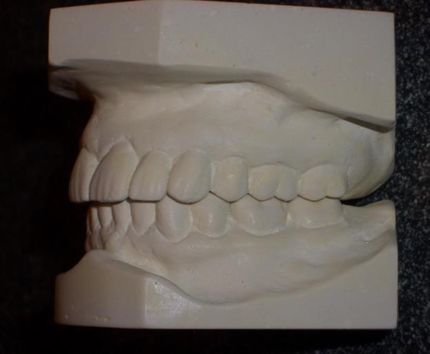
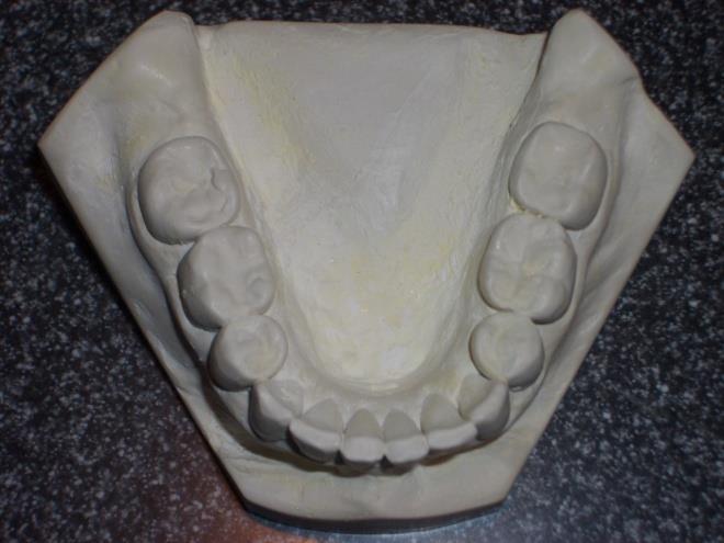
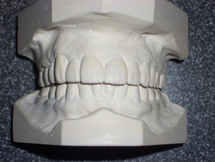
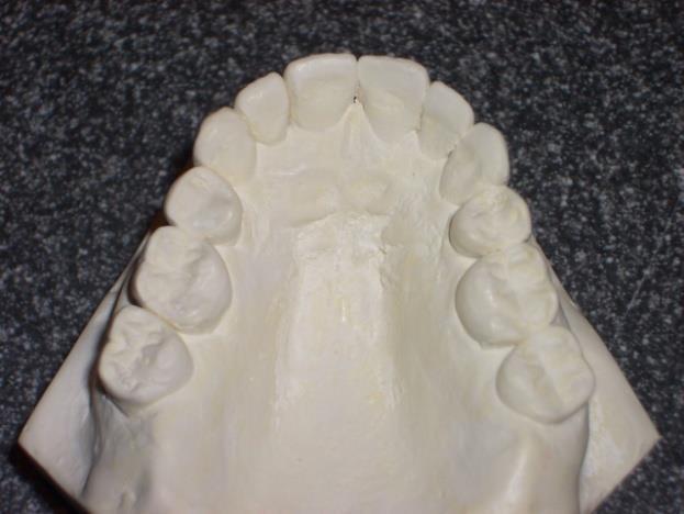
32 Patient s Models

33 The Orthotic Appliance
34 Treatment: January - February Initial Examination and diagnosis Seated bite plane. Height increased on right to balance posterior pivot appliance. Vertical balance against lower orthodontic retainer. Home care instructions provided Jaw stiffness better, no locking. Still has pain with function but reduced. Patient has sensitivity of upper teeth and jaw feels tired. Ear fullness gone. No referral pain to teeth or sinuses. 32 mm opening. Posterior orthotic occlusion balanced.
35 Treatment: March Patient still has slight pain and still deviating to the right 2 mm. Add anterior positioner to balance opening motion. Patient given mirror exercise and careful instruction by me and my Certified TMJ Assistant.

36 Treatment: April Patient doing midline exercises deviation to 1 mm right. Upper teeth are aching and neck hurts. She states that she is working too hard. Adjust anterior positioner Patient thinks she is doing well. A lot less pain. Midline opening with no deviation. Equilibrate teeth 11, 12, and 20. Observe for final equilibration in 3 weeks.
37 Treatment: May & November TMJ feeling better. Wearing plane only at night. Check, no equilibration necessary. Check in 6 months Patient reports opening with no pain at 44 mm. No deviation and no referral pain. Patient wearing plane at night. No problems. Check in 6 months
38 Treatment: Successful Result : Patient reports no pain. No tenderness on palpation. Opening at 44 mm with no deviation. No additional equilibration required. Check patient in 6 months. Patient Photo: Front View
39 In Summary After experiencing two rounds of orthodontia and coming to the office with a substantial fear of the need for surgery, this patient was able to achieve a successful result through careful diagnosis, orthotic mandibular repositioning and exercise. Photo of dentist and staff Doctor and his two Certified TMJ Assistants.
Temporomandibular Joint Disorders
 Temporomandibular Joint Disorders Introduction Temporomandibular joint disorders, or TMJ disorders, are a group of medical problems related to the jaw joint. TMJ disorders can cause headaches, ear pain,
TEMPORO-MANDIBULAR JOINT DISORDERS
 Disclaimer This movie is an educational resource only and should not be used to manage your dental health. All decisions about the management of TMJ Disorders must be made in conjunction with your Dental
Initial Doctor Questionnaire
 Initial Doctor Questionnaire DO NOT enter the patient in this study: if your patient does not have a TMD pain diagnosis if your patient does not need treatment at this time if you are not going to treat
CRANIAL NERVES. Dr. Amani A. Elfaki Associate Professor Department of Anatomy
 CRANIAL NERVES Dr. Amani A. Elfaki Associate Professor Department of Anatomy LEARNING OBJECTIVES Named the cranial nerves Identify the funcunal component of each cranial nerve Identify the effect of each
Muscles of mastication [part 1]
![Muscles of mastication [part 1]](/thumbs/76/73586850.jpg "Muscles of mastication [part 1]") Muscles of mastication [part 1] In this lecture well have the muscles of mastication, neuromuscular function, and its relationship to the occlusion morphology. The fourth determinant of occlusion is the
Muscles of mastication [part 1] In this lecture well have the muscles of mastication, neuromuscular function, and its relationship to the occlusion morphology. The fourth determinant of occlusion is the
Prosthetic Management of TMJ Disorders
 Prosthetic Management of TMJ Disorders Mohammed Alfarsi BDS, MDSc(Pros), PhD www.drmohdalfarsi.com com.+*()ا&%$ر"!. www Mohd@DrMohdAlfarsi.com @DrMohdAlfarsi DrMohdAlfarsi 056 224 2227 Overview Overview
Where is the Temporo-Mandibular Joint?
 TMJ Pain Open your jaw all the way and shut it. This simple movement would not be possible without the Temporo-Mandibular Joint (TMJ). It connects the temporal bone (the bone that forms the side of the
A Look at Two Syndromes: TEMPOROMANDIBULAR JOINT & CERVICOCRANIAL DYSFUNCTION IN THE EDS PATIENT. John Mitakides D.D.S., FAACP
 TEMPOROMANDIBULAR JOINT & CERVICOCRANIAL DYSFUNCTION IN THE EDS PATIENT John Mitakides D.D.S., FAACP A Look at Two Syndromes: How TMJ and CCD impact the EDS patient as they occur separately or together
Brain and spinal nerve. By: shirin Kashfi
 Brain and spinal nerve By: shirin Kashfi Nervous system: central nervous system (CNS) peripheral nervous system (PNS) Brain (cranial) nerves Spinal nerves Ganglions (dorsal root ganglions, sympathetic
INTRODUCTION: ANATOMY UNDERLYING CLINICAL TESTS OF CRANIAL NERVES
 INTRODUCTION: ANATOMY UNDERLYING CLINICAL TESTS OF CRANIAL NERVES CRANIAL NERVE I - OLFACTORY I - OLFACTORY NERVE - SMELL TEST: SMELL ODORS (note: not ammonia; pain in nasal cavity CN5 DAMAGE: LOSS OF
Anatomy and physiology of Temporomandibular Joint
 Anatomy and physiology of Temporomandibular Joint Temporomandibular joint (TMJ): It is the articulation of the condyle of the mandible, and the inter-articular disc; with the mandibular fossa (glenoid
Principle of Occlusion
 Principle of Occlusion Mohammed Alfarsi BDS, MDSc(Pros), PhD www.drmohdalfarsi.com com.+*()ا&%$ر"!. www Overview Principle of Occlusion Overview Principle of Occlusion Point centric Long centric Freedom
Up Date on TMD WHAT IS TMD? Temporomandibular Disorders (TMD)*: Donald Nixdorf DDS, MS
*: Donald Nixdorf DDS, MS") Up Date on TMD Donald Nixdorf DDS, MS Associate Professor Division of TMD and Orofacial Pain WHAT IS TMD? Temporomandibular Disorders (TMD)*: MUSCLE and JOINT DISORDERS * Temporomandibular Muscle and Joint
Up Date on TMD Donald Nixdorf DDS, MS Associate Professor Division of TMD and Orofacial Pain WHAT IS TMD? Temporomandibular Disorders (TMD)*: MUSCLE and JOINT DISORDERS * Temporomandibular Muscle and Joint
ChiroCredit.com Anatomy 226 INSTRUCTIONS/ASSIGNMENT FOR ANATOMICAL DISSECTION:
 ChiroCredit.com Anatomy 226 INSTRUCTIONS/ASSIGNMENT FOR ANATOMICAL DISSECTION: Once you click on the link to open the dissection module, the first thing you need to do is to be sure you can see all the
Upper Cross Syndrome: Assessment & Management in Family Practice HKDU Symposium Dec 2014
 Upper Cross Syndrome: Assessment & Management in Family Practice HKDU Symposium Dec 2014 Dr. Ngai Ho Yin Allen Family Medicine Specialist PGDipMusculoskeletal Medicine MBBS(HK), DCH(London), DFM(CUHK),
Anatomy and Physiology II. Review Spine and Neck
 Anatomy and Physiology II Review Spine and Neck Spine regions How many cervical vertibrae are there? 7 The curvature is the cervical region posterior? Concave posterior How many thoracic? And curvature?
U.S. DEPARTMENT OF HEALTH AND HUMAN SERVICES National Institutes of Health
 T M J D I S O R D E R S U.S. DEPARTMENT OF HEALTH AND HUMAN SERVICES National Institutes of Health CONTENTS 2 4 6 7 8 9 14 WHAT IS THE TEMPOROMANDIBULAR JOINT? WHAT ARE TMJ DISORDERS? WHAT CAUSES TMJ DISORDERS?
Temporomandibular Joint. Dr Noman ullah wazir
 Temporomandibular Joint Dr Noman ullah wazir Type of Joint TMJ is a Synovial joint between : The condylar head of the mandible. The mandibular fossa of squamous part of temporal bone. The joint cavity
Lab 16: PNS: Nerves and Autonomic NS Hamilton Answers to Pre- Lab Assignments
 Lab 16: PNS: Nerves and Autonomic NS Hamilton Answers to Pre- Lab Assignments Pre-Lab Activity 1: 1. a. olfactory nerve b. optic nerve c. oculomotor nerve d. abducens nerve e. trochlear nerve f. trigeminal
Neurological Assessment
 Neurological Assessment Name: Age: Gender: Date: History Review of history related to neurological system YES/NO If YES, provide details: General Neurological Mental Illness Neurological disease Severe
Anatomy and Physiology. Bones, Sutures, Teeth, Processes and Foramina of the Human Skull
 Anatomy and Physiology Chapter 6 DRO Bones, Sutures, Teeth, Processes and Foramina of the Human Skull Name: Period: Bones of the Human Skull Bones of the Cranium: Frontal bone: forms the forehead and the
Lec [8]: Mandibular nerve:
![Lec [8]: Mandibular nerve:](/thumbs/94/121295776.jpg "Lec [8]: Mandibular nerve:") Lec [8]: Mandibular nerve: The mandibular branch from the trigeminal ganglion lies in the middle cranial fossa lateral to the cavernous sinus. With the motor root of the trigeminal nerve [motor roots lies
Lec [8]: Mandibular nerve: The mandibular branch from the trigeminal ganglion lies in the middle cranial fossa lateral to the cavernous sinus. With the motor root of the trigeminal nerve [motor roots lies
Appendix 4. Case Summary Template. Social history. Patient (Name and Date of birth): Presenting complaints. History Medical history
: Presenting complaints. History Medical history") Appendix 4 Case Summary Template Patient (Name and Date of birth): Presenting complaints Chief complaint Subsidiary complaints History of complaints Patient s expectations History Medical history Medical
Appendix 4 Case Summary Template Patient (Name and Date of birth): Presenting complaints Chief complaint Subsidiary complaints History of complaints Patient s expectations History Medical history Medical
TMJ Disorder & Sleep Conditions: The Effects on Your Body
 TMJ Disorder & Sleep Conditions: The Effects on Your Body TMJ Disorders: Temporomandibular Joint Disorders The mandible, or jaw, is the movable part of the head involving important functions of daily life,
Human Anatomy and Physiology - Problem Drill 07: The Skeletal System Axial Skeleton
 Human Anatomy and Physiology - Problem Drill 07: The Skeletal System Axial Skeleton Question No. 1 of 10 Which of the following statements about the axial skeleton is correct? Question #01 A. The axial
Concepts of occlusion Balanced occlusion. Monoplane occlusion. Lingualized occlusion. Figure (10-1)
") Any contact between teeth of opposing dental arches; usually, referring to contact between the occlusal surface. The static relationship between the incising or masticatory surfaces of the maxillary or
Any contact between teeth of opposing dental arches; usually, referring to contact between the occlusal surface. The static relationship between the incising or masticatory surfaces of the maxillary or
Examination and Diseases of Cranial Nerves
 Cranial nerve evaluation is an important part of a neurologic exam. There are some differences in the assessment of cranial nerves with different species but the general principles are the same. Going
Establish a Healthy TMJ
 TW TMJ Establish a Healthy TMJ Prior to Restorative, Orthodontic or Prosthetic Treatment Dr. Brock Rondeau, D.D.S., I.B.O., D.A.B.C.P. Author: Dr. Brock Rondeau is one of North America s most sought after
Arrangement of the artificial teeth:
 Lecture Prosthodontic Dr. Osama Arrangement of the artificial teeth: It s the placement of the teeth on a denture with definite objective in mind or it s the setting of teeth on temporary bases. Rules
Temporal region. temporal & infratemporal fossae. Zhou Hong Ying Dept. of Anatomy
 Temporal region temporal & infratemporal fossae Zhou Hong Ying Dept. of Anatomy Temporal region is divided by zygomatic arch into temporal & infratemporal fossae. Temporal Fossa Infratemporal fossa Temporal
Arthrogenous disorders of the TMJ
 Arthrogenous disorders of the TMJ Seena Patel DMD, MPH Assistant Professor, Associate Director of Oral Medicine Arizona School of Dentistry & Oral Health, A.T. Still University, Mesa, AZ Southwest Orofacial
The ASE Example Case Report 2010
 The ASE Example Case Report 2010 The Requirements for Case Presentation in The Angle Society of Europe are specified in the Appendix I to the Bylaws. This example case report exemplifies how these requirements
Response Type axium Adult Comprehensive Oral Examination (COE)
") Page 1 1. RADIOGRAPHIC EVALUATION 1001 Panoramic image (PAN) Maxillary sinuses Nasal cavity TMJ complex Mandibular canal visualization?bone anomalies (eg. radiopacity/radiolucency) Soft tissue abnormalities
Page 1 1. RADIOGRAPHIC EVALUATION 1001 Panoramic image (PAN) Maxillary sinuses Nasal cavity TMJ complex Mandibular canal visualization?bone anomalies (eg. radiopacity/radiolucency) Soft tissue abnormalities
Sample Case #1. Disclaimer
 ABO Sample Cases Disclaimer Sample Case #1 The following sample questions and answers were composed and vetted by a panel of experts in orthodontics and are intended to provide an example of the types
Fellowship. Digital Case Documentation Format for Fellowship Case Presentation. OCCLUSION CONNECTIONS Dot Dentistry
 Digital Case Documentation Format for Case Presentation TMD Candidate s Name: CLAYTON A. CHAN, D.D.S. Case No.: 1 Our Disciplines ORTHOPEDIC Patient s Initials: CL (EXAMPLE CASE) RESTORATIVE OCCLUSION
Biology 218 Human Anatomy. Adapted from Martini Human Anatomy 7th ed. Chapter 6 The Skeletal System: Axial Division
 Adapted from Martini Human Anatomy 7th ed. Chapter 6 The Skeletal System: Axial Division Introduction The axial skeleton: Composed of bones along the central axis of the body Divided into three regions:
Parotid Gland. Parotid Gland. Largest of 3 paired salivary glands (submandibular; sublingual) Ramus of Mandible. Medial pterygoid.
 Ramus of Mandible. Medial pterygoid.") Parotid region Parotid Gland Largest of 3 paired salivary glands (submandibular; sublingual) Ramus of Mandible Medial pterygoid Cross section of mandible Masseter D S SCM Parotid Gland Mastoid Process
Parotid region Parotid Gland Largest of 3 paired salivary glands (submandibular; sublingual) Ramus of Mandible Medial pterygoid Cross section of mandible Masseter D S SCM Parotid Gland Mastoid Process
Case Report. Orthognathic Correction of Class II Open Bite. Using the Piezoelectric System and MatrixORTHOGNATHIC Plating System.
 Case Report Orthognathic Correction of Class II Open Bite. Using the Piezoelectric System and MatrixORTHOGNATHIC Plating System. Orthognathic Correction of Class II Open Bite. Using the Piezoelectric System
PERIPHERAL NERVOUS SYSTEM
 CHAPTER 13 PERIPHERAL NERVOUS SYSTEM Functional division of nervous system = afferent info to the CNS ascending spinal cord = efferent info from CNS descending spinal cord somatic skin, muscles visceral
Structure Location Function
 Frontal Bone Cranium forms the forehead and roof of the orbits Occipital Bone Cranium forms posterior and inferior portions of the cranium Temporal Bone Cranium inferior to the parietal bone forms the
Chapter 7 Part A The Skeleton
 Chapter 7 Part A The Skeleton Why This Matters Understanding the anatomy of the skeleton enables you to anticipate problems such as pelvic dimensions that may affect labor and delivery The Skeleton The
Temporomandibular Joint
 6 Temporomandibular Joint Most human bones are joined with one another. These connections of bones to each other are termed articulations or joints. Some joints are immovable as in the articulations of
Removable appliances
 Removable appliances Melinda Madléna DMD, PhD associate professor Department of Pedodontics and Orthodontics Faculty of Dentistry Semmelweis University Budapest Classification of the orthodontic anomalies
Infratemporal fossa: Tikrit University college of Dentistry Dr.Ban I.S. head & neck Anatomy 2 nd y.
 Infratemporal fossa: This is a space lying beneath the base of the skull between the lateral wall of the pharynx and the ramus of the mandible. It is also referred to as the parapharyngeal or lateral pharyngeal
TMJ UNDERSTANDING SYNDROME SPECIAL REPORT By Paul R. White, D.D.S. Special Report: Understanding TMJ Syndrome
 SPECIAL REPORT Special Report: Understanding TMJ Syndrome UNDERSTANDING TMJ SYNDROME By Paul R. White, D.D.S. 804.715.1647 www.smilerichmond.com 804.715.1647 www.smilerichmond.com 1 UNDERSTANDING TMJ SYNDROME
Upper arch. 1Prosthodontics. Dr.Bassam Ali Al-Turaihi. Basic anatomy & & landmark of denture & mouth
 1Prosthodontics Lecture 2 Dr.Bassam Ali Al-Turaihi Basic anatomy & & landmark of denture & mouth Upper arch Palatine process of maxilla: it form the anterior three quarter of the hard palate. Horizontal
Neuromuscular Dentistry: Transcutaneous Electrical Nerve Stimulation and Orthotic Solutions in Full Mouth Reconstruction
 Cronicon OPEN ACCESS EC DENTAL SCIENCE Case Report Neuromuscular Dentistry: Transcutaneous Electrical Nerve Stimulation and Orthotic Solutions in Full Mouth Reconstruction Mario Guiang* Guiang Dental,
For the following questions, indicate the letter that corresponds to the SINGLE MOST APPROPRIATE ANSWER
 GROSS ANATOMY EXAMINATION May 15, 2000 For the following questions, indicate the letter that corresponds to the SINGLE MOST APPROPRIATE ANSWER 1. Pain associated with an infection limited to the middle
Introduction to Occlusion and Mechanics of Mandibular Movement
 Introduction to Occlusion and Mechanics of Mandibular Movement Dr. Pauline Hayes Garrett Department of Endodontics, Prosthodontics, and Operative Dentistry University of Maryland, Baltimore Assigned reading
Peripheral Nervous System Dr. Gary Mumaugh
 Peripheral Nervous System Dr. Gary Mumaugh Spinal Nerves Overview Thirty-one pairs of spinal nerves are connected to the spinal cord No special names; numbered by level of vertebral column at which they
Case Report: Long-Term Outcome of Class II Division 1 Malocclusion Treated with Rapid Palatal Expansion and Cervical Traction
 Case Report Case Report: Long-Term Outcome of Class II Division 1 Malocclusion Treated with Rapid Palatal Expansion and Cervical Traction Roberto M. A. Lima, DDS a ; Anna Leticia Lima, DDS b Abstract:
Vivid Journal of Dental Sciences
 International Scientific Peer Reviewed Journals Vivid Journal of Dental Sciences Article Id: 2018100002 *Corresponding author Kengo Torii, Department of General Dentistry, School of Life Dentistry, Nippon
STRAIGHT TALK about CROOKED TEETH: Key #4, Healthy TMJ Function
 STRAIGHT TALK about CROOKED TEETH: Key #4, Healthy TMJ Function This is fourth in a series of articles by Derek Mahony, BDS, MDSc and S. Kent Lauson, DDS, MS (Orthodontists) Their book was written with
Biology 323 Human Anatomy for Biology Majors Week 10; Lecture 1; Tuesday Dr. Stuart S. Sumida. Cranial Nerves and Soft Tissues of the Skull
 Biology 323 Human Anatomy for Biology Majors Week 10; Lecture 1; Tuesday Dr. Stuart S. Sumida Cranial Nerves and Soft Tissues of the Skull FOREBRAIN MIDBRAIN HINDBRAIN Forebrain: Cerebrum Perception,
Photobiomodulation in Head and Neck Pain. Gerry Ross, DDS. Tottenham, Ontario Canada
 Photobiomodulation in Head and Neck Pain Gerry Ross, DDS Tottenham, Ontario Canada Disclosure: Dr. Ross provides education and training for Zolar Surgical Diode lasers, for which he receives a fee. No
TRAUMA TO THE FACE AND MOUTH
 Dr.Yahya A. Ali 3/10/2012 F.I.C.M.S TRAUMA TO THE FACE AND MOUTH Bailey & Love s 25 th edition Injuries to the orofacial region are common, but the majority are relatively minor in nature. A few are major
Oral cavity landmarks
 By: Dr. Ahmed Rabah Oral cavity landmarks The knowledge of oral anatomy and physiology will help the operator and provides enough landmarks to act as positive guide during denture construction. This subject
Dr.Sepideh Falah-kooshki
 Dr.Sepideh Falah-kooshki MAXILLA Premaxillary/median palatal suture (radiolucent). Incisive fossa and foramen (radiolucent). Nasal passages (radiolucent). Nasal septum (radiopaque). Anterior nasal spine
JAMSS Speed-to-Treat Protocol For treatment of jaw joint and muscle sprain/strain injuries
 JAMSS Speed-to-Treat Protocol For treatment of jaw joint and muscle sprain/strain injuries INTRODUCTION What is Jaw Joint and Muscle Sprain/Strain (JAMSS)? Jaw Joint and Muscle Sprain/Strain (JAMSS) is
Temporomandibular Joint Disorders
 Temporomandibular Joint Disorders Temporomandibular Joint : Is a synovial joint located between the condyle (head of the mandible) and the glenoid fossa (inferior surface of the squamous part of Temporal
Horizontal jaw relations: The relationship of mandible to maxilla in a
 Horizontal relations Horizontal jaw relations: The relationship of mandible to maxilla in a horizontal plane (in anteroposterior and side to side direction). a- Protruded or forward relation. b-lateral
Dr. Sami Zaqout, IUG Medical School
 The skull The skull is composed of several separate bones united at immobile joints called sutures. Exceptions? Frontal bone Occipital bone Vault Cranium Sphenoid bone Zygomatic bones Base Ethmoid bone
Bones of the skull & face
 Bones of the skull & face Cranium= brain case or helmet Copyright The McGraw-Hill Companies, Inc. Permission required for reproduction or display. The cranium is composed of eight bones : frontal Occipital
Parotid Gland, Temporomandibular Joint and Infratemporal Fossa
 M1 - Anatomy Parotid Gland, Temporomandibular Joint and Infratemporal Fossa Jeff Dupree Sanger 9-057 jldupree@vcu.edu Parotid gland: wraps around the mandible positioned between the mandible and the sphenoid
Cranial nerves.
 Cranial nerves eaglezhyxzy@163.com Key Points of Learning Name Components Passing through Peripheral distribution Central connection Function Cranial nerves Ⅰ olfactory Ⅱ optic Ⅲ occulomotor Ⅳ trochlear
TMJ Parametro Classico
 TMJ Parametro Classico Total Temporomandibular Joint Prosthesis 2 Personalized total TMJ replacement system (Parametro Classic & Parametro Saddle ) Patient Information in English This patient information
Neurological Assessment Part 1
 Neurological Assessment Part 1 MOTOR EXAMINATION: Look at bulk, contour and symmetry of individual muscles: muscles of face upper arm arm thigh lower leg Look for atrophy--may help to localize the site
2 skull, vertebral column, thoracic cage
 CHAPTER 7-SKELTON FILL-IN NOTES 2 skull, vertebral column, thoracic cage 3 Fig. 7.1 pg. 199 4 I. Skull: A. : Encloses and the brain - 8 bones B. : 14 bones Cranium A. Forehead (brain) Anterior part of
Total Prosthetic Replacement of the Temporomandibular Joint (TMJ)
") Total Prosthetic Replacement of the Temporomandibular Joint (TMJ) VERSION CONTROL Version: 2.0 Ratified by: Governing Body Date ratified: 13 November 2013 Name of originator/author: Name of responsible
Total Prosthetic Replacement of the Temporomandibular Joint (TMJ) VERSION CONTROL Version: 2.0 Ratified by: Governing Body Date ratified: 13 November 2013 Name of originator/author: Name of responsible
Diana Rose. Clinical Case Report Competition. West Coast College of Massage Therapy, Victoria. Second Place Winner. Winter 2009
 Massage Therapists Association of British Columbia Clinical Case Report Competition West Coast College of Massage Therapy, Victoria Winter 2009 Second Place Winner Diana Rose Reduction of temporomandibular
Basic Anatomy and Physiology of the Lips and Oral Cavity. Dr. Faghih
 Basic Anatomy and Physiology of the Lips and Oral Cavity Dr. Faghih It is divided into seven specific subsites : 1. Lips 2. dentoalveolar ridges 3. oral tongue 4. retromolar trigone 5. floor of mouth 6.
Fundamental & Preventive Curvatures of Teeth and Tooth Development. Lecture Three Chapter 15 Continued; Chapter 6 (parts) Dr. Margaret L.
 Dr. Margaret L.") Fundamental & Preventive Curvatures of Teeth and Tooth Development Lecture Three Chapter 15 Continued; Chapter 6 (parts) Dr. Margaret L. Dennis Proximal contact areas Contact areas are on the mesial and
Fundamental & Preventive Curvatures of Teeth and Tooth Development Lecture Three Chapter 15 Continued; Chapter 6 (parts) Dr. Margaret L. Dennis Proximal contact areas Contact areas are on the mesial and
The Temporomandibular joint: Anatomy, Mechanics, Pathology. Aditya Bahel, DO
 The Temporomandibular joint: Anatomy, Mechanics, Pathology Aditya Bahel, DO Outline Anatomy Mechanics and function Indications for TMJ imaging MR Protocols and pitfalls Pathology Treatment options Anatomy
Nightguards, Splints, and Orthotics
 Nightguards, splints, and orthotics are three different oral appliances that are regularly used to support and protect the jaw joints, muscles, and teeth through various structural conditions and injuries.
Cranial Nerves Exam. 1. To learn how to examine the functions of the 12 pairs of cranial nerves.
 Cranial Nerves Exam [Purpose] 1. To learn how to examine the functions of the 12 pairs of cranial nerves. 2. To understand the function of the 12 pairs of cranial nerves. [Principle] The cranial nerves
Orofacial pain and temporomandibular joint disorder patient history and questionnaire. Name: Sex: M F Date of Birth: / / Age:
 Orofacial pain and temporomandibular joint disorder patient history and questionnaire Date: / / Name: Sex: M F Date of Birth: / / Age: Occupation: Physician: Dentist: Referred by: Chief Complaint/Concern:
3. The Jaw and Related Structures
 Overview and objectives of this dissection 3. The Jaw and Related Structures The goal of this dissection is to observe the muscles of jaw raising. You will also have the opportunity to observe several
Lab Activity 19 & 20. Cranial Nerves General Senses. Portland Community College BI 232
 Lab Activity 19 & 20 Cranial Nerves General Senses Portland Community College BI 232 Cranial Nerves Nerves that originate from the brain rather than the spinal cord Part of the peripheral nervous system
Shadeguides Finding the Centric Relation The Kois Deprogrammer
 Maciej Zarow Shadeguides Finding the Centric Relation The Kois Deprogrammer 21 Jun 2018 Finding the centric relation might sometimes seem hard, but with appropriate devices, such as the Kois Deprogrammer
 www.oralradiologists.com CONE BEAM CT REPORT CASE XXXX Patient information Patient Name: - Referring Doctor: - Patient DOB: - Scan Date: [Start date] Reason for Exam: Maxillary facial pain Doctor Notes:
CONQUER YOUR JAW PAIN
 CONQUER YOUR JAW PAIN We think it s time you got back to living again, don t you? PRESENTED BY: What is Temporomandibular Joint Disorder (TMD, TMJ)? The joint that connects your jaw to the bones of your
ALTERNATE OCCLUSAL SCHEMES
 ALTERNATE OCCLUSAL SCHEMES The same basic concepts need to be applied to all occlusal schemes. Some challenges include missing teeth, transposed teeth, crossbites, and anterior open bites. POSTERIOR CROSSBITES
KJLO. A Sequential Approach for an Asymmetric Extraction Case in. Lingual Orthodontics. Case Report INTRODUCTION DIAGNOSIS
 KJLO Korean Journal of Lingual Orthodontics Case Report A Sequential Approach for an Asymmetric Extraction Case in Lingual Orthodontics Ji-Sung Jang 1, Kee-Joon Lee 2 1 Dream Orthodontic Clinic, Gimhae,
Conventional radiograph verses CT for evaluation of sagittal fracture of mandibular condyle
 Case Report: Conventional radiograph verses CT for evaluation of sagittal fracture of mandibular condyle Dr Anjali Wadhwa, Dr Gaurav Shah, Dr Shweta Sharma, Dr Anand Bhatnagar, Dr Pallavi Malaviya NIMS
ADJUSTABLE HERBST APPLIANCE - OASYS HINGE ACRYLIC
 P SYSTEMS The delivery visit should confirm the fit and comfort of the appliance, as well as the patients ability to insert and remove the appliance properly and without difficulty. It may be necessary
Fixed appliances II. Dr. Káldy Adrienn, Semmeweis University
 Fixed appliances II. Dr. Káldy Adrienn, Semmeweis University Head gear/facebow Delair mask/ face mask Fixed Class II. correctors Lip bumper Eva plate Nance appliance Pearl appliance Habbit crib Applied
Deprogrammers made. Deprogrammers can be used for... How do deprogrammers work?
 Deprogrammers made asy Easy Deprogrammers made Deprogrammers can be used for... Headache sufferers Bruxers and/or clenchers Muscle relaxation Determining proper joint position Protection against wear Diagnosis
Early Mixed Dentition Period
 REVIEW ARTIC CLE AODMR The Effects of a Prefabricated Functional Appliance in Early Mixed Dentition Period Toshio Iwata 1, Takashi Usui 2, Nobukazu Shirakawa 2, Toshitsugu Kawata 3 1 Doctor of Philosophy
DEVELOPING ANALOGUE/SUBTITUTE FOR THE MANDIBULAR DENTURE BEARING AREA. Dr Muhammad Rizwan Memon FCPS Assistant Professor
 DEVELOPING ANALOGUE/SUBTITUTE FOR THE MANDIBULAR DENTURE BEARING AREA Dr Muhammad Rizwan Memon FCPS Assistant Professor Crest of Residual Ridge Buccal Shelf Shape of supporting structure Mylohyoid Ridge
Basic Functional Anatomy of the Gnathostomatic System and its Clinical Implications. Eugene Santucci DDS, MA, FACD
 Basic Functional Anatomy of the Gnathostomatic System and its Clinical Implications Eugene Santucci DDS, MA, FACD NOTHING IS MORE FUNDAMENTAL TO TREATING PATIENTS THAN KNOWING ANATOMY Jeffery P. Okeson
University of Palestine. Midterm Exam 2013/2014 Total Grade:
 Course No: DNTS2208 Course Title: Head and Neck Anatomy Date: 09/11/2013 No. of Questions: (50) Time: 1hour Using Calculator (No) University of Palestine Midterm Exam 2013/2014 Total Grade: Instructor
BUCCAL MUCOSA RIDGING AND TONGUE INDENTATION: INCIDENCE AND ASSOCIATED FACTORS
 Bull. Tokyo dent. Coll., Vol. 40, No. 2, pp. 71 78, May, 1999 71 Original Article BUCCAL MUCOSA RIDGING AND TONGUE INDENTATION: INCIDENCE AND ASSOCIATED FACTORS KATIUSKA PIQUERO, TOMOHIKO ANDO and KAORU
Unit VIII Problem 3 Neuroanatomy: Brain Stem, Cranial Nerves and Scalp
 Unit VIII Problem 3 Neuroanatomy: Brain Stem, Cranial Nerves and Scalp - Brain stem: It is connected to the cerebellum and cerebral hemispheres. Rostral end of brain stem: diencephalon is the area which
Oral cavity : consist of two parts: the oral vestibule and the oral cavity proper. Oral vestibule : is slit like space between.
 Oral cavity Oral cavity : consist of two parts: the oral vestibule and the oral cavity proper Oral vestibule : is slit like space between the teeth, buccal gingiva, lips, and cheeks 1 Oral cavity Oral
Significant improvement with limited orthodontics anterior crossbite in an adult patient
 VARIA Significant improvement with limited orthodontics anterior crossbite in an adult patient Arzu Ari-Demirkaya Istanbul, Turkey Summary Objectives. Orthodontic treatment is known to last as long as
Cranial Nerves. Steven McLoon Department of Neuroscience University of Minnesota
 Cranial Nerves Steven McLoon Department of Neuroscience University of Minnesota 1 Course News Change in Lab Sequence Week of Oct 2 Lab 5 Week of Oct 9 Lab 4 2 Sensory and Motor Systems Sensory Systems:
Bones Ethmoid bone Inferior nasal concha Lacrimal bone Maxilla Nasal bone Palatine bone Vomer Zygomatic bone Mandible
 splanchnocranium - Consists of part of skull that is derived from branchial arches - The facial bones are the bones of the anterior and lower human skull Bones Ethmoid bone Inferior nasal concha Lacrimal
The future of health is digital
 Dated: XX/XX/XXXX Name: XXXXXXXX XXXXXXXXXXX Birth Date: XX/XX/XXXX Date of scan: XX/XX/XXXX Examination of the anatomical volume: The following structures are reviewed and evaluated for bilateral symmetry,
OCCLUSION: PHYSIOLOGIC vs. NON-PHYSIOLOGIC
 Oral Anatomy and Occlusion Prosthodontic Component OCCLUSION: PHYSIOLOGIC vs. NON-PHYSIOLOGIC By: Dr. Babak Shokati, DDS, MSc. MSc. Prosthodontics Definition of Masticatory System by The Academy of Prosthodontics
Samantha W. Chou, D.M.D N. Southport Ave. Chicago, Illinois Phone: Fax:
 Samantha W. Chou, D.M.D. 2325 N. Southport Ave. Chicago, Illinois 60614 Phone: 312-608-6881 Fax: 773-296-0601 Samanthawchou@gmail.com What is our role as the dentist? "We live in a culture in which people