MR: Finger and Thumb Injuries
|
|
|
- Mabel Floyd
- 6 years ago
- Views:
Transcription

1 MR: Finger and Thumb Injuries Laura W. Bancroft, M.D. Professor of Radiology University of Central Florida Florida State University

2 Outline Normal anatomy of the fingers and thumb MR imaging protocols MRI findings of sports injuries of the fingers and thumb
3 NORMAL ANATOMY
- distal to proximal Clavero JA et al. Extensor Mechanism of the Fingers: MR Imaging Anatomic Correlation. RadioGraphics 2003; 23:593-611.")
4 I -DIP joint II - middle phalanx III -PIP joint IV - proximal phalanx V -MCP joint VI - dorsum of hand VII -wrist extensor compartment VIII - extrinsic extensor muscles Zone specific Anatomy (Verdan)- distal to proximal Clavero JA et al. Extensor Mechanism of the Fingers: MR Imaging Anatomic Correlation. RadioGraphics 2003; 23:
5 terminal tendon triangular ligament med/lat conjoined tendons Clavero JA et al. Extensor Mechanism of the Fingers: MR Imaging Anatomic Correlation. RadioGraphics 2003; 23:
6 med/lat conjoined tendons lateral slip medial slip central slip Clavero JA et al. Extensor Mechanism of the Fingers: MR Imaging Anatomic Correlation. RadioGraphics 2003; 23:

7 retinacular ligament oblique fibers transverse fibers interosseous muscle sagittal band Clavero JA et al. Extensor Mechanism of the Fingers: MR Imaging Anatomic Correlation. RadioGraphics 2003; 23:
8 Central slip Extensor Tendon
9 Conjoined tendons Extensor Tendon

10 Transverse retinacular ligaments Extensor Tendon
11 FDS FDS palmar FDS lateral

12 FDP FDP FDP FDS FDS palmar FDS lateral
13 Flexor Pulley System Injury Flexor tendons pass through a fibro-osseous canal Extends from the head of the metacarpals to the DIP joints 2 thumb pulleys A1 and A2
14 Flexor Pulley System Injury A2 and A4 Largest Prevent bowstringing
15 Flexor Pulley System Injury

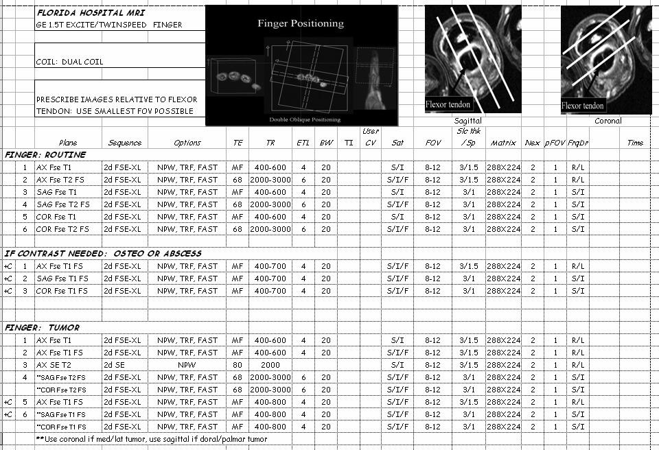
16 Magnetic Resonance Imaging Coil selection - critical to quality imaging of the hand and fingers Small extremity coil AKA - elbow coil

17 Magnetic Resonance Imaging Small loop coil AKA Digit coil Allow very small FOV
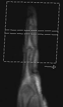
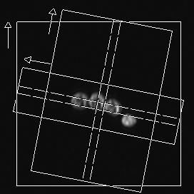
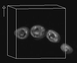
18 Finger Positioning Double Oblique Positioning
19 S
20 Trauma

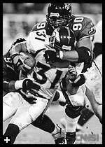
21 Finger and Thumb Injuries Very common in athletes 9% of all sports-related injuries Hand is characteristically in front of the athlete Absorbs the initial contact

22 Finger and Thumb Injuries Hands are used in a majority of sports Many competitive team sports Fingers and thumb - most often injured
23 Fingers and Thumb Injuries Common in sports with a high risk of falling Skiing Biking Gymnastics In-line skating

24 Fingers and Thumb Injuries In-line skating >50% of injuries involve hand/wrist Football Hand and wrist injuries account for 15-20%

25 Finger and Thumb Injuries Minor injuries Finger sprain Major injuries Fracture or dislocation Hard to prevent

26 Imaging Radiographs Computed Tomography Magnetic Resonance Imaging

27 Hyperextension Injury Capsular and Volar Plate Disruption
28 Hyperextension Injury Capsular and Volar Plate Disruption Volar Plate Thick fibrocartilaginous structure Prevents hyperextension Palmaraspect of PIP joint Distal firm attachment

29 Jersey Finger Disruption of the FDP from the volar base of the distal phalanx Finger is pulled or forced into extension while the DIP is actively flexed

30 Attempting to grab someone by the jersey while making a tackle Football Rugby Ring finger - 75% of cases Localized pain and swelling Inability to flex DIP joint Jersey Finger

31 Jersey Finger Radiographs or other imaging studies may be useful May depict a bony avulsion Site of attachment of the FDP Volar aspect of the base of the distal phalanx

32 Mallet Finger Occurs with forced flexion of the extended DIP joint Results in: Stretching or tearing of the extensor tendon substance Avulsion fracture Extensor tendon insertion Dorsal base of the distal phalanx

33 Mallet Finger Extensor tendon tear or bony avulsion Bony avulsion of dorsal base of the distal phalanx

34 Mallet Finger Classic mechanism of injury Tip of the extended finger struck by a ball Softball, baseball, or basketball Often referred to as a Jammed Finger
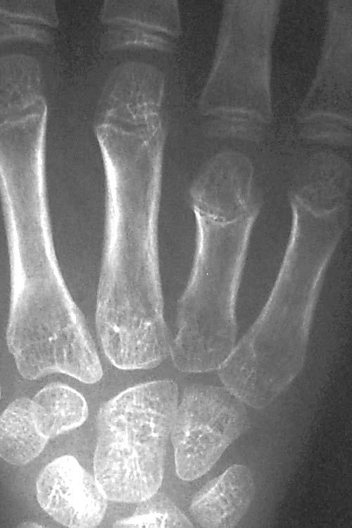
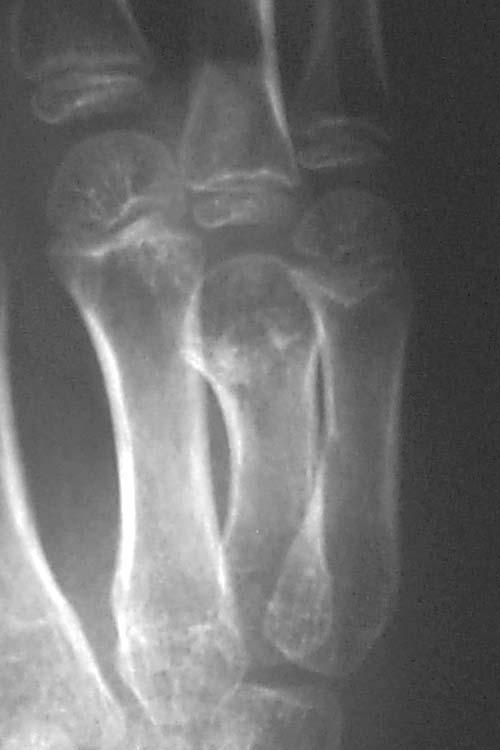
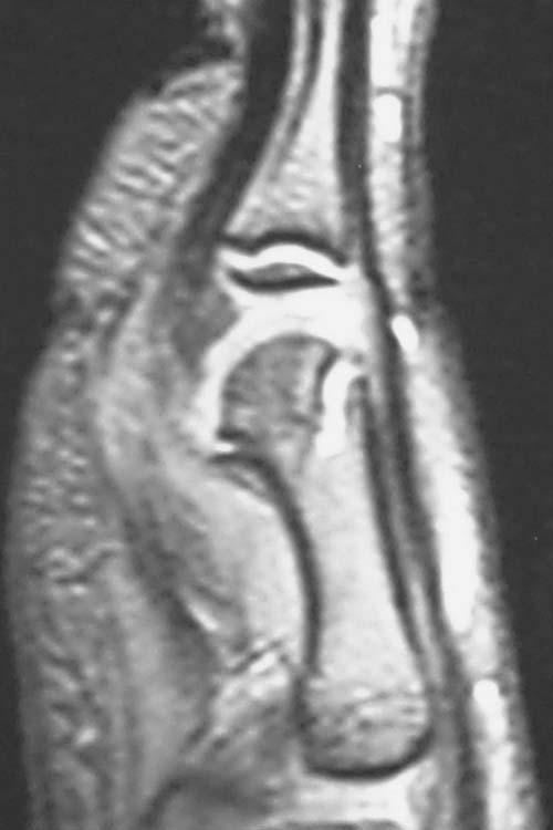
35 Applications: Physeal Injury MR imaging accurately depicts the physeal anatomy Cartilage sensitive sequences are especially useful in mapping physeal bars
36
37
38
39
40

41 Flexor Tendon Zones I between FDS and FDP attachments II FDS attachment to palmar fold III A1 pulley to retinaculum IV carpal tunnel V forearm proximal to retinaculum I II III
42 Flexor Tenosynovitis MR imaging useful Increased signal within the tendon sheath fluid sensitive sequences Enhancement Post-gadolinium images

43 Flexor Tenosynovitis Cortisone injections injected directly into the region of concern 90% improve with non-operative treatment
44 Tendon Evaluation Normal tendons demonstrate low signal intensity on all pulse sequence MR accurately depicts tendon morphology and the gap for severed tendons MR is also useful for pulley injuries.

45 Diagnosis Clinical Exam Evaluation of FDS (examine individual because of separate muscle slip to each tendon)

46 Diagnosis Clinical Exam Evaluation of FDP ( Examine together since tendons share common muscle belly, FDP to index separate)

47 Diagnosis Clinical Exam Squeeze test- squeeze volar mid-forearm and assess flexion of digits
 Squeeze test- squeeze volar mid-forearm and assess flexion of digits Tenodesis effect: fingers should flex with passive wrist")
48 Diagnosis Clinical Exam Observation- Cascade effect of digits Evaluation of FDS (examine individual because of separate muscle slip to each tendon) Evaluation of FDP ( Examine together since tendons share common muscle belly, FDP to index separate) Squeeze test- squeeze volar mid-forearm and assess flexion of digits Tenodesis effect: fingers should flex with passive wrist extension
49 Flexor Digitorum Profundus Avulsion Often missed or ignored because flexion at PIP and MCP still intact Young male athlete, ring finger most common
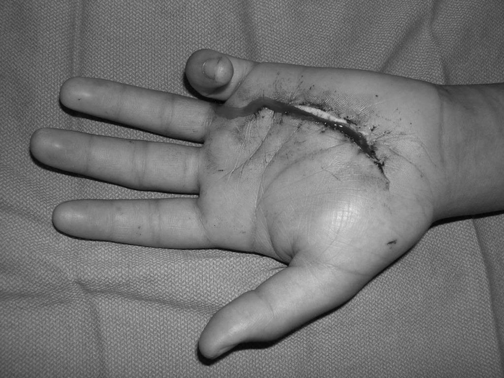
50 Flexor Tendon Tears Commonly result from sports-related injuries May occur anywhere along the course of the tendons Localized pain and swelling Inability to flex the IP joints

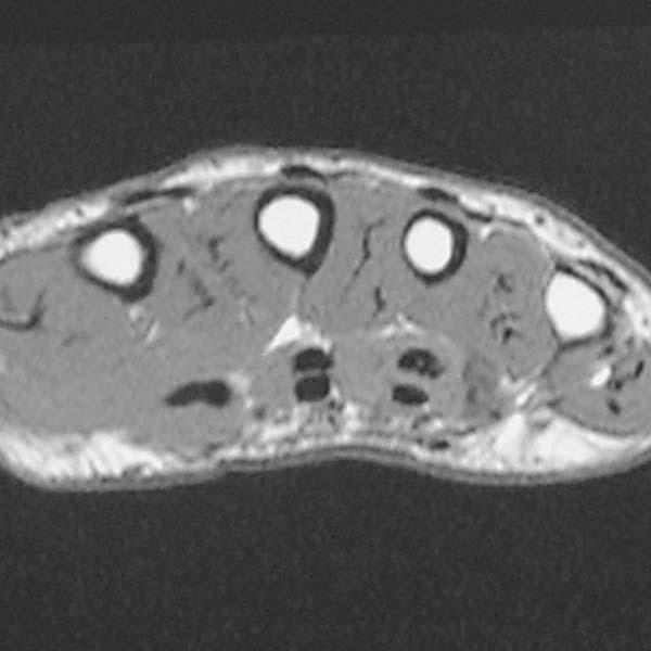
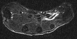
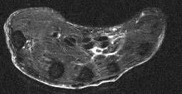
51 Flexor Tendon Tears Often difficult to diagnose and fully characterize clinically MR imaging - a noninvasive technique to identify: Site of tear Degree of retraction of torn fibers
52


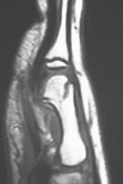
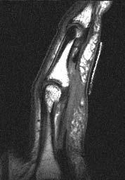
53 Flexor digitorum profundus and superficialis rupture
54
55

56 Flexor Digitorum Profundus Avulsion Leddy and Packer classification: Type I: Tendon retracts into palm- repair within 7-10 days Courtesy of Dr. Peter Murray
57
58
59 Flexor Digitorum Profundus Avulsion Leddy and Packer classification: Type II: Small bony fragment avulsedusually trapped proximally at A3 pulleyrepair in first 6 wks possible III- large bony fragment avulsed- usually trapped at A4 pulley- ORIF From Strickland J, Green s Hand Surgery

60 Courtesy of Dr. Peter Murray

61 Courtesy of Dr. Peter Murray

62 Courtesy of Dr. Peter Murray
63 Flexor Digitorum Profundus Avulsion Leddy and Packer classification: Type III- large bony fragment avulsed- usually trapped at A4 pulley- ORIF From Strickland J, Green s Hand Surgery

64 Flexor Pulley System Injury Flexor pulley system is divided into: 5 annular pulleys 3 cruciform pulleys Numbered from proximal to distal

65 Flexor Pulley System Injury A2 pulley - most important to flexor tendon function Injury typically begins with the A2 pulley Followed sequentially by the A3 and A4 pulley Rarely - A1 pulley

66 Flexor Pulley System Injury Injuries are seen in rock climbers and in other sports resulting in forced extension of a flexed finger Account for approximately 30% of all hand injuries in rock climbers Schoffl VR et al. Injuries to the finger flexor pulley system in rock climbers. J Hand Surg 2006; 31:

67 Flexor Pulley System Injury Crimp Position DIP joints - extended PIP joints flexed MCP joints extended Carpus slightly extended

68 Flexor Pulley System Injury Hanging Finger Position Flexed DIP joints PIP joints MCP joints Schoffl VR et al. Injuries to the finger flexor pulley system in rock climbers. J Hand Surg 2006; 31:

69 Pulley Injury - Grades I pulley strain II - A4 rupture or partial rupture A2/A3 III A2/A3 rupture IV Multiple ruptures Single rupture with lumbrical ms or collateral ligament injury Schoffl VR et al. Injuries to the finger flexor pulley system in rock climbers. J Hand Surg 2006; 31:
70 MRI (3T) A2 PULLEY RUPTURES Sensitivity: 87.5 % Specificity: 100 % Positive predictive value (PPV): 100 % Negative predictive value (NPV): 95.2 %
71
72

73 Courtesy of Dr. Peter Murray Reconstruct pulley


74 Courtesy of Dr. Peter Murray Reconstruct pulley


75 Courtesy of Dr. Peter Murray Reconstruct flexor tendon


76 Courtesy of Dr. Peter Murray Reconstruct flexor tendon

77 Courtesy of Dr. Peter Murray

78 Courtesy of Dr. Peter Murray
79 THUMB
80 Extensor pollicis longus/brevis Normal anatomy thumb
81 flexor pollicis longus Normal anatomy thumb


82 pulleys Normal anatomy thumb
83 flexor pollicis longus Normal anatomy thumb
84 Adductor aponeurosis Ulnar collateral ligament Normal anatomy thumb


85 Normal anatomy thumb radial collateral ligament

86 IML=Intermetacarpal lig. POL=Posterior oblique lig. DRL=Dorsoradial lig. ECRL=Ext. carpi radialis longus APL=Abductor pollicis longus

87 Bennett fracture 2 part intra-articular fracture/dislocation of base of 1st metacarpal Small fragment of 1st metacarpal continues to articulate with trapezium Lateral retraction of 1st metacarpal by abductor pollicis longus
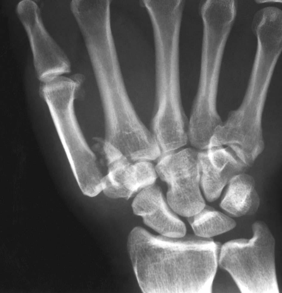
88

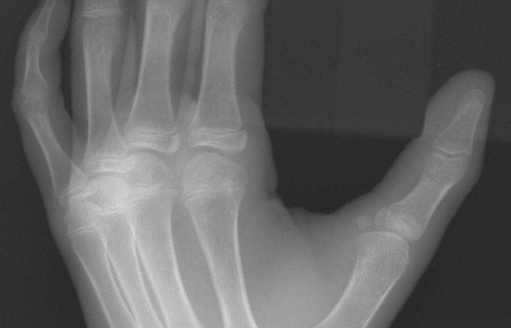
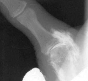
89 Rolando Fracture Originally described - Y-shaped 3-fragment fracture Extended to the articular surface Today the eponym is widely used for any comminuted intra-articular fracture at the base of the thumb
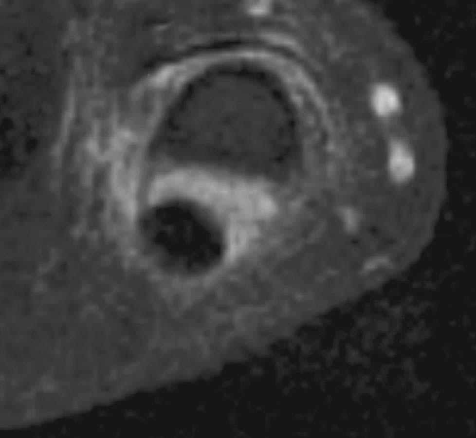
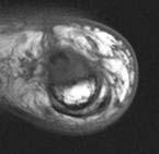
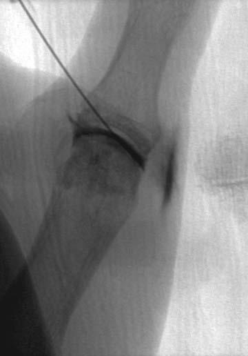
90 Gamekeeper s Thumb Disruption of the ulnar collateral ligament of the 1 st MCP joint Result of an acute radial or valgus stress on the thumb
91 Gamekeeper s Thumb Injury can occur in the form of: An avulsion fracture Isolated ligament rupture Combined fracture and ligament tear

92 Gamekeeper s Thumb Commonly referred to as Skier s Thumb Most commonly seen in snow skier s Fall holding a ski pole causing forced abduction and extension of the thumb

93 Gamekeeper s Thumb Radiographs may be useful May depict a small avulsion fracture Ulnar aspect of the base of the 1 st proximal phalanx Attachment of the UCL
94

95 Gamekeeper s Thumb Stress radiographs Neutral radiographs Radiographs with abduction and extension Greater than 30 degrees difference Abnormal - UCL disruption

96

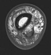
97 Gamekeeper s Thumb MR or MR arthrography Accurately demonstrate the osseous and soft tissue structures about the MCP joint Including the UCL
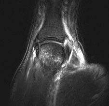
98
99
100
101 Gamekeeper s thumb If the fracture fragment is nondisplaced - splinting of the thumb may lead to healing and restoration of joint stability In most patients surgical repair is preferable
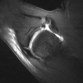
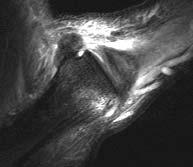
102 Stener Lesion Torn UCL displaces superficial to the adductor aponeurosis Prevents spontaneous ligament healing 29% of UCL injuries

103 Stener Lesion MR imaging can depict UCL Adductor Aponeurosis Operative intervention Normal anatomic apposition Healing of the displaced UCL
104
105 UCL UCL


106 UCL Adductor aponeurosis UCL Adductor aponeurosi

107 Stener Lesion If a Stener lesion is present Only operative intervention Normal anatomic apposition Healing of the displaced UCL


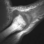
108 FSE PD FSE T2 FS RCL avulsion
109 FSE PD FSE T2 FS RCL avulsion


110 FSE PD FSE T2 FS RCL avulsion
111 Conclusion Knowledge of the mechanism of injury and various injury patterns of osseous and soft tissue injury may direct the appropriate imaging studies for the fingers and thumb Imaging features of infection Review of the most common benign and malignant tumors of the fingers and thumb
5/8/2017. Finger Injuries in Football. Tendon Injuries of the Hand and Wrist in Football Steve Kronlage, MD Andrews Institute Gulf Breeze, Florida
 Finger Injuries in Football Tendon Injuries of the Hand and Wrist in Football Steve Kronlage, MD Andrews Institute Gulf Breeze, Florida A jammed finger is an injury (at very least a torn ligament) A swollen
FINGER INJURIES. Chapter 24, pgs ,
 FINGER INJURIES Chapter 24, pgs 727 730, 741 743 1. Demonstrate mastery of anatomical references to the hand and fingers. 2. Compare and contrast Mallet Finger, Swan Neck Deformity and Boutonnière Deformity.
Hand injuries. The metacarpal bones may fracture through the base, shaft or the neck.
 Hand injuries Metacarpal injuries The metacarpal bones may fracture through the base, shaft or the neck. Shaft fractures; these are caused by direct trauma which may cause transverse # of one or more metacarpal
Musculoskeletal Imaging of the Digits. Arash David Tehranzadeh, MD UCSD MSK Radiology May 11 th, 2006
 Musculoskeletal Imaging of the Digits Arash David Tehranzadeh, MD UCSD MSK Radiology May 11 th, 2006 Musculoskeletal Imaging of the Digit Anatomy & Internal Derangement The Extensor System The Flexor System
Assessing hand ligaments and tendons lesions using MRI
 Assessing hand ligaments and tendons lesions using MRI Award: Certificate of Merit Poster No.: C-0691 Congress: ECR 2017 Type: Educational Exhibit Authors: A. M. Benitez Vazquez, M. I. Rossi Prieto, C.
Introduction to Ultrasound Examination of the Hand and upper
 Introduction to Ultrasound Examination of the Hand and upper Emil Dionysian, M.D. Ultrasound of upper ext. Upside Convenient Opens another exam dimension Can be like a stethoscope Helps 3-D D visualization
Fractures and dislocations of the fingers
 Chapter 1 Fractures and dislocations of the fingers Felix S. Chew, M.D., and Catherine Maldjian, M.D. Case 1 1 Phalangeal tuft avulsion fracture 31-year-old woman injured in a ground-level fall. Lateral
Kinesiology of The Wrist and Hand. Cuneyt Mirzanli Istanbul Gelisim University
 Kinesiology of The Wrist and Hand Cuneyt Mirzanli Istanbul Gelisim University Bones The wrist and hand contain 29 bones including the radius and ulna. There are eight carpal bones in two rows of four to
Wrist and Hand Anatomy
 Wrist and Hand Anatomy Bone Anatomy Scapoid Lunate Triquetrium Pisiform Trapeziod Trapezium Capitate Hamate Wrist Articulations Radiocarpal Joint Proximal portion Distal portion Most surface contact found
Main Menu. Wrist and Hand Joints click here. The Power is in Your Hands
 1 The Wrist and Hand Joints click here Main Menu K.5 http://www.handsonlineeducation.com/classes/k5/k5entry.htm[3/23/18, 1:40:40 PM] Bones 29 bones, including radius and ulna 8 carpal bones in 2 rows of
Physical therapy of the wrist and hand
 Physical therapy of the wrist and hand Functional anatomy wrist and hand The wrist includes distal radius, scaphoid, lunate, triquetrum, pisiform, trapezium, trapezoid, capitate, and hamate. The hand includes
Clinical Orthopaedic Rehabilitation Volume 1 and 2
 Clinical Orthopaedic Rehabilitation Volume 1 and 2 COURSE DESCRIPTION This program is a practical, clinical guide that provides guidance on the evaluation, differential diagnosis, treatment, and rehabilitation
Hand & Wrist Injuries. DR MA Manjra
 Hand & Wrist Injuries DR MA Manjra 1 Background Up to 25% of all athletic injuries General population Sport people Sport specific Position specific Multifaceted Time of season Level of athlete Parents
Index. radiologic.theclinics.com. Note: Page numbers of article titles are in boldface type.
 Index Note: Page numbers of article titles are in boldface type. A Acromioclavicular joint injuries in football players, 318, 319 ALPSA. See Anterior labroligamentous periosteal sleeve avulsion. Anterior
ARM Brachium Musculature
 ARM Brachium Musculature Coracobrachialis coracoid process of the scapula medial shaft of the humerus at about its middle 1. flexes the humerus 2. assists to adduct the humerus Blood: muscular branches
Trapezium is by the thumb, Trapezoid is inside
 Trapezium is by the thumb, Trapezoid is inside Intercarpal Jt Radiocarpal Jt Distal Middle Proximal DIP PIP Interphalangeal Jts Metacarpalphalangeal (MCP) Jt Metacarpal Carpometacarpal (CMC) Jt Trapezium
10/10/2014. Structure and Function of the Hand. The Hand. Osteology of the Hand
 Structure and Function of the Hand 19 bones and 19 joints are necessary to produce all the motions of the hand The Hand Dorsal aspect Palmar aspect The digits are numbered 1-5 Thumb = #1 Little finger
Muscles of the hand Prof. Abdulameer Al-Nuaimi
 Muscles of the hand Prof. Abdulameer Al-Nuaimi a.alnuaimi@sheffield.ac.uk abdulameerh@yahoo.com Thenar Muscles Thenar muscles are three short muscles located at base of the thumb. All are innervated by
CASE ONE CASE ONE. RADIAL HEAD FRACTURE Mason Classification. RADIAL HEAD FRACTURE Mechanism of Injury. RADIAL HEAD FRACTURE Imaging
 CASE ONE An eighteen year old female falls during a basketball game, striking her elbow on the court. She presents to your office that day with a painful, swollen elbow that she is unable to flex or extend
MCQWeek2. All arise from the common flexor origin. The posterior aspect of the medial epicondyle is the common flexor origin.
 MCQWeek2. 1. Regarding superficial muscles of anterior compartment of the forearm: All arise from the common flexor origin. The posterior aspect of the medial epicondyle is the common flexor origin. Flexor
Hand Anatomy A Patient's Guide to Hand Anatomy
 Hand Anatomy A Patient's Guide to Hand Anatomy Introduction Few structures of the human anatomy are as unique as the hand. The hand needs to be mobile in order to position the fingers and thumb. Adequate
divided by the bones ( redius and ulna ) and interosseous membrane into :
 and interosseous membrane into :") fossa Cubital Has: * floor. * roof : - Skin - superficial fasica - deep fascia ( include bicipital aponeurosis ) Structures within the roof : -cephalic and basilic veins -and between them median cubital
fossa Cubital Has: * floor. * roof : - Skin - superficial fasica - deep fascia ( include bicipital aponeurosis ) Structures within the roof : -cephalic and basilic veins -and between them median cubital
1. A 40-year-old male has dislocated his right 2 nd MCP. You have pulled as hard as you can but cannot reduce the dislocation. The problem is likely:
 CHAPTER 50 HAND 2 OCTOBER 2013 1. A 40-year-old male has dislocated his right 2 nd MCP. You have pulled as hard as you can but cannot reduce the dislocation. The problem is likely: A. He is a gamer and
Ligaments of Elbow hinge: sagittal plane so need lateral and medial ligaments
 Ligaments of Elbow hinge: sagittal plane so need lateral and medial ligaments Ulnar Collateral ligament on medial side; arising from medial epicondyle and stops excess valgus movement (lateral movement)
Trigger Digits, Mallet Finger & Metacarpal Injuries. Joseph P. McCormick, M.D. Affinity Orthopaedics & Sports Medicine 2013
 Trigger Digits, Mallet Finger & Metacarpal Injuries Joseph P. McCormick, M.D. Affinity Orthopaedics & Sports Medicine 2013 Overview Trigger Digits: diagnosis and treatment Bonus: approach in children Mallet
This presentation is the intellectual property of the author. Contact them for permission to reprint and/or distribute.
 The Stiff Hand: Boutonniere & Sylvia Dávila, PT, CHT San Antonio, Texas Extensor Mechanism Central slip inserts into base of the middle phalanx Lateral bands lie dorsal to the PIP joint center of rotation
Nerves of Upper limb. Dr. Brijendra Singh Professor & Head Department of Anatomy AIIMS Rishikesh
 Nerves of Upper limb Dr. Brijendra Singh Professor & Head Department of Anatomy AIIMS Rishikesh 1 Objectives Origin, course & relation of median & ulnar nerves. Motor & sensory distribution Carpal tunnel
Pediatric Musculoskeletal Ultrasound: Cases reviewed and lessons learned
 Pediatric Musculoskeletal Ultrasound: Cases reviewed and lessons learned Jessica Leschied, MD Sections of Pediatric and Musculoskeletal Radiology C.S. Mott Children s Hospital University of Michigan Ann
ACUTE HAND INJURIES FOR THE PRIMARY CARE PHYSICIAN
 Vincent Shaw, MD, FAAFP Program Director Baton Rouge General Family Medicine Residency Program Baton Rouge General Sports Medicine Fellowship ACUTE HAND INJURIES FOR THE PRIMARY CARE PHYSICIAN Disclosures
Biceps Brachii. Muscles of the Arm and Hand 4/4/2017 MR. S. KELLY
 Muscles of the Arm and Hand PSK 4U MR. S. KELLY NORTH GRENVILLE DHS Biceps Brachii Origin: scapula Insertion: radius, fascia of forearm (bicipital aponeurosis) Action: supination and elbow flexion Innervation:
Wrist & Hand Ultrasonography 대구가톨릭대학교병원재활의학과 권동락
 Wrist & Hand Ultrasonography 대구가톨릭대학교병원재활의학과 권동락 Dorsal Wrist Evaluation (1 st Compartment) EPB APL Transverse View APL, abductor pollicis longus; EPB, extensor pollicis brevis Dorsal Wrist Evaluation
Wrist and Hand Complaints
 Wrist and Hand Complaints Charles S. Day, M.D., M.B.A. Chief, Hand & Upper Extremity Surgery St. Elizabeth s Medical Center Tufts University School of Medicine Primary Care Internal Medicine 2018 Outline
Anatomy of the Hand and Nomenclature. R K Kankate Specialist Registrar St.George s Hospital
 Anatomy of the Hand and Nomenclature R K Kankate Specialist Registrar St.George s Hospital Bony skeleton muscles and ligaments nervous, arterial and venous system Ossification of bones: carpus Most carpal
Wrist and Hand Anatomy/Biomechanics
 Wrist and Hand Anatomy/Biomechanics Kristin Kelley, DPT, OCS, FAAOMPT Orthopaedic Manual Physical Therapy Series Charlottesville 2017-2018 Orthopaedic Manual Physical Therapy Series 2017-2018 Anatomy -
Anatomy - Hand. Wrist and Hand Anatomy/Biomechanics. Osteology. Carpal Arch. Property of VOMPTI, LLC
 Wrist and Hand Anatomy/Biomechanics Kristin Kelley, DPT, OCS, FAAOMPT The wrist The metacarpals The Phalanges Digit 1 thumb Digit 5 digiti minimi Anatomy - Hand Orthopaedic Manual Physical Therapy Series
The Forearm, Wrist, Hand and Fingers. Contusion Injuries to the Forearm. Forearm Fractures 12/11/2017. Oak Ridge High School Conroe, Texas
 The Forearm, Wrist, Hand and Fingers Oak Ridge High School Conroe, Texas Contusion Injuries to the Forearm The forearm is constantly exposed to bruising and contusions in contact sports. The ulna receives
The hand is full with sweat glands, activated at times of stress. In Slide #2 there was a mistake where the doctor mentioned lateral septum twice.
 We should only know: Name, action & nerve supply Layers - Skin - Superficial fascia - Deep fascia The hand is full with sweat glands, activated at times of stress. Deep fascia In Slide #2 there was a mistake
THE EPIDEMIOLOGY OF HAND EMERGENCIES
 THE EPIDEMIOLOGY OF HAND EMERGENCIES Dr. Adel Abdel Aziz Senior Emergency Physician Honorary Senior Clinical Lecturer, University of Southampton Training Program Director Emergency Medicine/ Health Education
Clinical examination of the wrist, thumb and hand
 Clinical examination of the wrist, thumb and hand 20 CHAPTER CONTENTS Referred pain 319 History 319 Inspection 320 Functional examination 320 The distal radioulnar joint.............. 320 The wrist.......................
Hand and Wrist Editing file. Color Code Important Doctors Notes Notes/Extra explanation
 Hand and Wrist Editing file Color Code Important Doctors Notes Notes/Extra explanation Objectives Describe the anatomy of the deep fascia of the wrist & hand (flexor & extensor retinacula & palmar aponeurosis).
Exam of the Injured Hand and Wrist. Christina M. Ward, MD Regions Hospital TRIA Woodbury
 Exam of the Injured Hand and Wrist Christina M. Ward, MD Regions Hospital TRIA Woodbury Disclosures We have no disclosures that are pertinent to this presentation Terminology Ring Long Index Small Thumb
Elbow/Wrist/Hand Pointers
 Elbow/Wrist/Hand Pointers Elbow Injuries -break elbow into 4 quadrants -Lateral -Medial -Posterior -Anterior 1. Lateral Epicondylosis (Tennis Elbow) a. extensor supinator tendinopathy b. repetitive gripping/wrist
Hand & Wrist Casey G. Batten MD Assistant Clinical Professor UCSF Sports Medicine
 Hand & Wrist Casey G. Batten MD Assistant Clinical Professor UCSF Sports Medicine Topics: Scaphoid Fracture Scapholunate Separation TFCC Injury Thumb Ulnar Collateral Lig (UCL) Injury Extensor Injury /
RHEUMATOID HAND. History Pain Loss of function Neck pain. Diminished ADL assessment:
 RHEUMATOID HAND History Pain Loss of function Neck pain Diminished ADL assessment: Using toothbrush, hairbrush, knife, fork Dressing bra, Pulling up trousers / stockings Operating remote control Hobbies
Wrist & Hand Assessment and General View
 Wrist & Hand Assessment and General View Done by; Mshari S. Alghadier BSc Physical Therapy RHPT 366 m.alghadier@sau.edu.sa http://faculty.sau.edu.sa/m.alghadier/ Functional anatomy The hand can be divided
Ulnar Collateral Ligament Injuries of the Thumb Game Keeper s Thumb A Patient's Guide to Ulnar Collateral Ligament Injuries of the Thumb
 Ulnar Collateral Ligament Injuries of the Thumb Game Keeper s Thumb A Patient's Guide to Ulnar Collateral Ligament Injuries of the Thumb 1 Introduction Injury to the ulnar collateral ligament of the thumb
Andrew B. Stein, MD Boston University Medical Center May 2 & 3, 2016
 Andrew B. Stein, MD Boston University Medical Center andrew.stein@bmc.org Work Related Workshop WorkInjuries Related Injuries Workshop Tendon injuries may be obvious or subtle History (mechanism of injury)
Upper Limb- Sports Medicine II
 Upper Limb- Sports Medicine II I. Palpation A. With patient sitting, supine, & prone, palpate for pain, specific tenderness, swelling, effusion, local hyperthermia B. Bony Palpation 1. Carpal Bones (8)
The hand. it's the most important subject of the upper limb because it has a clinical importance. the palm of the hand**
 Today at 12:48 AM The hand it's the most important subject of the upper limb because it has a clinical importance. the palm of the hand** -the palmar aponeurosis located in the palm of the hand which is
Small muscles of the hand
 By the name of Allah Small muscles of the hand Revision: The palmar aponeurosis is triangular in shape with apex and base. It is divided into 4 bands that radiate to the medial four fingers. Dupuytren
Common. Common Hand Problems in Elite Athletes
 Common Hand Problems in Elite Athletes Fred Corley M.D. Dept. of Orthopaedic Surgery UTHSCSA I have no disclosures concerning this talk. The University of Texas Health Science Center @ San Antonio - Orthopaedics
HAND EXAMINATION & COMMON INJURIES OF THE HAND. Majoring in Minors Conference th January 2013 Derriford Hospital
 HAND EXAMINATION & COMMON INJURIES OF THE HAND Majoring in Minors Conference 16-17 th January 2013 Derriford Hospital Objectives Anatomy & Terminology History Examination Look Feel Move Investigations
Fractures of the Hand in Children Which are simple? And Which have pitfalls??
 Fractures of the Hand in Children Which are simple? And Which have pitfalls?? Kaye E Wilkins DVM, MD Professor of Orthopedics and Pediatrics Departments of Orthopedics and Pediatrics University of Texas
The Forearm 2. Extensor & lateral Compartments of the Forearm
 The Forearm 2 Extensor & lateral Compartments of the Forearm 1-Lateral Fascial Compartment (at the lateral side of the forearm ) *Some books mention the lateral compartment contain just the Brachioradialis
DISCLOSURE LEARNING OBJECTIVES
 1 THE HAND & WRIST EXAM Matthew Silvis, MD Departments of Family and Community Medicine & Orthopedics and Rehabilitation PAFP Chesapeake Escape CME Conference July 28 th, 2015 DISCLOSURE I have no financial
The Elbow and the cubital fossa. Prof Oluwadiya Kehinde
 The Elbow and the cubital fossa Prof Oluwadiya Kehinde www.oluwadiya.com Elbow and Forearm Anatomy The elbow joint is formed by the humerus, radius, and the ulna Bony anatomy of the elbow Distal Humerus
Dynamic 22 Mhz ultrasound evaluation (HR-US) of the finger: a detailed didactic approach.
 of the finger: a detailed didactic approach.") Dynamic 22 Mhz ultrasound evaluation (HR-US) of the finger: a detailed didactic approach. Poster No.: C-2228 Congress: ECR 2014 Type: Educational Exhibit Authors: A. Muda, D. Orlandi, V. Prono, S. Migone,
Dynamic 22 Mhz ultrasound evaluation (HR-US) of the finger: a detailed didactic approach. Poster No.: C-2228 Congress: ECR 2014 Type: Educational Exhibit Authors: A. Muda, D. Orlandi, V. Prono, S. Migone,
Revisiting the Curtis Procedure for Boutonniere Deformity Correction
 180 Letter to Editor Revisiting the Curtis Procedure for Boutonniere Deformity Correction Lee Seng Khoo*, Vasco Senna-Fernandes Ivo Pitanguy Institute, Rua Dona Mariana 65, Botafogo, Rio De Janeiro, Brazil
1 Comprehensive Orthopaedic Review Hand and Wrist Tendinopathies Bernard F. Hearon, MD September 2, de Quervain Disorder Fritz de Quervain,
 1 Comprehensive Orthopaedic Review Hand and Wrist Tendinopathies Bernard F. Hearon, MD September 2, 2016 2 de Quervain Disorder Fritz de Quervain, Swiss surgeon, 1895 Stenosing tendinosis, 1st dorsal compartment
Spectrum Of MRI Findings In Sports And Work Related Injuries Of Fingers And Thumb
 Spectrum Of MRI Findings In Sports And Work Related Injuries Of Fingers And Thumb Poster No.: P-0127 Congress: ESSR 2016 Type: Scientific Poster Authors: G. Malik; Umm Al Quwain/AE Keywords: Anatomy, Musculoskeletal
Forearm and Wrist Regions Neumann Chapter 7
 Forearm and Wrist Regions Neumann Chapter 7 REVIEW AND HIGHLIGHTS OF OSTEOLOGY & ARTHROLOGY Radius dorsal radial tubercle radial styloid process Ulna ulnar styloid process ulnar head Carpals Proximal Row
Structure and Function of the Hand
 Structure and Function of the Hand Some say it takes a village to raise a child, but it takes 19 bones and 19 joints in the hand for it to function smoothly. The Hand Dorsal aspect 2 3 4 The digits are
31yo M with chronic basilar thumb and wrist pain that started after cross-country bicycle ride 5 yrs ago.
 31yo M with chronic basilar thumb and wrist pain that started after cross-country bicycle ride 5 yrs ago. EPL EPB APL Full-thickness tear involving the dorsal deltoid ligament of the first carpometacarpal
Office Orthopedics. No conflict of interest No financial disclosures 1/31/2018
 Office Orthopedics Amin Afsari DO Orthopedic Hand and Upper Extremity Surgery Orthopedic Institute of Wisconsin Midwest Orthopedic Specialty Hospital 1 No conflict of interest No financial disclosures
SPECIAL ARTICLE. Missed tendon injuries INTRODUCTION
 Archives of Emergency Medicine, 1991, 8, 87-91 SPECIAL ARTICLE Missed tendon injuries H. R. GULY Consultant in A & E, Derriford Hospital, Plymouth INTRODUCTION The timing of the repair of divided tendons
PIP Joint Injuries of the Finger A Patient's Guide to PIP Joint Injuries of the Finger
 PIP Joint Injuries of the Finger A Patient's Guide to PIP Joint Injuries of the Finger Introduction We use our hands constantly, placing them in harm's way continuously. Injuries to the finger joints are
[[Sally Leaning Towards Peter To Take Cold Hand]]
![[[Sally Leaning Towards Peter To Take Cold Hand]]](/thumbs/84/91174469.jpg "[[Sally Leaning Towards Peter To Take Cold Hand]]") In this lecture we will talk about the bones of the hand, and the muscles and contents of the forearm. *The hand bones are: - Carpal bones. -Metacarpals. -Phalanges. *The carpal bones (wrist bones): They
In this lecture we will talk about the bones of the hand, and the muscles and contents of the forearm. *The hand bones are: - Carpal bones. -Metacarpals. -Phalanges. *The carpal bones (wrist bones): They
The Muscular System. Chapter 10 Part C. PowerPoint Lecture Slides prepared by Karen Dunbar Kareiva Ivy Tech Community College
 Chapter 10 Part C The Muscular System Annie Leibovitz/Contact Press Images PowerPoint Lecture Slides prepared by Karen Dunbar Kareiva Ivy Tech Community College Table 10.9: Muscles Crossing the Shoulder
13 13/3/2012. Adel Muhanna
 13 13/3/2012 Adel Muhanna بسم هللا الرحمن الرحيم The Hand Extensor retinaculum: Deep fascia of anterior compartment of the wrist is thickened to form flexor retinaculum : a bridge that have 6 structures
Objectives. How your brain sees your hand. Surface anatomy Bones Joints Muscles Tendons Nerves Arteries
 Hand Therapy Review Course Washington University St. Louis, MO April 7 9, 2017 Over eighty percent of activities of daily living involve grasping or seizing objects with the hand. (Katz et al., 1970) Anatomy
Lab Activity 11: Group II
 Lab Activity 11: Group II Muscles Martini Chapter 11 Portland Community College BI 231 Origin and Insertion Origin: The place where the fixed end attaches to a bone, cartilage, or connective tissue. Insertion:
Shaun P. Garff, DO Physician of Sports Medicine
 Shaun P. Garff, DO Physician of Sports Medicine Speaker Disclosure I have no actual or potential conflicts of interest to disclose as it relates to this presentation. A little about me Born and raised
Elbow, Wrist & Hand Evaluation.
 Elbow, Wrist & Hand Evaluation www.fisiokinesiterapia.biz Common Injuries to the Elbow, Wrist, Hand & Fingers Lateral epicondylitis tennis elbow Medial epicondylitis golfer s s elbow, little league elbow
compartments of the forearm
 " forearm posterior compartment " compartments of the forearm Posterior Fascial compartment Muscles: ** The superficial group 1. Extensor carpi radialis brevis 2. Ex. digitorum 3. Ex. digiti minimi 4.
Wrist and Hand Injuries in the Athlete
 University of Kentucky UKnowledge Rehabilitation Sciences Faculty Publications Rehabilitation Sciences 2001 Wrist and Hand Injuries in the Athlete Timothy L. Uhl University of Kentucky, tluhl2@uky.edu
Management of Wrist and Hand Injuries
 Sunday General Session Management of Wrist and Hand Injuries Shaun Garff, DO Sports Medicine Physician Methodist Health System Dallas, Texas Educational Objectives By the end of this educational activity,
Classification of Established Volkmann s Ischemic Contracture and the Program for Its Treatment
 10 Classification of Established Volkmann s Ischemic Contracture and the Program for Its Treatment In spite of the advances made in preventive treatment of muscular ischemia at the forearm and hand, there
What you don t want to miss
 March 25, 2009 Vishal Michael Shah, M.D. What you don t want to miss Spectrum of Injuries Contusions Sprains Dislocations Fractures Lacerations Tendon Avulsions Ligament Tears Overuse Injuries FINGER
STRUCTURAL BASIS OF MEDICAL PRACTICE EXAMINATION 5 October 6, 2006
 STRUCTURAL BASIS OF MEDICAL PRACTICE EXAMINATION 5 October 6, 2006 PART l. Answer in the space provided. (8 pts) 1. Identify the structures. (2 pts) B C A. _pisiform B. _ulnar artery A C. _flexor carpi
Hand and wrist emergencies
 Chapter1 Hand and wrist emergencies Carl A. Germann Distal radius and ulnar injuries PEARL: Fractures of the distal radius and ulna are the most common type of fractures in patients younger than 75 years.
HAND INJURY REHAB CONCEPTS AND RETURN TO PLAY
 HAND INJURY REHAB CONCEPTS AND RETURN TO PLAY DAVID COLVIN, PT, DPT, OCS, MS, ATC Presentation Overview Discuss common hand and finger injuries/rehabilitation in baseball UCL of the Thumb Tear Rehab comparisons
LECTURE 8 HANDS: BONES AND MUSCLES
 LECTURE 8 HANDS: BONES AND MUSCLES WRIST AND HAND - Human hand can do power grip and precision grip - Thumb is 90 to the rest of the hand can do fine actions - Often able to do power actions o Take tools
The Rheumatoid Hand Deformities & Management. Dr. Anirudh Sharma Resident Department of Orthopedics
 + The Rheumatoid Hand Deformities & Management Dr. Anirudh Sharma Resident Department of Orthopedics + Why is Rheumatoid Arthritis important? + RA is a very debilitating disease median life expectancy
Swan-Neck Deformity. Introduction. Anatomy
 Swan-Neck Deformity Introduction Normal finger position and movement occur from the balanced actions of many important structures. Ligaments support the finger joints. Muscles hold and move the fingers.
Interesting Case Series. Zone I Flexor Tendon Injuries
 Interesting Case Series Zone I Flexor Tendon Injuries Evgenios Evgeniou, MBBS, MRCS, a and Harriet Walker, MBBS, MRCS b a North Bristol NHS Trust, Bristol, United Kingdom, b Plymouth Hospitals NHS Trust,
Acknowledgements. Methods of Learning. The Dorsal Interossei 11/2/17. Special thanks to:
 The Dorsal Interossei Mike Cricchio, MBA, OT/L, CHT Site Manager UFHealth Hand and Upper Extremity criccm@gmail.com Acknowledgements Special thanks to: Michelle Darnell, DPT, PT Kimberly Hellriegel, OTR/L
Supplied in part by the musculocutaneous nerve. Forms the axis of rotation in movements of pronation and supination
 Anatomy: Upper limb (15 questions) 1. Latissimus Dorsi: Is innervated by the dorsal scapular nerve Lies above feres major muscle Medially rotates the humerus All of the above 2. Supinator muscle is: Deep
Nerves of the upper limb Prof. Abdulameer Al-Nuaimi. E. mail:
 Nerves of the upper limb Prof. Abdulameer Al-Nuaimi E-mail: a.al-nuaimi@sheffield.ac.uk E. mail: abdulameerh@yahoo.com Brachial plexus Median nerve After originating from the brachial plexus in the axilla,
MLT Muscle(s) Patient Position Therapist position Stabilization Limb Position Picture Put biceps on slack by bending elbow.
 Patient Position Therapist position Stabilization Limb Position Picture Put biceps on slack by bending elbow.") MLT Muscle(s) Patient Position Therapist position Stabilization Limb Position Picture Put biceps on slack by bending elbow. Pectoralis Minor Supine, arm at side, elbows extended, supinated Head of Table
MLT Muscle(s) Patient Position Therapist position Stabilization Limb Position Picture Put biceps on slack by bending elbow. Pectoralis Minor Supine, arm at side, elbows extended, supinated Head of Table
Reversing PIP Joint Contractures:
 Reversing PIP Joint Contractures: Applicability of the Digit Widget External Fixation System John M. Agee M.D. Reversing PIP Joint Contractures: Applicability of the Digit Widget External Fixation System
MR IMAGING OF THE WRIST
 MR IMAGING OF THE WRIST Wrist Instability Dissociative Pattern apparent on routine radiographs Non-dissociative Stress / positional radiographs Dynamic fluoroscopy during stress Arthrography MRI / MR arthrography
Hand Fractures: When is closed treatment OK? Epidemiology in USA: Metacarpal fractures: Page 1
 Hand Fractures: When is closed treatment OK? Robert J Strauch MD Professor of Orthopaedic Surgery Columbia University New York City Epidemiology in USA: 2009 Distal radius fx s: 16/10,000 Phalangeal fx
Lecture 9: Forearm bones and muscles
 Lecture 9: Forearm bones and muscles Remember, the region between the shoulder and the elbow = brachium/arm, between elbow and wrist = antebrachium/forearm. Forearm bones : Humerus (distal ends) Radius
Anatomy Workshop Upper Extremity David Ebaugh, PT, PhD Workshop Leader. Lab Leaders: STATION I BRACHIAL PLEXUS
 Anatomy Workshop Upper Extremity David Ebaugh, PT, PhD Workshop Leader Lab Leaders: STATION I BRACHIAL PLEXUS A. Posterior cervical triangle and axilla B. Formation of plexus 1. Ventral rami C5-T1 2. Trunks
Wrist & Hand Injury in Sports
 Wrist & Hand Injury in Sports Jennifer Allen,PT,DPT,OCS,SCS,CHT Return to Play Criteria, Clinical Pearls, & Rehab Considerations PBATS Baseball Medicine Conference 2018 Disclosures Wrist & Hand Injury
Kristin Kelley, DPT, OCS, FAAOMPT Orthopaedic Manual Physical Therapy Series Charlottesville Trauma/Fractures
 WRIST/HAND PATHOLOGY Kristin Kelley, DPT, OCS, FAAOMPT Orthopaedic Manual Physical Therapy Series Charlottesville 2017-2018 Trauma/Fractures Hook of Hamate Fractures Triangular Fibrocartilage Complex (TFCC)
Ankle Tendons in Athletes. Laura W. Bancroft, M.D.
 Ankle Tendons in Athletes Laura W. Bancroft, M.D. Outline Protocols Normal Anatomy Tendinopathy, partial and complete tears Posterior tibial, Flexor Hallucis Longus, Achilles, Peroneal and Anterior Tibial
Trauma/Fractures WRIST/HAND PATHOLOGY. TFCC Injury. Hook of Hamate Fracture. Property of VOMPTI, LLC
 WRIST/HAND PATHOLOGY Kristin Kelley, DPT, OCS, FAAOMPT Orthopaedic Manual Physical Therapy Series Charlottesville 2017-2018 Trauma/Fractures Hook of Hamate Fractures Triangular Fibrocartilage Complex (TFCC)
FOOSH It sounded like a fun thing at the time!
 FOOSH It sounded like a fun thing at the time! Evaluating acute hand and wrist injuries Larry Collins, MPAS, PA-C, ATC, DFAAPA Assistant Professor, Physician Assistant Program Assistant Professor, Department
FOOSH It sounded like a fun thing at the time!
 FOOSH It sounded like a fun thing at the time! Evaluating acute hand and wrist injuries Larry Collins, MPAS, PA-C, ATC, DFAAPA Assistant Professor, Physician Assistant Program Assistant Professor, Department