Ankle Tendons in Athletes. Laura W. Bancroft, M.D.
|
|
|
- Brittney Strickland
- 6 years ago
- Views:
Transcription
1 Ankle Tendons in Athletes Laura W. Bancroft, M.D.
2 Outline Protocols Normal Anatomy Tendinopathy, partial and complete tears Posterior tibial, Flexor Hallucis Longus, Achilles, Peroneal and Anterior Tibial Tendons Post-surgical change of tendons
3 Protocols Conventional MRI Sagittal T1 and FSE PD FS Axial FSE PD and FSE T2 FS Coronal DESS FOV 14 cm
4 Protocols Conventional MRI Sagittal T1 and FSE PD FS Axial FSE PD and FSE T2 FS Coronal DESS FOV 14 cm
5 Protocols Conventional MRI Sagittal T1 and FSE PD FS Axial FSE PD and FSE T2 FS Coronal DESS FOV 14 cm
6 Protocols MR Arthrogram 0.1 cc Gad in 20 cc saline/lidocaine/contrast Sagittal, coronal and axial T1 FS Sagittal FSE T2 FS FOV 14 cm
7 Normal Anatomy
8 Posterior tibial t. Flexor digitorum t. Flexor hallucis longus t. FSE T2 FS
9 Anterior tibialis t. Extensor hallucis longus t. Extensor digitorum longus t. FSE T2 FS
10 Peroneal longus t. Peroneal brevis t. FSE T2 FS
11 Plantaris t. Achilles t. FSE T2 FS
12 Deltoid l. Anterior tibiofibular l. Posterior tibiofibular l. FSE T2 FS
13 Anterior talofibular l. Posterior talofibular l. FSE T2 FS
14 PTT FSE T2 FS
15 Sustentaculum talus FHL FSE T2 FS

16 FSE T2 FS Peroneus brevis t.

17 Deltoid ligament Spring ligament (dorsal calcaneonavicular)

18 PTT FDL T1W

19 Spring ligament ST FHL T1W
20 Achilles t. T1W Plantar fascia
21 Sinus tarsi T1W

22 Cervical and interosseous l. T1W

23 Root of extensor retinaculum Posterior talofibular l. T1W
24 Peroneus longus t. Peroneus brevis t. T1W

25 Anterior tibiofibular l. Posterior tibiofibular l. Anterior talofibular l. Posterior talofibular l. Calcaneofibular l.

26 Posterior intermalleolar l. Posterior inferior tibiofibular l. Transverse tibiofibular l. Deltoid l. Posterior talofibular l. Calcaneofibular l.
27 Posterior inferior tibiofibular l. Transverse tibiofibular l. Posterior talofibular l. T1W
28 Plantar fascia T1W
29 * Deltoid l. PTT FDL FHL T1W
30 Posterior Tibial Tendon Origin tibia and fibula (posteromedial borders) Attachment navicular, medial cuneiform Function inversion and plantar flexion of foot
31 Posterior Tibial Tendon Pathology Usually degenerative tendinopathy leads to tears Rare injury in athletes Sports with rapid change of direction / cutting sports PTT is subject to great stress after heel strike as hindfoot moves from loaded eversion to increasing inversion Jacoby SM.et al. Acute posterior tibial tendon tear in an ice-hockey player. Foot Ankle Intern 2008; 29: PD
32 Posterior Tibial Tendon Pathology Usually degenerative tendinopathy leads to tears Rare injury in athletes Sports with rapid change of direction / cutting sports PTT is subject to great stress after heel strike as hindfoot moves from loaded eversion to increasing inversion Jacoby SM.et al. Acute posterior tibial tendon tear in an ice-hockey player. Foot Ankle Intern 2008; 29: T1
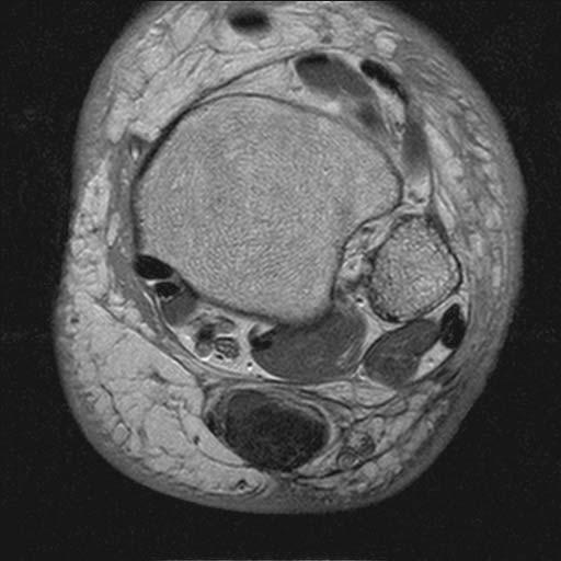
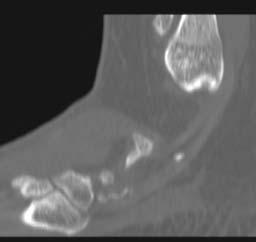
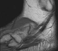
33 * * * T1W T1W Accessory navicular and PTT tendinopathy


34 * * * T1W T1W Accessory navicular and PTT tendinopathy

35 Posterior Tibial Tendon Pathology Tenosynovitis precipitated by training errors Pronated feet Direct medial ankle trauma Systematic metabolic diseases Jacoby SM.et al. Acute posterior tibial tendon tear in an ice-hockey player. Foot Ankle Intern 2008; 29: PD

36 Posterior Tibial Tendon Pathology Tenosynovitis precipitated by training errors Pronated feet Direct medial ankle trauma Systematic metabolic diseases Jacoby SM.et al. Acute posterior tibial tendon tear in an ice-hockey player. Foot Ankle Intern 2008; 29: PDT2

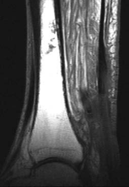
37 PD PTT thinning and tibial spurring

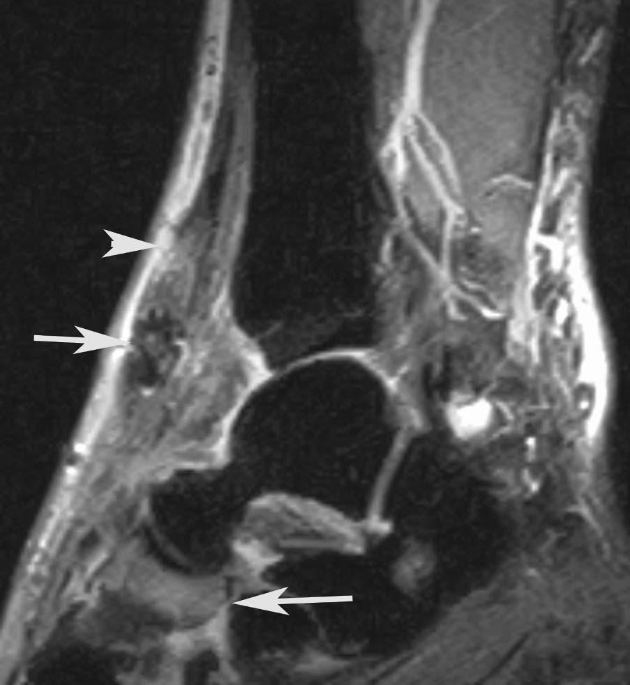
38 PTT split tear and tenosynovitis FSE T2 FS


39 T1W PTT split tear FSE T2 FS

40 PTT split tear FSE PD FS
41 Flexor Hallucis Longus Tendon Origin posteromedial 1/3 of fibula Attachment base of distal phalanx of hallux Function flexes all joints of hallux, plantar flexes ankle

42 Flexor Hallucis Longus Tenosynovitis Os Trigonum Syndrome Tenosynovitis in ballerinas dancing on point Posterior impingement due to os trigonum syndrome PD
43 Flexor Hallucis Longus Tenosynovitis Os Trigonum Syndrome Tenosynovitis in ballerinas dancing on point Posterior impingement due to os trigonum syndrome PD

44 Flexor Hallucis Longus Tenosynovitis Os Trigonum Syndrome PD T2

45 Flexor Hallucis Longus Rupture DS PD
46 Achilles Tendon Origin gastrocnemius, soleus and plantaris muscles Attachment calcaneus Function plantar flexes foot

47 Achilles Tendon Disorders 7-9% annual incidence of Achilles disorders in top-level runners Most common cause of Achilles tendon injury - training errors Sudden increase in intensity Changes of terrain or surface Changes in training schedules Inappropriate footwear DS Heckman. Tendon Disorders of the Foot and Ankle, Part 2 Achilles Tendon Disorders. Am J Sports Medicine. 2009; 37, PD
48 Achilles Tendon Disorders 7-9% annual incidence of Achilles disorders in top-level runners Most common cause of Achilles tendon injury - training errors Sudden increase in intensity Changes of terrain or surface Changes in training schedules Inappropriate footwear DS Heckman. Tendon Disorders of the Foot and Ankle, Part 2 Achilles Tendon Disorders. Am J Sports Medicine. 2009; 37, T1

49 Achilles Tendon Tears Lower extremity malalignment Hyperpronated or cavus foot Forefoot varus FSE PD FS DS Heckman. Tendon Disorders of the Foot and Ankle, Part 2 Achilles Tendon Disorders. Am J Sports Medicine. 2009; 37,


50 T1W FSE PD FS Achilles near complete rupture


51 T1W FSE PD FS Achilles rupture


52 T1W FSE PD FS Achilles rupture


53 T1W FSE T2 FS Achilles rupture

54 T1W Achilles rupture

55 T1W FSE PD FS Achilles rupture


56 T1W FSE T2 FS Achilles rupture
57 Peroneal Brevis Tendon Origin fibula (lateral lower 2/3) and intermuscular septa Attachment 5 th metatarsal base Function plantar flexion, eversion of foot
58 Peroneal Tendon Pathology Persistent lateral ankle symptoms after "typical" ankle sprain Acute tears of peroneus brevis >> longus Often coexistent with peroneal instability Subluxation Foot is dorsiflexed and peroneal muscles strongly contract = eversion force Peroneal instability and tearing Ballet dancing, skiing, soccer, tennis, American football, running, basketball and ice skating Cerrato RA et al. Peroneal tendon tears, surgical management and its complications. Foot Ankle Clin. 2009; 14:


59 T1W Peroneus brevis split tear

60 T1W Peroneus brevis split tear and peroneus longus tendinopathy
61 T1W Peroneus brevis split tear and peroneus longus tendinopathy
62 T1W Partial thickness Achilles tendon and peroneus brevis split tear

63 Partial thickness Achilles tendon and peroneus brevis split tear PD
64 Peroneal Tendon Pathology Persistent lateral ankle symptoms after "typical" ankle sprain Acute tears of peroneus brevis >> longus Often coexistent with peroneal instability Subluxation Foot is dorsiflexed and peroneal muscles strongly contract = eversion force Peroneal instability and tearing Ballet dancing, skiing, soccer, tennis, American football, running, basketball and ice skating Want XT el al. Normal variants and diseases of the peroneal tendons and superior peroneal retinaculum: MR imaging features. RadioGraphics. 2005; 25:

65 Retinacular stripping, peroneal split tear and subluxation PD Cerrato RA et al. Peroneal tendon tears, surgical management and its complications. Foot Ankle Clin. 2009; 14: Want XT el al. Normal variants and diseases of the peroneal tendons and superior peroneal retinaculum: MR imaging features. RadioGraphics. 2005; 25:

66 PD Retinacular rupture and peroneal subluxation
67 Peroneal Longus Tendon Origin lateral condyle of tibia, head and proximal 2/3 of fibula, intermuscular septum Attachment lateral/plantar medial cuneiform and 1 st metatarsal base Function plantar flex hallux, plantar flex and evert the foot

68 Fragmented os peroneum
69 T1W Fragmented os peroneum
70 T1W FSE PD FS Peroneus longus tear
71 T1W FSE PD FS Peroneus longus tear


72 FSE PD FS Peroneus longus tear and edema in tubercle
73 Anterior Tibial Tendon Origin body of tibia Attachment medial cuneiform and 1 st metatarsal Function dorsiflexion and inversion of foot
74 * T1W Anterior tibialis tendon tear


75 T1W Anterior tibialis tendon tear FSE T2 FS

76 Anterior tibialis tendon tear
77 Postsurgical


78 T1W FSE PD FS Achilles rupture after reattachment
79 T1W Achilles rerupture

80 T1W FSE PD FS Achilles rerupture

81 Achilles rerupture FSE PD FS

82 T1W Achilles repair - ulceration
83 T1W FSE T2 FS Achilles repair - ulceration
84 FSE PD FS Achilles FSE T2 FS Failed FHL transfer for Achilles rupture

85 FHL FSE T2 FS FSE PD FS Failed FHL transfer for Achilles rupture
86 FHL FSE T2 FS FSE PD FS Achilles Failed FHL transfer for Achilles rupture

87 FSE T2 FS FSE PD FS Failed FHL transfer for Achilles rupture


88 T1W Calcaneal osteotomy with FDL transfer

89 T1W Calcaneal osteotomy with FDL transfer

90 T1W FSE PD FS ATT Retear
91 T1W T1 FS +Gd Infected ATT graft

92 T1W T1 FS +Gd Infected ATT graft

93 * T1 FS +Gd T1 FS +Gd Infected ATT graft
94 Conclusion Posterior tibial tendon injuries are rare injury in athletes, and are most common in sports involving rapid change of direction Flexor hallucis longus tenosynovitis can be seen in ballerinas on point, due to posterior impingement Achilles tendinopathy and tears are relatively common in top-level runners, and are most often due to training errors
95 Conclusion Peroneal tendon tears are most often peroneus brevis split tears, but subluxations or dislocations can occur with flexor retinacular disruption Anterior tibial tendon tears most often occur at the level of the extensor retinaculum, and less commonly at the myotendinous junction
The Leg. Prof. Oluwadiya KS
 The Leg Prof. Oluwadiya KS www.oluwadiya.sitesled.com Compartments of the leg 4 Four Compartments: 1. Anterior compartment Deep fibular nerve Dorsiflexes the foot and toes 2. Lateral Compartment Superficial
Copyright 2004, Yoshiyuki Shiratori. All right reserved.
 Ankle and Leg Evaluation 1. History Chief Complaint: A. What happened? B. Is it a sharp or dull pain? C. How long have you had the pain? D. Can you pinpoint the pain? E. Do you have any numbness or tingling?
Clarification of Terms
 Clarification of Terms The plantar aspect of the foot refers to the role or its bottom The dorsal aspect refers to the top or its superior portion The ankle and foot perform three main functions: 1. shock
~, /' ~::'~ EXTENSOR HALLUCIS LONGUS. Leg-anterolateral :.:~ / ~\,
 TIBIALIS ANTERIOR Lateral condyle of tibia, upper half of lateral surface of tibia, interosseous membrane Medial side and plantar surface of medial cuneiform bone, and base of first metatarsal bone Dorsiflexes
BLUE SKY SCHOOL OF PROFESSIONAL MASSAGE AND THERAPEUTIC BODYWORK Musculoskeletal Anatomy & Kinesiology KNEE & ANKLE MUSCLES
 BLUE SKY SCHOOL OF PROFESSIONAL MASSAGE AND THERAPEUTIC BODYWORK Musculoskeletal Anatomy & Kinesiology KNEE & ANKLE MUSCLES MSAK201-I Session 3 1) REVIEW a) THIGH, LEG, ANKLE & FOOT i) Tibia Medial Malleolus
Musculoskeletal Ultrasound Technical Guidelines. VI. Ankle
 European Society of MusculoSkeletal Radiology Musculoskeletal Ultrasound Technical Guidelines VI. Ankle Ian Beggs, UK Stefano Bianchi, Switzerland Angel Bueno, Spain Michel Cohen, France Michel Court-Payen,
بسم هللا الرحمن الرحيم
 بسم هللا الرحمن الرحيم Laboratory RHS 221 Manual Muscle Testing Theory 1 hour practical 2 hours Dr. Ali Aldali, MS, PT Department of Physical Therapy King Saud University Talocrural and Subtalar Joint
Anatomy of Foot and Ankle
 Anatomy of Foot and Ankle Surface anatomy of the ankle & foot Surface anatomy of the ankle & foot Medial orientation point medial malleous sustentaculum tali tuberosity of navicular TA muscle TP muscle
Ankle Injuries. Resident Guidebook. Achilles tendon sprain/tear. Peroneal tendinopathy Peroneal subluxation. Extensor Hallucis Longus Tenosynovitis
 Ankle Injuries Achilles tendon sprain/tear Peroneal tendinopathy Peroneal subluxation Extensor Hallucis Longus Tenosynovitis Weber Fracture Stress fracture Calcaneal bursitis Calcaneal fracture Base of
ANKLE PLANTAR FLEXION
 ANKLE PLANTAR FLEXION Evaluation and Measurements By Isabelle Devreux 1 Ankle Plantar Flexion: Gastrocnemius and Soleus ROM: 0 to 40-45 A. Soleus: Origin: Posterior of head of fibula and proximal1/3 of
The Lower Limb VI: The Leg. Anatomy RHS 241 Lecture 6 Dr. Einas Al-Eisa
 The Lower Limb VI: The Leg Anatomy RHS 241 Lecture 6 Dr. Einas Al-Eisa Muscles of the leg Posterior compartment (superficial & deep): primary plantar flexors of the foot flexors of the toes Anterior compartment:
Section Three: The Leg, Ankle, and Foot Lecture: Review of Clinical Anatomy, Patterns of Dysfunction and Injury, and
 Section Three: The Leg, Ankle, and Foot Lecture: Review of Clinical Anatomy, Patterns of Dysfunction and Injury, and Treatment Implications for the Leg, Ankle, and Foot Levels I and II Demonstration and
ANKLE JOINT ANATOMY 3. TALRSALS = (FOOT BONES) Fibula. Frances Daly MSc 1 CALCANEUS 2. TALUS 3. NAVICULAR 4. CUBOID 5.
 Fibula. Frances Daly MSc 1 CALCANEUS 2. TALUS 3. NAVICULAR 4. CUBOID 5.") ANKLE JOINT ANATOMY The ankle joint is a synovial joint of the hinge type. The joint is formed by the distal end of the tibia and medial malleolus, the fibula and lateral malleolus and talus bone. It is
ANKLE JOINT ANATOMY The ankle joint is a synovial joint of the hinge type. The joint is formed by the distal end of the tibia and medial malleolus, the fibula and lateral malleolus and talus bone. It is
Therapeutic Foot Care Certificate Program Part I: Online Home Study Program
 Therapeutic Foot Care Certificate Program Part I: Online Home Study Program 1 Anatomy And Terminology Of The Lower Extremity Joan E. Edelstein, MA, PT, FISPO Associate Professor of Clinical Physical Therapy
Review relevant anatomy of the foot and ankle. Learn the approach to examining the foot and ankle
 Objectives Review relevant anatomy of the foot and ankle Learn the approach to examining the foot and ankle Learn the basics of diagnosis and treatment of ankle sprains Overview of other common causes
Main Menu. Ankle and Foot Joints click here. The Power is in Your Hands
 1 The Ankle and Foot Joints click here Main Menu Copyright HandsOn Therapy Schools 2009 K.8 http://www.handsonlineeducation.com/classes/k8/k8entry.htm[3/27/18, 1:40:03 PM] Ankle and Foot Joint 26 bones
SURGICAL AND APPLIED ANATOMY
 Página 1 de 9 Copyright 2001 Lippincott Williams & Wilkins Bucholz, Robert W., Heckman, James D. Rockwood & Green's Fractures in Adults, 5th Edition SURGICAL AND APPLIED ANATOMY Part of "47 - ANKLE FRACTURES"
Prevention and Treatment of Injuries. Anatomy. Anatomy. Tibia: the second longest bone in the body
 Prevention and Treatment of Injuries The Ankle and Lower Leg Westfield High School Houston, Texas Anatomy Tibia: the second longest bone in the body Serves as the principle weight-bearing bone of the leg.
Ultrasound of Mid and Hindfoot Pathology
 Ultrasound of Mid and Hindfoot Pathology Levon N. Nazarian, M.D. Professor of Radiology Thomas Jefferson University Hospital Disclosures None relevant to this presentation Educational Objective Following
The Lower Limb VII: The Ankle & Foot. Anatomy RHS 241 Lecture 7 Dr. Einas Al-Eisa
 The Lower Limb VII: The Ankle & Foot Anatomy RHS 241 Lecture 7 Dr. Einas Al-Eisa Ankle joint Synovial, hinge joint Allow movement of the foot in the sagittal plane only (1 degree of freedom): dorsiflexion:
Introduction to Anatomy. Dr. Maher Hadidi. Laith Al-Hawajreh. Mar/25 th /2013
 Introduction to Anatomy Dr. Maher Hadidi Laith Al-Hawajreh 22 Mar/25 th /2013 Lower limb - The leg The skeleton of the leg is formed by two bones: 1) Medial: Tibia 2) Lateral: Fibula The two bones are
Leg. Dr. Heba Kalbouneh Associate Professor of Anatomy and Histology
 Leg Dr. Heba Kalbouneh Associate Professor of Anatomy and Histology Skin of the Leg Cutaneous Nerves Medially: The saphenous nerve, a branch of the femoral nerve supplies the skin on the medial surface
5.1 Identify, describe the attachments of and deduce the actions of the muscles of the thigh:
 5.1 Identify, describe the attachments of and deduce the actions of the muscles of the thigh: Anterior group Proximal attachment Distal attachment Sartorius ASIS» Upper part of shaft tibia (middle surface)»
MUSCLES OF THE LOWER LIMBS
 MUSCLES OF THE LOWER LIMBS Naming, location and general function Dr. Nabil khouri ROLES THAT SHOULD NOT BE FORGOTTEN Most anterior compartment muscles of the hip and thigh Flexor of the femur at the hip
موسى صالح عبد الرحمن الحنبلي أحمد سلمان
 8 موسى صالح عبد الرحمن الحنبلي أحمد سلمان 1 P a g e Today we will talk about a new region, which is the leg. And as always, we will start with studying the sensory innervation of the leg. What is the importance
Imaging of Ankle and Foot pain
 Imaging of Ankle and Foot pain Pramot Tanutit, M.D. Department of Radiology Faculty of Medicine, Prince of Songkla University 1 Outlines Plain film: anatomy Common causes of ankle and foot pain Exclude:
Anterior Impingement
 Anterior Impingement Ziali Sivardeen BMedSci, (MRCS), AFRCS, FRCS (Tr & Orth) Consultant Trauma and Orthopaedic Surgeon (Shoulder, Knee and Sports Injuries) Aims Causes of Anterior Ankle Pain Ankle Impingement
Ankle impingement syndromes - pictorial review.
 Ankle impingement syndromes - pictorial review. Poster No.: P-0148 Congress: ESSR 2015 Type: Educational Poster Authors: R. D. T. Mesquita, J. Pinto, J. L. Rosas, A. Vieira ; Porto/PT, 1 2 2 3 1 1 3 Matosinhos/PT,
Ankle impingement syndromes - pictorial review.
 Ankle impingement syndromes - pictorial review. Poster No.: P-0148 Congress: ESSR 2015 Type: Educational Poster Authors: R. D. T. Mesquita, J. Pinto, J. L. Rosas, A. Vieira ; Porto/PT, 1 2 2 3 1 1 3 Matosinhos/PT,
17/10/2017. Foot and Ankle
 17/10/2017 Alicia M. Yochum RN, DC, DACBR, RMSK Foot and Ankle Plantar Fasciitis Hallux Valgus Deformity Achilles Tendinosis Posterior Tibialis Tendon tendinopathy Stress Fracture Ligamentous tearing Turf
Physical Examination of the Foot & Ankle
 Inspection Standing, feet straight forward facing toward examiner Swelling Deformity Flatfoot (pes planus and hindfoot valgus) High arch (pes cavus and hindfoot varus) Peek-a-boo heel Varus Too many toes
First & second layers of muscles of the sole
 The FOOT First & second layers of muscles of the sole introduction The muscles acting on the foot can be divided into two distinct groups; extrinsic and intrinsic muscles. The extrinsic muscles arise from
What Happens to the Paediatric Flat Foot? Peter J Briggs Freeman Hospital Newcastle upon Tyne
 What Happens to the Paediatric Flat Foot? Peter J Briggs Freeman Hospital Newcastle upon Tyne We don t know!! Population Studies 2300 children aged 4-13 years Shoe wearers Flat foot 8.6% Non-shoe wearers
musculoskeletal system anatomy muscles of foot sheet done by: dina sawadha & mohammad abukabeer
 musculoskeletal system anatomy muscles of foot sheet done by: dina sawadha & mohammad abukabeer Extensor retinaculum : A- superior extensor retinaculum (SER) : originates from the distal ends of the tibia
11/2/17. Lateral Collateral Complex Medial Collateral Complex Distal Tibiofibular Syndesmosis Spring Ligament
 Andrew J Grainger Leeds, UK Lateral Collateral Complex ial Collateral Complex Distal Tibiofibular Syndesmosis Spring Ligament Brief anatomy review Scan tips and tricks Pathological appearances andrewgrainger@nhs.net
Muscles of the Hip 1. Tensor Fasciae Latae O: iliac crest I: lateral femoral condyle Action: abducts the thigh Nerve: gluteal nerve
 Muscles of the Hip 1. Tensor Fasciae Latae O: iliac crest I: lateral femoral condyle Action: abducts the thigh Nerve: gluteal nerve 2. Gluteus Maximus O: ilium I: femur Action: abduct the thigh Nerve:
Understanding Leg Anatomy and Function THE UPPER LEG
 Understanding Leg Anatomy and Function THE UPPER LEG The long thigh bone is the femur. It connects to the pelvis to form the hip joint and then extends down to meet the tibia (shin bone) at the knee joint.
Knee, Ankle, and Foot: Normal and Abnormal Features with MRI and Ultrasound Correlation. Disclosures. Outline. Joint Effusion. Suprapatellar recess
 Knee, Ankle, and Foot: Normal and Abnormal Features with MRI and Ultrasound Correlation Jon A. Jacobson, M.D. Professor of Radiology Director, Division of Musculoskeletal Radiology University of Michigan
Anatomy of Ankle & Foot. Chang-Hyung Lee, M.D., Ph.D. Physical Medicine & Rehabilitation Samsung Medical Center
 Anatomy of Ankle & Foot Chang-Hyung Lee, M.D., Ph.D. Physical Medicine & Rehabilitation Samsung Medical Center Ankle Introduction Most frequently injured major joint 3 main articulation: distal tibiofibular
MR Imaging of Ankle Anatomy and Review of Common Ankle Pathologies
 MR Imaging of Ankle Anatomy and Review of Common Ankle Pathologies Poster No.: C-0284 Congress: ECR 2015 Type: Educational Exhibit Authors: E. Flaherty, D. Stedman, M. Chen, G. Bean, R. Loredo; San Antonio,
The Spring Ligament, PTT Tear, and Adult Acquired Flatfoot Deformity On MRI
 The Spring Ligament, PTT Tear, and Adult Acquired Flatfoot Deformity On MRI (Part 2) By William Renner, M.D. This and other topics will be discussed in: The posterior tibial tendon is the primary stabilizer
CHAPTER 80 BASIC CONSIDERATIONS
 Página 1 de 32 Copyright 2001 Lippincott Williams & Wilkins Loeser, John D. Bonica's Management of Pain, 3rd Edition CHAPTER 80 BASIC CONSIDERATIONS Part of "CHAPTER 80 - Pain in the Leg, Ankle, and Foot"
EDL EHL. Extensor Hallucis Longus L5 Extensor Digitorum longus L5,1 Peroneus Tertius L5 1 Extensor Digitorum Brevis S1,2 [like intrinsic muscle]
![EDL EHL. Extensor Hallucis Longus L5 Extensor Digitorum longus L5,1 Peroneus Tertius L5 1 Extensor Digitorum Brevis S1,2 [like intrinsic muscle]](/thumbs/78/77875930.jpg "EDL EHL. Extensor Hallucis Longus L5 Extensor Digitorum longus L5,1 Peroneus Tertius L5 1 Extensor Digitorum Brevis S1,2 [like intrinsic muscle]") ANATOMY OF ANKLE AND FOOT Lateral aspect: [Dorsal medial to lateral= dorsal under extensor retinaculum] Tibialis Anterior EHL Artery [Dorsal pedal A] and Anterior tibial N EDL Peroneus Tertius Behind the
ANATOMY OF ANKLE AND FOOT Lateral aspect: [Dorsal medial to lateral= dorsal under extensor retinaculum] Tibialis Anterior EHL Artery [Dorsal pedal A] and Anterior tibial N EDL Peroneus Tertius Behind the
Anatomy MCQs Week 13
 Anatomy MCQs Week 13 1. Posterior to the medial malleolus of the ankle: The neurovascular bundle lies between Tibialis Posterior and Flexor Digitorum Longus The tendon of Tibialis Posterior inserts into
The plantar aponeurosis
 Anatomy of the foot The plantar aponeurosis Is a triangular thickening of the deep fascia Its apex is attached to the medial and lateral tubercles of the calcaneum. The base of the aponeurosis divides
Title. Issue Date Right.
 NOSITE: Nagasaki University's c Title uthor(s) Dynamic supination and hindfoot var tendons of both peroneus longus and Matsubayashi, Shohei; Tsujimoto, i Citation cta medica Nagasakiensia, 61(2), p Issue
Extraarticular Lateral Ankle Impingement
 Extraarticular Lateral Ankle Impingement Poster No.: C-1282 Congress: ECR 2016 Type: Educational Exhibit Authors: C. Cevikol; Keywords: Trauma, Diagnostic procedure, MR, CT, Musculoskeletal system, Musculoskeletal
Anatomy & Physiology. Muscles of the Lower Limbs.
 Anatomy & Physiology Muscles of the Lower Limbs http://www.ishapeup.com/musclecharts.html Muscles of the Lower Limbs Among the strongest muscles in the body. Because pelvic girdle is composed of heavy,
The Foot. Dr. Wegdan Moh.Mustafa Medicine Faculty Assistant Professor Mob:
 The Foot Dr. Wegdan Moh.Mustafa Medicine Faculty Assistant Professor Mob: 0127155717 The skeleton of the foot Cutaneous innervations Sole of foot layers of muscles First layer -Abductor hallucis -Flexor
Anatomy and evaluation of the ankle.
 Anatomy and evaluation of the ankle www.fisiokinesiterapia.biz Ankle Anatomical Structures Tibia Fibular Talus Tibia This is the strongest largest bone of the lower leg. It bears weight and the bone creates
Clin Podiatr Med Surg 19 (2002) Index
 Index") Clin Podiatr Med Surg 19 (2002) 335 344 Index Note: Page numbers of article titles are in bold face type. A Accessory soleus muscle, magnetic resonance imaging of, 300 Achilles tendon injury of, magnetic
Clin Podiatr Med Surg 19 (2002) 335 344 Index Note: Page numbers of article titles are in bold face type. A Accessory soleus muscle, magnetic resonance imaging of, 300 Achilles tendon injury of, magnetic
Ultrasound Evaluation of Posteromedial Ankle Pathology. Andrew C Cordle, M.D., Ph.D. 9/21/2018
 Ultrasound Evaluation of Posteromedial Ankle Pathology Andrew C Cordle, M.D., Ph.D. 9/21/2018 Overview: Pathology of the Posteromedial Ankle Flexor Tendon Pathology Accessory Navicular Bone Pathology Tarsal
Feet First. Michael K. Cooper, DO FACOFP Family Practice/OMM St John Clinic - Claremore OOA 2018 Annual Convention
 Feet First Michael K. Cooper, DO FACOFP Family Practice/OMM St John Clinic - Claremore OOA 2018 Annual Convention Disclaimer I have no conflict of interest. I am not on any pharmaceutical company payroll
THE JOURNAL OF NUCLEAR MEDICINE Vol. 56 No. 3 March 2015 Rauscher et al.
 Supplemental Figure 1 Correlation analysis of tracer between and subsequent as assessed by SUV max in focal lesions (A). x-axis displays quantitative values as obtained by, and y-axis displays corresponding
Dr Nabil khouri MD. MSc. Ph.D
 Dr Nabil khouri MD. MSc. Ph.D Foot Anatomy The foot consists of 26 bones: 14 phalangeal, 5 metatarsal, and 7 tarsal. Toes are used to balance the body. Metatarsal Bones gives elasticity to the foot in
Joints and muscles of the foot. Architecture of the foot. Sándor Katz M.D.,Ph.D.
 Joints and muscles of the foot. Architecture of the foot. Sándor Katz M.D.,Ph.D. Ankle (talocrural) joint type: hinge Talocrural joint - medial collateral ligament Medial collateral = deltoid ligament
Posterior Tibialis Tendon Dysfunction & Repair
 1 Posterior Tibialis Tendon Dysfunction & Repair Surgical Indications and Considerations Anatomical Considerations: The posterior tibialis muscle arises from the interosseous membrane and the adjacent
OVERUSE AND SPORTS-RELATED INJURIES. Overuse and sports-related injuries of the ankle and hindfoot: MR imaging findings
 OVERUSE ND SPORTS-RELTED INJURIES CHPTER 10 Overuse and sports-related injuries of the ankle and hindfoot: MR imaging findings Elizabeth S. Sijbrandij 1, d P.G. van Gils 1, Eduard E. de Lange 2 From the
Index. Clin Sports Med 23 (2004) Note: Page numbers of article titles are in boldface type.
 Note: Page numbers of article titles are in boldface type.") Clin Sports Med 23 (2004) 169 173 Index Note: Page numbers of article titles are in boldface type. A Achilles enthesopathy, calcaneal spur with, 133 clinical presentation of, 135 136 definition of, 131
Clin Sports Med 23 (2004) 169 173 Index Note: Page numbers of article titles are in boldface type. A Achilles enthesopathy, calcaneal spur with, 133 clinical presentation of, 135 136 definition of, 131
MRI of the Ankle and Foot
 Acta Radiológica Portuguesa, Vol.XX, nº 79, pág. 55-63, Jul.-Set., 2008 MRI of the Ankle and Foot Mark Anderson University of Virginia Health Sciences Center, Charlottesville, Virginia discuss the basic
통증물리치료학및 실습 CH 10. 근육및인대손상재활. Gachon University Department of Physical Therapy. Hwi-young Cho, PT, PhD
 통증물리치료학및 실습 CH 10. 근육및인대손상재활 Gachon University Department of Physical Therapy Hwi-young Cho, PT, PhD Sprain & Strain http://www.youtube.com/watch?v=2mo- 4B_qz6c Sprain Ligament Strain Muscle & Tendon Sprain
2017 SAFSA CONGRESS PROGRAMME
 2017 SAFSA CONGRESS PROGRAMME THURSDAY, MAY 25 07h45 07h55: WELCOME & INTRODUCTIONS Forefoot I: Hallux Valgus and Lesser Toes (08h00-10h00 Lectures) 08h00 08h30: Surgical Management of Hallux Valgus Rippstein,
Pelvic cavity. Gross anatomy of the lower limb. Walking. Sándor Katz M.D.,Ph.D.
 Pelvic cavity. Gross anatomy of the lower limb. Walking. Sándor Katz M.D.,Ph.D. Lower limb Pelvic girdle Free lower extremity Hip bone Definitive fusion of the Y- shaped growth plate occurs 16th -18th
CHAPTER 17. The Foot, Ankle, and Lower Leg KEY TERMS OBJECTIVES
 CHAPTER 17 The Foot, Ankle, and Lower Leg KEY TERMS Achilles tendon anterior compartment compartment syndrome cramp deep posterior compartment extrinsic muscles intrinsic muscles lateral longitudinal arch
Muscles of the lower extremities. Dr. Nabil khouri MD, MSc, Ph.D
 Muscles of the lower extremities Dr. Nabil khouri MD, MSc, Ph.D Posterior leg Popliteal fossa Boundaries Biceps femoris (superior-lateral) Semitendinosis and semimembranosis (superior-medial) Gastrocnemius
Ankle and hindfoot Note medial malleolus, lateral malleolus, inferior tibiofibular joint, talocrural joint and subtalar joint form the 3 joint complex
 Session 4 Look at the ankle (talocrural joint) and the subtalar joint (hind foot) Anatomy of the joints Muscles and how the joints move (biomechanics) Structure of tendons and Achilles tendinitis Some
Introduction. The primary function of the ankle and foot is to absorb shock and impart thrust to the body during walking.
 The ankle 1 Introduction The primary function of the ankle and foot is to absorb shock and impart thrust to the body during walking. OSTEOLOGRY The term ankle refers primarily to the talocrural joint,
Biokinesiology of the Ankle Complex
 Rehabilitation Considerations Following Ankle Fracture: Impact on Gait & Closed Kinetic Chain Function Disclosures David Nolan, PT, DPT, MS, OCS, SCS, CSCS I have no actual or potential conflict of interest
Recognizing common injuries to the lower extremity
 Recognizing common injuries to the lower extremity Bones Femur Patella Tibia Tibial Tuberosity Medial Malleolus Fibula Lateral Malleolus Bones Tarsals Talus Calcaneus Metatarsals Phalanges Joints - Knee
None to report. Transverse tendon rupture Partial or complete Acute Focally increased signal Edema Hemorrhage
 None to report Kate O Mara, DO AOCR Annual Conference April 21, 2015 Review pertinent anatomy Discuss typical appearance of injured ligaments & tendons Consider commonly associated injuries Recognize injury
Arthroscopy Of the Ankle.
 Arthroscopy Of the Ankle www.fisiokinesiterapia.biz Ankle Arthroscopy Anatomy Patient setup Portal placement Procedures Complications Anatomy Portals Anterior Anteromedial Anterolateral Anterocentral Posterior
Copyright 2012 by The McGraw-Hill Companies, Inc. All rights reserved. McGraw-Hill/Irwin
 CHAPTER 8: THE LOWER EXTREMITY: KNEE, ANKLE, AND FOOT KINESIOLOGY Scientific Basis of Human Motion, 12 th edition Hamilton, Weimar & Luttgens Presentation Created by TK Koesterer, Ph.D., ATC Humboldt State
Case Report Painful Os Peroneum Syndrome: Underdiagnosed Condition in the Lateral Midfoot Pain
 Case Reports in Radiology Volume 2016, Article ID 8739362, 4 pages http://dx.doi.org/10.1155/2016/8739362 Case Report Painful Os Peroneum Syndrome: Underdiagnosed Condition in the Lateral Midfoot Pain
TENDON TRANSFER IN CAVUS FOOT
 TENDON TRANSFER IN CAVUS FOOT Cavovarus deformity is defined by fixed equinus of the forefoot on the hindfoot, resulting in a pathologic elevation of the longitudinal arch, with either a fixed or flexible
Outline. Ankle/Foot Anatomy Ankle Sprains Ottawa Ankle Rules DDx: The Sprain That Wasn t
 Ankle Injuries Outline Ankle/Foot Anatomy Ankle Sprains Ottawa Ankle Rules DDx: The Sprain That Wasn t Anatomy: Ankle Mortise Bony Anatomy Lateral Ligament Complex Medial Ligament Complex Ankle Sprains
Impingement Syndromes of the Ankle. Noaman W Siddiqi MD 5/4/2006
 Impingement Syndromes of the Ankle Noaman W Siddiqi MD 5/4/2006 Ankle Impingement Overview Clinical DX Increasingly recognized cause of chronic ankle pain Etiology can be soft tissue or osseous Professional
Ankle Sprains and Their Imitators
 Ankle Sprains and Their Imitators Mark Halstead, MD Dr. Mark Halstead is the Associate Professor of the Departments of Orthopedics and Pediatrics at Washington University School of Medicine; Director of
Posterior Ankle Impingement: Don t Get Pinched
 Posterior Ankle Impingement: Don t Get Pinched 11 th Annual Sports Medicine Continuing Education Conference Gregory P Witkowski, MD Orthopaedic Trauma and Foot/Ankle Surgery Disclosures I have nothing
Foot. Dr. Heba Kalbouneh Associate Professor of Anatomy and Histology
 Foot Dr. Heba Kalbouneh Associate Professor of Anatomy and Histology Dorsum of the Foot Sole of the Foot Plantar aponeurosis It is a triangular thickening of deep fascia in the sole of the foot Attachments:
Sports Injuries of the Foot and Ankle. Mark McEleney, MD University of Iowa College of Medicine Refresher Course for the Family Physician 4/4/2018
 Sports Injuries of the Foot and Ankle Mark McEleney, MD University of Iowa College of Medicine Refresher Course for the Family Physician 4/4/2018 I. Objectives A. By the end of the lecture attendees will
(vii) Clinical examination of the foot and ankle
 Clinical examination of the foot and ankle") (vii) Clinical examination of the foot and ankle Howard Davies Chris Blundell Abstract Examination of the foot and ankle can appear to be highly complicated, but if broken down into the component parts
(vii) Clinical examination of the foot and ankle Howard Davies Chris Blundell Abstract Examination of the foot and ankle can appear to be highly complicated, but if broken down into the component parts
Lower Limb Nerves. Clinical Anatomy
 Lower Limb Nerves Clinical Anatomy Lumbar Plexus Ventral rami L1 L4 Supplies: Abdominal wall External genitalia Anteromedial thigh Major nerves.. Lumbar Plexus Nerves relation to psoas m. : Obturator n.
ii ANKLE INJURIES SPECIFIC TRAINING AFTER INJURY TO THE FOOT OR ANKLE
 40 Ankle injuries are among the most common injuries in sport. Ankle sprain (which is a mechanism rather than a diagnosis) is the most common injury in virtually all epidemiological studies. Being the
The Muscular System. Chapter 10 Part D. PowerPoint Lecture Slides prepared by Karen Dunbar Kareiva Ivy Tech Community College
 Chapter 10 Part D The Muscular System Annie Leibovitz/Contact Press Images PowerPoint Lecture Slides prepared by Karen Dunbar Kareiva Ivy Tech Community College Table 10.14: Muscles Crossing the Hip and
Lateral ankle pain: what is the problem?
 Lateral ankle pain: what is the problem? Poster No.: C-1121 Congress: ECR 2013 Type: Educational Exhibit Authors: J. LEE, S. J. Lee, H. J. Choo, H. W. Jeong, Y.-M. Park, S. J. Choi; Busan/KR Keywords:
Muscles of Lesson Five. Muscular Nomenclature and Kinesiology - Two. Muscles of Lesson Five, cont. Chapter 16
 Chapter 16 Muscular Nomenclature and Kinesiology - Two Lessons 5-6 Muscles of Lesson Five Iliopsoas (psoas major, iliacus) Hip outward rotators (piriformis, gemellus superior, gemellus inferior, obturator
BIOMECHANICS OF ANKLE FRACTURES
 BIOMECHANICS OF ANKLE FRACTURES William R Reinus, MD MBA FACR Significance of Ankle Fractures Most common weight-bearing Fx 70% of all Fxs Incidence is increasing Bimodal distribution Men 15-24 Women over
Peggers Super Summaries: Foot Injuries
 Lisfranc Injury ANATOMY Roman arch with recessed 2 nd MT base AP medial side of intermediate cuneiform to 2 nd MT base Oblique medial side of lateral cuneiform with 3 rd MT base and 4 th with medial boarder
Dorsal surface-the upper area or top of the foot. Terminology
 It is important to learn the terminology as it relates to feet to properly communicate with referring physicians when necessary and to identify the relationship between the anatomical structure of the
Cavus Foot: Subtle and Not-So-Subtle AOFAS Resident Review Course September 28, 2013
 Cavus Foot: Subtle and Not-So-Subtle Course September 28, 2013 Matthew M. Roberts, MD Associate Professor of Clinical Orthopaedic Surgery Co-Chief, Foot and Ankle Service Hospital for Special Surgery Disclosure
The University Of Jordan Faculty Of Medicine FOOT. Dr.Ahmed Salman Assistant Prof. of Anatomy. The University Of Jordan
 The University Of Jordan Faculty Of Medicine FOOT Dr.Ahmed Salman Assistant Prof. of Anatomy. The University Of Jordan Tarsal Tunnel Syndrome Due to compression of Tibial nerve as it travels through the
حسام أبو عوض. - Ahmad. 1 P a g e
 - 9 حسام أبو عوض - - Ahmad 1 P a g e In the last lecture, we finished discussing the superficial part of the posterior compartment and the popliteus muscle of the deep layer[reminder: The entire posterior
1. A worker falls from a height and lands on his feet. Radiographs reveal a fracture of the sustentaculum tali. The muscle passing immediately
 1. A worker falls from a height and lands on his feet. Radiographs reveal a fracture of the sustentaculum tali. The muscle passing immediately beneath it that would be adversely affected is the: fibularis
Anatomy of the lower limb
 Anatomy of the lower limb Arches & sole of the foot Dr. Hayder ARCHES OF THE FOOT The foot as a mechanical unit performs two major functions: - It acts as a pliable platform to support the body weigh during
This presentation is the intellectual property of the author. Contact them for permission to reprint and/or distribute.
 Introduction Compartment Syndromes of the Leg Related to Athletic Activity Mark M. Casillas, M.D. Consequences of a misdiagnosis persistence of a performance limitation loss of function/compartment loss
13/05/14. Lower Limb Injuries Below the Knee
 Lower Limb Injuries Below the Knee Dr Peter Friis Brisbane Orthopaedic & Sports Medicine Centre At least 1 previous injury or reduced lower limb funcjon score had a significant increased risk of sustaining
THE LOWER EXTREMITY EXAM FOR THE FAMILY PRACTITIONER
 THE LOWER EXTREMITY EXAM FOR THE FAMILY PRACTITIONER Melinda A. Scott, D.O. Orthopedic Associates of Dayton Board Certified in Primary Care Sports Medicine GOALS Identify landmarks necessary for exam of
Shane A. Shapiro, M.D. Assistant Professor, Orthopedic Surgery Mayo Clinic 2012 MFMER slide MFMER slide-3
 Ultrasound Foot and Ankle Pathology Disclosures None relevant Shane A. Shapiro, M.D. Assistant Professor, Orthopedic Surgery Mayo Clinic Florida @ShaneShapiroMD 2012 MFMER slide-2 Foot and Ankle Fundamentals
Sports Injuries of the Ankle and Ankle Arthritis. Mr Amit Amin Consultant Foot and Ankle Surgeon Parkside Hospital
 Sports Injuries of the Ankle and Ankle Arthritis Mr Amit Amin Consultant Foot and Ankle Surgeon Parkside Hospital Impingement Painful mechanical limitation of full ankle movement secondary to osseous