The Pharynx. Dr. Nabil Khouri MD. MSc, Ph.D
|
|
|
- Constance Charles
- 6 years ago
- Views:
Transcription
1 The Pharynx Dr. Nabil Khouri MD. MSc, Ph.D
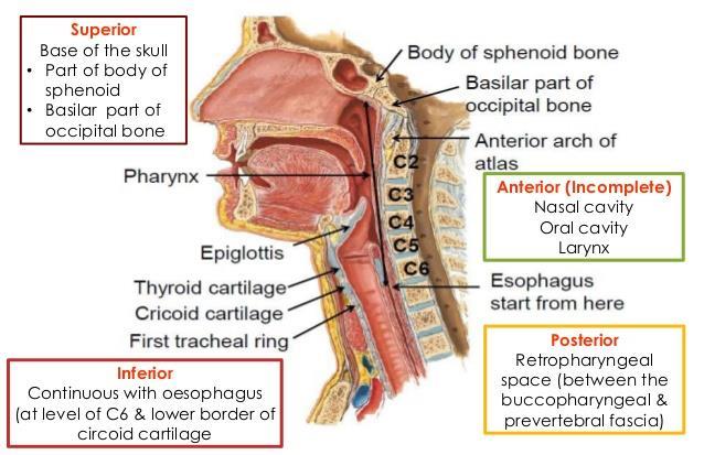
2 Introduction The pharynx is the Musculo-fascial halfcylinder that links the oral and nasal cavities in the head to the larynx and esophagus in the neck Common pathway for air and food Attached above to the base of the skull and is continuous below with the esophagus Ends at the level of vertebra C6
 & the inferior border of C6 vertebra (posteriorly) Dimensions: 12-14cm long Width : 3.5 cm (at its base) 1.")
3 Conical fibromuscular tube upper part of the air & food passages Location: Behind the nasal cavity, oral cavity and larynx Extend from the base of the skull to the inferior border of cricoid cartilage (anteriorly) & the inferior border of C6 vertebra (posteriorly) Dimensions: 12-14cm long Width : 3.5 cm (at its base) 1.5 cm (pharyngo-oesophageal junction narrowest part)
4
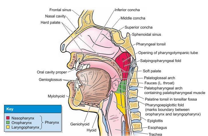
5 Region of Pharynx The walls of the pharynx are attached anteriorly to the margins of the nasal cavities, oral cavity, and larynx Subdivided into three regions having opening of Nasopharynx : posterior apertures (choanae) of the nasal cavities Oropharynx : posterior opening of the oral cavity (oropharyngeal isthmus) Laryngopharynx: superior aperture of the larynx (laryngeal inlet) opens
6
7
8

9 Oro-Pharynx
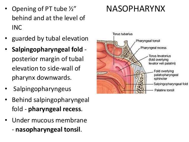
10 Pharynx In addition to these openings pharyngeal cavity is related anteriorly to the posterior one-third of the tongue posterior aspect of the larynx pharyngotympanic tubes open into the lateral walls of the nasopharynx Lingual, pharyngeal, and palatine tonsils - the deep surface of the pharyngeal walls Separated from the posteriorly positioned vertebral column by a thin retropharyngeal space containing loose connective tissue
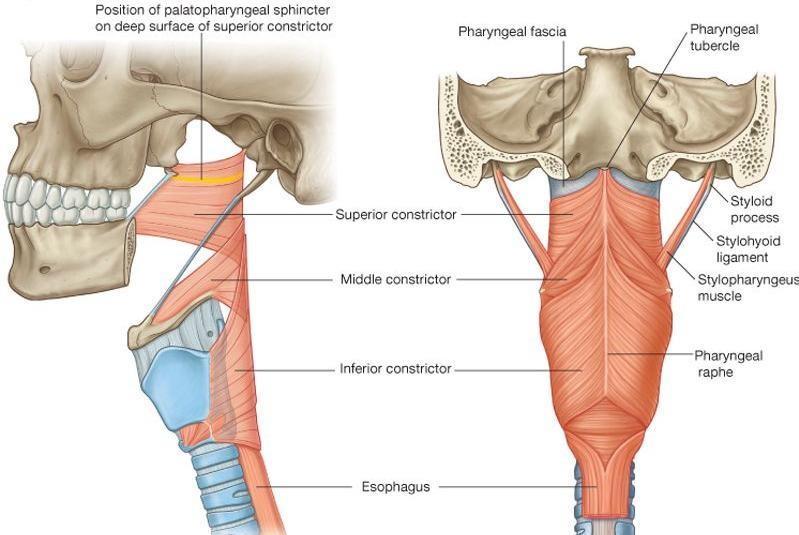
11 Skeletal framework 2 sides of the pharyngeal wall are welded together posteriorly in the midline by a vertically oriented cord-like ligament - pharyngeal raphe that extend From pharyngeal tubercle on the base of the skull To the level of cervical vertebra (C6) : blends with connective tissue in the posterior wall of the esophagus Irregular C-shaped line of pharyngeal wall attachment on the base of the skull Begins at the posterior margin of the medial plate of the pterygoid process of the sphenoid bone, Then it is attached just inferior to the cartilaginous part of the pharyngotympanic tube Then it passes onto the petrous part of the temporal bone to reach pharyngeal tubercle
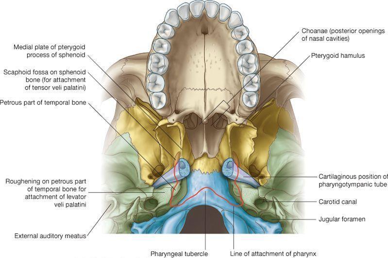
12
13 Bony Attachment for the lateral pharyngeal walls Vertical line of attachment for the lateral pharyngeal walls is discontinuous and in three parts. First part Begins superiorly on the posterior edge of the medial pterygoid plate - inferior to pharyngotympanic tube onto the pterygoid hamulus line descends along the pterygomandibular raphe Second part begins on the lower aspect of the stylohyoid ligament line continues onto the lesser horn of hyoid bone then turns and runs posteriorly along the entire upper surface of the greater horn of the hyoid where it terminates Third part begins superiorly on the superior tubercle of the thyroid cartilage descends along the oblique line to the inferior tubercle Then over the cricothyroid muscle along a tendinous thickening of fascia to the cricoid cartilage where it terminates

14 Lesser and greater horn of the hyoid bone
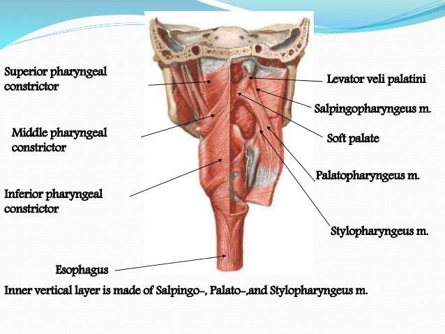
15 Pharyngeal wall Formed by skeletal muscles and by fascia Gaps between the muscles are reinforced by the fascia Organized into two groups based on the orientation of muscle fibers Constrictor muscles circular: Superior, middle & inferior Longitudinal muscles vertical: Stylopharyngeus, Salpingopharyngeus, and Palatopharyngeus
16
17 Constrictor muscles
18
19 Constrictor muscles The 3 constrictor muscle on each side - major contributors to the structure of the pharyngeal wall Superior, Middle and inferior Posteriorly, the muscles from each side are joined together by the pharyngeal raphe Anteriorly, these muscles attach to bones and ligaments related to the lateral margins of the nasal and oral cavities and the larynx. Constrict or narrow the pharyngeal cavity. Innervated by the pharyngeal branch of the vagus nerve [X]
20 Superior constrictors Upper part of the pharyngeal cavity Attached anteriorly to the pterygoid hamulus, pterygomandibular raphe, and adjacent bone of the mandible. Muscle fans out posteriorly and joins with its partner muscle from the other side at the pharyngeal raphe Palatopharyngeal sphincter Special band of muscle originating from anterolateral surface of the soft palate Circles the inner aspect of the pharyngeal wall Functions: constricts during swallowing - prominent ridge on the deep aspect of the pharyngeal wall
 Side of the posterior part of the")
21 Superior constrictor muscle Origin (from above to downwards) Pterygoid hamulus Pterygomandibular raphe Medial surface of mandible at posterior end of mylohyoid line (near the attachment of pterygomandibular raphe) Side of the posterior part of the tongue
22 Middle constrictors Attached to the lower aspect of the stylohyoid ligament lesser horn of the hyoid bone, and the entire upper surface of the greater horn of the hyoid Muscles fan out posteriorly and attach to the pharyngeal raphe Posterior part of the middle constrictors overlaps the superior constrictors


23 Middle constrictor muscle Origin Lower part of stylohyoid ligament Lesser cornu of hyoid bone Upper border of greater cornu of hyoid bone
24 Inferior constrictors Attach anteriorly to the oblique line of the thyroid cartilage and the cricoid cartilage. There are ligament that spans between these two attachments to cartilage - crosses the cricothyroid muscle Spread out posteriorly and attach to the pharyngeal raphe Posterior part of the inferior constrictors overlaps the middle constrictors Inferiorly, the muscle fibers blend with and attach into the wall of the esophagus Narrowest part of the pharyngeal cavity

25 Inferior constrictor muscle 1.Thyropharnygeus Arise from thyroid cartilage oblique line and inferior tubercle of thyroid cartilage A tendinous band that crossed the cricothyroid muscle & is attached above to the inferior tubercle of thyroid cartilage 1.Cricopharyngeus Cricoid cartilage behind the origin of cricothyroid muscle
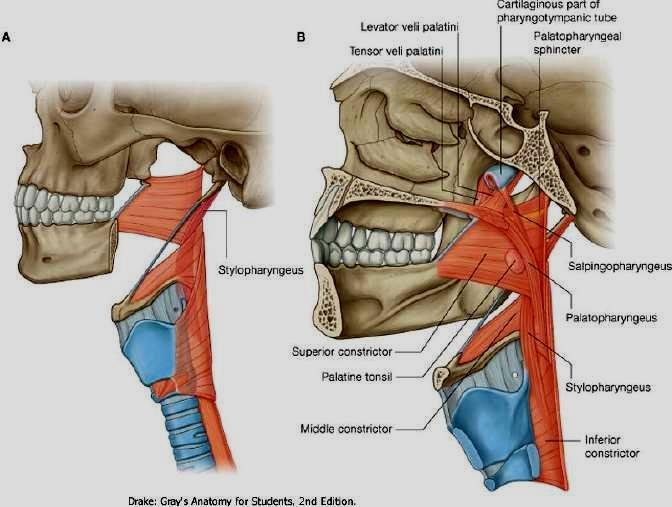
26 Longitudinal muscles Named according to their origins Stylopharyngeus: styloid process of the temporal bone Salpingopharyngeus: cartilaginous part of the pharyngotympanic tube Palatopharyngeus: soft palate Function elevate the pharyngeal wall, or during swallowing, pull the pharyngeal wall up and food bolus into the esophagus Nerve Supply: Except stylopharyngeus (Glossopharyngeal nerve ) both the muscle is supplied by Vagus Nerve
27 Longitudinal muscles Stylopharyngeus cylindrical stylopharyngeus muscle medial surface of the styloid process descends between the superior and middle constrictor muscles fan out on, and blend with, the deep surface of the pharyngeal wall. Salpingopharyngeus small muscle originating from the inferior aspect of the pharyngotympanic tube descending on, and blending into the deep surface of the pharyngeal wall Palatopharyngeus in addition to being a muscle of the pharynx, is also a muscle of the soft palate attached to the upper surface of the palatine aponeurosis passes posteriorly and inferiorly to blend with the deep surface of the pharyngeal wall

28 1. Stylopharyngeus From styloid process, passes through the gap between superior & middle constrictor Run downward to the inner surface of middle and inferior constrictor Stylopharyngeus muscle

29 2. Palatopharyngeus Descends from the sides of palate Runs longitudinally on the internal aspect of 3 constrictors Palatopharyngeus muscle


30 3. Salpingopharyngeus Descends from the auditory tube Merges with the palatopharyngeus muscle Salpingopharyngeus muscle
31 Fascia Pharyngeal fascia is separated into two layers, which sandwich the pharyngeal muscles between them Buccopharyngeal fascia thin layer coats the outside of the muscular part of the wall component of the pretracheal layer of cervical fascia Pharyngobasilar fascia thicker layer lines the inner surface Fascia reinforces the pharyngeal wall where muscle is deficient above the level of the superior constrictor - reinforced externally by muscles of the soft palate

32 Pharyngeal Spaces Situated behind the pharynx & extending from the base of skull to the bifurcation of trachea Divided into 2 lateral compartments [spaces of Gillette] by a fibrous raphe Has retropharyngeal nodes disappear at 3-4 years Infection can pass down behind the oesophagus into the mediastinum

33 Artery Supply
34 Artery Supply Upper parts of the pharynx the ascending pharyngeal artery; the ascending palatine and tonsillar branches of the facial artery; numerous branches of the maxillary and the lingual arteries. Lower parts of the pharynx pharyngeal branches from the inferior thyroid artery

35 Veins and Lymphatic Drainage
36 Veins and Lymphatic Drainage Veins of the pharynx form a plexus Superiorly - pterygoid plexus in the infratemporal fossa Inferiorly - facial and internal jugular veins Lymphatics drain into the deep cervical nodes and include retropharyngeal, paratracheal, and infrahyoid nodes
![Nerves Motor and most sensory innervation - branches of the vagus [X] & glossopharyngeal [IX] nerves This 2 nerved forms a plexus in the outer fascia of the pharyngeal wall, consisting of the](/docs-images/75/71793327/images/37-0.jpg "pharyngeal branch of the vagus nerve [X]; branches from the external laryngeal nerve from the superior laryngeal branch of the vagus nerve [X]; pharyngeal branches of the glossopharyngeal nerve")
37 Nerves Motor and most sensory innervation - branches of the vagus [X] & glossopharyngeal [IX] nerves This 2 nerved forms a plexus in the outer fascia of the pharyngeal wall, consisting of the pharyngeal branch of the vagus nerve [X]; branches from the external laryngeal nerve from the superior laryngeal branch of the vagus nerve [X]; pharyngeal branches of the glossopharyngeal nerve [IX]
38 Motor innervation All muscles of pharynx are supplied by the cranial accesory nerve through branches of vagus except stylopharyngeus muscle[ pharyngeal branches of glossopharyngeal nerve] Inferior constrictor muscle has additional supply from the external and recurrent laryngeal nerves Sensory innervation Afferent fibers travel through: 1.Glossopharyngeal nerve (mostly) 2. Vagus nerve (partly) 3. Maxillary nerve through pterygopalatine ganglion (nasopharynx Only) 4. Lesser palatine and glossopharyngeal nerve (soft palate & tonsil) Autonomic innervation Parasympathetic (secretomotor) 1.Greater petrosal nerve (branch of facial nerve) 2.Lesser palatine branches of pterygopalatine ganglion Generally, all the nerves supplying the pharynx form a plexus (pharyngeal plexus) which chiefly lies on the middle constrictor muscle: 1.Pharyngeal branch of vagus carrying cranial accesory nerve fibers 2.Pharyngeal branch of glossopharyngeal nerve 3.Pharyngeal branch of the superior sympathetic ganglion Fibers from pharyngeal plexus also supplies the muscles of soft palate except tensor veli palatini (by Mandibular nerve) 3 6
Prevertebral Region, Pharynx and Soft Palate
 Unit 20: Prevertebral Region, Pharynx and Soft Palate Dissection Instructions: Step1 Step 2 Step 1: Insert your fingers posterior to the sternocleidomastoid muscle, vagus nerve, internal jugular vein,
The PHARYNX. Dr. Nabil Khouri MD Ph.D
 The PHARYNX Dr. Nabil Khouri MD Ph.D PHARYNX Fibromuscular tube lined with mucous membrane extends from base of skull to lower border of cricoid cartilage (C-6). 12-14 cm long At the lower border of cricoid
Dr.Ban I.S. head & neck anatomy 2 nd y. جامعة تكريت كلية طب االسنان املرحلة الثانية
 جامعة تكريت كلية طب االسنان التشريح مادة املرحلة الثانية أ.م.د. بان امساعيل صديق 6102-6102 1 The Palate The palate forms the roof of the mouth and the floor of the nasal cavity. It is divided into two
Structure and Nerve Supply of The Larynx
 Kingdom of Bahrain Arabian Gulf University College of Medicine and Medical sciences Structure and Nerve Supply of The Larynx This presentation was originally prepared by: Dr. Kumar Notes were added by:
SCHOOL OF ANATOMICAL SCIENCES Mock Run Questions. 4 May 2012
 SCHOOL OF ANATOMICAL SCIENCES Mock Run Questions 4 May 2012 1. With regard to the muscles of the neck: a. the platysma muscle is supplied by the accessory nerve. b. the stylohyoid muscle is supplied by
Anatomy 2. Parotid bed (V.imp): meaning that gland is sleeping on structures and they are:
: meaning that gland is sleeping on structures and they are:") Anatomy 2 Parotid Gland: "refer to previous sheet for extra details." Its pyramidal in shape, apex is toward pharynx. Its Medial surface is divided into Anterio-medial and posterio-medial and its posterio-medial
Anatomy 2 Parotid Gland: "refer to previous sheet for extra details." Its pyramidal in shape, apex is toward pharynx. Its Medial surface is divided into Anterio-medial and posterio-medial and its posterio-medial
Upper Respiratory Tract
 Upper Respiratory Tract Lectures Objectives Describe the structure of nasal cavity including nasal septum. Describe the structure of lateral wall of nasal cavity including conchae and meatuses. Locate
The Digestive System in the Head and Neck
 The Digestive System in the Head and Neck The Mouth The Lips The lips are two fleshy folds that surround the oral orifice They are covered on the outside by skin and are lined on the inside by mucous membrane
Larynx. Rudimentary. Behind the posterior surface : -stylopharyngeus - salpingopharyngeus -platopharyngeus
 Larynx The larynx is an organ that provides a protective sphincter at the inlet of the air passages and is responsible for voice production. It extends from C3-C6: *Posterior: the pharynx *Lateral: the
Mohammad Mohtaseb. Nour Hussein. Faisal Nimri
 2 Mohammad Mohtaseb Nour Hussein Faisal Nimri Muscles of the tongue The tongue is a muscular organ and contains intrinsic and extrinsic muscles. The intrinsic muscle contains vertical, oblique, and transverse
THE INTERIOR OF THE PHARYNX. By Dr. Muhammad Imran Qureshi
 THE INTERIOR OF THE PHARYNX By Dr. Muhammad Imran Qureshi The Cavity The cavity of the pharynx is divided into: 1. The Nasal part (called Nasopharynx) 2. The Oral part (called the Oropharynx), 3. And the
The Neck the lower margin of the mandible above the suprasternal notch and the upper border of the clavicle
 The Neck is the region of the body that lies between the lower margin of the mandible above and the suprasternal notch and the upper border of the clavicle below Nerves of the neck Cervical Plexus Is formed
- Reem Akiely. -Wardeh Al-Swalmeh. - Mohammad Al-Muhtaseb. 1 P a g e
 -2 - Reem Akiely -Wardeh Al-Swalmeh - Mohammad Al-Muhtaseb 1 P a g e The palate: * Hard palate * Soft palate the Uvula: is a muscular structure present In the midline of the soft palate (اللهاة) The Hard
Subdivided into Vestibule & Oral cavity proper
 Extends from the lips to the oropharyngeal isthmus The oropharyngeal isthmus: Is the junction of mouth and pharynx. Is bounded: Above by the soft palate and the palatoglossal folds Below by the dorsum
Respiratory System. Cambridge University Press Concise Anatomy for Anaesthesia Andreas G. Erdmann Excerpt More information
 Respiratory System 1 The mouth DESCRIPTION The mouth extends from the lips (anterior) to the isthmus of the fauces (posterior). There are two sections: Vestibule slit-like cavity between the cheeks/lips
The Ear The ear consists of : 1-THE EXTERNAL EAR 2-THE MIDDLE EAR, OR TYMPANIC CAVITY 3-THE INTERNAL EAR, OR LABYRINTH 1-THE EXTERNAL EAR.
 The Ear The ear consists of : 1-THE EXTERNAL EAR 2-THE MIDDLE EAR, OR TYMPANIC CAVITY 3-THE INTERNAL EAR, OR LABYRINTH 1-THE EXTERNAL EAR Made of A-AURICLE B-EXTERNAL AUDITORY MEATUS A-AURICLE It consists
Tikrit University collage of dentistry Dr.Ban I.S. head & neck anatomy 2 nd y. Lec [5] / Temporal fossa :
![Tikrit University collage of dentistry Dr.Ban I.S. head & neck anatomy 2 nd y. Lec [5] / Temporal fossa :](/thumbs/88/115294566.jpg "Tikrit University collage of dentistry Dr.Ban I.S. head & neck anatomy 2 nd y. Lec [5] / Temporal fossa :") Lec [5] / Temporal fossa : Borders of the Temporal Fossa: Superior: Superior temporal line. Inferior: gap between zygomatic arch and infratemporal crest of sphenoid bone. Anterior: Frontal process of the
Lec [5] / Temporal fossa : Borders of the Temporal Fossa: Superior: Superior temporal line. Inferior: gap between zygomatic arch and infratemporal crest of sphenoid bone. Anterior: Frontal process of the
Temporal region. temporal & infratemporal fossae. Zhou Hong Ying Dept. of Anatomy
 Temporal region temporal & infratemporal fossae Zhou Hong Ying Dept. of Anatomy Temporal region is divided by zygomatic arch into temporal & infratemporal fossae. Temporal Fossa Infratemporal fossa Temporal
Dr.Ban I.S. head & neck anatomy 2 nd y. جامعة تكريت كلية طب االسنان املرحلة الثانية أ.م.د. بان امساعيل صديق 6102/6102
 جامعة تكريت كلية طب االسنان التشريح مادة املرحلة الثانية أ.م.د. بان امساعيل صديق 6102/6102 Parotid region The part of the face in front of the ear and below the zygomatic arch is the parotid region. The
Anatomy: head and Neck (6 questions) 1. Prevertebral Flexor Musculature (lying in front of the vertebrae) include all, EXCEPT: Longus Colli.
 1. Prevertebral Flexor Musculature (lying in front of the vertebrae) include all, EXCEPT: Longus Colli.") Anatomy: head and Neck (6 questions) 1. Prevertebral Flexor Musculature (lying in front of the vertebrae) include all, EXCEPT: Longus Colli. Rectus Capitis Anterior. Rectus Capitis Lateralis. Rectus Capitis
Anatomy: head and Neck (6 questions) 1. Prevertebral Flexor Musculature (lying in front of the vertebrae) include all, EXCEPT: Longus Colli. Rectus Capitis Anterior. Rectus Capitis Lateralis. Rectus Capitis
The Neck. BY: Lina Abdullah & Rahaf Jreisat
 The Neck BY: Lina Abdullah & Rahaf Jreisat Boundaries of the Neck: generally from base of the skull to root of the neck Superior margin :From superior nuchal line of occipital bone up to mastoid process
Veins of the Face and the Neck
 Veins of the Face and the Neck Facial Vein The facial vein is formed at the medial angle of the eye by the union of the supraorbital and supratrochlear veins. connected through the ophthalmic veins with
ORAL CAVITY, ESOPHAGUS AND STOMACH
 ORAL CAVITY, ESOPHAGUS AND STOMACH 1 OBJECTIVES By the end of the lecture you should be able to: Describe the anatomy the oral cavity, (boundaries, parts, nerve supply). Describe the anatomy of the palate,
Oral cavity : consist of two parts: the oral vestibule and the oral cavity proper. Oral vestibule : is slit like space between.
 Oral cavity Oral cavity : consist of two parts: the oral vestibule and the oral cavity proper Oral vestibule : is slit like space between the teeth, buccal gingiva, lips, and cheeks 1 Oral cavity Oral
Chapter 26: The temporomandibular joint, pharynx and larynx. The Temporomandibular Joint. Ligaments. (a) Capsular
 Capsular") Chapter 26: The temporomandibular joint, pharynx and larynx The Temporomandibular Joint This is a synovial joint of a condyloid (modified hinge) variety between the condyle of the mandible and the mandibular
Chapter 26: The temporomandibular joint, pharynx and larynx The Temporomandibular Joint This is a synovial joint of a condyloid (modified hinge) variety between the condyle of the mandible and the mandibular
Anatomy of the Pharynx and Oesophagus
 Anatomy of the Pharynx and Oesophagus EMBRYOLOGY Cephalocaudal and lateral folding result in the formation of an endodermally lined primitive gut. In its cephalic part this forms a blind ending tube, the
Infratemporal fossa: Tikrit University college of Dentistry Dr.Ban I.S. head & neck Anatomy 2 nd y.
 Infratemporal fossa: This is a space lying beneath the base of the skull between the lateral wall of the pharynx and the ramus of the mandible. It is also referred to as the parapharyngeal or lateral pharyngeal
Dr.Noor Hashem Mohammad Lecture (5)
") Dr.Noor Hashem Mohammad Lecture (5) 2016-2017 If the mandible is discarded, the anterior part of this aspect of the skull is seen to be formed by the hard palate. The palatal processes of the maxillae
Dr.Noor Hashem Mohammad Lecture (5) 2016-2017 If the mandible is discarded, the anterior part of this aspect of the skull is seen to be formed by the hard palate. The palatal processes of the maxillae
Anatomy of the Airway
 Anatomy of the Airway Nagelhout, 5 th edition, Chapter 26 Morgan & Mikhail, 5 th edition, Chapter 23 Mary Karlet, CRNA, PhD Airway Anatomy The airway consists of the nose, pharynx, larynx, trachea, and
Mohammad Hisham Al-Mohtaseb. Lina Mansour. Reyad Jabiri. 0 P a g e
 2 Mohammad Hisham Al-Mohtaseb Lina Mansour Reyad Jabiri 0 P a g e This is only correction for the last year sheet according to our record. If you already studied this sheet just read the yellow notes which
Nose, Nasal cavity, Paranasal Sinuses & Pharynx
 Nose, Nasal cavity, Paranasal Sinuses & Pharynx Respiratory block-anatomy-lecture 2 Editing file Objectives At the end of the lecture, the students should be able to: Describe the boundaries of the nasal
THYROID & PARATHYROID. By Prof. Saeed Abuel Makarem & Dr. Sanaa Al-Sharawy
 THYROID & PARATHYROID By Prof. Saeed Abuel Makarem & Dr. Sanaa Al-Sharawy 1 OBJECTIVES By the end of the lecture, the student should be able to: Describe the shape, position, relations and structure of
PTERYGOPALATINE FOSSA
 PTERYGOPALATINE FOSSA Outline Anatomical Structure and Boundaries Foramina and Communications with other spaces and cavities Contents Pterygopalatine Ganglion Especial emphasis on certain arteries and
Anterior triangle of neck
 Anterior triangle of neck Dept. of Anatomy Zhou Hong Ying Outline boundary and subdivisions of ant. triangle contents of the triangle Muscles: suprahyoid muscles, infrahyoid muscles Nerves: CNⅩ, CNⅪ, CNⅫ,
Neck-2. Dr. Heba Kalbouneh Associate Professor of Anatomy and Histology
 Neck-2 ` Dr. Heba Kalbouneh Associate Professor of Anatomy and Histology Triangles of the neck Side of the neck Midline Lower border of mandible Line between angle of mandible and mastoid Superior nuchal
Thyroid gland. importance. relations and connections. external laryngeal nerves. malformations.
 Thyroid gland 1. Recognize and understand the coverings of the thyroid gland and their clinical importance. 2. Recognize and understand the main parts of the thyroid gland and their locations, relations
Tympanic Bulla Temporal Bone. Digastric Muscle. Masseter Muscle
 Superior view Hyoid Bone The hyoid bone does not articulate with any other bones. It is held in place by ligaments to the styloid process of the temporal bone and the thyroid cartilage of the larynx. It
Lecture 01. The Thyroid & Parathyroid Glands. By: Dr Farooq Khan PMC Date: 12 th March. 2018
 Lecture 01 The Thyroid & Parathyroid Glands By: Dr Farooq Khan PMC Date: 12 th March. 2018 INTRODUCTION LAYERS OF THE NECK The neck has four major compartments or layer which are enclosed by an outer musculofascial
Anatomy of Oral Cavity DR. MAAN AL-ABBASI
 Anatomy of Oral Cavity DR. MAAN AL-ABBASI By the end of this lecture you should be able to: 1. Differentiate different parts of the oral cavity 2. Describe the blood and nerve supply of mucosa and muscles
Lec [8]: Mandibular nerve:
![Lec [8]: Mandibular nerve:](/thumbs/94/121295776.jpg "Lec [8]: Mandibular nerve:") Lec [8]: Mandibular nerve: The mandibular branch from the trigeminal ganglion lies in the middle cranial fossa lateral to the cavernous sinus. With the motor root of the trigeminal nerve [motor roots lies
Lec [8]: Mandibular nerve: The mandibular branch from the trigeminal ganglion lies in the middle cranial fossa lateral to the cavernous sinus. With the motor root of the trigeminal nerve [motor roots lies
The Skull and Temporomandibular joint II Prof. Abdulameer Al-Nuaimi. E. mail:
 The Skull and Temporomandibular joint II Prof. Abdulameer Al-Nuaimi E-mail: a.al-nuaimi@sheffield.ac.uk E. mail: abdulameerh@yahoo.com Temporal fossa The temporal fossa is a depression on the temporal
The Thoracic wall including the diaphragm. Prof Oluwadiya KS
 The Thoracic wall including the diaphragm Prof Oluwadiya KS www.oluwadiya.com Components of the thoracic wall Skin Superficial fascia Chest wall muscles (see upper limb slides) Skeletal framework Intercostal
Larynx - cartilaginous structure holding the vocal folds which protrude into airstream
 1! Larynx - cartilaginous structure holding the vocal folds which protrude into airstream 2! Flow increase - like thumb over garden hose Pressure drop - narrower space forces pressure drop due to speed
Chapter 10: Anatomy of the pharynx and esophagus. P. Beasley. Embryological development
 Chapter 10: Anatomy of the pharynx and esophagus P. Beasley Embryological development During the development of the embryo, a process of cephalocaudal and lateral folding takes place with the result that
Thyroid and Parathyroid Glands
 Thyroid and Parathyroid Glands Please view our Editing File before studying this lecture to check for any changes. Color Code Important Doctors Notes Notes/ explanation Objectives: By the end of the lecture,
OBJECTIVE: To obtain a fundamental knowledge of the root of the neck with respect to structure and function
 The root of the neck Jeff Dupree, Ph.D. e mail: jldupree@vcu.edu OBJECTIVE: To obtain a fundamental knowledge of the root of the neck with respect to structure and function READING ASSIGNMENT: Moore and
Neck of Condylar. Process. Anterior Border of Ramus. Mandibular. Foramen. Posterior Border of Ramus Incisive Fossa.
 Learning Outcomes The Mandible Surface Anatomy Muscle Attachments The (FOM) Muscles of the FOM The Tongue Muscles of the Tongue The Submandibular Region Submandibular Gland Sublingual Gland Lingual The
Bony orbit Roof The orbital plate of the frontal bone Lateral wall: the zygomatic bone and the greater wing of the sphenoid
 Bony orbit Roof: Formed by: The orbital plate of the frontal bone, which separates the orbital cavity from the anterior cranial fossa and the frontal lobe of the cerebral hemisphere Lateral wall: Formed
Cranial Nerve VII - Facial Nerve. The facial nerve has 3 main components with distinct functions
 Cranial Nerve VII - Facial Nerve The facial nerve has 3 main components with distinct functions Somatic motor efferent Supplies the muscles of facial expression; posterior belly of digastric muscle; stylohyoid,
Anatomy of the Thyroid Gland
 Anatomy of the Thyroid Gland Introduction Nomenclature G, thyreos= shield, eidos= like Location Root of the neck ventrally (C5-T1) Function endocrine gland that secretes: Thyroxine (T4) T3 Calcitonin LWW,
Anatomy of the Thorax
 Anatomy of the Thorax A) THE THORACIC WALL Boundaries Posteriorly by the thoracic part of the vertebral column Anteriorly by the sternum and costal cartilages Laterally by the ribs and intercostal spaces
Pharyngeal apparatus. - At the third week, it is a 3 layered structure: ectoderm, mesoderm and endoderm. This is called trilaminar disc
 Pharyngeal apparatus Remember from the first year embryology - The embryo was disc shaped in the second week of development (this is called embryonic disc) and it is a 2 layered disc (composed of two layers)---bilaminar
04 Development of the Face and Neck. Development of the Face Development of the neck
 04 Development of the Face and Neck Development of the Face Development of the neck Development of the face Overview of facial development The fourth week ~ the twelfth week of prenatal development Between
Parotid Gland, Temporomandibular Joint and Infratemporal Fossa
 M1 - Anatomy Parotid Gland, Temporomandibular Joint and Infratemporal Fossa Jeff Dupree Sanger 9-057 jldupree@vcu.edu Parotid gland: wraps around the mandible positioned between the mandible and the sphenoid
Respiratory System. Functional Anatomy of the Respiratory System
 Respiratory System Overview of the Respiratory System s Job Major Duty Respiration Other important aspects ph control Vocalization Processing incoming air Protection Metabolism (ACE) What structures allow
The Respiratory System
 PowerPoint Lecture Slide Presentation by Vince Austin Human Anatomy & Physiology FIFTH EDITION Elaine N. Marieb The Respiratory System Dr Nabil Khouri. MD, Ph.D Respiratory System Consists of a conducting
Structure Location Function
 Frontal Bone Cranium forms the forehead and roof of the orbits Occipital Bone Cranium forms posterior and inferior portions of the cranium Temporal Bone Cranium inferior to the parietal bone forms the
The Larynx. Prof. Dr.Mohammed Hisham Al-Muhtaseb
 The Larynx Prof. Dr.Mohammed Hisham Al-Muhtaseb The Larynx Extends from the middle of C3 vertebra till the level of the lower border of C6 Continue as Trachea Above it opens into the laryngo-pharynx Suspended
Maxilla, ORBIT and infratemporal fossa. Neophytos C Demetriades MD, DDS, MSc Associate professor European University of Cyprus School of Medicine
 Maxilla, ORBIT and infratemporal fossa Neophytos C Demetriades MD, DDS, MSc Associate professor European University of Cyprus School of Medicine MAXILLA Superior, middle, and inferior meatus Frontal sinus
Bisection of Head & Nasal Cavity 頭部對切以及鼻腔. 解剖學科馮琮涵副教授 分機
 Bisection of Head & Nasal Cavity 頭部對切以及鼻腔 解剖學科馮琮涵副教授 分機 3250 E-mail: thfong@tmu.edu.tw Outline: The structure of nose The concha and meatus in nasal cavity The openings of paranasal sinuses Canals, foramens
A. The supraclavicular nerves supply sensory fibers to the skin of the clavicular area
 YR 1 GROSS ANATOMY WRITTEN EXAM 2 -- October 10, 1997. CHOOSE THE SINGLE BEST ANSWER FOR QUESTIONS 1-42. 1. Each of the following statements is CORRECT EXCEPT: A. The supraclavicular nerves supply sensory
Nose & Mouth OUTLINE. Nose. - Nasal Cavity & Its Walls. - Paranasal Sinuses. - Neurovascular Structures. Mouth. - Oral Cavity & Its Contents
 Dept. of Human Anatomy, Si Chuan University Zhou hongying eaglezhyxzy@163.com Nose & Mouth OUTLINE Nose - Nasal Cavity & Its Walls - Paranasal Sinuses - Neurovascular Structures Mouth - Oral Cavity & Its
Temporal fossa Infratemporal fossa Pterygopalatine fossa Terminal branches of external carotid artery Pterygoid venous plexus
 Outline of content Temporal fossa Infratemporal fossa Pterygopalatine fossa Terminal branches of external carotid artery Pterygoid venous plexus Boundary Content Communication Mandibular division of trigeminal
Basic Anatomy and Physiology of the Lips and Oral Cavity. Dr. Faghih
 Basic Anatomy and Physiology of the Lips and Oral Cavity Dr. Faghih It is divided into seven specific subsites : 1. Lips 2. dentoalveolar ridges 3. oral tongue 4. retromolar trigone 5. floor of mouth 6.
Pharynx. Muscles of Pharynx
 Pharynx A funnel shaped fibromuscular tube that extends from the base of the skull & continues below with the esophagus at the level of C6 in the neck. It is divided into 3 parts: (1) Nasal: nasopharynx;
Bones Ethmoid bone Inferior nasal concha Lacrimal bone Maxilla Nasal bone Palatine bone Vomer Zygomatic bone Mandible
 splanchnocranium - Consists of part of skull that is derived from branchial arches - The facial bones are the bones of the anterior and lower human skull Bones Ethmoid bone Inferior nasal concha Lacrimal
Dr.Ban I.S. head & neck anatomy 2 nd y جامعة تكريت كلية طب االسنان مادة التشريح املرحلة الثانية أ.م.د. بان امساعيل صديق 6102/6102
 جامعة تكريت كلية طب االسنان مادة التشريح املرحلة الثانية أ.م.د. بان امساعيل صديق 6102/6102 Pterygopalatine fossa: The pterygopalatine fossa is a cone-shaped depression, It is located between the maxilla,
APRIL
 APRIL - 2003 OCTOBER - 2003 February 2009 [KU 652] Sub. Code : 4131 FIRST B.D.S DEGREE EXAMINATION (Modified Regulations III) Paper I HUMAN ANATOMY, HISTOLOGY AND EMBRYOLOGY Time : Three hours
Anatomy Sheet: Oral cavity Done by: rasha Rakan edited by: khansaa Mahmoud
 Anatomy Sheet: Oral cavity Done by: rasha Rakan edited by: khansaa Mahmoud The oral cavity has 2 parts: 1. Oral vestibule: outer part that consists of outside the teeth, between the teeth, the cheeks and
Gross Anatomy of the. TEMPORAL BONE, EXTERNAL EAR, and MIDDLE EAR
 Gross Anatomy of the TEMPORAL BONE, EXTERNAL EAR, and MIDDLE EAR M1 Gross and Developmental Anatomy 9:00 AM, December 11, 2008 Dr. Milton M. Sholley Professor of Anatomy and Neurobiology Assignment: Head
For the following questions, indicate the letter that corresponds to the SINGLE MOST APPROPRIATE ANSWER
 GROSS ANATOMY EXAMINATION May 15, 2000 For the following questions, indicate the letter that corresponds to the SINGLE MOST APPROPRIATE ANSWER 1. Pain associated with an infection limited to the middle
Gross Anatomy of the. TEMPORAL BONE, EXTERNAL EAR, and MIDDLE EAR. Assignment: Head to Toe Temporomandibular Joint (TMJ)
") Gross Anatomy the TEMPORAL BONE, EXTERNAL EAR, and MIDDLE EAR M1 Gross and Developmental Anatomy 9:00 AM, December 11, 2008 Dr. Milton M. Sholley Pressor Anatomy and Neurobiology Assignment: Head to Toe
Gross Anatomy the TEMPORAL BONE, EXTERNAL EAR, and MIDDLE EAR M1 Gross and Developmental Anatomy 9:00 AM, December 11, 2008 Dr. Milton M. Sholley Pressor Anatomy and Neurobiology Assignment: Head to Toe
Anatomy #9. Rashed AL-Jomared. The Cranial Nerves IX. Amneh Hazaimeh & Alanood Bostanji
 Anatomy #9 The Cranial Nerves IX Rashed AL-Jomared Amneh Hazaimeh & Alanood Bostanji السالم عليكم This lecture talks about the cranial nerves IX & X:: *Glossopharyngeal nerve : The nerve gets out of the
THE THORACIC WALL. Boundaries Posteriorly by the thoracic part of the vertebral column. Anteriorly by the sternum and costal cartilages
 THE THORACIC WALL Boundaries Posteriorly by the thoracic part of the vertebral column Anteriorly by the sternum and costal cartilages Laterally by the ribs and intercostal spaces Superiorly by the suprapleural
Dr. Sami Zaqout Faculty of Medicine IUG
 Auricle External Ear External auditory meatus The Ear Middle Ear (Tympanic Cavity) Auditory ossicles Internal Ear (Labyrinth) Bony labyrinth Membranous labyrinth External Ear Auricle External auditory
Anatomic Relations Summary. Done by: Sohayyla Yasin Dababseh
 Anatomic Relations Summary Done by: Sohayyla Yasin Dababseh Anatomic Relations Lecture 1 Part-1 - The medial wall of the nose is the septum. - The vestibule lies directly inside the nostrils (Nares). -
Skull-2. Norma Basalis Interna Norma Basalis Externa. Dr. Heba Kalbouneh Associate Professor of Anatomy and Histology
 Skull-2 Norma Basalis Interna Norma Basalis Externa Dr. Heba Kalbouneh Associate Professor of Anatomy and Histology Norma basalis interna Base of the skull- superior view The interior of the base of the
Oral Cavity and Pharynx. The Oral Cavity. The oral cavity is divided into two major portions: the vestibule and the cavum oris.
 11 Oral Cavity and Pharynx Persons who specialize in the care and treatment of the oral cavity have a great responsibility. The oral cavity participates actively in respiration, nutrition, and excretion
University of Palestine. Midterm Exam 2013/2014 Total Grade:
 [ Course No: DNTS2208 Course Title: Head and Neck Anatomy Date: 17/11/1024 No. of Questions: (52) Time: 2hours Using Calculator (No) University of Palestine Midterm Exam 2013/2014 Total Grade: Instructor
Cranial nerves.
 Cranial nerves eaglezhyxzy@163.com Key Points of Learning Name Components Passing through Peripheral distribution Central connection Function Cranial nerves Ⅰ olfactory Ⅱ optic Ⅲ occulomotor Ⅳ trochlear
Chapter 64: Anatomy. Daniel O. Graney, Guy J. Petruzzelli, Eugene N. Myers. Pharynx
 Chapter 64: Anatomy Daniel O. Graney, Guy J. Petruzzelli, Eugene N. Myers Pharynx The pharynx (Fig. 64-1) is a common aerodigestive tract, which is subdivided anatomically into the nasopharynx, oropharynx,
REVIEW/PREVIEW OF HEAD AND NECK ANATOMY FOR ENT EXAM
 REVIEW/PREVIEW OF HEAD AND NECK ANATOMY FOR ENT EXAM - 2017 PALPATE CAROTID ARTERY: AT LEVEL OF CAROTID BIFURCATION VERTEBRAL LEVEL C4 Sternocleidomastoid Muscle INTERNAL CAROTID EXTERNAL CAROTID COMMON
Omran Saeed. Luma Taweel. Mohammad Almohtaseb. 1 P a g e
 2 Omran Saeed Luma Taweel Mohammad Almohtaseb 1 P a g e I didn t include all the photos in this sheet in order to keep it as small as possible so if you need more clarification please refer to slides In
Cranial Nerves IX-X (Glossopharyngeal & Vagus Nerves)
") Cranial Nerves IX-X (Glossopharyngeal & Vagus Nerves) Please view our Editing File before studying this lecture to check for any changes. Color Code Important Doctors Notes Notes/Extra explanation Objectives
Cranial Nerves IX-X (Glossopharyngeal & Vagus Nerves) Please view our Editing File before studying this lecture to check for any changes. Color Code Important Doctors Notes Notes/Extra explanation Objectives
Mediastinum and pericardium
 Mediastinum and pericardium Prof. Abdulameer Al-Nuaimi E-mail: a.al-nuaimi@sheffield.ac.uk E. mail: abdulameerh@yahoo.com The mediastinum: is the central compartment of the thoracic cavity surrounded by
University of Palestine. Midterm Exam 2013/2014 Total Grade:
 Course No: DNTS2208 Course Title: Head and Neck Anatomy Date: 09/11/2013 No. of Questions: (50) Time: 1hour Using Calculator (No) University of Palestine Midterm Exam 2013/2014 Total Grade: Instructor
Trigeminal Nerve Worksheets, Distributions Page 1
 Trigeminal Nerve Worksheet #1 Distribution by Nerve Dr. Darren Hoffmann Dental Gross Anatomy, Spring 2013 We have drawn out each of the branches of CN V in lecture and you have an idea now for their basic
The Respiratory System
 The Respiratory System Respiration Includes Pulmonary ventilation Air moves in and out of lungs Continuous replacement of gases in alveoli (air sacs) External respiration Gas exchange between blood and
Lies in front and sides of the neck. Consists of two lobe connected anterior to the trachea by an isthmus.
 THYROID GLAND 1 Lies in front and sides of the neck. Consists of two lobe connected anterior to the trachea by an isthmus. A small pyramidal lobe projects upwards from the left lobe in 40% of cases. The
Newly identified thin membranous tissue in the deep infratemporal region
 Anat Sci Int (2012) 87:136 140 DOI 10.1007/s12565-012-0135-0 ORIGINAL ARTICLE Newly identified thin membranous tissue in the deep infratemporal region Kojiro Takezawa Ikuo Kageyama Received: 12 January
Dr. Sami Zaqout Faculty of Medicine IUG
 The Nose External Nose Nasal Cavity External Nose Blood and Nerve Supplies of the External Nose Blood Supply of the External Nose The skin of the external nose Branches of the ophthalmic and the maxillary
Lecturer: Ms DS Pillay ROOM 2P24 25 February 2013
 Lecturer: Ms DS Pillay ROOM 2P24 25 February 2013 Thoracic Wall Consists of thoracic cage Muscle Fascia Thoracic Cavity 3 Compartments of the Thorax (Great Vessels) (Heart) Superior thoracic aperture
Please refer back to the slides as these are extra notes only. Slide 2 -The Larynx is a Box of cartilage.
 [ANATOMY #3] 1 بسم رلاهللا Please refer back to the slides as these are extra notes only. Slide 2 -The Larynx is a Box of cartilage. -The lower border of c6 is the lower border of cricoid cartilage. -The
ANTERIOR CERVICAL TRIANGLE (Fig. 2.1 )
") 2 Neck Anatomy ANTERIOR CERVICAL TRIANGLE (Fig. 2.1 ) The boundaries are: Lateral: sternocleidomastoid muscle Superior: inferior border of the mandible Medial: anterior midline of the neck This large triangle
2 Neck Anatomy ANTERIOR CERVICAL TRIANGLE (Fig. 2.1 ) The boundaries are: Lateral: sternocleidomastoid muscle Superior: inferior border of the mandible Medial: anterior midline of the neck This large triangle
Syllabus: 6 pages (Page 6 lists corresponding figures for Grant's Atlas 11 th & 12 th Eds.)
") PLEURAL CAVITY AND LUNGS Dr. Milton M. Sholley SELF STUDY RESOURCES Essential Clinical Anatomy 3 rd ed. (ECA): pp. 70 81 Syllabus: 6 pages (Page 6 lists corresponding figures for Grant's Atlas 11 th &
PLEURAL CAVITY AND LUNGS Dr. Milton M. Sholley SELF STUDY RESOURCES Essential Clinical Anatomy 3 rd ed. (ECA): pp. 70 81 Syllabus: 6 pages (Page 6 lists corresponding figures for Grant's Atlas 11 th &
Oral Cavity, Soft Palate, Pharynx, and Larynx; Development of the Face and Palate
 Oral Cavity, Soft Palate, Pharynx, and Larynx; Development of the Face and Palate Think on this. The ability to eat and drink safely and efficiently is fundamental to our quality of life. The wide variety