Cover Page. The handle holds various files of this Leiden University dissertation.
|
|
|
- Ross Stanley
- 6 years ago
- Views:
Transcription

1 Cover Page The handle holds various files of this Leiden University dissertation. Author: Embden, Daphne van Title: Facts and fiction in hip fracture treatment Issue Date:
2 Facts and fiction in hip fracture treatment Daphne van Embden
3 Cover design: M. van Embden ISBN: Copyright 2015 D. van Embden All rights reserved. No part of this thesis may be reproduced, stored in a retrieval system or transmitted in any form or by any means without prior permission of the author or the copyright-owning journals for previously published chapters. Layout and printing: Optima Grafische Communicatie Grants: Part of thesis was funded by a grant of the Anna Fonds NOREF. Printing of this thesis has been financially supported by: Nederlandse Vereniging voor Traumachirurgie, Raad van Bestuur Medisch Centrum Haaglanden-Bronovo, Stiching Extra-Curriculaire Activiteiten Haagse Chirurgen, Traumacentrum West, ChipSoft
4 Facts and fiction in hip fracture treatment Proefschrift ter verkrijging van de graad van Doctor aan de Universiteit Leiden, op gezag van Rector Magnificus prof.mr. C.J.J.M. Stolker, volgens besluit van het College voor Promoties te verdedigen op woensdag 17 februari 2016 klokke uur door Daphne van Embden geboren te Rotterdam in 1984
5 Promotor: Prof. Dr. I.B. Schipper Co-promotores: Dr. S.A.G. Meylaerts, Medisch Centrum Haaglanden-Bronovo Dr. S.J. Rhemrev, Medisch Centrum Haaglanden-Bronovo Promotiecommissie: Prof. Dr. R.G.H. H. Nelissen Prof. Dr. R.S. Breederveld Prof. Dr. H.J. Ten Duis, Universitair Medisch Centrum Groningen
6 Contents Chapter 1 Introduction, aims and outline of the thesis 9 Personality of the fracture Chapter 2 Trochanteric Femoral Fracture Classification: relevance of the fracture line angle, a radiological study 21 Chapter 3 The comparison of two classifications for trochanteric femur fractures: the AO/ASIF classification and the Jensen classification 35 Chapter 4 The reliability of a simplified Garden classification for intra-capsular hip fractures 49 Chapter 5 The Pauwels classification for intra-capsular hip fractures: is it reliable? 59 Chapter 6 The value of a CT-scan compared to radiographs in the classification and treatment plan of trochanteric fractures 67 Personalized hip fracture treatment Chapter 7 Fixation device related rotational influences in femoral neck and trochanteric fractures: a radio stereometric analysis 81 Chapter 8 Radiographic fracture features predicting failure of internal fixation of displaced femoral neck fractures 99 Chapter 9 The surgeon s eye: a prospective analysis of the anteversion in the placement of hemiarthroplasties after a femoral neck fracture 115 Chapter 10 The medial femoral neck fracture: is there still a place for conservative treatment? 125
7 Chapter 11 General discussion: From fracture pattern to patient tailored treatment 139 Chapter 12 Summary and conclusions 151 Chapter 13 Samenvatting en conclusies 159 Chapter 14 List of publications and presentations 167 Chapter 15 Curriculum Vitae 173 Chapter 16 Acknowledgements 177
8
9
10 Chapter 1 Introduction, aims and outline of the thesis
11
12 Chapter 1 11 Introduction As the number of hip fracture patients has increased dramatically over the years, the need for high quality, multidisciplinary and patient centred fracture treatment continues to grow. General international demographics show that the average age of male hip fracture patients is 69 and 79 for female patients. 1 About 75% of all hip fracture patients is female. Most patients suffer from a hip fracture after a low energy trauma such as a fall, whereas in the young patients (under 50 years) more sports related and high energy trauma mechanisms are seen. 2 The total number of deaths occurring in the first year after an osteoporotic fracture was 143,000 in the EU in 2010 and around 50% of these patients had a hip fracture. An overall increase of 32% in hip fracture patients in the European Union (EU) is expected by the year 2025, resembling 199,432 patients per year. The average incidence of hip fractures in the Netherlands is 275 per 100,000 (368/ 100,000 women, 164/100,000 men). An increase of the number of patients with a hip fracture of 40% is expected by the year 2025, which would result in almost 24,000 patients a year. The predicted growing incidence would cause a 30% (246 million Euro) increase of health care costs in the Netherlands by the year , 4 Anatomy Hip fractures are typically fractures of the proximal femur. The proximal femur consists of a femoral head, neck and trochanteric area, which comprises a lesser and greater trochanter. The hip joint capsula is a strong ligamentous structure attached to the intertrochanteric line incorporating the femoral head and neck. Fractures of the femoral head and neck are therefore named intra-capsular fractures. Extra-capsular proximal femur fractures are trochanteric fractures (fractures within the margin of the lesser or greater trochanter) or subtrochanteric fractures. Subtrochanteric fractures are defined as the area from the lesser trochanter to 5 centimetres distally and are more common to result from a high-energy trauma mechanism, but may in the elderly very well occur after a low-energy fall. Vascular anatomy The vascular anatomy of the hip is another important anatomic factor influencing hip fracture treatment. Arteries of the proximal femur are divided into three parts: the extra-capsular arterial ring located at the base of the femoral neck, the ascending cervical branches of the extra-capsular arterial ring on the surface of the femoral neck and the arteries of the round ligament. The extra-capsular arterial ring is formed by a large branch of the medial femoral circumflex artery and by branches of the lateral femoral
13 12 Chapter 1 circumflex artery. In femoral neck fractures, especially in displaced fractures, the vascularization of the femoral head is at risk. The most important blood supply, provided by the intra-osseous cervical vessels that cross the marrow spaces from distally, is disrupted in case of a displaced femoral neck fracture. Alternative ways of blood supply such as the ligamentum teres and the branches of the extra-capsular arterial ring are not sufficient in many elderly patients. Insufficient post-traumatic blood supply in the hip may result in avascular necrosis (AVN). AVN rarely occurs in extra-capsular fractures. 5 Classification Intra-capsular fractures The Pauwels classification (1935) 6 was the first biomechanical classification of femoral neck fractures. In the Pauwels classification the fracture line angle is used to identify three groups of femoral neck fractures, based on the shearing angle of the fracture line of the distal fragment. It was suggested by Pauwels that a greater vertical shear is related to an increase of the incidence of non-union in femoral neck fractures as it increasingly interferes with the blood supply of the femoral neck. In daily practice, the Garden-classification (1961) 7 is still the most frequently used classification for femoral neck fractures. It is based on the amount of fracture displacement. Four types of fractures are distinguished: Garden grade I is an incomplete femoral neck fracture, with valgus impaction 8, Garden grade II is a complete but non-displaced fracture; a Garden grade III fracture is a complete and partially displaced fracture with alignment of the femoral neck relative to the neck in varus deformity and Garden grade IV is a complete fracture with complete displacement. The Garden grade I and II fractures are considered non-displaced and Garden grade III and IV fractures are considered displaced and are believed to be associated with higher complication rates. Both the Garden and the Pauwels classification are commonly used in literature, treatment guidelines, research, and pre-operative planning. The 31-B AO classification 9, which consists of nine subtypes, incorporating both fracture line and fracture displacement, is less frequently used for femoral neck fractures. Extra-capsular fractures Trochanteric femoral fractures are the most common type of extra-capsular hip fractures and account for 34-46% of the total number of hip fractures. 10 The number of patients with a trochanteric fracture is increasing faster than that of the femoral neck fractures. This might in part be due to the fact that the trochanteric fracture type seems to be more associated with osteoporosis than femoral neck fractures. 11, 12 A number of classification systems have been developed for trochanteric hip fractures. In 1949 Evans 13 described
14 Chapter 1 13 an anatomical classification based on the number of fragments and whether or not the lesser trochanter is split off as a separate fragment, which was later revised by Jensen. 14 The AO-classification of Müller 9 for trochanteric fractures is comprehensive when only used for subdivision into 31A1, A2 and A3. Currently, no single classification system for trochanteric fractures is unanimously accepted, because most classifications show low inter- and intra-observer agreement and are therefore considered unreliable Treatment options Non-operative treatment In the Netherlands, impacted or non-displaced femoral neck fractures are sometimes treated non-operatively. Non-operative treatment may be considered for non-displaced femoral neck fractures of healthy patients and patients who can support weight on the fractured hip during walking. This type of non-operative treatment could result in secondary displacement of the fracture in around one-third of the patients. The patients that suffer from secondary displacement of a femoral neck fracture will be treated by (hemi-)arthroplasty because it is likely to have caused a disruption of the blood supply of the head of the femur. Head-preserving treatment results in high rates of non-union or AVN. Non-operative treatment of trochanteric fractures is uncommon in the Western world but could be considered when no operative treatment facilities are available or when 18, 19 the patient is terminally ill, e.g. as a result of an advanced malignancy. Surgical treatment Femoral neck fractures can be treated by internal fixation or by hemi- or total arthroplasty. It has been proven that internal fixation is associated with less perioperative complications but more fixation failures and subsequent reoperations than arthroplasties. 20 However, many studies on these rates fail to report important fracture criteria such as fracture classification. It is therefore, despite the large numbers of studies on the topic, still not clear what the best treatment is for the different subtypes of fractures. When preservation of the femoral head is intended, non-displaced intra-capsular fractures can be treated with either a sliding hip screw (e.g. dynamic hip screw: DHS) or three cannulated screws (CS). In displaced femoral neck fractures, most surgeons tend to choose for hemi-arthroplasty in the elderly patients (above 75 years). 21 In patients younger than 75 and in good health, preservation of the femoral head is generally intended, even when some dislocation might have occurred. Younger healthy patients are less prone to AVN because of a better vascular status. Furthermore, the alternative,
15 14 Chapter 1 arthroplasty, is in many cases associated with major revision surgery after a period of years. 22 Trochanteric fractures, both stable and unstable, are commonly fixated using extramedullary implants such as a Dynamic Hip Screw or intramedullary devices such as the Gamma-nail System or Proximal Femoral Nail Antirotation (PFNa). Currently, sliding hip screw devices are most commonly used for the stable fractures such as the type AO 31-A1 fractures and intramedullary devices are most commonly used for AO 31-A3 fractures. The optimal treatment device for the AO 31-A2 fractures still is topic of debate. Recent studies showed some advantages of the more expensive intramedullary nails, although most of these studies did not analyse for the separate fracture subtypes For the AO 31-A3fractures, which consist of the transverse and reversed trochanteric fractures, consensus exists on implant type: this fracture is best treated by intramedullary implant. In studies the AO 31-A3 fracture was proven to be a biomechanical different type of fracture compared to the type A1 and A2. For instance, treating an A3 fracture by extramedullary implant leads to high rates of fixation failure, since the hip screw does 26, 27 not cross the primary fracture line. Fixation failure All above mentioned implants are associated with fixation related complications such as cut-out of the implant, AVN and delayed- or non-union. Fixation related complications are reported in up to 30% of the proximal femur fractures. They tend to vary strongly, depending on fracture type and choice of treatment: 4% in non-displaced femoral neck fractures 28 and up to 30% in displaced femoral neck fractures. 22 In trochanteric fractures reoperation rates are reported between 2% and 30%. 24, 25, 29 Many of these complications relate to the biomechanical characteristics of both the fracture and the fixation device and to patient or surgeons related factors such as the quality of the bone, operation technique and fracture reduction.
16 Chapter 1 15 Aims and outline of this thesis The first aim of this thesis is to provide a better understanding of fracture patterns and fracture classification, in other words: the personality of the fracture. The second aim is to personalize hip fracture treatment: What fracture, patient or surgeons characteristics may lead to improvement of hip fracture care? In order to achieve our aims we have tried to answer the questions outlined below. Personality of the fracture In Chapter 2 increased insight in the trochanteric fracture anatomy was intended by quantifying the properties of the fracture line in terms of the fracture line angle and its anatomical location. We aimed to answer the question: What anatomical fracture properties of trochanteric fractures may lead to an improved classification that is more appropriate for guiding treatment and outcome? An ideal fracture classification system should provide information on fracture pattern and stability, and, more importantly, it should guide the choice of treatment. In order to be of clinical value a classification should have a high degree of reproducibility. The reliability of the most frequently used classifications for proximal femur fractures were studied in Chapters 3, 4, 5 and 6. These studies intended to answer the following questions: What is the agreement among surgeons on current fracture classifications for proximal femoral fractures? What is the agreement among surgeons on choice of treatment and fracture stability based on fracture classification? Does agreement of fracture classification and agreement on choice of treatment on trochanteric fractures improve with additional computed tomography (CT) analysis of the fracture? Personlized hip fracture treatment Although not scientifically substantiated so far, rotational instability appears to play a significant role in fixation failure. In Chapter 7 the amount of rotational instability in hip fractures, related to type of fracture and modern implants was studied in order to answer the question: Is it possible to create a migration profile, in terms of rotation and shortening and identify those patients at risk for fixation failure? Chapter 8 presents the results of a retrospective cohort study concerning the pre- and post-operative radiographic fracture characteristics in relation to patient age and the occurrence of reoperation. The following question was studied:
17 16 Chapter 1 What patient and fracture properties of femoral neck fractures might attribute to fixation failure? The surgeons intra-operative estimations of the femoral anteversion angle during placement of a hemi-arthroplasty are relevant for the post-operative outcome of femoral neck fractures. These estimations are studied in Chapter 9. The study aimed to answer the question: How well does the surgeon intra-operatively estimate a femoral anteversion angle during placement of a hemi-prosthesis? In Chapter 10 a systematic review regarding the treatment dilemmas in non-displaced femoral neck fracture intends to answer the question: When should a surgeon treat a non-displaced femoral neck fracture non-operatively?
18 Chapter 1 17 References 1. Kannus P, Parkkari J, Sievänen H, Heinonen A, Vuori I, Järvinen M. Epidemiology of hip fractures. Bone Jan; 18(1 Suppl): 57S-63S. 2. Al-Ani AN1, Neander G, Samuelsson B, Blomfeldt R, Ekström W, Hedström M. Risk factors for osteoporosis are common in young and middle-aged patients with femoral neck fractures regardless of trauma mechanism. Acta Orthop Feb; 84(1): Hernlund E, Svedbom A, Ivergård M, Compston J, Cooper C, Stenmark J, McCloskey EV, Jönsson B, Kanis JA. Osteoporosis in the European Union: medical management, epidemiology and economic burden. A report prepared in collaboration with the International Osteoporosis Foundation (IOF) and the European Federation of Pharmaceutical Industry Associations (EFPIA). Arch Osteoporos. 2013; 8(1-2): Lanting LC (VeiligheidNL), Stam C (VeiligheidNL), Hertog PC den (VeiligheidNL), Brugmans MJP (VeiligheidNL). Neemt het aantal mensen met heupfracturen toe of af? In: Volksgezondheid Toekomst Verkenning, Nationaal Kompas Volksgezondheid. Bilthoven: RIVM, <http: //www. nationaalkompas.nl> Nationaal Kompas Volksgezondheid\Gezondheidstoestand\Ziekten en aandoeningen\bewegingsstelsel en bindweefsel\heupfractuur, 10 september DeLaMora SN1, Gilbert M. Introduction of intracapsular hip fractures: anatomy and pathologic features. Clin Orthop Relat Res Jun; (399): Rockwood & Green s Fractures in Adults. 6th Edition 2006 Lippincott Williams & Wilkins. 6. Pauwels F. Der Schenkelhalsbruch. Ein mechaizisches Problem. F. Enke, Stuttgart, Garden RS. Low-angle fixation in fractures of the femoral neck. J Bone Joint Surg (Br) 1961; 43: Klyver H, Scavenius M, Iversen BF. Formation of impacted fractures of the femoral neck: an experimental study. Clin Biomech Jul; 10(5): Muller ME, Nazarian S, Koch P et al. The comprehensive classification of fractures of the long bones. Berlin: Springer, Michaelsson K, Weiderpass E, Farahmand BY et al. Differences in risk factor patterns between cervical and trochanteric hip fractures. Swedish Hip Fracture Study Group. Osteoporos Int 1999; 10: Kannus P, Parkkari J, Sievänen H, Heinonen A, Vuori I, Järvinen M. Epidemiology of hip fractures. Bone Jan; 18(1 Suppl): 57S-63S. 12. Koval KJ, Aharonoff GB, Rokito AS, Lyon T, Zuckerman JD. Patients with femoral neck and intertrochanteric fractures. Are they the same? Clin Orthop Relat Res Sep; (330): Evans EM. The treatment of trochanteric fractures of the femur. J Bone Joint Surg Br 1949; 31B: Jensen J S. Classification of trochanteric fractures. Acta Orthop Scand 1980a; (51): Jin WJ, Dai LY, Cui YM et al. Reliability of classification systems for intertrochanteric fractures of the proximal femur in experienced orthopaedic surgeons. Injury 2005; 36: Pervez H, Parker MJ, Pryor GA et al. Classification of trochanteric fracture of the proximal femur: a study of the reliability of current systems. Injury 2002; 33: , 17. Schipper IB, Steyerberg EW, Castelein RM et al. Reliability of the AO/ASIF classification for pertrochanteric femoral fractures. Acta Orthop Scand 2001; 72: Parker MJ, Handoll HH. Conservative versus operative treatment for extracapsular hip fractures. Cochrane Database Syst Rev. 2000; (2): CD Review. Update in: Cochrane Database Syst Rev. 2000; (4): CD
19 18 Chapter McNamara P, Sharma K. Surgery or palliation for hip fractures in patients with advanced malignancy? Age Ageing. 1997; 26: Parker MJ1, Gurusamy K. Cochrane Database Syst Rev Oct 18; (4): CD Internal fixation versus arthroplasty for intracapsular proximal femoral fractures in adults 21. Weil NL, van Embden D, Hoogendoorn JM. European Journal of Trauma and Emergency Surgery (2014): 1-7, October 23, Parker MJ, Raghavan R, Gurusamy K. Incidence of fracture-healing complications after femoral neck fractures. Clin Orthop Relat Res May; 458: Barton TM, Gleeson R, Topliss C, Greenwood R, Harries WJ, Chesser TJ. A comparison of the long gamma nail with the sliding hip screw for the treatment of AO/OTA 31-A2 fractures of the proximal part of the femur: a prospective randomized trial. J Bone Joint Surg Am Apr; 92(4): Parker MJ, Bowers TR, Pryor GA. Sliding hip screw versus the Targon PF nail in the treatment of trochanteric fractures of the hip: a randomised trial of 600 fractures. J Bone Joint Surg Br Mar; 94(3): Zeng C, Wang YR, Wei J, Gao SG, Zhang FJ, Sun ZQ, Lei GH. Treatment of trochanteric fractures with proximal femoral nail antirotation or dynamic hip screw systems: a meta-analysis. J Int Med Res. 2012; 40(3): Haidukewych GJ, Israel TA, Berry DJ. Reverse obliquity fractures of the intertrochanteric region of the femur. J Bone Joint Surg Am 2001; 83-A: Brammar TJ, Kendrew J, Khan RJ, Parker MJ. Reverse obliquity and transverse fractures of the trochanteric region of the femur; a review of 101 cases. Injury Jul; 36(7): ) 28. Conn KS, Parker MJ. Undisplaced intracapsular hip fractures: results of internal fixation in 375 patients. Clin Orthop Relat Res 2004; (421): Review. 29. Schipper IB, Marti RK, van der Werken C. Unstable trochanteric femoral fractures: extramedullary or intramedullary fixation. Review of literature. Injury Feb; 35(2): Review.
20
21
22 Chapter 2 Trochanteric Femoral Fracture Classification: relevance of the fracture line angle, a radiological study Daphne van Embden Mark S. Gaston Lucy A. Bailey A Hamish R.W. Simpson Int Journal of Orthopaedics 2015 April 23 2(2):
23 22 Chapter 2 Abstract Aim The aim of this study was to evaluate the trochanteric fracture line in terms of the fracture line angle and anatomical location. Methods The preoperative AP radiographs of 164 patients with trochanteric fractures were obtained. Measurements were made of: (1) the angle between the mid-shaft femoral axis and the fracture line, (2) the intersection point of the fracture line with the greater trochanter. Results An increase in comminution correlated with an increased fracture line angle. The angle of the fracture line relative to the femoral shaft showed a mean of 43º (SD 10), but a range from 19º to 146º. Conclusion This study provides information on the fracture line properties of trochanteric fractures and demonstrates a massive range in fracture line inclination and fragment size. Engineering modeling studies have indicated that the measurements described in this study have a major bearing on fracture stability. These findings can be applied to improve classifications for stable and unstable trochanteric fractures.
24 Chapter 2 23 Introduction The trochanteric femoral fracture is still regarded as a major orthopaedic challenge as high rates of failure of fixation occur. 1-4 To optimise fracture fixation, the fracture pattern needs to be understood. 5 A number of classification systems have been developed for trochanteric hip fractures. In 1949 Evans described an anatomical classification based on the number of fragments and whether or not the lesser trochanter is split off as a separate fragment. 6 The AO-classification of Müller is comprehensive but is difficult to apply in detail in the clinical setting. 7 Currently, no single classification system for trochanteric fractures is unanimously accepted because most show low inter- and intra-observer agreement and are therefore considered unreliable Moreover, classification of trochanteric fractures is often considered of low clinical relevance because classifying the fracture does not indicate a prognosis or guide treatment, since both stable and unstable fractures are fixated with a sliding hip screw (SHS) or an intramedullary device (IM). 11 Studies assessing new implants or comparing existing implant types rarely use fracture classification systems despite their possible value. 12 Reverse type trochanteric fractures with a reversed oblique fracture line have been shown to be a biomechanically different type of fracture and are for this sub type intramedullary nailing has been recommended. 13 In addition, clinical studies suggest that the integrity of the lateral wall is a factor in trochanteric fracture stability which indicates that the site where the fracture line breeches the lateral cortex is important. 14 Therefore, the aim of this study was to evaluate the variation in anatomy of the trochanteric fracture line, in particular its inclination and the integrity of the lateral wall was assessed. Patients and Methods All pre-operative antero-posterior (AP) radiographs of the hip and pelvis and postoperative AP hip radiographs of femoral trochanteric fracture patients treated by SHS at the Royal Infirmary of Edinburgh over a 6 month period were analyzed. The radiographs were not standardized, but the images were obtained in routine clinical practice and therefore the ones available to the treating orthopaedic surgeon. The radiographs were digitized with a high-resolution flat-bed scanner especially designed to scan radiographs (UMAX Powerlook 2100XL). 15 The images were imported into Image J, a Java image processing program, and parameters were recorded by 2 orthopaedic residents and confirmed by two orthopaedic consultants. Each image was corrected for magnification error by recording the barrel width of the SHS (Dynamic Hip System, DePuy Synthes, Switzerland) on the post-operative image.
25 24 Chapter 2 Figure 1 Jensen classification of Evans classification The real width of this was known and was not affected by rotation on the radiographs, as it was a cylinder. The use of known SHS dimensions to correct for magnification has been reported previously. 16 Magnification was then corrected for the pre-operative image by measuring the smallest femoral neck width on the post-operative radiographs and the smallest femoral neck width on the preoperative radiograph. Any difference in the preoperative film was corrected throughout all measurements made on this image. Data that needed correction for magnification from eight fractures were excluded from analysis because of poor postoperative radiograph quality. All fractures were classified according the AO/ASIF classification and Jensen s modification of the Evans classification. (Figure 1) Fractures that showed a sub-trochanteric extension (fracture extending distally outside trochanteric area as defined in the AO/ASIF classification) 7 were excluded. Measurements of the fractured femur were taken from the pre-operative AP scanned radiograph (Figure 2a and Figure 2b). In particular, the greater trochanter was scrutinized to determine whether the lateral wall was intact and the greater trochanter was measured to assess whether the, fracture line was in the proximal, middle or distal one-third of the greater trochanter (Figure 3). If the fracture was displaced or communited, the fracture line was ascertained from the proximal end of the distal fragment of the fractured femur. If the height of the greater trochanter was difficult to assess due to it being fractured, its height was estimated from the contra-lateral femur on the pelvic radiograph. The area of the greater and lesser trochanter fragments was measured using a pixilation technique (Image J ). The AP area of the lesser trochanter fragment was calculated and the percentage of the width of the bone that this fracture fragment extended across the femur (the intrusion distance) was measured. Accuracy was assessed using repeat measurements (N=10), yielding a 3.5 % RSD (relative standard deviation) for the linear measures, 2.2 % RSD for the angular measures and 10.1% for the area measurements. Data was collected and analysed using statistical computer software SPSS version 14. Statistical significance accepted at p< 0.05 (ANOVA)

 (LT-FL: length of the lesser trochanter line to the point where it intersects with line the fracture line was not")
26 Chapter 2 25 Shaft axis Integrity of lateral wall: intersection point of fracture line with greater trochanter Fracture line angle Fracture line Figure 2a Measurements made using Image J on preoperative radiographs Figure 2b Measurements made using Image J on preoperative radiographs FL: fracture line GT and LT: lines that represent the length of the greater and lesser trochanter GT-FL length of GT to the point where it intersects with the fracture line (FL) (LT-FL: length of the lesser trochanter line to the point where it intersects with line the fracture line was not included in this study)
. The results of classification according to the Jensen s modification of the Evans grading and the AO/ASIF are shown in Table 1.")
27 26 Chapter 2 Figure 3 Lateral wall integrity: Fracture line crossing proximal, middle and distal one-third of the greater trochanter. Results There were 31 male and 133 female patients. The mean age was 80.5 years (S.D. 12.7). The results of classification according to the Jensen s modification of the Evans grading and the AO/ASIF are shown in Table 1. All patients could be classified with both Table 1 Jensen s modification of the Evans grading and the AO/ASIF classification Jensen s modification Count Type 1: Two part undisplaced 15 (9%) Type 2: Two part displaced 46 (28%) Type 3: Three part, loss of posterolateral support 21 (13%) Type 4: Three part, loss of medial support 38 (23%) Type 5: Four part 44 (27%) Total 164 AO classification Count AO31-1.1: Fractures along intretrochanteric line 14 (9%) AO31-1.2: Fractures through greater trochanter 40 (24%) AO31-1.3: Fractures below lesser trochanter 9 (6%) AO31-2.1: One intermediate fragment (lesser trochanter) 21 (13%) AO31-2.2: Intermediate fragments 37 (23%) AO31-2.3: More than 2 intermediate fragements 38 (23%) AO31-3.1: Simple, oblique 0 (0%) AO31-3.2: Simple, transverse 0 (0%) AO31-3.3: Reversed oblique, with medial fragment 5 (3%) Total 164

28 Chapter 2 27 Number of fractures Fracture line angle (degrees) Figure 4 Frequency plot for the fracture line angle. Figure 5 Frequency plot for the fracture line angle of simple two part fracture compared two multifragmentary fractures

29 28 Chapter 2 Figure 6 Frequency plot for the angle between the mid-shaft axis and the fracture line in relation to AO/ASIF classification Table 2 Fracture line crossing proximal, middle and distal one-third of the greater trochanter. AO classification Proximal 1/3 rd Number of fractures Middle 1/3rd Number of fractures Distal 1/3rd Number of fractures Total A1 28 (46%) 30 (42%) 0 (0%) 58 A2 33 (54%) 41 (58%) 16 (89%) 90 A3 0 (0%) 0 (0%) 2 (11%) 2 Total Jensen classification Proximal 1/3 rd Number of fractures Middle 1/3rd Number of fractures Distal 1/3rd Number of fractures Total Type 1 10 (16%) 4 (6%) 0 (0%) 14 Type 2 19 (31%) 23 (32%) 0 (0%) 42 Type 3 3 (5%) 11 (16%) 3 (17%) 17 Type 4 18 (30%) 15 (21%) 3 (17%) 36 Type 5 11 (18%) 18 (25%) 12 (67%) 41 Total Excluded data (n=14) in this table: the fracture line crossing proximal (n=2) of, or not crossing with the greater trochanter (n=4). Two of these four fractures were reversed oblique fractures. Eight fractures were excluded due to poor quality of the post-operative radiograph (n=8).
. The angle of the fracture line to the femoral shaft showed a median of 43º and a mean of 45º (S.D. 17º) with an extensive range from 19º to 90º for those of standard obliquity and 105º to 146º for those of reversed obliquity.")
30 Chapter 2 29 Figure 7 The intrusion of the medial fragment or fractured lesser trochanter into the fracture complex classification systems and the fractures with subtrochanteric extensions were excluded. The mean length of the fracture line was 74 mm (S.D. 13). The angle of the fracture line to the femoral shaft showed a median of 43º and a mean of 45º (S.D. 17º) with an extensive range from 19º to 90º for those of standard obliquity and 105º to 146º for those of reversed obliquity. Figure 4 shows the distribution of angle of the fracture line with the femoral shaft. Excluding the reversed oblique fractures, the mean angle of the two-part fractures was 41º (S.D. 8), of three-parts was 43º (S.D. 10) and of the fractures with fouror more parts 46º (S.D. 13). The distribution for simple 2-part fracture is compared to comminuted fractures with three-parts of more in Figure 5. An increase in comminution correlated with an increased fracture line angle (p=0.048, ANOVA). The fracture line angle is presented according to the AO/ASIF classification in Figure 6. In the 156 fractures that could be analyzed (8 excluded due to poor post-operative X-ray image quality), 63 fractures (40%) had an intact lateral wall, i.e. the fracture line intersected the proximal third of the greater (N=61) trochanter or passed proximal to the greater trochanter (N=2). These included 29 two-part and 34 three-part or more part fractures. The lateral wall integrity for the fractures was classified according to the AO and Jensen classification. There was a tendency of more distal intersection of the trochanter as the fracture becomes more unstable (Table 2).
31 30 Chapter 2 The sizes of medial and lateral fragments have major implications for load sharing. The area of the lateral fragment on the AP radiograph had a mean of 15.1 cm² (SD 7.8 cm²) with a range from 3.6 cm² to 35.3 cm². The medial fragment had a mean area size of 7.4 cm² (SD 5.2 cm²) with a range from 1.3 cm² to 29.6 cm². The lateral fragment had a larger mean area size than the medial fragment (p<0.05). The intrusion distances along the fracture line showed a mean of 70% intrusion of the lateral fragment. The medial fragment extended at most 60% into the fracture complex and 62% of the fractures with a lesser trochanter fragment extended to 25% percent of the fracture line. (Figure 7) Discussion It remains unclear what implant should be used for the different subtypes of trochanteric fractures. Most surgeons agree that simple two- part fractures (AO-A1) should be treated with a SHS. Reverse obliquity fractures (AO-A3) should be considered as biomechanically unstable. Their tendency for medial displacement caused by the reversed oblique course of the fracture line results in fixation failure rates of up to 56% when a conventional sliding hip screw device is used. 12,13 This is because the lag screw does not cross the primary fracture line and controlled collapse of the fracture with the head of the femur sliding on to the metaphysis, promotes separation rather than impaction of the fracture. 13, This group of fractures is routinely treated with an intramedullary device (IMN). Some patterns are considered unstable such as four-part fractures and fractures with 6, medial cortical comminution but the evidence for these assertions is absent or weak. Although, certain subtypes of trochanteric fractures have different biomechanical properties, the current classifications are rarely used for clinical purposes and prospective randomized studies comparing the SHS and IM-nail have failed to show differences between the implants. 12 This lack of difference, may be because the aspects of the fracture anatomy that affect the mechanical stability have not been taken into account. Recently, Goffin et al 14 using a finite element model have shown that the predicted chance of fixation failure with a SHS increases considerably when the lesser trochanter fragment intrusion distance reaches 40%. Our data shows that 53% of the patients with 3-part fractures or 4-part fractures fall into the category of an intrusion distance of 20% - 60% and we recommend that future studies on proximal femoral fractures should include this variable. Based on the known biomechanical properties of trochanteric fractures and currently used classifications, we believed there might be a role for using the angle of the fracture line and its position in grading the stability of the trochanteric fractures. In this study we provide a more detailed analysis of these fracture line characteristics.
32 Chapter 2 31 We have demonstrated that the fracture line crosses the upper third of the greater trochanter in only 50% of two-part fractures. In these patients, it would be expected that the integrity of the lateral wall is maintained and that after fixation, collapse of the fracture would be expected to be small. These findings regarding lateral wall integrity are of interest considering the study of Gotfried et al. 22 concerning the key role of an intact lateral wall in the stabilization of trochanteric fractures. In addition, Gotfried et al. have commented that fixation failure is often caused by perioperative fracturing and instability of the lateral wall. 22 In order to improve our care for patients with trochanteric fractures, new studies, comparing or introducing new implants, should take the different subtypes of trochanteric fractures into account. A clinically relevant and reliable classification system would be of value for selecting the optimal implant and evaluating new implants. Our study has shown that it may be of value to incorporate the inclination of the fracture line into trochanteric femoral fracture classification systems. The main limitation of this study that the used radiographs were not standardized. This was pragmatic and these would be the standard images available to the treating orthopedic surgeon. CT scanning would enable further definition of the fracture anatomy, but these are not routinely available. Despite above mentioned limitation, we conclude that this study provides information on the fracture line properties of trochanteric fractures and shows a wide variation in the inclination of the fracture line even within current subtypes and a lack of categorization of lateral wall integrity with current classification systems.
33 32 Chapter 2 References 1. Bannister GC, Gibson AG, Ackroyd CE et al. The fixation and prognosis of trochanteric fractures. A randomized prospective controlled trial. Clin Orthop Relat Res 1990; Leung KS, So WS, Shen WY et al. Gamma nails and dynamic hip screws for peritrochanteric fractures. A randomised prospective study in elderly patients. J Bone Joint Surg Br 1992; 74: Michaelsson K, Weiderpass E, Farahmand BY et al. Differences in risk factor patterns between cervical and trochanteric hip fractures. Swedish Hip Fracture Study Group. Osteoporos Int 1999; 10: Morris AH, Zuckerman JD. National Consensus Conference on Improving the Continuum of Care for Patients with Hip Fracture. J Bone Joint Surg Am 2002; 84-A: Topliss CJ, Jackson M, Atkins RM. Anatomy of pilon fractures of the distal tibia. J Bone Joint Surg Br 2005; 87: Evans EM. The treatment of trochanteric fractures of the femur. J Bone Joint Surg Br 1949; 31B: Muller ME, Nazarian S, Koch P et al. The comprehensive classification of fractures of the long bones. Berlin: Springer, Jin WJ, Dai LY, Cui YM et al. Reliability of classification systems for intertrochanteric fractures of the proximal femur in experienced orthopaedic surgeons. Injury 2005; 36: Pervez H, Parker MJ, Pryor GA et al. Classification of trochanteric fracture of the proximal femur: a study of the reliability of current systems. Injury 2002; 33: Schipper IB, Steyerberg EW, Castelein RM et al. Reliability of the AO/ASIF classification for pertrochanteric femoral fractures. Acta Orthop Scand 2001; 72: Embden van D, Rhemrev SJ, Meylaerts SA et al. The comparison of two classifications for trochanteric femur fractures: the AO/ASIF classification and the Jensen classification. Injury 2010 Apr; 41(4): Parker MJ, Bowers TR, Pryor GA. Sliding hip screw versus the Targon PF nail in the treatment of trochanteric fractures of the hip: a randomised trial of 600 fractures. J Bone Joint Surg Br 2012 Mar; 94(3): Haidukewych GJ, Israel TA, Berry DJ. Reverse obliquity fractures of the intertrochanteric region of the femur. J Bone Joint Surg Am 2001; 83-A: Goffin JM, Pankaj P, Simpson AH. A computational study on the effect of fracture intrusion distance in three- and four-part trochanteric fractures treated with Gamma nail and sliding hip screw. J Orthop Res 2014 Jan; 32(1): Chen SK, Hollender L. Digitizing of radiographs with a flatbed scanner. J Dent 1995; 23: Simpson AH, Varty K, Dodd CA. Sliding hip screws: modes of failure. Injury 1989; 20: Bendo JA, Weiner LS, Strauss E et al. Collapse of intertrochanteric hip fractures fixed with sliding screws. Orthop Rev 1994; Suppl: Gundle R, Gargan MF, Simpson AH. How to minimize failures of fixation of unstable intertrochanteric fractures. Injury 1995; 26: Dimon JH, Hughston JC. Unstable intertrochanteric fractures of the hip. J Bone Joint Surg Am 1967; 49: Gotfried Y. Percutaneous compression plating of intertrochanteric hip fractures. J Orthop Trauma 2000; 14:
34 Chapter Sarmiento A, Williams EM. The unstable intertrochanteric fracture: treatment with a valgus osteotomy and I-beam nail-plate. A preliminary report of one hundred cases. J Bone Joint Surg Am 1970; 52: Gotfried Y. The lateral trochanteric wall: a key element in the reconstruction of unstable pertrochanteric hip fractures. Clin Orthop Relat Res 2004;
35
36 Chapter 3 The comparison of two classifications for trochanteric femur fractures: the AO/ASIF classification and the Jensen classification Daphne van Embden Steven J. Rhemrev Sven A.G. Meylaerts Gert R. Roukema Injury Apr;41(4):
37 36 Chapter 3 Abstract Aim This study compares the reproducibility of two classifications for trochanteric femur fractures: the Jensen classification and the AO/ASIF classification. Furthermore we evaluated the agreement on fracture stability, choice of osteosynthesis, fracture reduction and the accuracy of implant positioning. Methods In order to calculate the inter-, and intra-observer variability ten observers classified 50 trochanteric fractures. Results The inter-observer agreement of the AO/ASIF classification and the Jensen classification was κ0.40 and κ0.48. The kappa coefficient of the intra-observer reliability of the AO/ ASIF classification was κ0.43 and κ0.56 for the Jensen classification. Preoperative agreement on fracture stability and type of implant showed kappa values of κ0.39 and κ0.65. The postoperative agreement on choice of implant, fracture reduction and position of the implant was κ0.17, κ0.29 and κ0.22, respectively. Conclusion Both classifications showed poor reproducibility. This study suggests that the definition of stability of trochanteric fractures remains controversial, which possibly complicates the choice of osteosynthesis.
38 Chapter 3 37 Introduction An ideal fracture classification system should provide information on fracture stability, and, more importantly, it should guide the choice of treatment and the classification should have a high degree of reproducibility. Trochanteric femoral fracture treatment is considered to be common practise and the fractures account for approximately half of all hip fractures. 1 The reliability of the two most frequently used classifications, the Jensen modification of Evans classification 2 and the AO/ASIF classification, have been assessed in a limited number of studies. 3-8 It is not well known whether or not surgeons agree on the definition of stability of these fractures or the choice of fixation. The Evans classification ( ), modified by Jensen ( ), describes the location of the fracture line and the stability of the fracture. The more recently developed AO/ ASIF classification 10 is designed to provide prognostic information on achieving and maintaining reduction of the fracture. The goal of this study was to assess the inter-observer reliability and intra-observer reproducibility of two frequently used classifications for trochanteric femur fractures, the Jensen modification of the Evans classification and the AO/ASIF classification. Furthermore, the agreement among observers on fracture stability, choice of osteosynthesis, fracture reduction, and position of the implant was evaluated. Patients and methods We randomly selected 50 anterior-posterior (AP) and lateral view preoperative radiographs of patients that were admitted from June 2006 to April 2007 with a fracture of the trochanteric region in our level 1 trauma centre. The quality of all radiographs was representative and initial choice of treatment was based on these radiographs. The observers group consisted of five trauma surgeons and five surgical residents with special interest for orthopaedic trauma. The observers were asked to classify independently the fractures according to the Jensen modification of the Evans classification and the AO/ASIF classification. All participants were familiar with both classifications and each questionnaire was provided with a diagram of the different types of fractures. The Jensen modification of the Evans classification (Figure 1) consists of five subtypes: type 1: undisplaced 2-part fracture, type 2: displaced 2-part fracture, type 3: 3-part fracture without posterolateral support due to dislocated of the greater trochanter fragment, type 4: 3-part fracture without medial support due to a dislocated lesser trochanter fragment and type 5: 4-part fracture without posterolateral and medial support. The AO/ASIF classification for trochanteric femur fractures (Müller, ) is build up
39 38 Chapter 3 Figure 1 Jensen s modification of the Evans classification Figure 2 The AO/ASIF classification for trochanteric femur fractures, proposed by Müller et al
40 Chapter 3 39 by three groups of possible types of fractures and then according to increasing fracture severity divided in the subgroups A, B or C. (Figure 2) The observers were provided as much time as needed for accurate assessment. The participants were not allowed to discuss their findings with others and they were not informed about the re-assessment of the radiographs. Three months after the initial assessment, each observer was asked to assess the same set of radiographs in a different order. In both sessions the observers were asked whether they considered the trochanteric fracture as stable or unstable, without providing them a definition. In the first session the preferred type of implant was determined. The observers could choose between a Dynamic Hip Screw or intramedullary device such as the Gamma-Nail In the second session we provided the observers with postoperative radiographs of the same fractures as shown in both sessions. The observers were asked whether they would have used the type of osteosynthesis as shown on the postoperative radiograph and, whether they considered the fracture reduction and the position of the implant adequate. Statistical analysis was performed by calculating the Cohen kappa value using SPSS 14.0 statistical software for intra-observer reliability. In order to calculate the multi-rater kappa for the inter-observer agreement the statistical method of Fleiss was used. 11 We interpreted the kappa value coefficient according to the guidelines proposed by Landis and Koch: less than 0.00 poor reliability, 0.00 to 0.20 slight reliability, 0.21 to 0.40 fair reliability, 0.41 to 0.60 moderate reliability, 0.61 to 0.80 substantial agreement and 0.81 to 1.00 almost perfect agreement. 12 The Student s T-test was used to compare mean kappa coefficients between the trauma surgeons and residents. Results The mean age of the 50 subjects was 80 (SD 12.7). Thirteen patients were male and 37 female. Table 1 shows the fractures classified by the authors using the AO/ASIF classification and Jensen classification. The intra-, and inter-observer agreement on both classifications was not significantly different between the trauma surgeons and residents (p>0.05). The kappa values are depicted in table 2 and 3. The inter-observer kappa of all observers regarding the fracture stability was 0.39 (SE 0.05) in the first session and 0.56 (SE 0.1) in the second session. The inter-observer kappa value of the trauma surgeons was 0.34 (SE 0.08) and 0.76 (SE 0.25). The residents scored 0.44 (SE 0.08) in the first session and 0.52 (SE 0.08) in the second session. The kappa coefficient of the intra-observer agreement on the stability of the trochanteric fractures
41 40 Chapter 3 Table 1 Classification of 50 trochanteric fractures by the authors according the Jensen classification and the AO/ ASIF classification Jensen N (%) Type 1 7 (14) Type 2 6 (12) Type 3 10 (20) Type 4 7 (14) Type 5 20 (40) Total 50 AO/ASIF N (%) A1.1 6 (12) A1.2 8 (16) A1.3 0 (0) A2.1 2 (4) A2.2 4 (8) A (30) A3.1 2 (4) A3.2 4 (8) A3.3 9 (18) Total 50 Table 2 Inter-observer agreement Session 1- Session 2 Kappa SE AO/ASIF (0.01) Trauma surgeons (0.02) Residents (0.02) AO excluding subgroups (0.02) Trauma surgeons (0.04) Residents (0.04) Jensen classification (0.02) Trauma surgeons (0.03) Residents (0.03)
42 Chapter 3 41 Table 3 Intra-observer agreement Observer AO/ASIF (SE) AO/ASIF (SE) Jensen (SE) Excluding subgroups All 0.43 (0.08) 0.71 (0.08) 0.56 (0.09) Trauma surgeons 0.42 (0.08) 0.72 (0.08) 0.50 (0.09) Residents 0.43 (0.08) 0.69 (0.08) 0.61 (0.08) was 0.59 (SE 0.1) for all observers. The trauma surgeons scored 0.64 (SE 0.1) and the residents 0.50 (SE 0.1). The preoperative agreement on the choice of implant showed a kappa value for all observers of 0.65 (SE 0.04). The trauma surgeons showed a kappa coefficient for interobserver reliability of 0.63 (SE 0.06) and the residents of 0.70 (SE 0.06). Postoperatively the trauma surgeons and residents considered 15% and 18% of the fractures were treated with an inappropriate type of implant, and their agreement showed a kappa value of 0.17 (SE 0.08). The inter-observer agreement on postoperative fracture reduction showed a kappa coefficient of 0.29 (SE 0.07) and on position of the implant it was 0.22 (SE 0.05). Discussion In this study the reliability of two commonly used classifications for trochanteric femur fractures, the AO/ASIF classification and the Jensen classification, was compared. We found a poor reliability for the AO/ASIF classification and only a moderate reliability for the Jensen classification. Furthermore, our study showed that the reproducibility of the AO/ASIF classification improved when subgroups of the classification were not provided.
43 42 Chapter 3 These classifications have been studied before in a limited number of studies and showed similar results. 3-8 However, several limitations weakened the available data because these studies were conducted in a smaller number of observers with statistical restrictions. In the present study a SPSS syntax file was used, specially developed to calculate the inter-observer kappa in a larger group of observers. There are several reasons to explain the disappointing reliability of these classifications. Our results showed a high rate (22%) of reversed oblique fractures, possibly because the study was performed in a level 1 trauma centre and these high energy fractures were more common. The variability coefficient of 0.67 (SE 0.08) for this subgroup for the AO classification showed substantial agreement and possibly implies that this particular fracture has a better understanding of instability than others. Because, trochanteric fractures of the reversed oblique type are not separately classified with the Jensen modification of the Evans classification, the large number of reversed oblique fractures in our study might have given an underestimated inter-, and intra-observer kappa value for this classification. This type of fracture is regarded as unstable and suffers from high complication rates (26%). 13 We therefore believe that the Type R: Reversed fracture, as originally used in the Evans grading, should be re-introduced to further improve agreement of this classification. 7 Besides the poor reliability of the fracture classification systems, the results of this study also showed low agreement on appointing a trochanteric fracture as stable or unstable. Surgeons often refer to trochanteric fractures in these terms but an exact definition lacks. Certain characteristics are generally considered unstable such as the reversed oblique fractures, four-part fractures and all fractures with medial cortical comminution but evidence for these assumptions are absent or weak. 9, 14, 15 Consequently, as shown in this study, there is little agreement on what type of implant to use in the case of an unstable fracture. According to most studies A1-fractures are considered stable and frequently treated with a Dynamic Hip Screw. The A2 and A3 types are considered unstable and usually treated with an intramedullary device. However, at the moment it is still widely questioned what type of implant is best to use in both stable and unstable trochanteric fractures, especially in the types A1.3, A2.1 and A3. 13, In this study the observers classified a total of 24 type A1.3 fractures and the proposed implant was a DHS in 10 patients and an intramedullary device in 14 patients. As for the type A2.1 fractures, a DHS was chosen in only 3 out of 24 patients. Better agreement was observed for the reversed oblique fracture, as the observers proposed a DHS for only 2 out of a total of 98 fractures that were classified as type A3. It is of great interest whether these findings also imply that a better clinical outcome could be expected in these specific groups that score highly on agreement. If that is the case we could postulate that the accurate treatment modality has been used. However, to obtain these data further research has to be conducted.
44 Chapter 3 43 Our results also indicated low agreement on fracture reduction and adequate implant positioning. In this study we found a better post-operative agreement on fracture reduction for fractures treated with a DHS than with an intramedullary device (κ0.39, SE 0.14 vs. κ0.24 SE 0.04), which we cannot explain. The agreement on the position of the DHS was poor (κ0.14, SE 0.18) and slightly better for the intramedullary implant (κ0.22, SE 0.07). These results suggest that at present there is little insight on the biomechanical properties of the trochanteric fracture and that it remains unclear, whether an unstable fracture is likely to lose its reduction and how fixation failure will occur. This confusion on fracture stability might be explained by contradicting reports in literature. For instance, some established authors provided conflicting advice on whether the medial structural integrity is crucial. 7 More recent studies by Palm 23 and Gotfried 10 imply a key role of an intact lateral wall in the stabilization and fixation of these fractures. Palm suggested that the integrity of the lateral trochanteric wall was an important predictor of re-operation and according to Gotfried 10 fixation failure was also caused by fracture and instability of the lateral wall. These studies suggest that current classifications might focus on less important fracture characteristics and might need to be revised. In the more complex type of trochanteric fractures adequate radiological evaluation could be the answer to evaluate an adequate treatment plan and reliable fracture classification. The value of computed tomography (CT) has been studied for different type of fractures with complicated fracture patterns such as tibial plateau or calcaneal fractures and proved to be superior to plain radiography However, for trochanteric fractures of higher complexity improvement of the reliability of fracture classifications was never assessed with CT in a clinical study. It is possible that better understanding of the fracture type and improved pre-operative planning will in higher agreement and improved clinical outcome. The major disadvantage in our study is the relatively large group of surgical residents in the group of reviewers. The low agreement on fracture classification and treatment might be explained by their lack of surgical experience. Other studies investigating the reliability of fracture classifications have used high numbers of residents before and did show experience improves the reliability of a classification. 8, 26, 27 The agreement of our residents on both classifications is lower, but failed to be significant. We have included experienced and less experienced observers because in clinical practise both are involved in fracture classification and treatment. A well designed and reliable classification system should be applicable by both orthopaedic surgeons and surgical residents. In conclusion, this study demonstrated that none of the widely used classification systems for trochanteric fractures accurately identified those fractures likely to have uneventful healing. Consequently there was no consensus on the choice of treatment in most cases. Moreover, the definition and agreement on a successful operation lacked and further blurred the complete appreciation of these fractures. In order to improve
45 44 Chapter 3 current fracture management, a classification system should be newly developed by obtaining more insight on the fracture characteristics, its biomechanical properties and understanding thereof, and a definition of successful fracture reduction.
46 Chapter 3 45 References 1. Michaelsson K, Weiderpass E, Farahmand B Y, Baron J A, Persson P G, Ziden L, Zetterberg C, Ljunghall S. Differences in risk factor patterns between cervical and trochanteric hip fractures. Swedish Hip Fracture Study Group. Osteoporos Int 1999; (10): Jensen J S. Classification of trochanteric fractures. Acta Orthop Scand 1980a; (51): Andersen E, Jorgensen L G, Hededam L T. Evans classification of trochanteric fractures: an assessment of the interobserver and intraobserver reliability. Injury 1990a; (21): De Boeck H. Classification of hip fractures. Acta Orthop Belg 1994; (60 Suppl 1): Gehrchen P M, Nielsen J O, Olesen B. Poor reproducibility of Evans classification of the trochanteric fracture. Assessment of 4 observers in 52 cases. Acta Orthop Scand 1993a; (64): Jin W J, Dai L Y, Cui Y M, Zhou Q, Jiang L S, Lu H. Reliability of classification systems for intertrochanteric fractures of the proximal femur in experienced orthopaedic surgeons. Injury 2005a; (36): Pervez H, Parker M J, Pryor G A, Lutchman L, Chirodian N. Classification of trochanteric fracture of the proximal femur: a study of the reliability of current systems. Injury 2002b; (33): Schipper I B, Steyerberg E W, Castelein R M, van Vugt A B. Reliability of the AO/ASIF classification for pertrochanteric femoral fractures. Acta Orthop Scand 2001a; (72): Evans E M. The treatment of trochanteric fractures of the femur. J Bone Joint Surg Br 1949; (31B): Muller M E, Nazarian S, Koch P, Schatzker J. The comprehensive classifaction of fractures of the long bones. Springer, Berlin 1990a. 11. Fleiss J L. Statistical Methods for Rates and Proportions. John Wiley and Sons, New York Landis J R, Koch G G. The measurement of observer agreement for categorical data. Biometrics 1977; (33): Haidukewych G J, Israel T A, Berry D J. Reverse obliquity fractures of the intertrochanteric region of the femur. J Bone Joint Surg Am 2001a; (83-A): Dimon J H, Hughston J C. Unstable intertrochanteric fractures of the hip. J Bone Joint Surg Am 1967; (49): Sarmiento A, Williams E M. The unstable intertrochanteric fracture: treatment with a valgus osteotomy and I-beam nail-plate. A preliminary report of one hundred cases. J Bone Joint Surg Am 1970; (52): Gotfried Y. The lateral trochanteric wall: a key element in the reconstruction of unstable pertrochanteric hip fractures. Clin Orthop Relat Res 2004a; Hardy D C, Descamps P Y, Krallis P, Fabeck L, Smets P, Bertens C L, Delince P E. Use of an intramedullary hip-screw compared with a compression hip-screw with a plate for intertrochanteric femoral fractures. A prospective, randomized study of one hundred patients. J Bone Joint Surg Am 1998; (80): Palm H, Jacobsen S, Sonne-Holm S, Gebuhr P. Integrity of the lateral femoral wall in intertrochanteric hip fractures: an important predictor of a reoperation. J Bone Joint Surg Am 2007b; (89): Sadowski C, Lubbeke A, Saudan M, Riand N, Stern R, Hoffmeyer P. Treatment of reverse oblique and transverse intertrochanteric fractures with use of an intramedullary nail or a 95 degrees screw-plate: a prospective, randomized study. J Bone Joint Surg Am 2002; (84-A): Beaule P E, Dorey F J, Matta J M. Letournel classification for acetabular fractures. Assessment of interobserver and intraobserver reliability. J Bone Joint Surg Am 2003; (85-A):
47 46 Chapter Chan P S, Klimkiewicz J J, Luchetti W T, Esterhai J L, Kneeland J B, Dalinka M K, Heppenstall R B. Impact of CT scan on treatment plan and fracture classification of tibial plateau fractures. J Orthop Trauma 1997; (11): Doornberg J, Lindenhovius A, Kloen P, van Dijk C N, Zurakowski D, Ring D. Two and three-dimensional computed tomography for the classification and management of distal humeral fractures. Evaluation of reliability and diagnostic accuracy. J Bone Joint Surg Am 2006; (88): Haapamaki V V, Kiuru M J, Koskinen S K. Ankle and foot injuries: analysis of MDCT findings. AJR Am J Roentgenol 2004; (183): Humphrey C A, Dirschl D R, Ellis T J. Interobserver reliability of a CT-based fracture classification system. J Orthop Trauma 2005; (19): Mustonen A O, Koskinen S K, Kiuru M J. Acute knee trauma: analysis of multidetector computed tomography findings and comparison with conventional radiography. Acta Radiol 2005; (46): Kreder H J, Hanel D P, McKee M, Jupiter J, McGillivary G, Swiontkowski M F. Consistency of AO fracture classification for the distal radius. J Bone Joint Surg Br 1996; 78(5): Martin J S, Marsh J L. Current classification of fractures. Rationale and utility. Radiol Clin North Am 1997; 35(3):
48
49
50 Chapter 4 The reliability of a simplified Garden classification for intra-capsular hip fractures Daphne van Embden Steven J. Rhemrev Franklin Genelin Sven A.G. Meylaerts Gert R. Roukema Orthop Traumatol Surg Res Jun;98(4):405-8.
51 50 Chapter 4 Abstract Aim The Garden classification is used to classify intra-capsular proximal femur fractures. The reliability of this classification is poor and several authors advise a simplified classification of intra-capsular hip fractures into non-displaced and displaced fractures. However, this proposed simplified classification has never been tested for its reliability. We estimate that simplifying the classification of femoral neck fractures will lead to a higher inter-observer agreement. Material and Methods Ten observers, trauma surgeons and residents, from two different institutes classified 100 intra-capsular femoral neck fractures. The inter-observer agreements were calculated using the multi-rater Fleiss kappa. Results The inter-observer kappa for the Garden classification was An agreement of κ0.52 was observed if the Garden classification was simplified and the fractures were classified by our observers as non-displaced or displaced. No difference in reliability was seen for the use of the four-grade Garden classification as well as the simplified classification between trauma surgeons and residents. Conclusion Classification of intra-capsular hip fractures according to the four-grade Garden classification is unreliable. The reliability of classification improves when the Garden classification is simplified in a classification using the terms: non-displaced or displaced.
52 Chapter 4 51 Introduction The Garden-classification 1 is used frequently to classify femoral neck fractures. Several studies have investigated the reliability of the four-grade Garden classification and showed poor reliability, caused by difficult radiological distinction between different grades, especially grade I and II, and a limited clinical relevance in terms of predicting the likelihood of malunion or avascular necrosis Several authors have recommended a simplified classification being non-displaced and displaced fractures. 3, 8, 9 However, this proposed simplified classification, has never been tested for its reliability and applicability in clinical practise. In this study we assess the agreement of two classification systems for femoral neck fractures, the four-category Garden classification and we investigate the reliability of a simplified classification of femoral neck fractures into non-displaced and displaced fractures. Patients and methods Patients We retrospectively selected 100 anterior-posterior (AP) and lateral view preoperative radiographs in a period from December 2005 until February 2007 of patients that had been admitted with a femoral neck fracture, in the Medisch Centrum Haaglanden, The Hague, The Netherlands. The radiographs were selected randomly selected by date and the radiographs were numbered. In the radiographs name of the patient was printed in very small print. Since the selected radiographs were from a group of patients that was treated for a femoral neck fracture at least three years ago and the results of this study were not likely to be influenced by this fact, it was not considered a disadvantage. The quality of all radiographs was representative and initial decision on treatment had been made based on these radiographs. Methods The observers group consisted of five trauma surgeons and five surgical residents with special interest for orthopaedic trauma from two different medical centres in Europe. (Medisch Centrum Haaglanden, The Hague, The Netherlands and the Kardinal Schwarzenberg sches Krankenhaus, Schwarzach, Austria). Half of the observers group was from the Medisch Centrum Haaglanden, in The Netherlands and the other half were from the Kardinal Schwarzenberg sches Krankenhaus, in Austria. The observers were provided as much time as needed for accurate assessment. They were asked to classify the 100 fractures independently according to the Garden classification. The Garden classification 1 (Figure 1) consists of four subtypes: Garden grade I is an incomplete femoral

53 52 Chapter 4 Figure 1 Top: The four-grade Garden classification, questionnaire of the observers Bottom: Classification: non-displaced and displaced. neck fracture, with valgus impaction; Garden grade II is a complete but non-displaced fracture; Garden grade III fracture is a complete and partially displaced fracture with alignment of the femoral neck relative to the neck in varus deformity and Garden grade IV is a complete fracture with complete displacement. All participants were familiar with the classification and each questionnaire was provided with an overview of the four different types of fractures. Furthermore, we asked the observers to classify the fractures as non-displaced and displaced. In order to mimic the clinical situation best, we did not provide the observers with a definition in terms of a description or figure. The inter-observer reliability of the four-grade Garden classification and the simplified classification of femoral neck fractures into non-displaced and displaced fractures was calculated. We also calculated the expected inter-observer variability of the simplified two- grade Garden classification. In order to calculate this kappa-value, we used data of the classified fractures according to the four- grade Garden classification by the ten observers and paired the Garden grades I and II ( non-displaced ) and the Garden grade III and IV ( displaced ) together.
54 Chapter 4 53 Statistical analysis was performed by calculating the Cohen kappa value using SPSS 14.0 statistical software for intra-observer reliability. In order to calculate the multi-rater kappa for the inter-observer agreement the statistical method of Fleiss was used. 13 We interpreted the kappa value coefficient according to the guidelines proposed by Landis and Koch: less than 0.00 poor reliability, 0.00 to 0.20 slight reliability, 0.21 to 0.40 fair reliability, 0.41 to 0.60 moderate reliability, 0.61 to 0.80 substantial agreement and 0.81 to 1.00 almost perfect agreement. 14 Results The mean age of the 100 subjects was 81.4 (42-98, SD 9.9). Twenty-six patients were male and 74 female. The inter-observer agreement of the fractures classified by all observers using the four-grade Garden classification and the inter-observer kappa of the fractures that were classified according non-displaced and displaced for both trauma surgeons and residents, are presented in Table 1. No difference was seen between the trauma surgeons and residents as the four-grade Garden classification and the simplified classification ( non-displaced and displaced ) showed respectively for both type of observers fair and moderate agreement. The inter-observer reliability of the created two groups (Garden grade I and II fractures and Garden grade III and IV fractures together) based on the four-grade Garden classification was κ0.72. There was a higher agreement value if we calculated the expected agreement based on the four-grade Garden classification. We statistically grouped the Garden grade I and II fractures and grade III and IV fractures together and found an expected two-grade Kappa value of κ0.72, SE 0.04). In Table I you find the Kappa-values of the four-grade Garden classification and the Kappa-values of the measured non-displaced and displaced classification. Table 1 Inter-observer variability Kappa SE Four-grade Garden classification Trauma surgeons Residents Two-grade Garden classification measured (non-displaced and displaced) Trauma surgeons Residents Two-grade Garden classification expected Trauma surgeons Residents
55 54 Chapter 4 The observers classified a total of 230 fractures as Garden I or II. Nevertheless, there was a wide range of 39% to a 100% among observers classifying these as non-displaced. Discussion A reliable fracture classification for the femoral neck fractures should have a high degree of inter-observer agreement, provide information on the likelihood of complications such as non-union or avascular necrosis and should guide implant choice. The use of a simple non-displaced and displaced classification was suggested by several authors because of the low reliability of the four-grade Garden classification and was never tested for its inter-observer agreement. The classification of femoral neck fractures into non-dipslaced and displaced is believed to be of clinical relevance as it is incorporated in several treatment guidelines. In this study we confirm fair reliability of the four-grade Garden classification and its use in clinical practise should be avoided. 3-5;7-12 The interobserver reliability of the simplified classification of the fractures into non-dipslaced and displaced was moderate. Remarkably, we found higher agreement values if we calculated the expected agreement based on the four-grade Garden classification, statistically grouping the Garden grade I and II fractures and grade III and IV fractures together. It seems that a distinct description on when to consider a fracture non-displaced lacks, as some observers only classified 39% of the Garden grade I and II fractures as non-displaced. Thus, simplified classifications may be more practical but clear definitions should be at hand for use to render it more reliable. Furthermore, as this is a reliability study simply performed by using preoperative radiographs, it should be taken into account that ideally, in the clinical situation, intra-operative radiographs should be used to classify fractures. Although intra-operative radiographs should be considered as gold standard for logistical reasons and patient benefit it is very valuable to be able to classify and make up a treatment plan before entering the operation room. Our results can be compared to the results shown by Blundell et al. 15 Their results showed higher inter-observer agreement after simplifying the AO-classification of intracapsular hip fractures of the proximal femur, by classifying the fractures into non-displaced (B1.1, B1.2 and B1.3), basal (B2.1) and displaced (B2.2, B2.3, B3.1, B3.2, B3.3). Another frequently used classification is the Pauwels classifications that consist of three-types of fractures, taking the angle of the fracture line into account. Although, it is used commonly, it has been proven that the Pauwels classification suffers from poor inter-observer reliability. 16 In this study we assess the reliability of the proposed simplified classification of femoral neck fractures in non-displaced and displaced fractures in a number of 100 patients, for the first time. The limitation of this study is regarding the fact that the observers classify-
56 Chapter 4 55 ing the non-displaced and displaced fractures were not provided with a clear definition or image, so we could not investigate whether the agreement would increase if these were at hand. Despite, our results do suggests that the kappa value of a simple nondisplaced and displaced classification of femoral neck fractures could be improved if a clear definition to the observer is provided. Conclusions A poor reliability of the four-category Garden classification was confirmed and for clinical or research purposes this classification should not be used. Better reliability was found for the classification of femoral neck fractures simply as non-displaced or displaced.
57 56 Chapter 4 References 1. Garden RS. Low-angle fixation in fractures of the femoral neck. J Bone Joint Surg (Br) 1961; 43: Barnes R, Brown JT, Garden RS. Subcapital fractures of the femur. A prospective review. J Bone Joint Surg (Br) 1976; 58: Beimers L, Kreder HJ, Berry GK. Subcapital hip fractures: the Garden classification should be replaced, not collapsed. Can J Surg 2002; 45: Eliasson P, Hansson LI, Karrholm J. Displacement in femoral neck fractures. A numerical analysis of 200 fractures. Acta Orthop Scand 1988; 59: Frandsen PA, Andersen E, Madsen F, Skjodt T. Garden s classification of femoral neck fractures. An assessment of inter-observer variation. J Bone Joint Surg (Br) 1988; 70: Hogh J, Jensen J, Lauritzen J. Dislocated femoral neck fractures. A follow-up study of 98 cases treated by multiple AO (ASIF) cancellous bone screws. Acta Orthop Scand 1982; 53: McCabe J, O Farrell D, O Byrne J. Observer variation in the radiographic classification of femoral neck fractures [abstract]. J Bone Joint Surg (Br) 1994; 76 (Suppl II-III): Parker MJ. Garden grading of intracapsular fractures: meaningful or misleading? Injury 1993; 24: Parker MJ. Prediction of fracture union after internal fixation of intracapsular femoral neck fractures. Injury 1994; 25 Suppl 2: B3-B Scavenius M, Ibsen A, Ronnebech J, Aagaard H. Inter- and intra-observer variation in the assessment of femoral neck fractures according the Garden classification. Acta Orthop Scand 1996; 67 (Suppl 267): Thomsen NOB, Jensen CM, Skovgaard N. Observer variation in the radiographic classification of fractures of the neck of the femur using Garden s system. Int Orthop 1996; 20: Zlowodzki M, Bhandari M, Keel M. Perception of Garden s classification for femoral neck fractures: an international survey of 298 orthopaedic trauma surgeons. Arch Orthop Trauma Surg 2005; 125: Fleiss JL. Statistical Methods for Rates and Proportions. 2 ed. New York, John Wiley and Sons, Landis JR, Koch GG. The measurement of observer agreement for categorical data. Biometrics 977; 33: Blundell CM, Parker MJ, Pryor GA, Hopkinson-Woolley J, Bhonsle SS. Assessment of the AO classification of intracapsular fractures of the proximal femur. J Bone Joint Surg Br Jul; 80(4): Van Embden D, Roukema GR, Rhemrev SJ, Genelin F, Meylaerts SA. The Pauwels classification for intracapsular hip fractures: is it reliable? Injury Nov; 42(11): Epub 2010 Dec 13.
58
59
60 Chapter 5 The Pauwels classification for intra-capsular hip fractures: is it reliable? Daphne van Embden* Gert R. Roukema* Steven J. Rhemrev Franklin Genelin Sven A.G. Meylaerts * Both authors made an equal contribution Injury Nov;42(11):
61 60 Chapter 5 Abstract Aim The Pauwels classification for the femoral neck fracture is still broadly used in literature and clinical practise. However, this classification has never been tested for its reliability in terms of inter-observer agreement. We assessed whether or not it is reliable to use the Pauwels classification in pre-operative planning. Methods Ten observers classified 100 intra-capsular femur fractures. The inter-observer agreement was calculated using the multi-rater Fleiss kappa. Results The Pauwels classification showed an inter-observer agreement of κ0.31 (0.01). Conclusion Classification of intra-capsular hip fractures according to the Pauwels classification using the Pauwels angle is unreliable and its use should be avoided.

62 Chapter 5 61 Introduction The Pauwels classification (1935) 1 was the first biomechanical classification of femoral neck fractures. In the Pauwels classification the fracture line angle is used to identify three groups of femoral neck fractures. It relates the shearing angle of the fracture line of the distal fragment. It was suggested by Pauwels that a greater vertical shear is related to an increase of the incidence of non-union or malunion in femoral neck fractures. The Pauwels grading consists of three grades. (Figure 1) The Pauwels angle is still broadly used in literature and for pre-operative planning. For example, in the Dutch guideline for the treatment of intracapsular fractures, it is advised, to treat a type III fracture with a Dynamic Hip Screw (DHS) and the type I and type II fractures by Cannulated Screw Fixating. 2 Despite its frequent use, studies have never investigated the reliability of a classification, such as the Pauwels classification that uses the inclination of the fracture line. This study investigates whether or not it is reliable to use this classification in pre-operative planning. Figure 1 Pauwels classification Methods We randomly selected 100 anterior-posterior (AP) and lateral view preoperative radiographs of patients that had been admitted with a femoral neck fracture from January 2008 to February 2009, in the Medisch Centrum Haaglanden, The Hague, The Netherlands. The quality of all radiographs was representative and initial decision on treatment had been made based on these radiographs. The observers group consisted of five trauma surgeons and five surgical residents with special interest for orthopaedic trauma from two different medical centres in
63 62 Chapter 5 Europe. (Medisch Centrum Haaglanden, The Hague, The Netherlands and the Kardinal Schwarzenberg sches Krankenhaus, Schwarzach, Austria) Half of the observers group was from the Medisch Centrum Haaglanden, in The Netherlands and the other half were from the Kardinal Schwarzenberg sches Krankenhaus, in Austria. The observers were provided as much time as needed for accurate assessment. They were asked to classify the 100 fractures independently according to the Pauwels classification. All participants were already familiar with the classification and each questionnaire was provided with an overview of the three different types of fractures. The following definitions were used (Figure 1): 1, 3 - Type I: Up to 30 degrees. The compressive forces are predominant. - Type II: 30 to 50 degrees. The shearing stress is present and may have a negative effect on the bone healing. - Type III: 50 degrees and more. The shearing stress dominates and is associated with a significant varus force. This results in fracture displacement and varus collapse. Statistical analysis was performed by calculating the Cohen kappa value using SPSS 14.0 statistical software for intra-observer reliability. In order to calculate the multi-rater kappa for the inter-observer agreement the statistical method of Fleiss was used. 4 We interpreted the kappa value coefficient according to the guidelines proposed by Landis and Koch: less than 0.00 poor reliability, 0.00 to 0.20 slight reliability, 0.21 to 0.40 fair reliability, 0.41 to 0.60 moderate reliability, 0.61 to 0.80 substantial agreement and 0.81 to 1.00 almost perfect agreement. 5 Results The mean age of the 100 subjects was 81.4 (SD 9.9). Twenty-seven patients were male and 74 female. The Pauwels classification showed an inter-observer agreement of κ0.31 (0.01) for all observers, κ 0.38 (0.04) for the surgeons and κ 0.27 (0.02) for the residents. Discussion We have assessed the reliability of the Pauwels classification and we found it to be fairly reliable (κ0.31). This first fracture classification system for femoral neck fractures, that is related to the shearing angle of the fracture line is still frequently mentioned in literature. No other study has investigated the inter-observer variability of the classification as tool in preoperative planning before. Studies did investigate the value of the Pauwels angle in predicting the likelihood of non-union and showed variable results.
64 Chapter 5 63 It was suggested that an increase in the fracture line angle is associated with an increase of the incidence in non- or malunion Parker and Dynan 12 investigated the predictive value of the Pauwels angle, and failed to show an increase in non-union in both the non-displaced or displaced fractures. Although, limited due to heterogeneity, other studies did not show an increase of the incidence of non- or malunion either. Parker 12 did show a significant association between the Pauwels angle and the Garden classification. He also implies that a more horizontal fracture line would increase the likelihood of fracture impaction and successful non-operative treatment of the Garden I femoral neck fracture. However, a number of studies have shown a unreliability of the Garden classification and its use should be limited to a simple non-displaced or displaced classification. Another clinical difficulty is, as mentioned by Bartoniceck 3, the frequent misinterpretation of the Pauwels classification and the difficulties of the measurement of the fracture line angle, pre- or after reduction of the fracture due to rotation of the femur. In this study preoperative anterior-posterior and lateral radiographs were provided to the observers. Although, frequently used for pre-operative planning, it is advised to use the Pauwels classification after reduction 18. Most studies in literature that implement the Pauwels angle use pre-reduction radiographs. Despite, the lack of evidence for the predictive value of the Pauwels angle it is still used in literature and preoperative planning. As shown in our study the Pauwels classification is unreliable with a low interobserver agreement and therefore its use should be avoided.
65 64 Chapter 5 References 1. Pauwels F. Der Schenkelhalsbruch. Ein mechaizisches Problem. F. Enke, Stuttgart, Richtlijn Behandeling van de proximale femurfractuur bij de oudere mens, 2e Trache/ Guideline for the treatment of the proximal femorl fracture. Nederlandse Vereniging voor Heelkunde/Dutch Surgical Society Bartonícek J. Pauwels classification of femoral neck fractures: correct interpretation of the original. J Orthop Trauma Jun-Jul; 15(5): Fleiss JL. Statistical Methods for Rates and Proportions. John Wiley and Sons, New York Landis JR, Koch GG. The measurement of observer agreement for categorical data. Biometrics 1977; (33): Bray TJ. Femoral neck fractures: clinical decision making. Clin Orthop 1997; 339: Bunata RE, Fahey JJ and Drennan DB. Factors influencing stability and necrosis of impacted femoral neck fractures. JAMA 1973; 223: Cassebaum WH and Nugent G. Predictability of bony union in displaced intracapsular fractures of the hip. J Trauma 1963; 3: Crawford HB. Impacted femoral neck fractures. Clin Ortlzop 1969; 66: Eklund J and Eriksson F. Fractures of the femoral neck: with special regard to the treatment and prognosis of stable abduction fractures. Acta Chir Scand 1964; 127: Flatmark AL and Lone T. The prognosis of abduction fractures of the neck of the femur. J Bone Joint Surg [Br] 1962; 44-B: Parker MJ, Dynan Y. Is Pauwels classification still valid? Injury Sep; 29(7): Hulth A. The inclination of the fracture surfaces and its relation to the results of healing in femoral neck fractures. Acta Chir Stand 1961; 121: Nilsson LT, Johansson A and Stromqvist B. Factors prediction healing complications in femoral neck fractures: 138 patients followed for 2 years. Acta Orthop Scafzd 1993; 64: Ohman U, Bjiirkegran N and Fahlstrom G. Fracture of the femoral neck, a five-year follow-up. Acta Chir Stand 1969; 135: Otremski I, Katz A, Dekel S, Salama R and Newman R J. Natural history of impacted subcapital femoral fractures and its relevance to treatment options. Injury 1990; 21: Raaymakers ELFB and Marti RK. Non-operative treatment of impacted femoral neck fractures: a prospective study of 170 cases. J Bone Joint Surg [Br]. 1991; 73-B: Garden RS. Low-angle fixation in fractures of the femoral neck. J Bone Joint Surg [Br] 1961; 43:
66
67
68 Chapter 6 The value of a CT-scan compared to radiographs in the classification and treatment plan of trochanteric fractures Daphne van Embden Merel M. Scheurkogel Inger B. Schipper Steven J. Rhemrev Sven A.G. Meylaerts Submitted Arch Orthop Trauma Surg
69 68 Chapter 6 Abstract Aim The clinical relevance of classification for trochanteric fractures is limited and little agreement exists on what type of implant should be used. It is unknown whether more advanced radio-diagnostics, such as CT, result in better agreement on the treatment. We assessed the effect of CT on agreement of classification and subsequent treatment for trochanteric fractures. Methods Eleven observers (5 radiologists, 4 trauma surgeons and 2 orthopaedic residents) assessed 30 radiographs and CTs of trochanteric fractures. Each rating included an assessment according to the AO-classification, Jensen classification and of the preferred type of implant. The inter-observer agreement of the AO-classification, Jensen classification and on the choice of implant was calculated. Results The inter-observer agreement was κ0.70 (SE 0.03) for radiographic assessment of the main groups of the AO-classification and κ0.68 (SE 0.03) for CT assessment. The agreement on choice of implant was κ0.63 (SE 0.05) if the choice was made with radiographs and κ0.69 (SE 0.05) with CTs. Six out of the 13 fractures were classified differently after assessment of the CT. Most corrections in choice of implant occurred for the assessment of A3 fractures. Conclusions This study confirmed that trochanteric fractures can be reliably classified on both radiographs and CT, according to the main groups of the AO-classification. The implementation of CT for trochanteric fractures does not lead to higher agreement on fracture classification or choice of treatment. Therefore, the clinical relevance of CT for classification of trochanteric fractures seems low. For specific subgroups such as A3 fractures, CT may be of value for adequate fracture classification and subsequent treatment strategies.
70 Chapter 6 69 Introduction A valid fracture classification system serves as an aid in treatment plan decision making. However, choices in treatment are difficult to make when classification systems are unreliable, leading to disagreement between clinicians on fracture type and possible treatment strategies. Explanations postulated for low reliability of fracture classification include ambiguous classification descriptions and difficulties in interpreting plain radiographs. The relevance of classification systems of trochanteric fractures for determination of preferred type of fracture fixation is low, since little agreement in literature exists on what type of implant to use for the stable and unstable fractures. 1, 2 The value of computed tomography (CT) for fracture classification has been studied for different types of complicated fracture patterns, such as tibial plateau fractures. CT proved to increase the agreement of surgeons on treatment plan 3 and CT is generally believed to lead to better understanding of the fracture pattern, resulting in improved pre-operative planning and is therefore most likely to ameliorate clinical outcome. 3-6 In this study, we evaluated and compared the reliability of classification of trochanteric fractures, assessed on both radiographs and CT. We also evaluated the agreement of clinicians on the treatment plan after assessment of these fractures on radiographs and CT. Material and Methods Thirty consecutive patients with a fracture in the trochanteric region were prospectively included in a teaching hospital in the period between January 2010 and February After the patient had signed the informed consent, a standard AP and lateral radiograph and additional CT were performed, according to a standard scanning-protocol of the fractured hip. Patients with a pathological fracture or subtrochanteric extension were excluded. Four trauma surgeons, 5 radiologists and 2 surgical residents with special interest in orthopaedic trauma were asked to classify these 30 AP- and lateral radiographs and 30 CT-scans. The observers were asked to classify the fractures both according to the Jensen modification of the Evans classification 7 and the 31-AO/ASIF classification. 8 (Figure 1) The assessments of the 31-AO/ASIF classification were used to extract the data of the AOmain group classification (fracture types A1, A2 and A3). All observers were familiar with both classifications. During classification sessions, an example of the classifications with a diagram of the different types of fractures was shown on each questionnaire.
71 70 Chapter 6 Figure 1 The AO/ASIF classification for trochanteric femur fractures Figure 2 Jensen modification of the Evans classification
72 Chapter 6 71 The Jensen modification of the Evans classification (Figure 2) consists of six subtypes: type 1: undisplaced 2-part fracture, type 2: displaced 2-part fracture, type 3: 3-part fracture without posterolateral support due to dislocated of the greater trochanter fragment, type 4: 3-part fracture without medial support due to a dislocated lesser trochanter fragment and type 5: 4-part fracture without posterolateral and medial support. The reversed type of fracture, type R, was included. The observers were allowed as much time for accurate assessment as needed. They were not allowed to discuss their findings with others. Only the surgeons and residents were asked for their proposed treatment plan after assessing the radiographs and CT-scans. They could choose between the most commonly used implants: the extramedullary implant dynamic hip screw (DHS) and the intramedullary implants: Gamma-nail/ PFNa or long Gamma-nail/PFNa. No additional information was given or asked. Statistical analysis was performed by calculating the Cohen kappa value using SPSS 18.0 statistical software for intra-observer reliability. In order to calculate an unweighted multi-rater kappa for the inter-observer agreement a SPSS syntax using the statistical method of Fleiss was used. 9 The number of patients to be included was estimated according to Shoukri. 10 We interpreted the kappa value coefficient according to the guidelines proposed by Landis and Koch: 0.00 to 0.20 poor reliability, 0.21 to 0.40 fair reliability, 0.41 to 0.60 moderate reliability, 0.61 to 0.80 substantial agreement and 0.81 to 1.00 almost perfect agreement. 11 The local ethical review board was consulted and approved our study according to their guidelines. Results Thirty patients were included with a mean age of 78 years (SD 13.0). Eleven patients were male (37%). In Table 1 the results of the inter-observer agreement of the AO-main group classification, and the Jensen classification are presented. AO-main group score a K-value of 0.70 (SE 0.03) for radiographs and 0.68 (SE 0.03) for CT, whereas the AO-classification including all subgroups had a kappa value of 0.34 (SE 0.02) and 0.27 (SE 0.02) and the Jensen classification had a kappa value for radiographs of 0.32 (SE 0.02) and 0.24 (SE 0.02) for CT. In Table 2 the results of the intra-observer agreement for the AO-main groups determined on radiographs and CT-scans are presented. The mean intra-observer agreement of the observers for the classification of the same fractures on radiograph and on CT was κ0.76 (SD 0.8).
73 72 Chapter 6 Table 1 Inter-observer agreement Radiographs CT Kappa 95-CI Kappa 95-CI AO-main groups (A1-A2-A3) All 0.70 [ ] 0.68 [ ] Radiologists 0.61 [ ] 0.55 [ ] Surgeons 0.76 [ ] 0.77 [ ] Jensen All 0.32 [ ] 0.24 [ ] Radiologists 0.41 [ ] 0.25 [ ] Surgeons 0.31 [ ] 0.26 [ ] Table 2 Intra-observer agreement for the AO-main groups on radiographs and CT scans. AO-main groups (A1-A2-A3) Intra-observer kappa (SE) Observer Radiograph vs. CT 1 radiologist 0.77 (0.11) 2 radiologist 0.80 (0.11) 3 radiologist 0.76 (0.11) 4 radiologist 0.70 (0.12) 5 radiologist 0.67 (0.13) 6 trauma surgeon 0.94 (0.06) 7 trauma surgeon 0.76 (0.11) 8 trauma surgeon 0.86 (0.09) 9 trauma surgeon 0.69 (0.12) 10 resident 0.71 (0.11) 11 resident 0.69 (0.12) Table 3 AO-main group ratings based on review of fracture radiographs and CT scans. AO-main group clasification on CT-scan A1 A2 A3 Total AO-main group classification A on radiographs A A Total
74 Chapter 6 73 Table 4 Non-corresponding ratings This Table shows the ratings that did not correspondent: the classification of the fractures that were reviewed by radiograph did not match the fracture classification based on the CT images. It concerns 11 different fractures. Furthermore, type of implant suggested by the observers is itemized. Rating Fracture Observer Radiograph CT Radiograph CT AO-main group classification Choice of implant 1 1 E R A3 A2 long IM-implant IM-implant 2 2 E R A3 A2 IM-implant IM-implant 3 3 B TS A1 A2 DHS IM-implant 4 3 C TS A1 A2 DHS IM-implant 5 3 D TS A1 A2 DHS IM-implant 6 4 F R A2 A3 long IM-implant IM-implant 7 5 B TS A1 A2 DHS DHS 8 5 D TS A1 A2 DHS DHS 9 6 B TS A2 A1 IM-implant DHS 10 6 D TS A2 A1 IM-implant DHS 11 6 E R A2 A1 IM-implant DHS 12 6 F R A2 A1 IM-implant DHS 13 7 E R A3 A2 long IM-implant IM-implant 14 8 C TS A1 A2 DHS DHS 15 8 D TS A1 A2 DHS IM-implant 16 8 F R A1 A2 IM-implant IM-implant 17 9 A TS A1 A2 DHS long IM-implant E R A1 A2 DHS DHS F R A2 A1 IM-implant DHS B TS A3 A1 IM-implant DHS D TS A3 A1 IM-implant IM-implant B TS A3 A1 IM-implant DHS R (Resident); TS (Trauma Surgeon); Observer A to F, the different observers DHS: dynamic hip screw IM: intramedullary implant In Table 3 the plain radiograph and CT corresponding and non-corresponding numbers of fractures, classified according to the AO classification, are presented in a crosstab. Six out of the 13 fractures were classified differently after assessment of the CT. Table 4 shows the 22 cases in which the plain radiograph ratings for fracture classification and treatment were adjusted, after reviewing the same fracture on CT. Proportionally, most corrections in choice of implant occurred for the assessment of A3 fractures. Different fracture classifications resulted in a different choice of implant in 18 out of the 22 ratings. The clinicians showed an inter-observer agreement on the choice of implant of κ0.62 (SE 0.05) if radiographs were used for assessment. The agreement based on CT assessment was κ0.69 (SE 0.05). Table 5 shows different ratings of the proposed fracture fixations in regard to the proposed AO-fracture classification.
75 74 Chapter 6 Table 5 Radiograph and CT based AO-fracture classification vs. choice of implant Implant DHS Y-nail or PFN Long Y-nail or PFN AO-main group A classification Radiographs A A Total Total Implant DHS Y-nail or PFN Long Y-nail or PFN AO-main group A classification CT A A Total Total Discussion As the number of hip fracture patients substantially increases over the years, the need for optimal fracture treatment becomes even more important. An increase of agreement on fracture classification and treatment, by surgeons and in literature might lead to an improvement of hip fracture treatment. It is generally accepted that CT improves the understanding of complex or intra-articular fracture patterns. 3-6 In other common osteoporotic fractures such as the distal radial fracture it was demonstrated that an increase of information gained by CT or 3D CT led 12, 13 to an increase in the quality of preoperative planning. Because the majority of trochanteric fractures cannot be considered simple fractures requiring standard diagnostic work-up and treatment, this study was designed to evaluate whether preoperative CT increases agreement of fracture classification and improved consensus on the choice of fracture fixation for trochanteric fractures. In our study the agreement on fracture type according to three classifications were compared after an assessment by radiograph and CT. The 31-AO-main group classification showed to be the most reliable classification, with a substantial agreement. All observers showed an intra-observer κ> 0.65 agreement, which shows that no substantial difference exists between assessment of a trochanteric fracture on radiograph or CT, if
76 Chapter 6 75 classified according to the AO-main group classification. These results were similar to those found by Chapman et al 14, who also investigated the classification of trochanteric fractures. Agreement on type of fracture fixation device showed substantial agreement. To date, in literature, trochanteric fracture classification did not influence implant choice, as it remains unclear, especially in the unstable fracture types, what implant is best to use for different type of trochanteric fractures. Currently, sliding hip screw devices are most commonly used for the stable fractures such as the type A1 fractures and intramedullary devices are most commonly used for A3 fractures. The optimal treatment device for the A2 fractures still is topic of debate. 15 Recent studies have showed some advantages of the more expensive intramedullary nails although most of these studies did not analyse 16, 17 for the separate fracture subtypes. In this study we have decided to simply ask the trauma surgeons and residents what implant they would use based on the fracture they had classified on the radiograph or CT. Our results show that these observers did change their choice of implant depending on whether the fracture was assessed on a radiograph or CT. In most of the cases where the classification of the fracture was changed by the residents and surgeons, this also resulted in a different choice of osteosynthesis (Table 4). Additional information on the fracture pattern and subsequent classification therefor proves to be of major importance for the specific choice of treatment. Our study also shows that most clinicians consistently propose their implants according to the AO classification. In the majority of the cases, type A1 fractures would be treated with a DHS, type A2 fractures with IM-nailing and a type A3 fracture by regular or long IM-nail depending on the distal extension of the fracture. This classification therefore does what it should do: it has a reasonably high agreement on fracture classification among clinicians and predicts treatment in an acceptable manner. For future studies investigating the subject of trochanteric fracture treatment, we recommend to incorporate this relative reliable fracture classification. Our results are supported by the findings of Palm et al. 18, 19 who describe in their paper the reliable use of treatment algorithms for hip fracture treatment. By accepting and using a universal treatment algorithm we might improve the applicability of studies regarding hip fracture treatment and improve the quality of care for this very common severe fracture. As shown in the results, classification of the complex A3 fractures is more challenging. In previous studies the AO 31-A3 fracture has proven to be a different type of fracture as compared to the type A1 and A2, in terms of mechanical stability. 20,21 For instance, treatment of A3 fractures with extramedullary implants may lead to high rates of fixation failure as the hip screw does not cross the primary fracture line and forces that result in varus cannot be withheld by these implants. 20,21 Although our data suggests no general
77 76 Chapter 6 advantages of preoperative CT scan for trochanteric fractures, an additional CT scan for the more complex A3 fractures might be of value, as these seem to be more difficult to classify and determine the choice of implant (Table 4): most alterations of implant chosen for treatment were made for A3 fractures. The A3 fracture suffers from higher complication rates up to 32%, mostly non-union or fixation failure. 20, 21 Performing a preoperative CT scan in order to gain information on fracture features such fracture line properties and amount of communition, could lead to improved preoperative planning and reduce the chance of malreduction, which is believed to be a major cause of fixation failure in these fractures. 3, 12, 13 Shen et al 22 assessed the influence of a preoperative CT study present during operation, on the length of operative procedure and demonstrated that a preoperative CT resulted in shorter operating times for intramedullary nailing for hip fractures. There was no additional value of performing a preoperative CT if extramedullary fixation was performed. This was explained by the presence of maximal surgical exposure if extramedullary fixation was performed, undoing the effect of the CT. In the A3 fractures, predominantly treated by intramedullary nailing, preoperative planning using a CT could therefore be of value. One of the limitations of this study is, that we have shown the observers the radiographs and the CT images separately and not as combined sets of radiodiagnostic images. We therefore did not truly investigate the clinical additional value of a CT-scan after a standard radiograph, which might for future studies be the more realistic clinical situation. Furthermore, the surgeons or residents reasons for choosing a specific type of osteosynthesis were not specified nor documented in this study. To conclude, the classification of trochanteric fractures on radiographs according to the AO-main groups is reliable: it has a reasonably high agreement among clinicians and it predicts treatment in an acceptable manner. In general for trochanteric fractures, there seems to be no increase of reliability if additional CT is used and CT scan of the fracture does not lead to better agreement on choice of implant. The clinicians in this study showed good agreement and are consistent regarding the fracture classification according to the AO-main groups and choice of implant, except for the more complex A3 trochanteric fractures. For this specific group of challenging fractures an additional CT may be of value for adequate fracture classification and subsequent treatment strategies.
78 Chapter 6 77 References 1. Embden van D, Rhemrev SJ, Meylaerts SA, Roukema GR. The comparison of two classifications for trochanteric femur fractures: the AO/ASIF classification and the Jensen classification. Injury Apr; 41(4): Schipper IB, Steyerberg EW, Castelein RM, van Vugt AB. Reliability of the AO/ASIF classification for pertrochanteric femoral fractures. Acta Orthop Scand Feb; 72(1): Chan P S, Klimkiewicz J J, Luchetti W T, Esterhai J L, Kneeland J B, Dalinka M K, Heppenstall R B. Impact of CT scan on treatment plan and fracture classification of tibial plateau fractures. J Orthop Trauma 1997; (11): Zhu Y, Yang G, Luo CF, Smith WR, Hu CF, Gao H, Zhong B, Zeng BF. Computed tomography-based Three-Column Classification in tibial plateau fractures: introduction of its utility and assessment of its reproducibility. J Trauma Acute Care Surg Sep; 73(3): Haapamaki V V, Kiuru M J, Koskinen S K. Ankle and foot injuries: analysis of MDCT findings. AJR Am J Roentgenol 2004; (183): Mustonen A O, Koskinen S K, Kiuru M J. Acute knee trauma: analysis of multidetector computed tomography findings and comparison with conventional radiography. Acta Radiol 2005; (46): Evans E M. The treatment of trochanteric fractures of the femur. J Bone Joint Surg Br 1949; (31B): Marsh JL, Slongo TF, Agel J, et al. Fracture and Dislocation Classification Compendium : Orthopaedic Trauma Association Classification, Database and Outcomes Committee. J Orthop Trauma. 2007; 21 Supplement 10 pp: S1-S Fleiss JL. Statistical Methods for Rates and Proportions. 2 ed. New York, John Wiley and Sons, Shoukri MM, Asyali MH. Sample size requirements for the design of reliability study: review and new results. Statistical Methods in Medical Research 2004; 13: Landis JR, Koch GG. The measurement of observer agreement for categorical data. Biometrics 1977; 33: Harness NG Ring D Zurakowski D Harris GJ Jupiter JB. The influence of three-dimensional computed tomography reconstructions on the characterization and treatment of distal radial fractures. J Bone Joint Surg Am Jun; 88(6): Avery DM 3rd, Matullo KS. Distal radial traction radiographs: interobserver and intraobserver reliability compared with computed tomography. J Bone Joint Surg Am Apr 2; 96(7): Chapman CB, Herrera MF, Binenbaum G, Schweppe M, Staron RB, Feldman F, Rosenwasser MP. Classification of intertrochanteric fractures with computed tomography: a study of intraobserver and interobserver variability and prognostic value. Am J Orthop (Belle Mead NJ) Sep; 32(9): Barton TM, Gleeson R, Topliss C, Greenwood R, Harries WJ, Chesser TJ. A comparison of the long gamma nail with the sliding hip screw for the treatment of AO/OTA 31-A2 fractures of the proximal part of the femur: a prospective randomized trial. J Bone Joint Surg Am Apr; 92(4): Parker MJ, Bowers TR, Pryor GA. Sliding hip screw versus the Targon PF nail in the treatment of trochanteric fractures of the hip: a randomised trial of 600 fractures. J Bone Joint Surg Br Mar; 94(3): Zeng C, Wang YR, Wei J, Gao SG, Zhang FJ, Sun ZQ, Lei GH. Treatment of trochanteric fractures with proximal femoral nail antirotation or dynamic hip screw systems: a meta-analysis. J Int Med Res. 2012; 40(3):
79 78 Chapter Palm H, Posner E, Ahler-Toftehøj HU, Siesing P, Gylvin S, Aasvang T, Holck K, Holtz KB. High reliability of an algorithm for choice of implants in hip fracture patients. Int Orthop Mar 28. [Epub ahead of print] 19. Palm H, Krasheninnikoff M, Holck K, Lemser T, Foss NB, Jacobsen S, Kehlet H, Gebuhr P. A new algorithm for hip fracture surgery. Reoperation rate reduced from 18 % to 12 % in 2,000 consecutive patients followed for 1 year. Acta Orthop Feb; 83(1): Haidukewych GJ, Israel TA, Berry DJ. Reverse obliquity fractures of the intertrochanteric region of the femur. J Bone Joint Surg Am 2001; 83-A: Brammar TJ, Kendrew J, Khan RJ, Parker MJ. Reverse obliquity and transverse fractures of the trochanteric region of the femur; a review of 101 cases. Injury Jul; 36(7): Shen J, Hu F, Zhang L, Tang P, Bi Z. Preoperative classification assessment reliability and influence on the length of intertrochanteric fracture operations. Int Orthop Apr; 37(4):
80
81
82 Chapter 7 Fixation device related rotational influences in femoral neck and trochanteric fractures: a radio stereometric analysis Daphne van Embden Guido A.N.L. Stollenwerck Lennard A.Koster Bart L. Kaptein Rob G.H.H. Nelissen Inger B. Schipper Bone Joint J Mar;97-B(3):391-7.
83 82 Chapter 7 Abstract The aim of this study was to quantify the stability of fracture-implant complex in fractures after fixation. A total of 15 patients with an undisplaced fracture of the femoral neck, treated with either a dynamic hip screw or three cannulated hip screws, and 16 patients with an AO31-A2 trochanteric fracture treated with a dynamic hip screw or a Gamma Nail, were included. Radiostereometric analysis was used at six weeks, four months and 12 months post-operatively to evaluate shortening and rotation. Migration could be assessed in ten patients with a fracture of the femoral neck and seven with a trochanteric fracture. By four months post-operatively, a mean shortening of 5.4 mm (-0.04 to 16.1) had occurred in the fracture of the femoral neck group and 5.0 mm (-0.13 to 12.9) in the trochanteric fracture group. A wide range of rotation occurred in both types of fracture. Right-sided trochanteric fractures seem more rotationally stable than left-sided fractures. This prospective study shows that migration at the fracture site occurs continuously during the first four post-operative months, after which stabilisation occurs. This information may allow the early recognition of patients at risk of failure of fixation.
84 Chapter 7 83 Introduction Undisplaced intracapsular fractures of the hip are often treated with either a sliding hip screw such as a Dynamic Hip Screw (DHS, DePuy Synthes, Oberdorf, Switzerland) or three cannulated screws (CS, DePuy Synthes). Extracapsular proximal femoral fractures or trochanteric fractures are most commonly treated with an extramedullary sliding hip screw device or an intramedullary (IM) nail, such as the Gamma Nail (GN, Gamma3 Trochanteric Nail 180, Stryker, Kiel, Germany). All these types of implants are associated with fixation-related complications, such as cut-out of the implant and delayed union. Fixation-related complications are reported in up to 30% of proximal femoral fractures. Complication rates vary depending on the type of fracture and the choice of treatment: 12% in undisplaced 1 and up to 30% in displaced fractures of the femoral neck. 2 In trochanteric fractures, failure of fixation is reported in between 2% 3 and 20% 4 of patients, and the incidence of fracture-related complications in transverse or reversed oblique trochanteric fractures (31-A3) 5 is 30% to 32%. 6, 7 Many of these complications relate to the biomechanical characteristics of both the fracture and the fixation device, and to the quality of the reduction and fixation Rotational instability of the fracture-implant complex is thought to be a significant cause of failure of fixation and may be a key predictor of the most common fixation-related complications. 11 However, the extent of rotational instability in hip fractures treated with modern implants has not been previously investigated in detail. Rotational stability is difficult to assess using standard imaging techniques. However, movement between the fracture fragments can be accurately measured by radiostereometric analysis (RSA). 12 Therefore, the aim of this study was to use RSA to quantify the movement of proximal femoral fracture fragments after fixation with the most commonly used methods of osteosynthesis (GN, DHS and CS). Patients and Methods Between April 2010 and April 2012, all patients aged over 60 years who were admitted to Leiden University Medical Center s departments of trauma and orthopaedic surgery with either an AO 31-B15 fracture of the femoral neck (Garden 13 grade 1 or 2, undisplaced intracapsular fracture) or an AO 31-A2 trochanteric fracture planned for osteosynthesis, were enrolled after providing written informed consent. Patients with severe arthritis of the involved hip, a pathological fracture, pre-existent immobility, or those who could not be reviewed post-operatively, were excluded. The study had approval from the local ethics committee of the Leiden University Medical Center.
85 84 Chapter 7 Figure 1 Diagram showing patient positioning in radiostereometric analysis (RSA) set-up. The RSA set-up consists of two synchronised x-ray tubes (A) and a calibration box (B). The x-ray films are positioned underneath this box (C). The hip is positioned at the intersection of the x-ray beams, so that a stereo image is created. All operations were performed by or under the direct supervision of an orthopaedic or trauma surgeon within two days of admission. Patients with a fracture of the femoral neck were randomly assigned to treatment with either a DHS or CS. Those with a trochanteric fracture were randomly assigned to treatment with either a DHS or a GN. Intra-operatively, after or during placement of the fixation device, between three and six spherical tantalum markers (1 mm diameter, Wennbergs Finmek AB, Gunnilse, Sweden) were inserted into each fragment at the medial and lateral side of the main fracture line, surrounding the implant. Micromotion of the fracture fragments along the three orthogonal axes (i.e., X, Y and Z) was tracked post-operatively. RSA radiographs were obtained within the first one to two days (T = 1), at six weeks (T = 2) (after full weightbearing mobilisation was achieved), four months (T = 3) and one year post-operatively (T = 4). Figure 1 shows the positioning of the patient in an RSA setup. Statistical analysis The RSA images were analysed with Model-based RSA software (version 3.34; RSAcore, Department. of Orthopaedics, LUMC, Leiden, The Netherlands) by a trained technician. Migration was calculated using the largest fracture fragment marker set possible (mean error of rigid-body fitting (ME) max 0.5 mm; condition number (CN) below 150 m-1). 12 The ME is a measure to assess the stability over time of markers in the rigid body, and the CN is a calculated number used to assess the distribution of markers in the rigid
.")
 micromotion of the markers and the 3D")
86 Chapter 7 85 Figure 2 Radiostereometric analysis radiograph presenting head and shaft markers (red dots). The black lines are created by the computer program in order to correlate the markers with a second radiograph (not presented). The yellow and green markers in this radiograph are box markers. These are used, together with the second radiograph, which is taken simultaneously but from another angle, to calculate three-dimensional (3D) micromotion of the markers and the 3D orientation of the cylindrical model (M) that represents the position of the proximal cannulated screw. Figure 3 Schematic view of the migration model based on an implant head screw. CRy: rotation about y-axis; Cy: shortening along the y-axis.
87 86 Chapter 7 body. 12 Measurements were made of translations along and rotations around the orthogonal axes. The axial system in which the migration is expressed was orientated such that the Y-axis was aligned with the longitudinal axis of the fixation material (i.e., parallel to the screw of the DHS/GN or to the most proximal CS) (Figure 2). The origin of this co-ordinate system was positioned in the centre of gravity of the markers in the femoral head. The migration calculations are given as translations and rotations of the femoral head with respect to the femoral shaft, using the immediate post-operative (T = 1) RSA acquisition as the baseline. This results in data representing rotation (CRy) and fracture shortening or collapse (Cy) (Figure 3). Left-sided hips were transformed to right-sided in order to analyse the results as one group. 12 Descriptive statistics were used to present the results. Due to the limited sample size, formal group comparisons were not feasible. Results In total, 31 patients were included: 15 consecutive patients with an undisplaced fracture of the femoral neck were treated with a DHS (n = 6) or a CS (n = 9), and 16 consecutive patients with a trochanteric fracture were treated with a DHS (n = 10) or a GN (n = 6) (Figure 4). In those with an undisplaced fracture of the femoral neck, ten patients (3 DHS, 7 CS) had adequate RSA data for final analysis. Three (1 DHS and 2 CS) did not have sufficient markers to calculate rotation or shortening at the fracture site, mostly caused by absence of adequately positioned medial markers. Two patients (both DHS) only had post-operative RSA data and were lost to follow-up. Both withdrew from the study as they were unable to visit the hospital. In those with a trochanteric fracture, seven (4 DHS, 3 GN) had adequate RSA data for analysis. In six patients, the markers in the fracture fragments were not sufficiently stable for the accurate analysis of migration (ME > 0.35 mm). 12 Two patients treated with a GN and one treated with a DHS had only one post-operative RSA examination and all three withdrew as they were unable to visit the hospital. The baseline characteristics of those with complete follow-up data are presented in Table 1. In undisplaced fractures of the femoral neck, both CRy and Cy remain stable after an initial migration up to the four month follow-up. The mean CRy at four months was 5.5 (-3.6 to 14.0 ). Although not statistically tested due to the small sample size, there did not seem to be obvious rotational differences between those treated with CS and DHS. A wide range of rotation, between and 11.6 in the first four months (Table 2), was mainly caused by patient 6 (-28.2 ) (Figure 5). This patient died six weeks postoperatively.
88 Chapter 7 87 n = 31 n = 15 AO 31-B1 non-displaced intracapsular fracture n = 16 AO 31-A2 trochanteric fracture n = 6 DHS n = 9 CS n = 10 DHS n = 6 Gamma nail - n = 3 could not be analysed due to technical reasons - n = 2 lost to follow-up after first post-operative radiograph - n = 3 no markers inserted - n = 3 could not be analysed due to technical reasons - n = 3 lost to follow-up after first post-operative radiograph n = 10 could be analysed n = 7 could be analysed n = 3 DHS n = 4 DHS n = 7 CS n = 3 Gamma nail Figure 4 Overview of the included and analysed patients with proximal femoral hip fractures Table 1 Patient characteristics Type of fracture Femoral neck N=10 Trochanteric N=7 Mean age (range) in years 72 (62-88) 79 (63-92) Sex (f/m) 6/4 5/2 Complications 1* 1** Mortality 1 0 RSA radiographs 6 weeks 4 months 1 year *delayed union and AVN reoperation after 6 months **superficial wound infection
89 88 Chapter 7 Table 2 Rotation and shortening This table presents the results of the rotation (CRy) and fracture shortening or collaps (Cy) as presented in Figure 3. Time Time Time Parameter Femoral neck fractures 6 weeks N=10 4 months N=7 1 year N=5 Rotation (CRy) degree Mean (S.D.) Minimum maximum 0.1 (10.9) (6.1) (3.5) Shortening (Cy) mm Mean (S.D.) Minimum maximum 5.3 (4.5) (5.8) (3.4) Time Time Time Parameter Trochanteric fractures 6 weeks N=7 4 months N=4 1 year N=6 Rotation (CRy) degree Mean (S.D.) Minimum maximum -4.7 (13.1) (15.8) (12.2) Shortening (Cy) mm Mean (S.D.) Minimum maximum 4.4 (3.9) (6.0) (5.0) The mean shortening (Cy) in undisplaced fractures of the femoral neck at four months was 5.4 mm (-0.04 to 16.1). The mean rotation (Cry) in trochanteric fractures was 10.6 (-28.1 to 6.1 ) at four months (Table 2). The mean shortening after four months was 5.0 mm (-0.13 to 12.9). Fractures treated with a DHS (n = 4) had a mean shortening 7.1 mm (4.6 to 10.7) after six weeks. Those treated with a GN (n = 3) had a mean shortening of 0.7 mm (0.3 to 1.3). Figure 6a illustrates the migration profiles in trochanteric fractures; and there was a difference in rotational stability between right- and left-sided fractures (Figure 6b). For shortening in the fractures of the femoral neck, no differences between the sides were seen. One patient (P1 in Figure 5) in the undisplaced fracture of the femoral neck group had delayed union and osteonecrosis (ON) of the femoral head that led to re-operation after 5.5 months. This patient had the most shortening compared with the other patients after both six weeks and four months and had rotation of the femoral head of > 10


90 Chapter 7 89 Figure 5 Graphs showing the migration profile of AO 31-B1 fractures of the femoral neck after fixation with cannulated screws or a dynamic hip screw. Treated with a DHS (*): P5, P7, P10. Treated with CS: P1, P2, P3, P4, P6, P8, P9. Patients P5, P6 and P7 only had two follow-up scans. P3 had one extra follow-up scan.
. Treated with a DHS(*): P1, P4, P6 and P7.")
 rotation of right-sided and left sided trochanteric fractures.")
91 90 Chapter 7 a Figure 6 a and Figure 6 b Graphs showing: a) the migration profile of AO 31-A2 trochanteric fracture after fracture fixation with DSH or Gamma Nail (GN). Treated with a DHS(*): P1, P4, P6 and P7. Treated with a GN: P2, P3 and P5. P7 only had two follow-up scans; and b) rotation of right-sided and left sided trochanteric fractures. Treated with a DHS (*): P1, P4, P6 and P7. Treated with a GN: P2, P3 and P5. P7 only had two follow-up scans.

92 b Chapter 7 91
93 92 Chapter 7 after six weeks. In another patient with a fracture of the femoral neck, it was suspected that two of the markers migrated into the articular cartilage of the hip but data from this patient could not be analysed for technical reasons. This patient had no symptoms related to this finding. In the trochanteric fracture group, one patient had a superficial wound infection. No other complications were encountered. Discussion This is the first time that RSA migration profiles, including measurements of rotational stability and shortening, have been presented in patients with a fracture of the hip treated with the most commonly used modes of osteosynthesis. We found considerable rotational and axial instability in both undisplaced fractures of the femoral neck and AO-31 A2 trochanteric fractures treated in this way. Our findings are relevant as rotation of the femoral head is believed to facilitate cut-out of the implant, as shown by Lustenberger et al. 11 Their assumption was supported by Baumgartner et al, 14 who showed the importance of optimal placement of the screw in the femoral head and emphasised the use of the tip-apex-distance (TAD). They showed that a TAD of > 20 mm to 25 mm predisposes to cut-out of the implant, which probably starts by rotation of the head around the screw. The biomechanical aspects of rotational stability have been the subject of research and debate for many years, especially in patients with a trochanteric fracture. 8, 11 Two separate types of rotation may occur: rotation of the screw with the medial fragment in respect to the lateral fragment, or rotation of the medial fragment around the hip screw of the DHS or the GN. In this study, we could not differentiate between these types of rotation. However, both the DHS and the GN, together with many other implants, have some form of anti-rotational mechanism that prevents rotation of the screw in the implant. In these implants, only rotation of the medial fragment around the hip screw can occur. We may therefore provisionally deduce that rotation of the medial fragment in a 31-A2 trochanteric fracture as measured in our study results from rotation of the fragment around the hip screw. This highlights the importance of the use of hip screws designed to prevent the rotation. Additionally, the anti-rotational effect of cement augmentation could be considered A remarkable finding in the results of this study was the difference in rotational stability of left and right-sided trochanteric fractures. No differences were found in the undisplaced fractures of the femoral neck as these were mainly treated with CSs. Mohan et al 18 described a higher rate of potentially unstable fixation in left-sided trochanteric fractures and explained this finding by the clockwise torque in the screw of the DHS. In unstable right sided-fractures the clockwise torque causes compression of the proximal
94 Chapter 7 93 fragment into the distal fragment. In left-sided fractures the buttress of the anterior spike (proximal fragment) does not occur, resulting in a potentially unstable fixation. Also, soft-tissue restraints such as the iliofemoral ligament will be tightened in rightsided trochanteric fractures due to the right-sided torque, which may result in increased tension of soft-tissue and subsequently less displacement of the fracture. Significantly higher complication rates of left-sided fractures have not been reported in clinical trials, therefore the extent of the relevance in daily clinical practice, is currently not known. Shortening could be a sign of prolonged micromotion within a fracture, a first sign of nonunion or ON, or could be a predictor of cut-out of an implant. 19 The overall results of our study show a limited mean shortening (Table 2). (After four months: undisplaced fractures of the femoral neck 5.4 mm; to 16.1 and trochanteric fractures 5.0 mm; to 12.9). The only patient in this study suffering from delayed union and subsequent re-operation had a maximum shortening of 16 mm after four months, which was the highest value of all the patients. This patient also had a large amount of rotation compared with other patients. The other patient with a similar migration profile died six weeks post-operatively. Although no statistical conclusions can be drawn, it may be that a high migration profile in the first six to 16 weeks after fixation may indicate actual or future problems with the healing of the fracture. For both migration parameters, shortening and rotation, a clear trend was displayed: regardless of the type of fracture or fixation, most migration happened within the first four months after operation, and primarily in the first six weeks. These results suggest that stabilisation of the fracture caused by consolidation starts after six weeks and will be completed for most patients within four months. Most fractures with uncomplicated healing show this same limited course of shortening and rotation. These findings are in-line with the assumption that continuous shortening after three months is a sign of nonunion. 19 Migration profiles, therefore, may help identify those at high risk for failure of fixation. A study group has previously used RSA for the assessment of fractures of the hip, mainly displaced fracture of the femoral neck In one of their studies, three undisplaced fractures of the femoral neck were treated with two cannulated screws and, similar to our findings, limited rotation was seen. 20 Despite the fact that the older studies mainly concerned displaced fractures of the femoral neck treated with hook-pin fixation or two cannulated screws, the pattern of migration were similar to that illustrated in Figures 5 and 6, with signs of stabilisation of the fracture after three to four months. The technique of RSA has evolved in the last 20 years as digital radiography and software improvements have led to more accurate calculation of rotational stability and results which are easier to interpret. In most previous studies, calculation of rotation was performed in the three orthogonal axes, which are difficult to interpret for clinical purposes, 19, 20 whereas the results of this study are expressed as translations and rota-
95 94 Chapter 7 tions in an orthogonal axial system aligned with the longitudinal axis of the fixation device. Thus, these results can be interpreted as migration of the fracture with respect to the fixation device and other fracture fragments. No double examinations were acquired to determine the precision of the RSA set-up in this study. The precision of the translational/rotational tracking of a tibial prosthesis using marker-based RSA measurements with the model-based RSA software and the same patient set-up in the same hospital was reported to be mm for translations and < 0.25 for rotations. 24 The use of RSA in fractures is less accurate than in vitro measurements or when used for arthroplasties. In the study of Ragnarsson et al 20 regarding hip fractures, translations of 0.5 (X), 0.4 (Y) and 1 mm (Z) and rotation of 1.2 (X), 1.4 (Y) and 0.5 (Z) were considered significant. The limitations of this study concern technical and logistical issues. We encountered some technical problems regarding the use of RSA in hip fracture surgery compared with its use in arthroplasty surgery: the implantation of markers in the different fracture fragments proved to be challenging due to limited access. Although RSA is a proven technique with high accuracy 25, the markers are less stable in fracture surgery. This may be due to the fact that it is more difficult to place the markers satisfactorily combined with micromotion of the fracture fragments and the low bone mineral density in the older patients who sustain these fractures. These problems resulted in fewer RSA acquisitions than anticipated being available for analysis. We also lost patients to follow-up, mostly because of the burden of their age, a well-known problem in studies of elderly patients with fractures of the hip. As many of these operations are undertaken as an emergency, some patients were not included due to unfamiliarity with placement of RSA markers. This is an extra challenge for RSA studies in fracture surgery compared with elective surgery. As a result the sample size of both groups was too small to draw statistical conclusions regarding migration profiles. Moreover, no reliable statements can be made concerning the different types of implant based on these data. Despite these limitations, the data presented in this study are valuable for understanding the biomechanics of hip fracture surgery and will be helpful in the design of future studies, especially considering that these migration profiles concerned only stable fractures. More pronounced differences in migration will probably be found when studies compare unstable fractures. The results may be used to develop a predictive rule for poor outcome after surgery. For joint arthroplasty surgery, RSA studies have been performed successfully and will help to define quality rules for hip arthroplasty surgery in the future. In conclusion, the RSA migration profiles showed that there is substantial translational instability in both un-displaced fractures of the femoral neck and AO-31 A2 trochanteric fractures treated with the most commonly used implants in the first four months after
96 Chapter 7 95 operation. Left-sided trochanteric fractures treated by DHS or intramedullary fixation, seem to be more rotationally unstable than right-sided fractures. Since rotation is most probably due to rotation of the medial fragment around the hip screw(s), systems which prevent rotation, or cement augmentation of the hip screw, may be valuable in elderly patients who sustain a fracture of the hip. Future research using RSA in patients who sustain a fracture of the hip may help develop risk profiles for adverse outcome and quality control to identify optimal reduction of the fracture and positioning of the implants.
97 96 Chapter 7 References 1. Conn KS, Parker MJ. Undisplaced intracapsular hip fractures: results of internal fixation in 375 patients. Clin Orthop Relat Res 2004;421: Parker MJ, Raghavan R, Gurusamy K. Incidence of fracture-healing complications after femoral neck fractures. Clin Orthop Relat Res 2007;458: Parker MJ, Bowers TR, Pryor GA. Sliding hip screw versus the Targon PF nail in the treatment of trochanteric fractures of the hip: a randomised trial of 600 fractures. J Bone Joint Surg [Br] 2012;94-B: Pajarinen J, Lindahl J, Michelsson O, Savolainen V, Hirvensalo E. Pertrochanteric femoral fractures treated with a dynamic hip screw or a proximal femoral nail; a randomised study comparing postoperative rehabilitation. J Bone Joint Surg [Br] 2005;87-B: Müller ME, Nazarian S, Koch P, Schatzker J. The comprehensive classification of fractures of the long bones. Springer-Verlag, Berlin/Heidelberg, Haidukewych GJ, Israel TA, Berry DJ. Reverse obliquity fractures of the intertrochanteric region of the femur. J Bone Joint Surg [Am] 2001;83-A: Sadowski C, Lübbeke A, Saudan M, et al. Treatment of reverse oblique and transverse intertrochanteric fractures with use of an intramedullary nail or a 95 degrees screw-plate: a prospective, randomized study. J Bone Joint Surg [Am] 2002;84-A: Schipper IB, Steyerberg EW, Castelein RM, et al. Treatment of unstable trochanteric fractures. Randomised comparison of the gamma nail and the proximal femoral nail. J Bone Joint Surg [Br] 2004;86-B: Hsu CE, Shih CM, Wang CC, Huang KC. Lateral femoral wall thickness. A reliable predictor of postoperative lateral wall fracture in intertrochanteric fractures. Bone Joint J 2013;95-B: Abram SG, Pollard TC, Andrade AJ. Inadequate three-point proximal fixation predicts failure of the Gamma nail. Bone Joint J 2013;95-B: Lustenberger A, Bekic J, Ganz R. [Rotational instability of trochanteric femoral fractures secured with the dynamic hip screw. A radiologic analysis]. Unfallchirurg 1995;98: (in German). 12. Valstar ER, Gill R, Ryd L, et al. Guidelines for standardization of radiostereometry (RSA) of implants. Acta Orthop 2005;76: Garden RS. Low-angle fixation in fractures of the femoral neck. J Bone Joint Surg [Br] 1961;43- B: Baumgaertner MR, Curtin SL, Lindskog DM, Keggi JM. The value of the tip-apex distance in predicting failure of fixation of peritrochanteric fractures of the hip. J Bone Joint Surg [Am] 1995;77- A: Mereddy P, Kamath S, Ramakrishnan M, Malik H, Donnachie N. The AO/ASIF proximal femoral nail antirotation (PFNA): a new design for the treatment of unstable proximal femoral fractures. Injury 2009;40: Mattsson P, Larsson S. Stability of internally fixed femoral neck fractures augmented with resorbable cement; a prospective randomized study using radiostereometry. Scand J Surg 2003;92: Mattsson P, Larsson S. Unstable trochanteric fractures augmented with calcium phosphate cement. A prospective randomized study using radiostereometry to measure fracture stability. Scand J Surg 2004;93: Mohan R, Karthikeyan R, Sonanis SV. Dynamic hip screw: does side make a difference? Effects of clockwise torque on right and left DHS. Injury 2000;31:
98 Chapter Ragnarsson JI, Kärrholm J. Stability of femoral neck fracture. Roentgen stereophotogrammetry of 29 hook-pinned fractures. Acta Orthop Scand 1991;62: Ragnarsson JI, Boquist L, Ekelund L, Kärrholm J. Instability and femoral head vitality in fractures of the femoral neck. Clin Orthop Relat Res 1993;287: Ragnarsson JI, Eliasson P, Kärrholm J, Lundström B. The accuracy of measurements of femoral neck fractures; conventional radiography versus roentgen stereophotogrammetric analysis. :Acta Orthop Scand 1992;63: Ragnarsson JI, Kärrholm J. Factors influencing postoperative movement in displaced femoral neck fractures: evaluation by conventional radiography and stereoradiography. J Orthop Trauma 1992;6: Ragnarsson JI, Hansson LI, Kärrholm J. Stability of femoral neck fractures; a postoperative roentgen stereophotogrammetric analysis. :Acta Orthop Scand 1989;60: Kaptein BL, Valstar ER, Stoel BC, Reiber HC, Nelissen RG. Clinical validation of model-based RSA for a total knee prosthesis. Clin Orthop Relat Res 2007;464: Madanat R, Moritz N, Larsson S, Aro HT. RSA applications in monitoring of fracture healing in clinical trials. Scand J Surg 2006;95:
99
100 Chapter 8 Radiographic fracture features predicting failure of internal fixation of displaced femoral neck fractures Nikki L. Weil Daphne van Embden Jochem M. Hoogendoorn Eur J Trauma Emerg Surg (2014): 1-7, October 23, 2014.
101 100 Chapter 8 Abstract Aim Fixation related complications of displaced femoral neck fractures treated by internal fixation are accompanied by high mortality and morbidity. The aim of this study is to investigate the pre- and postoperative radiographic fracture characteristics in relation to patient age and the occurrence of reoperation caused by fixation failure. Methods The preoperative radiographs of all patients presenting with a proximal femur fracture between January 2004 and December 2012 were retrospectively assessed for fracture type and dislocation (AP and lateral view). Patients with a displaced femoral neck fracture treated by closed reduction and internal fixation were included. The postoperative radiographs were assessed on adequate fracture reduction and correct position of the implant. Patient characteristics and outcome in terms of occurrence of fixation failure (implant break out, non-union) and reoperation rate were recorded. Results Hundred-and-forty-nine patients were admitted with a displaced femoral neck fracture and treated by internal fixation. Fixation failure was seen in 34 (23%) patients, 9 patients suffered from osteonecrosis. In total, 37 (25%) patients underwent reoperation caused by fixation related complications. Taking the different age categories into account 44% of the patients over 75 years suffered fixation failure, compared with 17% of the patients under 65 years. Postoperative incorrect reduction, with persisting dorso-ventral dislocation and/or lack of medial support resulted in reoperation in 37% of the patients, compared to 19% reoperations in patients with adequate reduction. Conclusion The results of this study show that patient age and fracture reduction are important predictors for reoperation. In the preoperative treatment plan, patient age should be taken into account and surgeons should strive for anatomical reduction. Patients over 75 should always undergo arthroplasty. In patients aged 65-75, conversion to arthroplasty should be strongly considered if anatomical reduction is impossible.
102 Chapter Introduction Hip fractures are associated with a 30% mortality rate in the first year postoperatively. 1 In this fragile population, reoperation is accompanied by a dramatic increase in morbidity and mortality rates, and should therefore be avoided by all means. Non-displaced femoral neck fractures are commonly treated by internal fixation. However, closed reduction and fixation of displaced fractures remains controversial, as it is related to high rates of fixation failure (10% to 33%) such as implant break-out or nonunion. 2 Despite advances in implant design and postoperative care these numbers have not changed over the years. 3,4 Some patient and fracture related factors associated with fixation failure such as improper fracture reduction and higher age have been identified but never investigated thoroughly. 5-7 It is generally accepted, for patients with a natural age above 80 years to treat a displaced fracture by (hemi-)arthroplasty. 8 Despite an international tendency of treating patients aged between 65 and 80 by (hemi-)arthroplasty, it remains controversial in terms of scientific evidence, whether femoral head preservation should be intended or (hemi)- arthroplasty should be considered For instance, for this group of patients depending on comorbidity, head preserving treatment is still incorporated in the Dutch guidelines. 8 The aim of this retrospective cohort study is to investigate predicting pre- and postoperative radiographic fracture characteristics and compare these to the reoperation rate caused by fixation failure and patient age. Methods All patients presenting in a large teaching hospital in The Hague, The Netherlands, between January 2004 and December 2012 due to a proximal femur fracture, were recorded. Based on the radiographs at admittance, fractures were classified as extracapsular (trochanteric) or intracapsular, and as non-displaced or displaced. Displacement was defined as dislocation on the anterior-posterior (AP) radiograph (Garden III/IV) and/or dislocation on the axial view. All patients with a displaced intracapsular fracture treated by three cancellous screws (CS) or dynamic hip screw (DHS) were included. The operative procedures could be performed by surgical residents, general surgeons, trauma surgeons or orthopedic surgeons. Gender, age at admission and ASA score (a global score that assesses the physical status of patients before surgery) 13 were recorded. Generally, all patients had a radiological follow up until one year after trauma or until reoperation or death occurred. In cases with a follow-up period of less than one year, the


103 102 Chapter 8 Figure 1 Garden angle: correct between degrees Figure 2 No post-operative medial support

.")
104 Chapter files of other specialisms were checked to see if the patient had any complaints. If not, the follow-up was set at one year. The following parameters were recorded for the preoperative radiographs. A displaced fracture was characterized by: 1) translation in AP view (Garden III/IV) and/or 2) dorsoventral translation (in lateral view) present in AP non-displaced fractures. Postoperative radiographs were analyzed for adequate fracture reduction and correct position of the screw(s). Fracture reduction was assessed according to 1) the Garden angle between degrees (Figure 1), 2) presence of medial support (Figure 2), and 3) dorso-ventral angulation between 5 degrees anteversion and 10 degrees retroversion (Figure 3) 8. Figure 3 Dorso-ventral dislocation, angle correct between 5 degrees anteversion and 10 degrees retroversion Figure 4 Positioning of the DHS, defined correct as the head screw in the central or caudal 1/3 part of the femoral neck in AP view and in axial view in the central part or dorsal 1/3 part of the femoral neck, with a tip-apex distance between 5-10mm


105 104 Chapter 8 Figure 5 Positioning of the CS, defined correct as a screw placed onto the calcar femoris in AP-view and in axial view a screw placed onto the dorsal cortex of the femoral neck, with a tip-apex distance between 5-10mm Figure 6 Tip-apex distance, correct between 5-10mm
106 Chapter Finally, implant position was assessed. For the CS, adequate positioning of the parallel screws was recorded. This was defined by a screw placed onto the calcar femoris in AP-view and in axial view a screw placed onto the dorsal cortex of the femoral neck. (Figure 4) 8 Correct placement of a DHS was defined by placement of the head screw in the central or caudal 1/3 part of the femoral neck in AP view and in axial view in the central part or dorsal 1/3 part of the femoral neck 8 (Figure 5) Finally, to complete the assessment of adequate implant positioning the tip-apex distance (correct between 5-10 mm) was calculated 14 (Figure 6). All radiographs taken during follow-up were reviewed for occurrence of implant breakout, non-union after one year and osteonecrosis of the femoral head. The complications, implant breakout and nonunion were collectively termed fixation failure. The number of patients needing reoperation (both revision surgery and implant removal for different reasons) was recorded. Reoperation-rate caused by fixation failure is used as primary outcome. Statistical analysis was performed with SPSS statistics Results Of all 2402 patients presenting with a proximal femur fracture, 149 patients were diagnosed with a displaced femoral neck fracture and treated by closed reduction and internal fixation. Patient characteristics are presented in Table 1. Preoperative displacement in AP-view was found in 116 (78%) patients, 33 (22%) fractures showed solely dorso-ventral dislocation without displacement in AP. Table 1 Mean (S.D.) Range 62 (13.8) Age Age <65 year Age year 93 (62%) 29 (20%) Age >75 year 27 (18%) Groups Sex f/m (%) 83 (56%)/ 66 (44%) ASA (24%) 85 (57%) 24 (16%) 4 (3%)
107 106 Chapter 8 Sixty-five (44%) patients were treated by DHS, 14 (9%) by DHS in combination with an antirotation screw and 70 were treated by 3 CS (47%). Fracture related complications Forty-four patients suffered from a fracture related complication. In 34 (23%) patients fixation failure occurred during follow up. Twenty-five patients suffered from break-out of the implant, in 6 patients this was combined with non-union and in two patients this was combined with osteonecrosis. Nine patients had non-union without break-out of the implant after one year. The mean time to a complication was 5 months (SD 3.8 months). Of all patients with fixation failure 27 underwent conversion to (hemi-)arthroplasty. One patient was treated by osteotomy and another patient suffered from loss of reduction directly postoperative and underwent a redo. Since, no normal postoperative radiograph could be obtained the quality of reduction and implant position could not be assessed. Three patients did not undergo re-operation, two patients were lost in follow-up after fixation failure occurred but before re-operation was performed and one patient had moved to another country before surgery. Ten (7%) patients suffered from osteonecrosis. Four patients underwent conversion to (hemi-)arthroplasty due to osteonecrosis alone. One received a shorter screw and one underwent removal of the implant. Four had no complaints but only signs of osteonecrosis on the radiograph and did not undergo a re-operation. Implant removal after consolidation, due to other reasons than fixation failure or osteonecrosis was performed in 8 patients. Complications, specified by age are listed in Table 2. With increasing age the reoperationrate increases from 17% in patients <65 years up to 44% in patients >75 years of age. Table 2 Complications listed by age category Complications by age Number of patients Fixation failure Osteonecrosis Reoperation < 65 years (17%) 7 (8%) 16 (17%) 65-75Years ASA 1-2 ASA (24%) 6 (25%) 1 (20%) 2 (7%) 2 (8%) 0 (0%) 9 (31%) 8 (33%) 1 (20%) > 75 years (41%) 1 (4%) 12 (44%) Total (23%) 10 (7%) 37 (25%) Taking the different age categories into account 44% of the patients over 75 underwent a reoperation, due to fixation failure compared to 17% of the patients under 65.
108 Chapter Fracture reduction and implant position In Table 3, incorrect fracture reductions and the occurrence of malreduction and reoperation are listed. Fracture reduction was performed perfect in 99 patients (67%). Twenty-eight (57%) of the 49 patients with postoperative malreduction had malreduction in lateral view (dorso-ventral displacement), 10 (36%) of them underwent a reoperation. Twenty-six (53%) had lack of medial support, in twelve (46%) reoperation was performed. Three (50%) out of 6 patients suffering malreduction in lateral view combined with lack of medial support underwent a reoperation. Table 3 Causes of malreduction Cause of malreduction Number of patients Reoperation Incorrect garden angle 2 0 No medial support 14 6 Incorrect dorso-ventral angulation 19 4 Incorrect dorso-ventral angulation + no medial support 6 3 Incorrect garden angle + no medial support 5 2 Incorrect garden angle + incorrect dorso-ventral angulation 2 2 Incorrect garden angle + incorrect dorso-ventral angulation + no medial support 1 1 Twenty-eight (57%) of the 49 patients with postoperative malreduction had malreduction in lateral view (dorsoventral displacement), 10 (36%) of them underwent a reoperation. Twenty-six (53%) had lack of medial support, in 12 (46%) reoperation was performed. Three (50%) out of 6 patients suffering malreduction in lateral view combined with lack of medial support underwent a reoperation In Table 4, the stabilization methods and the occurrence of fixation failure and reoperation are listed. Eighteen (28%) of the patients treated with a DHS suffered fixation failure, compared to 13 (19%) patients treated by CS. Seven (11%) patients treated with a DHS suffered from osteonecrosis compared to 1 (1%) patient treated with CS. Together leading to a reoperation in 23 (35%) patients treated with a DHS and a reoperation in 11 (16%) patients treated by CS.
109 108 Chapter 8 Table 4 The different stabilization techniques and de occurrence of complications N Age (mean) Perfect reduction* Perfect implant position* Perfect implant position + perfect reduction* Fixation failure Osteonecrosis Reoperation CS (64%) 49 (71%) 32 (46%) 13 (19%) 1 (1%) 11 (16%) DHS (69%) 39 (60%) 32 (49%) 18 (28%) 7(11%) 23 (35%) DHS + antirotation screw (64%) 6 (43%) 5 (36%) 3 (21%) 2 (14%) 3 (21%) * In one patient treated with CS the post-operative characteristics could not be assessed because of occurrence of fixation failure on the first radiograph post-operative. * Eighteen (28%) of the patients treated with a DHS suffered fixation failure, compared to 13 (19%) patients treated by CS. Seven (11%) patients treated with a DHS suffered from osteonecrosis compared to 1 (1%) patient treated with CS. Together leading to a reoperation in 23 (35%) patients treated with a DHS and a reoperation in 11 (16%) patients treated by CS.
110 Chapter Table 5 Overall complications listed according to perfect implant position and imperfect position and reduction Number of patients Fixation failure Osteonecrosis Reoperation Perfect implant position and reduction Imperfect implant position and/or reduction imperfect positioning led to fixation failure in 22 out of 79 patients. All patient with a fixation failure needed a reoperation * n = 148 because in one patient the post-operative characteristics could not be assessed because of occurrence of fixation failure on the first radiograph post-operative. Implant position was assessed as perfect according to the strict criteria in 94 (63%) patients. In Table 5, implant position and fracture reduction are combined. Correct implant position and correct reduction was seen in 69 patients (47%). Discussion In this study we show in a retrospective manner a detailed overview of patient and fracture related factors influencing treatment outcome if head preservation was attempted in the treatment of displaced femoral neck fractures. In our study, reoperation-rate caused by fixation failure is used as primary outcome measure, as it is a second surgery that is associated with high morbidity and mortality rates in this fragile patient population. Analysis showed 37 (25%) patients underwent reoperation caused by fixation failure or osteonecrosis. Fixation failure occurred in 23% of the patients. Obviously, this is a high percentage, but slightly less than the overall failure rate summarized in a meta-analysis, which shows a rate of 33%. 2 Clearly these are disappointing results that do not seem to have changed over the last decades. Despite an overall international tendency of treating the displaced femoral neck fractures more and more by (hemi-)arthoplasty, evidence on the causes of high fixation failure rates is lacking. By identifying pre-, and postoperative fracture characteristics we
111 110 Chapter 8 could select and improve the head preserving treatment of those patients that can and should be treated by osteosynthesis. Parker et al. showed that fracture displacement had some predictive value in occurrence rate of nonunion 15. It his study, fractures were included that seemed to be displaced in AP-view. In our study fractures were also included if dislocation only was seen in lateral view so in dorso-ventral direction as our study shows a high rate of reoperations (up to 50%) in patients with malreduction in lateral view and/or lack of medial support. These results suggest that these patients suffer from higher rates of fixation failure caused by malreduction in dorsal-ventral direction and/or lack of medial support. Therefore perioperatively adequate assessment of the lateral-axial view might be of value. Almazedi et al. studied this subject before and also concluded that fractures that appear nondisplaced in AP view require a lateral radiograph to preoperative classify the femoral neck fracture properly. 16 Our high rate of incorrect fracture reduction (33%) could be overstated as we used three combined criteria for assessment of perfect. The three single parameters, Garden angle (Figure 1), presence of medial support (Figure 2) and angulation between 5 degrees anteversion and 10 degrees retroversion (Figure 3), were adopted from the Dutch Guidelines for the treatment of proximal femur fracture, and although rarely investigated they seem to give a good idea on correct fracture reduction. Patient age seem to be another predictor of fixation failure of the fixated displaced femoral neck fracture. Before the publication of the Dutch guidelines for femoral neck fractures in 2007, which incorporates treatment of displaced femoral neck fractures in patients over 80 by hemi-arthroplasty 11, some patients above 80 were treated by internal fixation. In our study, still 17 patients above 80 were treated by internal fixation, they were operated on mainly before Above 75 years, 27 patients were operated and because of the high failure rate of 44%, we plead for (hemi-)arthroplasty in these patients. In our study a higher rate of complications, especially the occurrence of osteonecrosis, is seen in the group of patients treated by a DHS compared to the patients treated by CS. This could be partially explained by the slightly higher age (mean age of 65 years vs. 61 years) of the patients treated by DHS and a lower percentage of perfect implant position in the DHS group (60% vs. 71%). It is generally accepted that young patients (under 65) femoral head preservation should be intended but head preserving treatment in the group of patients aged years is clearly debatable as they suffer from a reoperation rate of 30% 2. Internationally there is a tendency to treat these fractures by arthroplasty, although not a lot of studies have been performed proving superiority of the (hemi-)arthroplasty over head-preserving treatment. As clinical studies regarding the elderly hip fracture population are very difficult to conduct this evidence might never be found and as a failure rate in an elderly and
112 Chapter fragile population of 30% is unacceptable, (hemi-)arthroplasty should be considered for all patients over the age of 75. Limitations of this study are the incomplete follow-up, the relatively small number of patients, the retrospective character of the study and the use of different stabilization techniques in one cohort. Especially, the number of patients suffering from a fracture with solely dorso-ventral dislocation is limited so no definitive conclusions can be made. Although this study presents data of a small retrospective patient population, which could be seen as a clinical audit, it is likely that these results are very well comparable to the results in other large teaching hospitals. In summary, this study shows a high complication rate of internal fixation of displaced femoral neck fractures, especially in patients older than 75. We also establish the high importance of anatomical reduction, especially in dorso-ventral direction (displacement seen in lateral view). We conclude that, in order to make a correct treatment plan, e.g. internal fixation or (hemi-)arthroplasty, the factor age and a proper lateral radiograph should be taken into account and performed.
113 112 Chapter 8 References 1. Bhandari M, Tornetta P 3rd, Hanson B, Swiontkowski MF. Optimal internal fixation for femoral neck fractures: multiple screws or sliding hip screws. J. Orthop. Trauma 2009 Jul; 23(6): La-Yao GL, Keller RB, Littenberg B, Wennberg JE, Outcomes after displaced fractures of the femoral neck. A meta-analysis of one hundred and six published reports. J Bone Joins Surg Am Jan; 76(1): Garden RS. Low-angle fixation of fractures of the femoral neck. J Bone Joint Surg Br. 1961; 43: Keating JF, Grant A, Masson M, Scott NW, Forbes JF, Displaced intracapsular hip fractures in fit, older people: a randomised comparison of reduction and fixation, bipolar hemiarthroplasty and total hip athroplasty. Health Technol Assess Oct; 9(41): Bosch U, Schreiber T, Krettek C, Reduction and fixation of displaced intracapsular fractures of the proximal femur. Clin Orthop Relat Res Jun; (399): Rodriguez-Merchan EC, Displaced intracapsular hip fractures: hemiarthroplasty or total arthroplasty? Clin Orthop Relat Res Jun; (399): Eisler J, Cornwall R, Strauss E, Koval K, Siu A, Gilbert M, Outcomes of elderly patients with nondisplaced femoral neck fractures. Clin Orthop Relat Res Jun; (399): Dutch Surgical Society Guideline treatment of proximal femoral fractures.http: // nl/uploads/gt/i9/gti9yrkuauxchvz1yb92sg/behandeling-van-de-proximale-femurfractuur-bijde-oudere-mens-2008.pdf. 9. Jackson M, Learmonth ID, The treatment of nonunion after intracapsular fracture of the proximal femur. Clin Orthop Relat Res Jun; (399): Tidermark J, Ponzer S, Svensson O, Söderqvist A, Törnkvist H, Internal fixation compared with total hip replacement for displaced femoral neck fractures in the elderly. A randomised, controlled trial. J Bone Joint Surg Br Apr; 85(3): Gjertsen JE, Vinje T, Lie SA, Engesaeter LB, Havelin LI, Furnes O Patient satisfaction, pain, and quality of life 4 months after displaced femoral neck fractures: a comparison of 663 fractures treated with internal fixation and 906 with bipolar hemiarthroplasty reported to the Norwegian Hip Fracture Register. Acta Orthop Oct; 79(5): Bhandari M, Devereaux PJ, Tornetta P 3rd, Swiontkowski MF, Berry DJ, Haidukewych G, et al, Operative management of displaced femoral neck fractures in elderly patients. An international survey. J Bone Joint Surg Am Sep; 87(9): Owens WD, Felts JA, Spitznagel EL Jr, ASA physical status classifications: a study of consistency of ratings. Anesthesiology Oct; 49(4): Baumgaertner MR, Curtin SL, Lindskog DM, Keggi JM, The Value of the Tip-Apex Distance in Predicting Failure of Fixation of Peritrochanteric Fractures of the Hip. J Bone Joint Surg Am Jul; 77(7): Parker MJ, Kendrew J, Gurusamy K, Radiological predictive factors in the healing of displaced intracapsular hip fractures. A clinical study of 404 cases. Hip Int Jul-Aug; 21(4): Almazedi B, Smith CD, Morgan D, Thomas G, Pereira G, Another fractured neck of the femur: do we need a lateral X-ray? Br J Radiol May; 84(1001):
114
115
116 Chapter 9 The surgeon s eye: a prospective analysis of the anteversion in the placement of hemiarthroplasties after a femoral neck fracture. Daphne van Embden Willem van Gijn Timothy J. van der Steenhoven Steven J. Rhemrev Hip Int Mar-Apr;25(2):127-30
117 116 Chapter 9 Abstract Aim Hip dislocation after hemiarthroplasty performed in elderly patients with a femoral neck fracture is associated with severe morbidity and costs. Optimal anteversion during the placement of the hemiarthroplasty might reduce the dislocation rate. We assessed the surgeons intraoperative visual estimations of the femoral anteversion. Methods The postoperative femoral anteversion of 20 consecutively performed hemiarthroplasties was measured on computer tomography and compared to the intraoperative visual estimations of the surgeon. Furthermore, the femoral anteversion of the contralateral non-fractured hip, which was considered the ideal anatomical reference, was recorded. Results The mean postoperative anteversion of the hemiarthroplasty was 20º (range 29º, S.D. 8.7). The mean femoral anteversion of the contralateral non operated femur was 14º (range 44º, S.D. 9.5). The average difference between the anteversion angle estimated by the surgeon and the CT-measured is 9º (1º to 18º). In 14 (70%) cases the measured angle was greater than desired. Conclusion The current operation technique in which the anteversion angle is estimated by the surgeon s eye shows relatively good intraoperative precision.
118 Chapter Introduction Hip dislocations occur in 2 to 6 percent of the patients with a femoral neck fracture treated by hemiarthroplasty. 1, 2 Dislocation of a hemiarthroplasty is rare but associated with mortality rates after 6 months up to 65% and significant costs. 2, 3 A small number of studies have been performed regarding hip dislocation after hemiarthroplasty and some factors are known to be predisposing for dislocation: type of approach, as the anterolateral approach is known to result in fewer dislocations 3 than a posterior approach and surgical technique. 1, 4, 5 Finally, femoral anteversion angle of the implant is believed to be of influence in the occurrence of dislocation of an implant and it is generally advised to place a prosthesis with an angle within the range of 10-20º. However, it is unclear whether the visual estimation by a surgeon regarding the femoral anteversion during the placement of a hemiprosthesis is reliable and within the intended 10-20º. In the study by Dorr et al. 6 a poor performance was seen of the operating surgeon, when performing total hip arthroplasty not hemiarthroplasty. Therefore, we assessed the quality of the surgeons visual estimations of the femoral anteversion during the placement of a hemiarthroplasty after a femoral neck fracture. Methods Twenty patients with a femoral neck fracture treated by hemiarthroplasty were prospective and consecutively included in a large teaching hospital in The Hague, The Netherlands in a period of one year. Informed consent was obtained. The operation was performed according the same surgical protocol, all surgeons used an anterolateral approach and a cemented unipolar type of implant was placed. We compared the visual estimations of the surgeon to the postoperatively measured femoral anteversion of the performed hemiarthroplasty measured by Computed Tomography (CT). Further, we measured the femoral anteversion of the contralateral non-fractured hip, which is considered the ideal anatomical reference. Intraoperative, the visual estimation of the femoral anteversion by the surgeon was recorded. Postoperatively the femoral anteversion of the pairs was measured accordingly Figure 1 and Figure 2. The area from above both hips to under the knees was scanned in one session while the patient s legs were fixated. Three CT-slices were selected: through the femoral head, through the femoral neck and through the posterior femoral condyles and these sliced were merged to one picture. The anteversion angle could then be measured between the line alongside the femoral condyles and the line through the centres of the femoral head and neck. Data was recorded and analysed using SPSS 17.0.


 and")
.")


119 118 Chapter 9 A B Figure 1 Femoral anteversion (Av): Angle between femoral neck axis (A) and condylar axis (B). Figure 2 CT images were used to measure the femoral anteversion
120 Chapter An unpaired T-test was performed to test for significance of the differences between the estimated femoral anteversion, the CT-measured femoral anteversion of the hemiarthroplasty and the femoral anteversion of the contralateral hip. Results We included 16 female and 3 male patients, with 20 displaced femoral neck fractures. The mean age was 80.1 (SD 7.1) with a range of 61 to 94 years. The twenty hemiprosthesis were placed by ten experienced surgeons. No cluster-effect was seen after performing a scatter plot as some surgeons performed more than one operation. No significant differences were seen between the groups (One-way ANOVA). The mean difference between the anteversion angle estimated by the surgeon and the CT-measured anteversion was 9º (-11º to +18º). In 14 cases the measured angle was greater than the desired. Results of the estimated and measured femoral anteversion are shown in Table I and Figure 3. Noticeable, in one patient both hips were fractured so the Shows the femoral anteversion vs. the estimated femoral anteversion by the surgeon. Line Plot regression Estimated femoral anteversion peroperative by surgeons Femoral anteversion maesured postoperatively by CT Figure 3 Scattergram of the surgeon s estimation and the postoperative femoral anteversion of the hemirarthroplasty. Seventeen dots are shown in this graphs because three patients had identical estimations combined with equal postoperative femoral anteversion.
121 120 Chapter 9 Table 1 Femoral anteversion of the anatomical reference (the femoral anteversion of the non-fractured femur), the estimated femoral anteversion by the sureon and the postoperative measured femoral anteversion of the hemiarthroplasty. Femoral anteversion Estimated Postoperatively (Absolute) difference between the estimated and postoperative femoral anteversion Patient ID of the non-fractured femur: anatomical reference (Degrees) intraoperative femoral anteversion (Degrees) measured femoral anteversion (Degrees) of the hemiarthroplasty (Degrees) Mean 13,68 15,00 19,50 8,60 (P <0,05; CI 6,51 10,69) Median 11,00 15,00 20,00 9,00 St. Deviation 9,54 3,97 8,72 4,47 Range Minimum Maximum
122 Chapter femoral anteversion of the contralateral hip after the second hemiarthroplasty could not be calculated. The femoral anteversion of the remaining 19 non-fractured contralateral femurs showed a mean of 14º (S.D. 9.5), with a range of 44º (-6º to +38º). Discussion This study assessed the performance of surgeons regarding the femoral anteversion for hemiarthroplasty after a femoral neck fracture for the first time. Intraoperative assessment of the femoral anteversion is challenging, especially in femoral neck fractures, as the anatomical references are limited to the (fractured) femoral neck and palpation of the epicondyles. The results of this study show that most surgeons desired anteversion of 15º and 70% of the postoperative angles were greater than desired. The absolute difference between the desired and measured anteversion was at most 18º and a mean difference was seen between the estimated and postoperative anteversion of 9º, which is quite precise. A similar study assessed the femoral anteversion of a cementless total hip arthroplasty and showed that in most cases this was not within the intended 10-20º. 6 In our study all implants were cemented and due to the cement, the stem is not restrained to anatomical osseous boundaries and less variation of the femoral anteversion is expected. Another difference between hemi-, and total hip arthroplasty, is that in hemiarthroplasty femoral anteversion is primarily achieved by positioning of the stem, whilst in total arthroplasty it can be adjusted by the anteversion of the cup. Only Pajarenet al 5. assessed the factors related to dislocation of a hemiarthroplasty after a femoral neck fracture and concluded that besides the surgical approach, technical factors such as the length of the residual femoral neck and the change in the offset of the hip predisposes dislocation. Femoral anteversion was suggested but not assessed in this study. In the present study the mean femoral anteversion of the contralateral hip was 14º. Anatomical studies showed a variance of femoral anteversion between 10º tot 40º. The in general advised anteversion of 15º seems to be a good resemblance of the ideal femoral anteversion and should be desired during the placement of a hemiarthroplasty for femoral neck fractures. On the other hand, the surgeons in our study have underestimated the intraoperative femoral anteversion, especially compared to the femoral anteversion of the contralateral femur, which increases the chance of dislocation. Hip dislocation occurs less often after an anterolateral approach, and if dislocation of the implant occurs it is most likely anterior dislocation. 7 Although, never investigated, one could suggest that an intraoperative anteversion angle of less than 15º could be advis-
123 122 Chapter 9 able. A limitation of this study is that our results might be influenced by the small group of patients and the relatively large group of surgeons. To conclude, the current operation technique in which the anteversion angle is estimated by the surgeon s eye shows relatively good intraoperative precision. Our results show a mean anteversion angle of 20º which is in the advised range of 10-20º.
124 Chapter References 1. Enocson A, Tidermark J, Törnkvist H, et al. Dislocation of hemiarthroplasty after femoral neck fracture: better outcome after the anterolateral approach in a prospective cohort study on 739 consecutive hips. Acta Orthop Apr; 79(2): Blewitt N, Mortimore S. Outcome of dislocation after hemiarthroplasty for fractured neck of the femur. Injury 1992; 23 (5): Sanchez-Sotelo J, Haidukewych GJ, Boberg CJ (2006) Hospitalcost of dislocation after primary total hip arthroplasty. J Bone Jt Surg Am 88: Varley J, Parker MJ. Stability of hip hemiarthroplasties. Int Orthop Oct; 28(5): Epub 2004 Aug Pajarinen J, Savolainen V, Tulikoura I, et al. Factors predisposing to dislocation of the Thompson hemiarthroplasty: 22 dislocations in 338 patients. Acta Orthop Scand Feb; 74(1): Dorr LD, Wan Z, Malik A, et al. A comparison of surgeon estimation and computed tomographic measurement of femoral component anteversion in cementless total hip arthroplasty. J Bone Joint Surg Am Nov; 91(11): Enocson A, Lapidus G, Törnkvist H, Tidermark J, Lapidus LJ. Direction of hip arthroplasty dislocation in patients with femoral neck fractures. Int Orthop Jun; 34(5):
125
126 Chapter 10 The medial femoral neck fracture: is there still a place for conservative treatment? Daphne van Embden Pieta Krijnen Inger B. Schipper Ned Tijdschr Geneeskd. 2014;158(0):A8105. Dutch.
127 126 Chapter 10 Abstract Non-displaced fractures of the femoral neck are generally internally fixated with preservation of the femoral head. The current guideline states that conservative treatment of non-displaced (impacted) femoral neck fractures may be considered in patients with a healthy patient profile and in patients who have already borne weight on the broken hip. This literature review shows that conservative treatment of patients with impacted hip fractures fails in approximately 30% of the cases. Most patients in whom conservative treatment has failed will receive a femoral neck prosthesis or total hip replacement. The placement of femoral neck prosthesis is known to carry a higher surgical and anaesthesiological risk compared to internal fixation of the non-displaced femoral neck fracture. Given the quality of surgical techniques and improvement in perioperative care, the operative risk of primary internal fixation is limited and direct internal fixation should be strongly considered for non-displaced femoral neck fractures in all patients whose life expectancy is longer than 2 weeks.

128 Chapter Introduction Annually, approximately 17,000 patients (2005) in the Netherlands and 615,000 patients in the European Union (2010), are admitted in the hospital with a hip fracture. This number is expected to continue to rise to reach 815,000 in Complications in hip fracture patients occur frequently and cause high morbidity rates. Moreover, 30% of these patients, older than 55, decease within one year after the fracture. 1, 2 Surgical fixation of hip fractures of hip fractures occurred even before World War II. As a result of introduction of new, improved, and often percutaneous surgical techniques, the treatment of hip fracture patients, and in particular those with a non-displaced femoral neck fracture, is a continuing subject of discussion. Classification of hip fractures Hip fractures can be classified as intra- or extra-capsular hip fractures. Intra-capsular hip fractures are known as femur neck fractures and femoral head fractures. Femoral neck fractures constitute about half of all hip fractures and are subdivided in non-displaced fractures (Garden classifications I and II) and displaced fractures (Garden classifications III and IV) (Figure 1). The impacted fracture, which occurs in 10-33% of patients with a hip fracture, is classified in the group of the non- or minimally displaced fractures as the head of the hip is in slight valgus. Garden grade I and II Garden grade III and IV Figure 1 Classification of neck of femur fractures Surgical treatment of femoral neck fractures If treated surgically, patients with non-displaced femoral neck fractures are usually treated using internal fixation, with the intention to preserve the femoral head. This implies that the fracture is treated with osteosynthesis, for example with a dynamic hip screw

129 128 Chapter 10 Figure 2 Femoral neck fracture treated with dynamic hip screw (DHS) (DHS) or three cannulated screws (CS) (Figure 2). These are short, low impact operations, even for the elderly patients. The most important complications include wound infection (<3%), break-out of the implant (2%), avascular necrosis of the femoral head (2-4%) and non-union (4 to 8.5%). A second surgery is performed in 8-15% of the patients. 3, 4 Young patients are treated by osteosynthesis, but in patients over years, in general, a total or hemi-prosthesis is used for treatment of displaced femoral neck fractures. In this group of older patients with displaced fractures, head preservation is prone to result in complications such as avascular necrosis, dislocation and non-union due to compromised and damaged vascularity. The placement of a prosthesis, especially a total hip replacement is a major operation with a high complication rates. The most common complications include dislocation (total hip arthroplasty: 9%; hemi-prosthesis: 3%) and revision of the prosthesis within one year (total hip replacement: 4%; hemi-prosthesis: 7%). Severe post-operative complications, such as pulmonary embolism, myocardial infarction, or a deep wound infection, are described, for approximately 25% of the operated patients. 5
130 Chapter Conservative treatment in non-displaced femoral neck fractures The guideline The treatment of proximal femur fractures in the elderly, which was drafted in 2008 by the Dutch Society for Surgery, states that conservative treatment of non-displaced femoral neck fractures may be considered in patients with a healthy patient profile, i.e. ASA (American Society of Anesthesiologists) class 1 or 2, and in patients who have already walked on the broken hip. 6 If conservative treatment for this fracture was intended, it is preferably, with early mobilization and by full load bearing. In clinical practice, the choice of whether or not to treat patients with non-displaced fractures or impacted fractures, non-operatively, often leads to debate. In The Netherlands, approximately 5% of the patients with a non-displaced femoral neck fracture is treated non-operative. This is especially the case for the disabled, elderly or demented patients or patients with a bad overall health (ASA class 3 or 4). 7 The conservative treatment may fail because of secondary displacement. In that case, surgical treatment will take place. As a result of the compromised blood supply to the femoral head due to fracture dislocation, after failure of the non-operative treatment younger patients will often receive a total hip replacement and in elderly patients a hemi-prosthesis will be placed. In this article we will provide an overview of current insight on the non-operative treatment of patients with a non-displaced femoral neck fracture. Selection of the articles In Medline we searched for all the studies published in Dutch, English or German with a combination of search terms for neck fracture and non-operative treatment (Figure 3). We also searched for relevant studies in the reference lists of the included articles of the Guideline. 6 We excluded letters to the editor, guidelines, recommendations, comments, editorials and patient descriptions. Studies with subject pathological fractures, periprosthetic fractures, stress fractures or fractures in children were also excluded.
131 130 Chapter 10 Geincludeerd Beoordeling Screening Identificatie Medline: (("Femoral Neck Fractures"[ti] OR "Femoral Neck Fracture"[ti] OR ("Femoral Neck"[ti] AND ("Fracture"[ti] OR "fractures"[ti])) OR "femoral neck fractures"[majr]) AND (conservative[tw] OR non-operative[tw] OR nonoperative[tw] OR conservativ*[tw] OR nonoperativ*[tw] OR non-operativ*[tw] OR management[tw])) OR (("Femoral Neck Fractures"[tw] OR "Femoral Neck Fracture"[tw] OR ("Femoral Neck"[tw] AND ("Fracture"[tw] OR "fractures"[tw])) OR "femoral neck fractures"[mesh] OR "Hip Fractures"[majr]) AND ("conservative treatment"[tw] OR "conservative therapy"[tw] OR "nonoperative management "[tw] OR conservative[ti] OR non-operative[ti] OR nonoperative[ti] OR conservativ*[ti] OR nonoperativ*[ti] OR non-operativ*[ti] OR management[ti]) AND hip[ti]) OR ((impacted[ti] OR intracapsular[ti] OR subcapital[ti]) AND ("Hip Fractures"[majr] OR "Hip Fracture"[ti] OR "Hip Fractures"[ti] OR "fracture of the hip"[ti]) AND ("conservative treatment"[tw] OR "conservative therapy"[tw] OR "non-operative management "[tw] OR conservative[ti] OR non-operative[ti] OR nonoperative[ti] OR conservativ*[ti] OR nonoperativ*[ti] OR non-operativ*[ti] OR management[ti])) AND (dutch[la] OR english[la] OR german[la]) n = 605 Totaal, zonder duplicaten n = 637 Beoordeeld op abstract en titel n = 637 Full-text artikelen beoordeeld op relevantie n = 65 Studies geincludeerd: n = 17 originele artikelen n = 2 reviews Referentielijsten en richtlijn n = 56 Exclusie o.b.v. 1) Type artikel: Letters to editors/ richtlijnen/ aanbevelingen/ commentaren /editorials/ case reports 2) Pathologische/ periprostethische/ stress fracturen 3) Leeftijd < 18 jaar n = 572 1) Conservatieve behandeling van niet-gedisloceerde collumfractuur 2) Afwezigheid een uitkomstparameter: secundaire dislocatie of instabiliteit, aspetsiche osteonecrose of avasculaire necrose (AVN), nonunion of pseudoartrose, ingreep verricht bij falen van de conservatieve behandeling, complicaties welke nietfractuurgerelateerd waren, mortaliteit. n = 46 Figure 3 (in Dutch) Schematic diagram of this literature study including the Medline search terms. After the selection and adding several publications 19 articles were included. Original publications Concerning the conservative treatment of patients with non-displaced femoral neck fractures we found a total of 17 original studies 8-24, and 2 reviews. 4, 25 Table 1 lists the main results of 1560 conservatively treated patients in the 17 original studies displayed. The scientific evidence of the original articles was low, there were two prospective cohort studies with evidence level 3. The remaining trials involved retrospective cohorts with evidence level 4 and 1 patient series with an evidence level of 5.26 Most of the studies were older than 20 years (see Table 1), whereby the usefulness of these studies to the standards of this time is debatable. For example, the patient characteristics, were not or
132 Chapter Table 1 Author Year Study type N Age (mean)º Sec. displacement N (%) After care Follow-up Level of evidence B Crawford* CS (8%) 4 months non-weight bared mean 3,5 years 5 AL Flatmark RC 51 4 (8%) 2-4 weeks non-weight bared 15 months -10 years 4 J W Hilleboe* RC (11%) weeks non-weight bared mean 2,5 years 4 BA Hansen* RC /82 ** 8 (19%) direct belast 3 years 4 G Bentley RC (16%) 8 weeks non-weight bared 3 years 4 M Famos* RC (32%) direct belast 4 J Jensen* RC (27%) weight-bared belast after 1-4 weeks 3-48 months 4 J Riedl* RC (9%) 10 days non-weight bared 4 J Manninger RC 64 8 (13%) 3 years 4 I Otremski* RC (9%) 94 patienten directly weight bared, 29 after 2 months (dep. weight) mean 3 years 4 P Cserháti* RC (20%) 3 months partially weight bared 3-7 years 4 ELFB Raaymakers* PC (30%) 8 weeks partially weight bared 2 years 3 J Tanaka* RC (37%) 2 weeks non-weight bared 6 months 4 L Helbig RC (52%) direct belast 6 years 4 CPM Verheyen* RC (46%) direct 4 Ma Shuqiang RC (37%) Traction until callus formation 4 JM Buord PC (33%) 48 uur non-weight bared 1 years 3 Total (25%) CS: case series, RC: retrospective cohort, PC: prospective cohort. * Articles named in Guideline. ** The mean age of the group without displacement was 74 years and the group with displacement had a mean of 82 years. º In the articles by AL Flatmark 5 and J Manninger 12 the different ages are presented in a figure and table (resp.). Level of evidence I: systematic review, II: randomized controlled trial, III: non-randomized prospective cohort, IV: non-randomized retrospective cohort study, V: case series. 26
133 132 Chapter 10 ill-defined. The earliest study dated from and 6 of the 17 enrolled original studies were published after Of the articles that are listed in the table, 13 studies were in the literature table of the Guideline. 6 Two other studies mentioned in the Guideline have not been selected for this review: one due to the Danish language and one because of the inclusion of the same group of patients in two studies. 19 Failure of conservative therapy: secondary dislocation and avascular necrosis In the selected literature the highest percentage of patients with secondary fracture dislocation of the fracture after non-operative treatment was 52%.21 The lowest rate was 8%, and was described in the two earliest studies. 8, 9 The six studies after 2000 (702 patients), all showed a dislocation rate of 30% or higher. Avascular necrosis was not described in five studies. The remaining 12 studies showed 9, 21 an avascular necrosis incidence ranging from 2-14% after non-operative treatment. The presence or absence of non-union or pseudoarthrosis was described in eight studies 9, 10, 15, 17, 18, 20, 21, 24 and occurred in one patient in three studies 9,18,21 these disorders were characterized as rare (incidence <1%). After care In four of the six studies published since 2000 rapid postoperative mobilization was described , 24 In older studies, the conservatively treated patients started to put weight on the broken hip until there were signs of consolidation. In one study, in which weight bearing was allowed depending on the position of fracture and the psychological state of the patient, no difference was found in the risk of secondary dislocation in early and late mobilization and an increased risk was seen for secondary displacement in the group of non-weight baring patients. 14 Operations after failure of non-operative therapy In 11 studies it was stated that an operation was performed after failure of the conservative therapy, but in the majority of the studies the operative treatment was not specified, nor were the results of these operations. In more recent studies, the main operative treatment after failure of conservative treatment was a total hip replacement 21, 23, 24 or hemi-prosthesis. Other complications Despite the impact of postoperative complications in the elderly patients, complications not related to the fracture, such as pneumonia, were only described in 5 of the 17 studies. 18, 20, 21, 23, 24 Mortality rates ranged from 2-19% and in a large part of the studies, the time of death after the fracture was not reported.
134 Chapter Reviews In a review of 2004, with an evidence level of 1, a group of 1887 patients with nondisplaced femoral neck fractures and mean age of 73 years, was treated with internal fixation and compared with a conservatively treated group of patients, obtained from literature. 4 The authors found a non-union and secondary dislocation rate of 4% after surgical treatment of a non-displaced femoral fracture. In the non-operative group, which was composed of nine studies with a total of 1,003 participants with an average age of 72 years, the risk of secondary displacement and non-union was 20%. The incidence of avascular necrosis after both treatment strategies was similar: 2% after internal fixation versus 3% after non-operative treatment. The Cochrane review of 2008, with an evidence level of 1, described five RCTs in which the outcomes of conservative and surgical treatment of patients with hip fractures were compared. 25 One trial involved the treatment of intra-capsular hip fractures, but the results of this study were never published. The authors of the Cochrane review argue that there is insufficient evidence to make a judgment about the benefits of surgical treatment compared to non-operative treatment. They conclude that the surgical treatment was introduced and became the preferred method before a good randomizedcontrolled trial (RCT) could be performed. They explained the lack of RCTs was in this review explained by the fact that non-operative therapy is associated with a much higher risk of secondary displacement. The role of age and comorbidity Patient age and comorbidity are important in the discussion about whether or not a non-displaced femoral neck fracture should be treated non-operatively. The influence of age and comorbidity on the risk of failure of non-operative treatment is described in two studies. 19, 23 These show that patients older than 70 have significantly greater risk of secondary dislocation of a non-operatively treated Garden I fracture. These studies also show that in patients with extensive comorbidity the risk of failure of conservative treatment increases. Other studies have shown that the functional outcome of patients with a hip fracture aged 90 years and older is better if they were operated. 27 Moreover, in disabled patients it seems that internal fixation of non-dislocated fractures results in better pain relief and self-reliance. 28 There are exceptions: in patients with a hip fracture and a very high surgical risk (ASA class 4 moribundus) who for example, must undergo volume resuscitation upon arrival at the emergency department, there is no difference in mortality and functional outcome between surgical and non-operative treatment of a hip fracture. 29 Further, terminally ill patients with an advanced malignancy after hip surgery rarely ever return home. In this category of patients, non-surgical treatment should be considered, as surgery might not be the preferred choice. 30
135 134 Chapter 10 Finally, most important, early mobilization seems to be the key factor in good patient outcome, both after surgery and as after non-operative treatment. 31 The supporters and opponents As is apparent from the foregoing discussion of the literature, the question whether or not patients with a non-displaced femoral neck fracture can be treated non-operatively is not easy to answer. The problem can be substantiated in several ways. Supporters of conservative treatment state that this treatment is associated with an acceptable risk of displacement. From our review of recent literature it shows that at least 30% of the conservatively treated patients will get a secondary displacement of the fracture and subsequently will undergo extensive surgery. This chance on secondary surgery rises in the presence of risk factors such as high age and comorbidity. On average, about one-third of the patients will, in the second instance, have to undergo a more extensive surgery after initial conservative treatment. This implies that an extensive operation and concomitant risks can be avoided successfully in two-thirds of the patients, by primary internal fixation of the still non-displaced fracture. Opponents of non-operative treatment state that the displacement risk after internal fixation is much lower than with non-operative treatment. 4 The surgical risk of internal fixation is low due to the simple surgical technique and improved pre-and postoperative care. 3,32 When secondary displacement occurs after non-operative treatment in most patients hemi-prosthesis will be placed. This procedure brings along higher complication risks. If the patient needs to undergo surgery because of secondary displacement, there is on average a longer duration of surgery, longer hospital stay, more complications and a higher 1-year mortality (19% for internal fixation; 26% in head-neck prosthesis). 3 Other considerations There is only one study on the cost-effectiveness of surgical versus conservative treatment of patients with hip fractures. It demonstrates that no cost advantage exists for surgical treatment of non-displaced femoral neck fractures. For all other types of hip fractures surgery is more cost-effective than non-operative treatment. 33 Limitations Most of the available literature is dated and of poor quality. Many follow-up data are lacking. Also, the characteristics of the patient population, such as comorbidity, are insufficiently described, so the question of which patients may or may not be treated
136 Chapter conservatively, remains unanswered. Further, there is a great diversity in the definition used of the non-displaced or impacted femoral neck fractures. Given the retrospective nature of most studies, there may have been selection biases. The incidence of secondary dislocation appears to be increasing over time, but this is undoubtedly an artifact due to incomplete registration of complications in earlier years. Since more than half of the studies are older than 20 years and the quality of the studies included has improved substantially in the course of time, the reliability of these earlier results is questionable. Conclusion Although the 2008 Guideline Treatment of proximal femur fractures in the elderly of the Dutch Society for Surgery states that the relatively young and healthy patients can be treated conservatively, clinical practice seems to show the opposite. Based on the currently available literature there is insufficient evidence to support the non-operative treatment of patients with non-displaced femoral neck fractures. However, the solid scientific evidence to support the primary operative treatment is also lacking. But we do know that higher patient age and more comorbidities, reduce the chances of successful conservative treatment. The development of the current percutaneous surgical techniques, the short duration of surgery, the improvement of perioperative care and the substantially higher surgical risk of the placement of prosthesis, render the primary internal fixation of a non-displaced femoral neck fracture a justifiable treatment for all age groups.
137 136 Chapter 10 References 1. Hernlund E, Svedbom A, Ivergård M, et al. Osteoporosis in the European Union: medical management, epidemiology and economic burden. A report prepared in collaboration with the International Osteoporosis Foundation (IOF) and the European Federation of Pharmaceutical Industry Associations (EFPIA). Arch Osteoporos. 2013; 8: Johnell O, Kanis JA. An estimate of the worldwide prevalence and disability associated with osteoporotic fractures. Osteoporos Int. 2006; 17: Parker MJ, White A, Boyle A. Fixation versus hemiarthroplasty for undisplaced intracapsular hip fractures. Injury. 2008; 39: Conn KS, Parker MJ. Undisplaced intracapsular hip fractures: results of internal fixation in 375 patients. Clin Orthop Relat Res. 2004; 421: Burgers PT, Van Geene AR, Van den Bekerom MP, et al. Total hip arthroplasty versus hemiarthroplasty for displaced femoral neck fractures in the healthy elderly: a meta-analysis and systematic review of randomized trials. Int Orthop. 2012; 36: Richtlijn Behandeling van de proximale femurfractuur bij de oudere mens. Utrecht: Nederlandse Vereniging voor Heelkunde; Zielinski SM, Meeuwis MA, Heetveld MJ, et al; Dutch femoral neck fracture investigator group. Adherence to a femoral neck fracture treatment guideline. Int Orthop. 2013; 37: Crawford HB. Conservative treatment of impacted fractures of the femoral neck. A report of fifty cases. J Bone Joint Surg Am. 1960; 42-A: Flatmark AL, Lone T. The prognosis of abduction fracture of the neck of the femur. J Bone Joint Surg Br. 1962; 44-B: Hilleboe JW, Staple TW, Lansche EW, Reynolds FC. The nonoperative treatment of impacted fractures of the femoral neck. South Med J. 1970; 63: Hansen BA, Solgaard S. Impacted fractures of the femoral neck treated by early mobilization and weight-bearing. Acta Orthop Scand. 1978; 49: Bentley G. Treatment of nondisplaced fractures of the femoral neck. Clin Orthop Relat Res. 1980; : Famos M, Regazzoni P, Allgöwer M. Femoral neck fractures: conservative or operative therapy in view of therapeutic results and rehabilitation (Duits). Soz Präventivmed. 1982; 27: Jensen J, Høgh J. Fractures of the femoral neck. A follow-up study after non-operative treatment of Garden s stage 1 and 2 fractures. Injury. 1983; 14: Riedl J, Kadletz R, Wanitschek P. Impacted femoral neck fractures (Duits). Unfallchirurgie. 1989; 15: Manninger J, Kazár G, Salacz T, Varga A. Nondisplaced (impacted) femoral neck fracture conservative or surgical treatment? (Duits). Unfallchirurgie. 1990; 16: Otremski I, Katz A, Dekel S, Salama R, Newman RJ. Natural history of impacted subcapital femoral fractures and its relevance to treatment options. Injury. 1990; 21: Cserháti P, Kazár G, Manninger J, Fekete K, Frenyó S. Non-operative or operative treatment for undisplaced femoral neck fractures: a comparative study of 122 non-operative and 125 operatively treated cases. Injury. 1996; 27: Raaymakers EL. The non-operative treatment of impacted femoral neck fractures. Injury. 2002; 33: C Tanaka J, Seki N, Tokimura F, Hayashi Y. Conservative treatment of Garden stage I femoral neck fracture in elderly patients. Arch Orthop Trauma Surg. 2002; 122: 24-8.
138 Chapter Helbig L, Werner M, Schneider S, Simank HG. Garden I femoral neck fractures: conservative vs operative therapy (Duits). Orthopade. 2005; 34: Verheyen CC, Smulders TC, van Walsum AD. High secondary displacement rate in the conservative treatment of impacted femoral neck fractures in 105 patients. Arch Orthop Trauma Surg. 2005; 125: Shuqiang M, Kunzheng W, Zhichao T, Mingyu Z, Wei W. Outcome of non-operative management in Garden I femoral neck fractures. Injury. 2006; 37: Buord JM, Flecher X, Parratte S, Boyer L, Aubaniac JM, Argenson JN. Garden I femoral neck fractures in patients 65 years old and older: is conservative functional treatment a viable option? Orthop Traumatol Surg Res. 2010; 96: Handoll HH, Parker MJ. Conservative versus operative treatment for hip fractures in adults. Cochrane Database Syst Rev. 2008; (3): CD Oxman AD. Checklists for review articles. BMJ. 1994; 309: Ooi LH, Wong TH, Toh CL, Wong HP. Hip fractures in nonagenarians a study on operative and non-operative management. Injury. 2005; 36: Hay D, Parker MJ. Hip fracture in the immobile patient. J Bone Joint Surg Br. 2003; 85: Gregory JJ, Kostakopoulou K, Cool WP, Ford DJ. One-year outcome for elderly patients with displaced intracapsular fractures of the femoral neck managed non-operatively. Injury. 2010; 41: McNamara P, Sharma K. Surgery or palliation for hip fractures in patients with advanced malignancy? Age Ageing. 1997; 26: Jain R, Basinski A, Kreder HJ. Nonoperative treatment of hip fractures. Int Orthop. 2003; 27: Sikand M, Wenn R, Moran CG. Mortality following surgery for undisplaced intracapsular hip fractures. Injury. 2004; 35: Parker MJ, Myles JW, Anand JK, Drewett R. Cost-benefit analysis of hip fracture treatment. J Bone Joint Surg Br. 1992; 74:
139
140 Chapter 11 General discussion: From the personality of the fracture to personalized treatment
141
142 Chapter The personality of the fracture Biomechanical properties of fractures and fracture patterns need to be understood in order to provide hip fracture patients with the optimal treatment. The fact that hip fracture patients suffer from significant rates of fixation related complications shows that there still is room for improvement. Many studies have been conducted and multiple reviews have been written concerning the best optimal treatment for the intra-capsular and extra-capsular hip fractures. With each new answer on this topic, new questions arise and many of them remain unanswered. 1, 2 A number of reasons can be pointed out for the fact that important questions have remained unanswered in hip fracture treatment. First, performing a prospective study involving hip fracture patients is difficult, but it can be done. 3, 4 Hip fracture patients comprise a heterogenetic group of patients, whom are mostly of high age, often have a number of comorbidities, and easily deteriorate after suffering from a hip fracture. It is especially difficult to obtain follow-up data in hip fracture studies, because of the large number of patients lost to follow-up due to inability to visit the hospital and mortality. Still, data of a large number of patients is required, in order to point out the small differences between implants and the various fracture types. A second problem regarding hip fracture treatment is that in many hip fracture studies the fractures have not been classified properly. Despite the fact that a significant number of studies have been performed, meta-analysis has been very difficult for both femoral neck and trochanteric fractures. Although most surgeons know there are different types of trochanteric fractures and treat different fracture types with specific implants, authors of large studies omit sub-classification of the results and conclusions for specific fracture types. 1, 5 Trochanteric fractures should be considered as fractures with a potentially complex fracture pattern and its classification has always been a subject of debate. In trochanteric fractures surgeons often refer to unstable and stable fractures, but these terms are undefined. Certain other fracture patterns, such as four-part fractures and all fractures with medial cortical comminution, serve as examples of unstable fractures, but also for these fractures the scientific substantiation of assumptions concerning treatment or outcome is either weak or absent. Some studies on hip fractures strongly suggest that the lateral cortex is as important for the stability of the trochanteric fracture as is medial communition. 6-8 In line with these findings, we show a strong correlation between an increasing fracture line angle of trochanteric fractures, increased communition and progressive instability (Chapter 2). The AO-classification for trochanteric fractures, also takes the fracture line properties into account. To date, classification according to the simplified AO-classification 9 (A1, A2 or A3) is the most reliable way of classifying trochanteric fractures. (Chapter 3) The usage of hip fracture treatment protocols, including implant selection based on
143 142 Chapter 11 classification has been proven valuable and we could use them for both clinical and scientific purposes. 10, 11 Improvement of hip fracture treatment could be achieved by prospectively comparing simple but comprehensive treatment protocols as opposed to classical implant comparison. Data including all clinical parameters and potential items influencing outcome could be collected in a collective hip fracture database to allow analysis of a more representative patient population. The surgeons performances and perioperative care could also be analyzed. These all-inclusive evaluation protocols could facilitate uniform national and international data comparison. In the future, this large amount of information may enhance the development of specific software modules, for example capable of analysing and classifying radiographs or CT-scans and implementing patient characteristics such as age and comorbidity (ASA). (Chapter 6) The result would be a computerized personalized hip fracture treatment proposition, based on relevant patient related, fracture related, implant related and surgeon related factors. Personalized hip fracture treatment: predicting fixation failure The major improvement in hip fracture treatment would be structural and significant reduction in fixation failure. The occurrence of fixation failure can be explained from four different views that also may present as causal factors: patient related factors, fracture related factors, implant related factors and surgeon related factors. In current hip fracture treatment, patient related factors, such as age, are great of influence in the treatment of intra-capsular fractures. (Chapter 8) For example, in national and international guidelines hemi-arthroplasty is advised in the older patient with more comorbidity. However, higher activity levels of the average older patients places higher demands on the implant with which the fracture is treated. These biology based demands are not accounted for in our age based protocols, in which we generally depict patients of 70 years and older as the elderly, assuming this coincides with lower activity levels. Another challenging group of patients are the very old, immobile and malnourished patients with, for example, a multi-fragmentary A3 trochanteric fracture. This patient has less physical demands after the fracture treatment and the treatment should be fast and as complication risk-free as possible, as the aim of the treatment is pain relief and possible regain of function. The main question remains whether or not we should regard this wide diversity of patients with a wide variety of fracture types, as one group of patients. Within the patient characteristics, bone density, might have an important role in relation to complications. Hip fracture patients have osteoporotic fractures due to their mostly age related low bone density. Bone density is of great influence in the stability of the fracture-implant complex and subsequent treatment outcome.
144 Chapter Two other important factors of influence on treatment outcome are the fracture and the implant, together the fracture-implant complex. Expensive implants have been developed and introduced but have failed to result in better outcome in clinical studies. The majority of these implants, such as intramedullary devices for trochanteric fractures, are still widely used. Many implants continue to exist as treatment option, not only because of their good results in fracture treatment demonstrated in valid clinical and biomechanical research. The influence of industry driven incentives and the surgeons preference for specific implants is however also substantial. For instance, in the Netherlands the intramedullary device is most commonly used in the more unstable trochanteric fractures, type A2 and A3 fractures. However, it has only been proven to be superior to the cheaper extra-medullary devices for treatment of the A3 fracture. (Chapter 3 and Chapter 6) Identification of factors predicting fracture instability could be of great value in the future of hip fracture treatment. Although never objectified before, it is believed that rotational instability of the hip fractures is the key in the most common modes of fixation failure. Rotation of the femoral head around the implant, in cases of suboptimal position of the hip screw, might cause the hip screw of the implant to cut out. 12 In our study (Chapter 7) we demonstrated a proof of principle: by the use of radio-stereometric analysis (RSA) a patient migration profile could be demonstrated. In the future this or similar techniques may be used in early prediction of fracture-implant complex related complications, since those with more rotation, shortening or prolonged micromotion will be patients prone to cut-out or non-union. Also, early identification of low risk patients could help in early discharge from follow-up controls, thereby reducing cost and patient burden. Next to these new insights we did find another remarkable result: there might be more instability in left-sided trochanteric fractures as compared to right side trochanteric fractures. This might seem surprising, but may have a simple explanation when one considers the clockwise torque that all of the commonly used implants have. In right sided fractures the clockwise torque causes compression of the head and neck fragment into the distal fragment, creating medial buttress. As the left side is mirrored, the clockwise torque pushes the distal fragment away resulting in loss of reduction. To the author s knowledge the possible difference between complication risk caused by instability between left and right sided trochanteric fractures have only been described once before by Mohan et al. 13 These claims were contradicted in a response by Pervez and Parker. 14 No difference in cut out rate was observed in the study by Pervez and Parker. However, their data was not complete and only 30 cut-outs were recorded in 1447 patients, which is less than 2% and therefore hardly a solid base for argumentations. Our observations give new support to the existence of these differences and will merit further research. The last relevant factor for patient outcome after hip fracture surgery is the surgeon and his/her surgical performance. Obtaining anatomical reduction of a displaced femoral
145 144 Chapter 11 fracture is proven to be of great significance in preventing fixation failure. In a retrospective cohort presented in this thesis (Chapter 8) the surgeons performance showed high rates of non-anatomical reduction in patients that had to be re-operated for fixation failure. Non-anatomical reduction may have multiple causes; it primarily indicates either the procedural difficulty of anatomical reduction of a displaced femoral neck fracture or the unawareness of the importance of anatomical reduction. In the Netherlands, historically the clinical relevance of potential difficulties of hip fracture surgery and their consequences might have been underestimated. Future guidelines should incorporate that these surgeries should be performed by or under the supervision of a certified (orthopaedic) trauma surgeon. In hemi-arthroplasty, the correct placement of the prosthesis with adequate femoral anteversion (10-20º), is also important in the prevention of postoperative hip dislocation, which is a serious complication in a fragile patient population. Although acceptable angles were achieved by the surgeons performing hemi-arthroplasties and the group of surgeons performing the hemi-arthroplasties in Chapter 9, existed of experienced surgeon, not all of them were specialised in trauma surgery. It might be of interest to see if the results have improved, since over time the hemi-arthroplasties operation has become exclusive to orthopaedic or trauma surgeons and residents. Strength and limitations of this thesis The strength of this thesis is that it addresses questions that are often asked in everyday clinical practice. The questions presented and answered in this thesis arose from clinical situations that still arise every day in our operating theatres, emergency departments and in (multidisciplinary) meetings. This thesis answers some of these questions and hopes to offer an objective and quantitative foundation for decisions currently made based solely on the surgeons personal experience. This thesis addresses an important health care subject. The topic of hip fractures is currently relatively underexposed in medical science. Nonetheless, the number of hip fracture patients continues to grow, and so does its large burden on our health care system. The studies in this thesis have a number of limitations and a number of flaws can be pointed out. In the first reliability study (Chapter 3) The comparison of two classifications for trochanteric femur fractures: the AO/ASIF classification and the Jensen classification the results were mainly interpreted as negative: classification of trochanteric fractures was considered unreliable. However, the follow up study (Chapter 6): The value of a CTscan compared to radiographs in the classification and treatment plan of trochanteric fractures showed that the imperfect classification of the fractures according the AO classification is still the most reliable we have. In other words, no classification system is flawless, and in the course of time, after presenting and discussing the first study in
146 Chapter international meetings and comparing it to other classifications, we concluded in the follow up study that the classification system is good enough and we asked ourselves if we can make it better by adding a CT? In this study we have asked the observers simply to classify CTs and radiographs of trochanteric fractures. This gave important insights in the value of radiographs and CT individually. As in clinical practise a radiograph is always performed first; further research should focus on the additional value of a CT after a radiograph. Some logistic limitations could be pointed out in the study (Chapter 2) Trochanteric Femoral Fracture Classification: relevance of the fracture line angle. The study was performed in The Royal Infirmary of Edinburgh, where we assessed non-digitized radiographs of a large number of trochanteric fractures. After digitizing, these had to be analysed for standardized measurements, which is difficult in non-standardized trochanteric fracture radiographs. The accuracy and reliability of these measurements would benefit from further validation. In the study (Chapter 7) Fixation device related rotational influences in femoral neck and trochanteric fractures: a radio stereometric analysis, we encountered problems regarding patient inclusion. The small number of hip fracture patients eligible for inclusion presented at the level 1 University Trauma Centre of Leiden, limited the scope of the study. However, valuable experience and results were gained in spite of the logistic difficulties of implementing an experimental technique in acute fracture surgery.
147 146 Chapter 11 Facts, fiction and clinical implications gathered from this thesis Facts Fact: An increase in fracture line angle is correlated with more communition in trochanteric fractures and therefor an increase of fracture instability. Implication: These findings can be applied to improve classifications for stable and unstable trochanteric fractures. Fact: The best way to classify trochanteric fractures is by using the three groups of the AO-classification: A1, A2 and A3. Implication: The routine use of the AO classification for trochanteric fractures should be incorporated in treatment protocols, so it can guide treatment and be used in future studies. Fact: The four grade Garden and Pauwels classification are not reliable and do not guide treatment. Implication: Femoral neck fractures should be classified as non-displaced or displaced. Fact: Fracture instability in both femoral neck fractures and trochanteric fractures ceases to exist 4 months postoperative. Implication: Patients with uneventful radiological and functional follow-up can be discharged from fracture follow-up after 4 months. Fact: RSA in hip fractures provides us with valuable information on fracture rotation, shortening and consolidation. Implication: This technique could be of future use in creating patient migration profiles, to allow early recognition of patients at risk of fixation failure. Fact: Patient age and fracture reduction are the most important predictors for reoperation after internal fixation of a displaced femoral neck fracture. Implication: Patients aged over 75 with a displaced femoral neck fracture should preferably undergo arthroplasty. In patients aged between years, if no anatomical reduction is achieved during internal fixation, conversion to arthroplasty should be considered. Fact: Postoperative incorrect reduction of a displaced femoral neck fracture, with persisting dorso-ventral dislocation results in higher reoperation rates compared to reoperations in patients with adequate reduction.
148 Chapter Implication: More clinical awareness of the high relevance of anatomical reduction in patients with a displaced femoral neck fracture treated with osteosynthesis is needed. Fact: In approximately 30% of the non-surgical treated patients with a non-displaced femoral neck fractures will suffer from secondary displacement of the fracture. Implication: Internal fixation of a non-displaced femoral neck fracture should always be considered. Fiction Fiction: Computed tomography (CT) results in an increase of agreement on the fracture pattern and treatment strategy in trochanteric fractures. Implication: CT should not be used in standard cases. However, CT may be of value for adequate fracture classification in the preoperative planning of communited, reversed or transverse (A3) trochanteric fractures. Fiction: Right sided and left sided trochanteric fractures are equally stable. Implication: Fracture fixation complexes of left sided trochanteric fractures seem more rotational unstable than right sided fractures. Counter-clockwise torque head screws for left sided hip fractures could reduce cut out rates. Prior to adaptations of implants for this purpose, the cut-out rates of large numbers of patients should be assessed in order to confirm the difference in rotational stability and its cause. Fiction: All surgeons show a good intraoperative precision regarding the anteversion angle during the placement of a hemiarthroplasty. Implication: Despite the relative good precision there is a high variance between surgeons. Because of the importance of the anteversion during placement of a hemiarthroplasty, which might lead to a lower rate of hip dislocation, these surgeries should be performed by or under supervision of a trauma surgeon.
149 148 Chapter 11 References 1. Parker MJ, Bowers TR, Pryor GA. Sliding hip screw versus the Targon PF nail in the treatment of trochanteric fractures of the hip: a randomised trial of 600 fractures. J Bone Joint Surg Br Mar; 94(3): Parker MJ, Gurusamy K. Internal fixation versus arthroplasty for intracapsular proximal femoral fractures in adults. Cochrane Database Syst Rev Oct 18; (4): CD Review. 3. Zielinski SM, Keijsers NL, Praet SF, Heetveld MJ, Bhandari M, Wilssens JP, Patka P, Van Lieshout EM; FAITH Trial Investigators. Functional outcome after successful internal fixation versus salvage arthroplasty of patients with a femoral neckfracture. J Orthop Trauma Dec; 28(12): e ) 4. van Embden D, Stollenwerck GA, Koster LA, Kaptein BL, Nelissen RG, Schipper IB. The stability of fixation of proximal femoral fractures: a radiostereometric analysis. Bone Joint J Mar; 97-B(3): Zeng C, Wang YR, Wei J, Gao SG, Zhang FJ, Sun ZQ, Lei GH. Treatment of trochanteric fractures with proximal femoral nail antirotation or dynamic hip screw systems: a meta-analysis. J Int Med Res. 2012; 40(3): Gotfried Y. The lateral trochanteric wall: a key element in the reconstruction of unstable pertrochanteric hip fractures. Clin Orthop Relat Res 2004; Gotfried Y. Integrity of the lateral femoral wall in intertrochanteric hip fractures: an important predictor of a reoperation. J Bone Joint Surg Am Nov; 89(11): ; author reply Palm H, Jacobsen S, Sonne-Holm S, Gebuhr P; Hip Fracture Study Group. Integrity of the lateral femoral wall in intertrochanteric hip fractures: an important predictor of a reoperation. J Bone Joint Surg Am Mar; 89(3): Muller ME, Nazarian S, Koch P et al. The comprehensive classification of fractures of the long bones. Berlin: Springer, Palm H, Posner E, Ahler-Toftehøj HU, Siesing P, Gylvin S, Aasvang T, Holck K, Holtz KB. High reliability of an algorithm for choice of implants in hip fracture patients. Int Orthop Jun; 37(6): Palm H, Krasheninnikoff M, Holck K, Lemser T, Foss NB, Jacobsen S, Kehlet H, Gebuhr P. A new algorithm for hip fracture surgery. Reoperation rate reduced from 18 % to 12 % in 2,000 consecutive patients followed for 1 year. Acta Orthop Feb; 83(1): Lustenberger A, Bekic J, Ganz R. Rotational instability of trochanteric femoral fractures secured with the dynamic hip screw. A radiologic analysis. Unfallchirurg 1995; 98(10): Mohan R, Karthikeyan R, Sonanis SV. Dynamic hip screw: does side make a difference? Effects of clockwise torque on right and left DHS. Injury 2000 Nov; 31(9): Pervez H, Parker MJ. Dynamic hip screw: does side make a difference? Effects of clockwise torque on the right and left DHS. Injury Jan; 33(1): 93; author reply 94.
150
151
152 Chapter 12 Summary and answer to the questions
153
154 Chapter Summary and answer to the questions The number of hip fracture patients is increasing dramatically. This elderly fragile group of patients suffer from great morbidity and mortality rates as hip fracture treatment is associated with significant rates of fracture and non-fracture related complications. Hip fractures are divided in two main groups of fractures, the intra-capsular and extracapsular fractures. Femoral neck fractures have simple fracture patterns and are classified best accordingly the preservation of the vitality of the head of the femur, which is at risk if the fracture is displaced resulting in avascular necrosis and union problems. Trochanteric fractures should be considered as fractures with complex fracture patterns. In both intra- and extra-capsular hip fractures, best treatment strategies are subject of debate. Personality of the fracture Chapter 2 evaluated the variation of the trochanteric fracture line, its inclination and the integrity of the lateral wall of the trochanter. We included the preoperative AP radiographs of 164 randomly selected patients with trochanteric fractures. Measurements were made of the angle between the mid-shaft femoral axis and the fracture line and the intersection point of the fracture line with the greater trochanter. We found that an increase in comminution correlated with an increased fracture line angle. This study provided information on the fracture line properties of trochanteric fractures and demonstrates a massive range in fracture line inclination and fragment size that theoretical studies have indicated will have a major bearing on fracture stability. Incorporation of the fracture line properties, such as the fracture line angle, may lead to an improved classifications for trochanteric fractures. In Chapters 3 to 6 the agreement of surgeons on fracture patterns, the classification and the treatment strategies of both femoral neck and trochanteric fractures were assessed. In Chapter 3 the reproducibility of two classifications for trochanteric femur fractures, the Jensen classification and the AO classification was compared. Furthermore, the agreement on fracture stability, choice of osteosynthesis, fracture reduction and the accuracy of implant positioning was evaluated. The inter-, and intra-observer variability of ten observers who classified 50 trochanteric fractures was calculated. The inter-observer agreement of the AO classification and the Jensen classification was κ0.40 and κ0.48. The kappa-value of the inter-observer agreement of the AO main groups was 0.71 (SE 0.08). The kappa coefficient of the intra-observer reliability of the AO classification was κ0.43 and κ0.56 for the Jensen classification. Preoperative agreement of the surgeons on fracture stability and type of implant showed kappa values of κ0.39 and κ0.65. The postoperative agreement on choice of implant, fracture reduction and position of the implant was κ0.17, κ0.29 and κ0.22, respectively. This study suggested that the defini-
155 154 Chapter 12 tion of stability of trochanteric fractures remains controversial, which caused difficulty in the choice of implant for the clinicians. This was confirmed by the low agreement between surgeons on the choice of implant. In Chapter 4 the assessment of the reliability of a simplified Garden classification for intracapsular hip fractures was performed. The Garden classification is used to classify intracapsular proximal femur fractures. The reliability of this classification was questioned and several authors advised a simplified classification of intra-capsular hip fractures into non-displaced and displaced fractures. However, this proposed simplified classification was never tested for its reliability before. We estimated that simplifying the classification of femoral neck fractures would lead to a higher inter-observer agreement. Ten observers were asked, to classify 100 intra-capsular femoral neck fractures. The inter-observer kappa for the Garden classification was An agreement of κ0.52 was observed if the Garden classification was simplified and the fractures were classified by our observers as non-displaced or displaced. No difference in the reliability was seen between trauma surgeons and residents. We concluded that classification of intra-capsular hip fractures according to the four-grade Garden classification is not useful due to low reproducibility. It should be simplified in a classification using the terms: non-displaced or displaced. The reliability of the other classification for intra-capsular hip fractures, the Pauwels classification, was tested in Chapter 5. The Pauwels classification for the femoral neck fracture is still broadly used in literature and clinical practise. However, this classification had never been tested for its reliability in terms of inter-observer agreement. We assessed whether or not it is reliable to use the Pauwels classification in pre-operative planning. Again, ten observers classified 100 intra-capsular femur fractures. The Pauwels classification showed an inter-observer agreement of κ0.31 (0.01), which is very low. We therefore concluded that classification of intra-capsular hip fractures according to the Pauwels classification is not recommended. In Chapter 3 it was shown that the clinical relevance of classification for trochanteric fractures was limited and little agreement existed on what type of implant should be used. In Chapter 6 the hypothesis that more complex radio-diagnostics such as CT results leads to better agreement on the treatment was tested. We assessed the effect of CT on agreement of classification and subsequent treatment for trochanteric fractures. We asked eleven observers (5 radiologists, 4 trauma surgeons and 2 orthopaedic residents) to assess 30 radiographs and CTs of trochanteric fractures. Each rating included an assessment according to the AO-classification, Jensen classification and of the preferred type of implant. The inter-observer agreement of the AO-classification, the Jensen classification and on the choice of implant was calculated. The inter-observer agreement was κ0.70 for radiographic assessment of the main groups of the AO-classification and κ0.68 for CT assessment. The agreement on choice of implant was κ0.63 if the choice was made with radiographs and κ0.69 with CTs. Remarkable is that 6 out of the 13 frac-
156 Chapter tures were classified differently after assessment of the CT. These results confirmed that trochanteric fractures can be reliably classified on both radiographs and CT, according to the main groups of the AO-classification. The implementation of CT for trochanteric fractures does not lead to higher agreement on fracture classification or choice of treatment and the clinical relevance of CT for classification of trochanteric fractures seem low. CT may be of value for adequate fracture classification and subsequent treatment strategies for specific subgroups such as A3 fractures, which should be considered as a biomechanical more complex type of fracture. Personalized hip fracture treatment In Chapter 7 a study is presented regarding fracture fixation related complications in both femoral neck fractures and trochanteric fractures. Rotational instability of the fractureimplant complex is thought to be a significant cause of fixation failure in these fractures and may even be a key denominator and predictor of most common fixation-related complications, such as cut-out and loss of reduction. However, the extent of rotational instability in hip fractures treated with modern implants has never been quantified in detail. Rotational instability is difficult to track using standard imaging techniques but can be measured by radio stereometric analysis (RSA). Fifteen patients with a nondisplaced femoral neck fracture, treated with either a dynamic hip screw (DHS) or three cannulated hip screws (CS), and 16 patients with an A2 trochanteric fracture treated with a DHS or an intramedullary nail (IM), were included in this study. Radio stereometric analysis (RSA) was used at 6 weeks, 4 months and 12 months post-operation to track shortening along the fixation material and rotation around the implant as a measure of postoperative fracture instability. We could measure migration in 10 patients with femoral neck fractures and 7 patients with trochanteric fractures. Until 4 months, a mean shortening of 5.4 mm (range: mm) was seen in the group with femoral neck fractures and 5.0 mm (range: mm) in the trochanteric fractures group. A wide range of rotation occurred in both fracture types until 4 months postoperative. In this prospective study we showed that fracture instability is present until 4 months, after which fracture stabilization occurs. Furthermore, more rotational instability was seen in left-sided trochanteric fractures than in right-sided fractures. This could possibly be explained by the clockwise torque used for the femoral head screw used in both intramedullary as extramedullary implants. This detailed information on fracture rotation, shortening and consolidation could be of future use in the early recognition of patients at risk of fixation failure. Chapter 8 presents another study regarding fixation related complications. It investigates the pre- and postoperative radiographic fracture characteristics in relation to patient age and the occurrence of reoperation due to fixation failure in displaced femoral neck fractures. The preoperative radiographs of 149 patients that presented with a displaced
157 156 Chapter 12 femoral neck fracture treated by closed reduction and internal fixation were included. The postoperative radiographs were assessed on adequacy of fracture reduction and correct position of the implant. Patient characteristics and outcome in terms of occurrence of reoperation and fixation failure (implant break out, non-union) and reoperation rate were recorded. Fixation failure was seen in 34 (23%) patients. In total, 37 patients underwent reoperation caused by fixation related complications. Taking the different age categories into account 44% of the patients >75 years suffered fixation failure, compared with 17% of the patients <65 years. Postoperative incorrect reduction, with persisting dorso-ventral dislocation and/or lack of medial support resulted in reoperation in 37% of the patients, compared to 19% reoperations in patients with adequate reduction. The results of this study showed that patient age and fracture reduction are very important predictors for reoperation after internal fixation of displaced femoral neck fractures. In the preoperative treatment plan, patient age should be taken in to account and surgeons should strive for anatomical reduction. Patients over 75 should always undergo arthroplasty for femoral neck fractures. In patients aged 65-75, conversion to arthroplasty must be strongly considered if anatomical reduction is impossible. Although frequently used for treatment, hemi-arthroplasty is also associated with complications. Hip dislocation is described in 2 to 6% of the patients with a femoral neck fracture treated by hemi-arthroplasty. Dislocation is associated with 6 months mortality rates up to 65%. The femoral anteversion angle of the implant is believed to be of influence in the occurrence of dislocation of an implant and it is generally advised to position the prosthesis with an anteversion angle of 10-20º. However, it is unclear whether the visual estimation by a surgeon regarding the femoral anteversion during the placement of a hemiprosthesis is reliable and within the intended 10-20º. Therefore, in Chapter 9 we assessed the quality of the surgeons visual estimations of the femoral anteversion during the placement of a hemi-arthroplasty after a femoral neck fracture. The postoperative femoral anteversion of 20 consecutively performed hemi-arthroplasties was measured on CT and compared to the intraoperative visual estimations of the surgeon. Furthermore, the femoral anteversion of the contralateral non-fractured hip, which was considered the ideal anatomical reference, was recorded. The results show a mean postoperative anteversion of the hemi-arthroplasty was 20º (range 29º, S.D. 8.7). The mean femoral anteversion of the contralateral non operated femur was 14º (range 44º, S.D. 9.5). The average difference between the anteversion angle estimated by the surgeon and the CT-measured is 9º (1º to 18º). In 14 (70%) cases the measured angle was greater than desired. We concluded in this study that the current operation technique in which the anteversion angle is estimated by the surgeon s eye shows relatively good intra-operative precision.
158 Chapter In Chapter 10, we reviewed the literature to find out whether or not non-operative treatment of the non-displaced femoral neck fracture should be considered and if so, what type of patient should be treated this way. This is a controversial question and subject of debate in many Dutch hospitals. According to the current Dutch guideline non-operative treatment may be considered for non-displaced femoral neck fractures of healthy patients and patients who have put weight on the fractured hip during walking. In literature we found a secondary fracture displacement rate of approximately 30%. If secondary displacement occurs, patients will commonly need to undergo arthroplasty. Arthroplasty, however, is associated with higher complication risks and mortality, compared to direct internal fracture fixation in patients with a non-displaced femoral neck fracture. Therefore, we concluded that internal fixation of should be considered for patients with a non-displaced femoral neck fracture and a life expectancy of more than 2 weeks.
159
160 Chapter 13 Samenvatting en antwoord op de vragen
161
162 Chapter Samenvatting en antwoord op de vragen Het aantal patiënten met een heupfractuur zal drastisch toenemen. De behandeling van een heupfractuur gaat gepaard met veel morbiditeit en mortaliteit als gevolg van een relatief hoge incidentie van fractuur gerelateerde en niet-fractuur gerelateerde complicaties. Heupfracturen zijn onderverdeeld in twee hoofdgroepen, de intra-capsulaire en extracapsulaire heupfracturen. Collumfracturen (intra-capsulaire heupfractuur) hebben een eenvoudig fractuurpatroon en kunnen het best worden in gedeeld aan de hand van de dislocatie. Als er sprake is van verplaatsing van de breuk wordt de vitaliteit van de kop van het femur bedreigd wat kan resulteren in avasculaire necrose en genezingsproblemen. Pertrochantere fracturen moeten worden beschouwd als fracturen met een complexer fractuurpatroon. Behandelstrategieen voor zowel intra- als extra-capsulaire heupfracturen is vaak onderwerp van discussie. Persoonlijkheid van de breuk In Hoofdstuk 2 werden de eigenschappen van de fractuurlijn van pertrochantere fracturen bestudeerd. Zo werd onder anderen de hoek van de fractuuurlijn en de integriteit van de laterale cortex van het trochanter complex bepaald. Op de preoperatieve voor-achterwaardse röntgenfoto s van 164 willekeurig geselecteerde patiënten met pertrochantere fracturen werden metingen uitgevoerd van de hoek tussen de midschachtfemorale-as en de fractuurlijn. Tevens werd het snijpunt van de breuklijn met het trochanter major werden bepaald. Er werd een correlatie gevonden tussen toename van de communitie van de fractuur en een grotere fractuurlijnhoek. Dit onderzoek geeft meer informatie over de eigenschappen van de fractuurlijn, de ruime variatie van de hoek van de fractuurlijn en de verschillende afmetingen van de fractuurfragmenten. De eigenschappen van de fractuurlijn, zoals de fractuurlijnhoek kunnen van belang zijn in de stabiliteit van de fractuur, wat kan leiden tot betere classificatie van pertrochantere fracturen. In de Hoofdstukken 3 tot 6 werd de overeenstemming van chirurgen over fractuurpatronen, classificatie en behandelstrategieën van zowel collum als pertrochantere heupfracturen behandeld. In Hoofdstuk 3 werd de reproduceerbaarheid van twee classificaties voor pertrochantere femurfracturen, de Jensen classificatie en de AO-classificatie vergeleken. Tevens werd de overeenstemming over de stabiliteit, de keuze van het type osteosynthese, de fractuurreductie en de nauwkeurigheid van de positionering van het implantaat geëvalueerd. De inter- en intra-observer variabiliteit van tien beoordelaars die 50 pertrochantere fracturen classificeerden werd berekend. De overeenstemming tussen de observers van de AO-classificatie en de Jensen classificatie was κ0.40 en κ0.48. De inter-observer over-
163 162 Chapter 13 eenstemming van de AO hoofdgroepen was κ0.71 (SE 0.08). De kappa-coëfficiënt van de intra-observer agreement van de AO-classificatie was κ0.43 en κ0.56 voor de Jensen classificatie. Preoperatieve overeenstemming van de chirurgen over de fractuurstabiliteit en het type implantaat toonde een kappa waarde van κ0.39 en κ0.65. De postoperatieve overeenstemming over de keuze van het implantaat, fractuurreductie en de positie van het implantaat was κ0.17, κ0.29 en κ0.22, respectievelijk. Deze studie suggereert dat de definitie van de stabiliteit van pertrochantere fracturen onduidelijk is, met als gevolg de verminderde overeenstemming van de clinici over de implantaatkeuze. In Hoofdstuk 4 werd de betrouwbaarheid van een vereenvoudigde Garden classificatie voor intra-capsulaire heupfracturen werd onderzocht. De Garden classificatie wordt gebruikt om collumfracturen te classificeren. De betrouwbaarheid van deze indeling werd in andere studies onderzocht en verschillende auteurs adviseerde een vereenvoudigde classificatie voor intra-capsulaire heupfracturen in wel-, of niet-gedisloceerde fracturen. In deze studie werd de voorgestelde vereenvoudigde indeling getest op de betrouwbaarheid. Tien observers werden gevraagd, 100 collumfracturen te classificeren. De inter-observer kappa voor de Garden classificatie was Een overeenstemming van κ0.52 werd gevonden als de Garden classificatie werd vereenvoudigd en de fracturen werden geclassificeerd door onze waarnemers als niet-verplaatste of verplaatst. Er werd geen verschil in de betrouwbaarheid gezien tussen trauma chirurgen en artsassistenten. We concludeerden dat de indeling van de intra-capsulaire heupfracturen volgens de Garden classificatie niet zinvol is vanwege de lage reproduceerbaarheid. Classificatie op basis van de termen: niet-verplaatste of verplaatst is meer betrouwbaar. De betrouwbaarheid van de Pauwels classificatie voor collumfracturen werd getest in Hoofdstuk 5. De Pauwels classificatie voor collumfracturen wordt nog frequent in de literatuur en de klinische praktijk gebruikt. Echter, deze indeling is nooit getest om zijn betrouwbaarheid in termen van inter-observer overeenstemming. Opnieuw werden tien observers gevraagd, 100 intra-capsulaire femurfractuur te classificeren. De Pauwels classificatie toonde een relatief lage inter-observer agreement van κ0.31 (0.01). Het gebruik van de Pauwels classificatie wordt daarom niet aanbevolen. In Hoofdstuk 3 werd aangetoond dat de klinische relevantie van de indeling van trochanteric fracturen beperkt was en er tevens een beperkte overeenstemming tussen clinici bestond over wat voor soort implantaat voor de verschillende fracturen gebruikt moest worden. In Hoofdstuk 6 wordt onderzocht of meer complexe radio-diagnostiek, zoals de CT zou leiden tot betere overeenstemming tussen de beoordelaars. Het effect van CT op de overeenstemming van de clinici over de indelingen en de daaropvolgende behandeling voor pertrochantere fracturen werd bestudeerd. We vroegen elf observers (5 radiologen, 4 trauma chirurgen en 2 orthopedie arts-assistenten) 30 röntgenfoto s en CT s van pertrochantere fracturen te beoordelen. De overeenstemming tussen de observers van de AO-classificatie, de Jensen classificatie en van de keuze van het im-
164 Chapter plantaat werd berekend. De overeenstemming tussen de observers was κ0.70 voor de beoordeling van de röntgenfoto s van hoofdgroepen van de AO-classificatie en κ0.68 voor CT beoordelingen. De overeenstemming over de keuze van het implantaat was κ0.63 als de keuze werd gemaakt met röntgenfoto s en κ0.69 met CT s. Opmerkelijk is dat 6 van de 13 fracturen werden anders werden geclassificeerd na beoordeling van de CT. Onze resultaten bevestigden dat pertrochantere fracturen op betrouwbare wijze kunnen worden ingedeeld op zowel röntgenfoto s en CT volgens de hoofdgroepen van de AO-classificatie. Het gebruik van CT voor pertrochantere fracturen leidt niet tot hogere overeenstemming van de fractuurindeling of behandeling dan bij gebruik van de röntgenfoto en daarom lijkt de klinische relevantie van de CT laag. CT kan van waarde zijn in de preoperatieve planning bij de behandeling van specifieke subgroepen zoals de biomechanische complexere A3 fracturen. De geïndividualiseerde behandeling van heupfracturen In Hoofdstuk 7 wordt een studie gepresenteerd over fixatie gerelateerde complicaties van zowel collumfracturen als pertrochantere fracturen. Er wordt verondersteld dat de rotatie-instabiliteit van het fractuur-implantaat complex een belangrijke oorzaak is van het uitbreken van een implantaat of het verlies van repositie. Echter, de mate van rotatie-instabiliteit in heupfracturen behandeld met moderne implantaten is nooit in detail gekwantificeerd. Rotatie instabiliteit is moeilijk vast te stellen met behulp van standaard beeldvormende technieken, maar kan worden gemeten door radio stereometrische analyse (RSA). Vijftien patiënten met niet-gedisloceerde collumfracturen, behandeld met een dynamisch heupschroef (DHS) of drie canule heup schroeven (CS) en 16 patiënten met een A2 pertrochantere femurfractuur behandeld met een DHS of een intramedullaire nagel (IM), werden in deze studie geïncludeerd. Zes weken, 4 maanden en 12 maanden postoperatief werd RSA gebruikt om de verkorting en rotatie van een fractuur als een maat voor instabiliteit te bepalen. In 10 patiënten met collumfracturen en 7 patiënten met pertrochantere fracturen konden de metingen worden gedaan. Tot 4 maanden postoperatief vond een gemiddelde verkorting van 5,4 mm (range: mm) plaats in de groep patiënten met collumfracturen en 5,0 mm (range: mm) in de groep patiënten met pertrochantere fracturen. Rotatie-instabiliteit vond in verschillende mate tot 4 maanden na de operatie plaats. In deze prospectieve studie hebben we aangetoond dat fractuurinstabiliteit binnen vier maanden postoperatief gestabiliseerd wordt. Tevens werd meer rotatie-instabiliteit gezien in linkszijdige pertrochantere fracturen dan in de rechtszijdige fracturen. Dit zou kunnen worden verklaard door de rechtsdraaiende schroefdraad van de schroeven gebruikt in zowel intramedullaire als extramedullaire implantaten. Deze gedetailleerde informatie over de rotatie en verkorting van heupfracturen kan worden gebruikt in de vroege herkenning van patiënten met verhoogd risico op fixatie gerelateerde complicaties.
165 164 Chapter 13 In Hoofdstuk 8 wordt een andere studie gepresenteerd met betrekking tot fixatie gerelateerde complicaties. De pre- en postoperatieve radiologische kenmerken van gedisloceerde collumfracturen met betrekking tot de leeftijd van de patiënt en het voorkomen van reoperatie als gevolg door het falen van de fixatie. De preoperatieve röntgenfoto s van 149 patiënten die zich presenteerden met een verplaatste collum fractuur en behandeld met interne fixatie werden beoordeeld op fractuurreductie en correcte positie van het implantaat. Kenmerken van de patiënt en het voorkomen van reoperatie door fixatie falen werden geregistreerd. Het falen van de fixatie werd in 34 (23%) patiënten waargenomen. In totaal ondergingen 37 patiënten een nieuwe operatie als gevolg van fixatie gerelateerde complicaties. 44% van de patiënten ouder dan 75 jaar onderging reoperatie als gevolg van het falen van de fixatie versus 17% patiënten jonger dan 65 jaar. Persisterende aanhoudende dorso-ventrale dislocatie en/of afwezige mediale afsteun leidde tot falen van de fixatie in 37% van de patiënten. Dit werd maar in 19% van de patiënten gezien met adequate dorso-ventrale reductie. De resultaten van deze studie laten zien dat leeftijd van de patiënt en fractuurrepositie belangrijke voorspellers zijn voor reoperatie na interne fixatie van gedisloceerde collumfracturen. In het preoperatieve plan, moet rekening worden gehouden met leeftijd van de patiënt en zal men moeten streven naar anatomische reductie. Bij patiënten ouder dan 75 moet altijd een prothese worden geplaatst in het geval van een verplaatste collumfractuur. Bij patiënten van jaar, moet de conversie naar een hemi-arthroplastiek sterk worden overwogen indien geen anatomische repositie mogelijk is. Hoewel vaak voor de behandeling met een kophalsprothese wordt gekozen, is ook hierbij sprake van complicaties. Een heupluxatie wordt in 2 tot 6% van de patiënten met een kophalsprothese na collumfractuur beschreven en is geassocieerd met een 6 maanden mortaliteit tot 65%. Een juiste anteversie hoek van het implantaat is een belangrijke factor in het voorkomen van de luxatie en in het algemeen wordt geadviseerd om de prothese te positioneren met een anteversie hoek van 10-20º. Het is echter onduidelijk of de visuele inschatting van een chirurg tijdens het plaatsen van een kophalsprothese betrouwbaar is en binnen de beoogde 10-20º. In Hoofdstuk 9 hebben we de kwaliteit van de visuele schattingen van de anteversie door de chirurg tijdens de plaatsing van een kophalsprothese beoordeeld. De postoperatieve anteversie van 20 achtereenvolgens geplaatste kophalsprotheses werd gemeten met CT en vergeleken met de intraoperatieve visuele schattingen van de chirurg. Bovendien werd de anteversie van de contralaterale niet-gebroken heup, die werd beschouwd als de ideale anatomische referentie, gemeten. De resultaten tonen een gemiddelde postoperatieve anteversie van de kophalsprothese van 20º (range 29º, SD 8.7). De gemiddelde femorale anteversie van de contralaterale niet-geopereerde femur was 14º (range 44º, SD 9.5). Het gemiddelde verschil tussen de anteversie hoek geschat door de chirurg en de CT-gemeten was 9º (1º tot 18º). In 14 (70%) gevallen was de gemeten hoek groter dan gewenst. Wij
166 Chapter concludeerden in deze studie dat er sprake is van een tamelijk goede intraoperatieve precisie met betrekking tot de antversiehoek, bij het plaatsen van een kophalsprothese. In Hoofdstuk 10 is een literatuuroverzicht van de huidige inzichten over de conservatieve behandeling van niet-gedisloceerde collumfracturen. Niet-gedisloceerde collumfracturen worden doorgaans kopsparend behandeld en intern gefixeerd. In de huidige richtlijn wordt vermeld dat conservatieve behandeling van niet-gedisloceerde (geïnclaveerde) collumfracturen kan overwogen bij patiënten met een gezond patiëntenprofiel en bij patiënten die de gebroken heup reeds hebben belast. Uit het literatuuroverzicht blijkt dat de conservatieve behandeling van een patiënt met een geïnclaveerde collumfractuur faalt in ongeveer 30% van de gevallen. In de meeste gevallen zal de patiënt waarbij conservatieve behandeling heeft gefaald, een kophalsprothese of totale heup prothese krijgen. De plaatsing van een kophalsprothese heeft een hoger chirurgisch en anaesthesiologisch operatierisico in vergelijking met de interne fixatie van de niet-gedisloceerde collumfractuur. Gezien de kwaliteit van de operatietechnieken en verbetering van perioperatieve zorg is het operatierisico beperkt en moet sterk worden overwogen een niet-gedisloceerde collumfractuur bij alle patiënten met een levensverwachting langer dan 2 weken, direct intern te fixeren.
167
168 Chapter 14 List of publications
169
170 Chapter Publications van Embden D, Scheurkogel MM, Schipper IB, Rhemrev SJ, Meylaerts SAG. The value of a CT-scan compared to radiographs in the classification and treatment plan of trochanteric fractures. Arch Orthop Trauma Surg, submitted. Van Embden D, Stollenwerck GA, Koster LA, Kaptein BL, Nelissen RG, Schipper IB. The stability of fixation of proximal femoral fractures: a radiostereometric analysis. Bone Joint J Mar 97-B(3): Van Embden D, Bailey LA, Gaston MS, Simspon AH. Trochanteric Femoral Fracture Classification: relevance of the fracture line angle, a radiological study. Int Journal of Orthopaedics 2015 Apr 23 2(2): Van Embden D, Krijnen P, Schipper IB The medial femoral neck fracture: is there still a place for conservative treatment? Ned Tijdschr Geneeskd. 2014; 158(0): A8105. Dutch. Weil NL, van Embden D, Hoogendoorn JM. Radiographic fracture features predicting failure of internal fixation of displaced femoral neck fractures. Eur J Trauma Emerg Surg Oct;41(5): Van Embden D, van Gijn W, van der Steenhoven T, Rhemrev S. Prospective analysis of the femoral anteversion in the placement of hemi-arthroplasties after a femoral neck fracture. Hip Int Mar-Apr; 25(2): Van Embden D, Rhemrev SJ, Genelin F, Meylaerts SAG, Roukema GR. The reliability of a simplified Garden classification for intra-capsular hip fractures. Orthop Traumatol Surg Res Jun; 98(4): Van Embden D, Roukema GR, Rhemrev SJ, Meylaerts SAG. The Pauwels classification for intracapsular hip fractures: is it reliable? Injury Nov; 42(11): Van Embden D, Rhemrev SJ. Diagnose in beeld. Een patiënte met een pijnlijke schouder en koude hand na val. Ned Tijdschr Geneeskd. 2010; 154: A791 (Dutch). Van Embden D, Rhemrev SJ, Meylaerts SAG, Roukema GR. The reliability of two commonly used classifications for trochanteric femur fractures: the AO/ASIF classification and Jensen classification. Injury Apr; 41(4):
171 170 Chapter 14 Van Embden D, Sturms LM, Poelhekke LMSJ, Hoogeveen JPM, Jukema GN. Epidemiologie en prehospitale triage van polytraumakinderen in de regio van Traumacentrum West-Nederland. Ned Tijds Traum. 2007; 15:
172
173
174 Chapter 15 Curriculum Vitae
175
176 Chapter Curriculum Vitae Daphne van Embden and her twin sister Fleur, were born on the 25 th of September 1984 in Rotterdam, The Netherlands. In 2002 she started to study Medicine at Leiden University. While attenting medical school she participated in a student exchange program at the Karoliska Instituted in Stockholm, Sweden, followed a research training program at the Cancer Research Institute in Salamanca, Spain and organized the Leiden International Medical Student Conference. In 2005 and 2006 she worked for the trauma registry of Trauma Centrum West (TCW). During this period she wrote the article Epidemiologie en prehospitale triage van polytraumakinderen in de regio van Traumacentrum West-Nederland which was awarded by the Dutch Trauma Society as best Dutch paper of the year. In 2006 she performed her science internship at the department of orthopaedics at the Royal Infirmary of Edinburgh (Prof. A.H.R.W. Simpson) and she started her internships at the Medisch Centrum Haaglanden, Westeinde. In 2008 she graduated and started working at the surgical department of the Medisch Centrum Haaglanden and the Leiden University Medical Centre both as a resident not in training, until she started with her surgical residency at the Medisch Centrum Haaglanden (Dr. J.C.A. de Mol van Otterloo and Dr. S.A.G. Meylaerts) in During the past years she continued performing medical research in the field of hip fractures and started working with Prof. I.B. Schipper. In 2014 she was the chairman of the organizing committee of the annual Surgeons Cup: Back to the Beach. Currently, Daphne is a resident (trauma) surgery at the Medisch Centrum Haaglanden-Bronovo. She works at the Westeinde hospital and lives in The Hague with her partner Bas Trietsch. In the winter of 2016 she will do an AO-trauma fellowship at the UCSF SFGH Orthopaedic Trauma Institute in San Francisco, United States (Prof. T. Miclau 3rd).
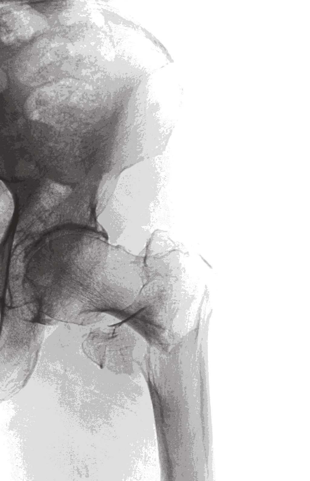
177
Cover Page. The handle holds various files of this Leiden University dissertation.
 Cover Page The handle http://hdl.handle.net/1887/38039 holds various files of this Leiden University dissertation. Author: Embden, Daphne van Title: Facts and fiction in hip fracture treatment Issue Date:
The Lateral Trochanteric Wall A Key Element in the Reconstruction of Unstable Pertrochanteric Hip Fractures
 CLINICAL ORTHOPAEDICS AND RELATED RESEARCH Number 425, pp. 82 86 2004 Lippincott Williams & Wilkins The Lateral Trochanteric Wall A Key Element in the Reconstruction of Unstable Pertrochanteric Hip Fractures
Cover Page. The handle holds various files of this Leiden University dissertation.
 Cover Page The handle http://hdl.handle.net/1887/38039 holds various files of this Leiden University dissertation. Author: Embden, Daphne van Title: Facts and fiction in hip fracture treatment Issue Date:
Pelvis injuries Fractures of the femur (proximal,shaft) Dr Tamás Bodzay
 Dr Tamás Bodzay") Pelvis injuries Fractures of the femur (proximal,shaft) Dr Tamás Bodzay Pelvis anatomy Pelvis function - axial load bearing - protection: abdominal, pelvic structures Pelvic injury mechanism Falling from
Pelvis injuries Fractures of the femur (proximal,shaft) Dr Tamás Bodzay Pelvis anatomy Pelvis function - axial load bearing - protection: abdominal, pelvic structures Pelvic injury mechanism Falling from
Technique Guide. DHS Blade. For osteoporotic bone.
 Technique Guide DHS Blade. For osteoporotic bone. Table of Contents Introduction Features and Benefits 2 Indications and Contraindications 4 Clinical Cases 5 Surgical Technique Implantation 6 Implant
9/24/2015. When Can I Use a SHS? When CAN T I Use a SHS? Sliding Hip Screw. Time proven. Technically simple. Cheap. Quick
 When Can I Use a SHS? Frank A. Liporace, MD Associate Professor Director of Orthopaedic Trauma Research Director of Orthopaedic Trauma Jersey City Medical Center New York University / Hospital for Joint
Assessment of Prognosis of Patients with Intertrochanteric Fractures Undergoing Treatment with PFN: An Observational Study
 Original article: Assessment of Prognosis of Patients with Intertrochanteric Fractures Undergoing Treatment with PFN: An Observational Study Gajraj Singh 1, Sandhya Gautam 2 1Assistant Professor, Department
Comparitive Study between Proximal Femoral Nailing and Dynamic Hip Screw in Intertrochanteric Fracture of Femur *
 Open Journal of Orthopedics, 2013, 3, 291-295 Published Online November 2013 (http://www.scirp.org/journal/ojo) http://dx.doi.org/10.4236/ojo.2013.37053 291 Comparitive Study between Proximal Femoral Nailing
Cover Page. The handle holds various files of this Leiden University dissertation
 Cover Page The handle http://hdl.handle.net/1887/38594 holds various files of this Leiden University dissertation Author: Haan, Melina C. den Title: Cell therapy in ischemic heart disease models : role
The Orthopaedic Enigma: A Simplified Classification
 ISPUB.COM The Internet Journal of Orthopedic Surgery Volume 3 Number 2 The Orthopaedic Enigma: A Simplified Classification D Iyer Citation D Iyer. The Orthopaedic Enigma: A Simplified Classification. The
ORIGINAL ARTICLE. INTER TROCHANTERIC # NECK FEMUR FIXATION WITH TFN 250 CASES. Prasad Vijaykumar Joshi, Chandrashekar Yadav.
 INTER TROCHANTERIC # NECK FEMUR FIXATION WITH TFN 250 CASES. Prasad Vijaykumar Joshi, Chandrashekar Yadav. 1. Assistant Professor. Department of Orthopaedics, Joshi Hospital Pvt. Ltd. Phaltan, Maharashtra.
Cover Page. The handle holds various files of this Leiden University dissertation.
 Cover Page The handle http://hdl.handle.net/1887/20570 holds various files of this Leiden University dissertation. Author: Zwaal, Peer van der Title: On rotator cuff tears : studies on evaluation, clinical
Femoral Neck Fractures- Gold standards and breaking news. Anna Ekman, MD, PhD Stockholm South hospital Sweden
 Femoral Neck Fractures- Gold standards and breaking news Anna Ekman, MD, PhD Stockholm South hospital Sweden Learning outcomes Learning Outcomes Epidemiology Preoperative planning Reduction techniques
A comparative study of 30 cases of trochanteric fracture femur treated with dynamic hip screw and proximal femoral nailing
 Original Article A comparative study of 30 cases of trochanteric fracture femur treated with dynamic hip screw and proximal femoral nailing Jaswinder Pal Singh Walia *, Himanshu Tailor**, H S Mann ***,
A Comparison of the Proximal Femoral Nail Antirotation Device and Dynamic Hip Screw in the Treatment of Unstable Pertrochanteric Fracture
 The Journal of International Medical Research 2010; 38: 1266 1275 A Comparison of the Proximal Femoral Nail Antirotation Device and Dynamic Hip Screw in the Treatment of Unstable Pertrochanteric Fracture
Aesculap Targon FN. Head Preserving Solution for Medial Femoral Neck Fractures. Aesculap Orthopaedics
 Aesculap Targon FN Head Preserving Solution for Medial Femoral Neck Fractures Aesculap Orthopaedics Targon FN Operating Technique Indications for Targon FN AO 3 B. AO 3 B.2 AO 3 B.3 Undisplaced intracapsular
DISLOCATION AND FRACTURES OF THE HIP. Dr Károly Fekete
 DISLOCATION AND FRACTURES OF THE HIP Dr Károly Fekete 1 OUTLINE Epidemiology Incidence Anatomy Patient s examination, clinical symptons Diagnosis Classification Management Special complications 2 EPIDEMIOLOGY,
JMSCR Vol. 03 Issue 08 Page August 2015
 www.jmscr.igmpublication.org Impact Factor 3.79 Index Copernicus Value: 5.88 ISSN (e)-2347-176x ISSN (p) 2455-0450 DOI: http://dx.doi.org/10.18535/jmscr/v3i8.08 Study of Functional and Radiological Outcome
Cover Page. The handle holds various files of this Leiden University dissertation
 Cover Page The handle http://hdl.handle.net/1887/23056 holds various files of this Leiden University dissertation Author: Velders, Matthijs A. Title: Optimization of care for ST-elevation myocardial infarction
Salvage of failed dynamic hip screw fixation of intertrochanteric fractures
 Injury, Int. J. Care Injured (2005) xxx, xxx xxx 1 www.elsevier.com/locate/injury 2 3 4 5 6 7 8 8 9 10 11 12 13 14 15 Salvage of failed dynamic hip screw fixation of intertrochanteric s G.Z. Said, O. Farouk
Comparison of two modality of fixation in unstable trochantric fractures in elderly patients
 Original article Comparison of two modality of fixation in unstable trochantric fractures in elderly patients 1Dr. Vipin Garg, 2 Dr. Anjul Agarwal 1MS Orhtopaedics, Assistant professor, Department of orthopaedics,
Intertrochanteric Femur Fractures. Alan Afsari, MD March 2014
 Intertrochanteric Femur Fractures Alan Afsari, MD March 2014 Topics epidemiology anatomy classification mechanism of injury patient assessment treatment rehabilitation complications Epidemiology 341k people
Randomized comparative study to evaluate the role of proximal femoral nail and dynamic hip screw in unstable trochanteric fractures
 International Journal of Research in Orthopaedics Mayi SC et al. Int J Res Orthop. 2016 Sep;2(3):75-79 http://www.ijoro.org Research Article DOI: http://dx.doi.org/10.18203/issn.2455-4510.intjresorthop20162618
HOW TO CITE THIS ARTICLE:
 A COMPARATIVE STUDY OF FUNCTIONAL OUTCOME BETWEEN DYNAMIC HIP SCREW AND PROXIMAL FEMORAL NAIL IN SURGICAL MANAGEMENT OF PER-TROCHANTERIC FRACTURES Umesh M. Shivanna 1, Girish H. Rudrappa 2 HOW TO CITE
The Journal of the Korean Society of Fractures Vol.16, No.1, January, 2003
 The Journal of the Korean Society of Fractures Vol16, No1, January, 2003 : 351 ( )463-712 TEL: (031) 780-5270/5271 FAX : (031) 708-3578 E-mail: bskima@netsgocom 16,, ( > 20mm ) 5, ) 20 % 1 ), 6,, 3 8 8
Journal of Orthopaedic Surgery and Research 2010, 5:62
 Journal of Orthopaedic Surgery and Research This Provisional PDF corresponds to the article as it appeared upon acceptance. Fully formatted PDF and full text (HTML) versions will be made available soon.
5/31/2018. Ipsilateral Femoral Neck And Shaft Fractures. Ipsilateral Neck-Shaft Fractures Introduction. Ipsilateral Neck-Shaft Fractures Introduction
 Ipsilateral Femoral Neck And Shaft Fractures Exchange Nailing For Non- Union Donald Wiss MD Cedars-Sinai Medical Center Los Angeles, California Introduction Uncommon Injury Invariably High Energy Trauma
PFN in trochanteric fractures: Clinico radiological outcome study
 2018; 4(2): 943-947 ISSN: 2395-1958 IJOS 2018; 4(2): 943-947 2018 IJOS www.orthopaper.com Received: 14-02-2018 Accepted: 15-03-2018 R Amarnath Senior Asst. Professor, Department of Orthopaedic Surgery,
Functional evaluation of proximal femoral fractures managed with cephalomedullary nailing by oxford hip score - A prospective study
 2017; 3(3): 980-985 ISSN: 2395-1958 IJOS 2017; 3(3): 980-985 2017 IJOS www.orthopaper.com Received: 13-05-2017 Accepted: 14-06-2017 Pranav Kothiyal Senior Resident, Department of Orthopaedics, SGRRIMHS,
ICUC Paper. The treatment of trochanteric fractures revisited: Pietro Regazzoni, Alberto Fernandez, Dominik Heim, Stephan M. Perren.
 The treatment of trochanteric fractures revisited: Pietro Regazzoni, Alberto Fernandez, Dominik Heim, Stephan M. Perren September 2016 An optimal treatment of hip fractures is crucial because of the great
Cover Page. The handle holds various files of this Leiden University dissertation.
 Cover Page The handle http://hdl.handle.net/1887/42781 holds various files of this Leiden University dissertation. Author: Elbaz M.S.M.M. Title: Three-dimensional in-vivo intra-cardiac vortex flow from
PFNA-II. Proximal Femoral Nail Antirotation.
 PFNA-II. Proximal Femoral Nail Antirotation. Surgical Technique This publication is not intended for distribution in the USA. Instruments and implants approved by the AO Foundation. PFNA-II. Proximal Femoral
Comparative Study of Fixation Devices for Intertrochanteric Fractures
 Comparative Study of Fixation Devices for Intertrochanteric Fractures C. Sticlaru * A. Davidescu Politehnica University of Timişoara Politehnica University of Timişoara Timişoara, România Timişoara, România
JMSCR Vol 05 Issue 07 Page July 2017
 www.jmscr.igmpublication.org Impact Factor 5.84 Index Copernicus Value: 83.27 ISSN (e)-2347-176x ISSN (p) 2455-0450 DOI: https://dx.doi.org/10.18535/jmscr/v5i7.29 Clinical Study of Osteosynthesis in Fracture
Clinical outcomes of muscle pedicle bone grafting (Meyer's Procedure) in cases of old displaced femur neck fractures: A Study Of 20 Cases
 in cases of old displaced femur neck fractures: A Study Of 20 Cases") ISPUB.COM The Internet Journal of Orthopedic Surgery Volume 10 Number 1 Clinical outcomes of muscle pedicle bone grafting (Meyer's Procedure) in cases of old displaced femur neck fractures: A Study Of
ISPUB.COM The Internet Journal of Orthopedic Surgery Volume 10 Number 1 Clinical outcomes of muscle pedicle bone grafting (Meyer's Procedure) in cases of old displaced femur neck fractures: A Study Of
Page Proof 1 of 5. Fig. E1-A The INTERTAN nail was short or long.
 Page 1 of 5 Fig. E1-A The INTERTAN nail was short or long. Fig. E1-B The sliding hip screw comes in different lengths, and is used with or without a trochanteric stabilizing plate. Page Proof 1 of 5 Page
The choice of internal fixator for fractures around the femoral trochanter depends on area classification
 DOI 10.1186/s40064-016-3206-1 RESEARCH Open Access The choice of internal fixator for fractures around the femoral trochanter depends on area classification Hiroaki Kijima 1,2*, Shin Yamada 1,2, Natsuo
Index. B Backslap technique depth assessment, 82, 83 diaphysis distal trocar, 82 83
 Index A Acromial impingement, 75, 76 Aequalis intramedullary locking avascular necrosis, 95 central humeral head, 78, 80 clinical and functional outcomes, 95, 96 design, 77, 79 perioperative complications,
Types of Plates 1. New Dynamic Compression Plate: Diaphyseal fracture: Radius, Ulna, Humerus, Rarely tibia
 Types of Plates 1. New Dynamic Compression Plate: DCP Diaphyseal fracture: Radius, Ulna, Humerus, Rarely tibia 1. Undercut adjacent to the holes low contact: less stress shield 2. Undercut at the undersurface
MRIMS Journal of Health Sciences 2016;4(1) pissn: , eissn:
 pissn: , eissn:") MRIMS Journal of Health Sciences 216;4(1) pissn: 2321-76, eissn: 2321-7294 http://www.mrimsjournal.com/ Original Article A comparative study of proximal femoral nailing and dynamic hip screw in the management
MRIMS Journal of Health Sciences 216;4(1) pissn: 2321-76, eissn: 2321-7294 http://www.mrimsjournal.com/ Original Article A comparative study of proximal femoral nailing and dynamic hip screw in the management
76 F: Plays tennis, lives independently, told she has weak bone
 Femoral Neck Fractures In the ELDERLY What to do, when and why Frank Liporace, MD Chairman & VP, Dept of Orthopaedics Chief of Trauma & Adult Reconstruction Jersey City Medical Center / RWJ Barnabas Health
Failed Subtrochanteric Fracture How I Decide What to Do?
 Failed Subtrochanteric Fracture How I Decide What to Do? Gerald E. Wozasek Thomas M. Tiefenboeck 5 October 2016, Washington Medical University of Vienna, Department of Trauma Surgery ordination @wozasek.at
INTERTROCHANTERIC FEMORAL FRACTURES TREATED BY DYNAMIC HIP SCREW
 29 INTERTROCHANTERIC FEMORAL FRACTURES TREATED BY DYNAMIC HIP SCREW Muhammad Ayoub Laghari, Asadullah Makhdoom, Pir Abdul latif Qureshi, Abbass Memon, Faheem Ahmed Memon, Professor Khaleeque Ahmed Siddiqui
Cover Page. The handle holds various files of this Leiden University dissertation.
 Cover Page The handle http://hdl.handle.net/1887/38506 holds various files of this Leiden University dissertation. Author: Nies, Jessica Annemarie Bernadette van Title: Early identification of rheumatoid
Review of Proximal Nail Antirotation (PFNA) and PFNA-2 Our Local Experience
 and PFNA-2 Our Local Experience") doi: 10.5704/MOJ.1107.001 Review of Proximal Nail Antirotation (PFNA) and PFNA-2 Our Local Experience WL Loo, M Med Orth, SYJ Loh, FRCS (Edin), HC Lee, FRCS (Edin) Department of Orthopaedic Surgery, Changi
doi: 10.5704/MOJ.1107.001 Review of Proximal Nail Antirotation (PFNA) and PFNA-2 Our Local Experience WL Loo, M Med Orth, SYJ Loh, FRCS (Edin), HC Lee, FRCS (Edin) Department of Orthopaedic Surgery, Changi
Multimodality imaging in chronic coronary artery disease Maureen M. Henneman
 Multimodality imaging in chronic coronary artery disease Maureen M. Henneman 080237 Henneman boek.indb 1 03-11-2008 10:56:25 The studies described in this thesis were performed at the Department of Cardiology
STUDY OF RESULTS OF ENDER NAILING AND CANNULATED CANCELLOUS SCREW IN THE TREATMENT OF INTERTROCHANTERIC FRACTURE FEMUR
 RESEARCH ARTICLE STUDY OF RESULTS OF ENDER NAILING AND CANNULATED CANCELLOUS SCREW IN THE TREATMENT OF INTERTROCHANTERIC FRACTURE FEMUR Bhavik Dalal, Tarkik Amin, Archit Gandhi, Rohit Shah Smt NHL Municipal
Proximal Humerus Fractures: contemporary perspectives
 Proximal Humerus Fractures: contemporary perspectives Diego L Fernandez M.D Professor of Orthopaedic Surgery Department of Orthopaedic Surgery Lindenhof Hospital, Berne, Switzerland www.diegofernandez.ch
Multimodality Imaging of Anatomy and Function in Coronary Artery Disease. Joanne D. Schuijf
 Multimodality Imaging of Anatomy and Function in Coronary Artery Disease Joanne D. Schuijf The research described in this thesis was performed at the departments of Cardiology and Radiology of the Leiden
The Mechanical Properties Of Fixing Greater Trochanter Or Lesser Trochanter In Complex Four Part Intertrochanteric Fractures
 The Mechanical Properties Of Fixing Greater Trochanter Or Lesser Trochanter In Complex Four Part Intertrochanteric Fractures Chih-Hui Chen 1, Kui-Chou Huang 2, Cheng-Kung Cheng, PhD 3, Hung-Chan Kao 4.
University of Groningen. Functional outcome after a spinal fracture Post, Richard Bernardus
 University of Groningen Functional outcome after a spinal fracture Post, Richard Bernardus IMPORTANT NOTE: You are advised to consult the publisher's version (publisher's PDF) if you wish to cite from
Trochanter Stabilization Plate for DHS Implants
 Extends DHS Plate Construct to Help Stabilize Greater Trochanter Trochanter Stabilization Plate for DHS Implants Surgical Technique Table of Contents Introduction Trochanter Stabilization Plate for DHS
Treatment of Femoral Intertrochanteric Fracture with Proximal Femoral Nail
 17, 1, 20041 Journal of the Korean Fracture Society Vol. 17, No. 1, January, 2004 Treatment of Femoral Intertrochanteric Fracture with Proximal Femoral Nail Bum-Soo Kim, M.D., Sogu Lew, M.D., Sang-Hun
Provision of Rotational Stability: Prevention of Collapse: Closed Fracture Reduction: Minimally Invasive Surgery with no Exposure of the Fracture:
 INTRODUCTION Percutaneous Compression Plating was developed by considering each of the stages in the surgical procedure for pertrochanteric fractures and the ways in which these might be improved. Primary
Importance of screw position in intertrochanteric femoral fractures treated by dynamic hip screw
 Orthopaedics & Traumatology: Surgery & Research (2010) 96, 21 27 ORIGINAL ARTICLE Importance of screw position in intertrochanteric femoral fractures treated by dynamic hip screw M. Güven a,, U. Yavuz
Internal fixation of femoral neck fractures
 Acta Orthop Scand 55, 423-429, 1984 Internal fixation of femoral neck fractures Compression screw compared with nail plate fixation In a prospective, randomized study of femoral neck fracture operations,
Use Of A Long Femoral Stem In The Treatment Of Proximal Femoral Fractures: A Report Of Four Cases
 ISPUB.COM The Internet Journal of Orthopedic Surgery Volume 5 Number 1 Use Of A Long Femoral Stem In The Treatment Of Proximal Femoral Fractures: A Report Of Four Cases C Yu, V Singh Citation C Yu, V Singh..
ABSTRACT INTRODUCTION. Hui Xie 1,*, Zhan Wang 2,*, Junji Zhang 3, Langhai Xu 2 and Bao Chen 1. Research Paper
 /, 2017, Vol. 8, (No. 47), pp: 82700-82704 Clinical outcome of dynamic hip locking plates and proximal femoral nails anti-rotation-asia for treating intertrochanteric femur fracture with lateral wall fractures
Type I : At the level of lesser trochanter Type II : Less than 2.5 cm below lesser trochanter. Type III : cm below lesser trochanter
 Type II : Major fracture line along the intertrochanteric line with communition in coronal plain. Type III : Fracture at the level of lesser trochanter with variable communition and extension in subtrochanteric
4/28/2010. Fractures. Normal Bone and Normal Ossification Bone Terms. Epiphysis Epiphyseal Plate (physis) Metaphysis
 Metaphysis") Fractures Normal Bone and Normal Ossification Bone Terms Epiphysis Epiphyseal Plate (physis) Metaphysis Diaphysis 1 Fracture Classifications A. Longitudinal B. Transverse C. Oblique D. Spiral E. Incomplete
Fractures Normal Bone and Normal Ossification Bone Terms Epiphysis Epiphyseal Plate (physis) Metaphysis Diaphysis 1 Fracture Classifications A. Longitudinal B. Transverse C. Oblique D. Spiral E. Incomplete
INTER- AND INTRA INDIVIDUAL VARIATION IN EARPRINTS LYNN M EIJERM AN
 INTER- AND INTRA INDIVIDUAL VARIATION IN EARPRINTS LYNN M EIJERM AN Inter- and intra-individual variation in earprints Meijerman, Lynn Thesis University Leiden With Ref. ISBN-10: 90-806456-9-9 ISBN-13:
Femoral Neck Fractures
 Femoral Neck Fractures Michael Monge, Harvard Medical School Agenda Epidemiology Normal anatomy of the femur Garden classifications Patients Summary 1 Epidemiology 1 250,000 yearly hip fractures in the
Cover Page. The handle holds various files of this Leiden University dissertation.
 Cover Page The handle http://hdl.handle.net/1887/23937 holds various files of this Leiden University dissertation. Author: Liu, Zhen Title: Exploring the interplay between TGF-β and VEGF signalling in
Echocardiographic evaluation of left ventricular function in ischemic heart disease. Sjoerd A. Mollema
 Echocardiographic evaluation of left ventricular function in ischemic heart disease Sjoerd A. Mollema The studies described in this thesis were performed at the Department of Cardiology of the Leiden University
designed to advance the treatment of hip fractures.
 designed to advance the treatment of hip fractures. introducing the tfn-advanced proximal femoral nailing system (tfna). the tfna system is a new system designed to solve a wide range of unmet needs for
Fractures of the tibia shaft treated with locked intramedullary nail Retrospective clinical and radiographic assesment
 ARS Medica Tomitana - 2013; 4(75): 197-201 DOI: 10.2478/arsm-2013-0035 Șerban Al., Botnaru V., Turcu R., Obadă B., Anderlik St. Fractures of the tibia shaft treated with locked intramedullary nail Retrospective
Citation for published version (APA): Gerritsma-Bleeker, C. L. E. (2005). Long-term follow-up of the SKI knee prosthesis s.n.
: Gerritsma-Bleeker, C. L. E. (2005). Long-term follow-up of the SKI knee prosthesis s.n.") University of Groningen Long-term follow-up of the SKI knee prosthesis Gerritsma-Bleeker, Catharina Louise Emilie IMPORTANT NOTE: You are advised to consult the publisher's version (publisher's PDF) if
University of Groningen Long-term follow-up of the SKI knee prosthesis Gerritsma-Bleeker, Catharina Louise Emilie IMPORTANT NOTE: You are advised to consult the publisher's version (publisher's PDF) if
DISCLOSURE FNFX ORIF OR ARTHROPLASTY? 11/21/2016 FEMORAL NECK FRACTURES: ORIF OR ARTHROPLASTY? ROYALTIES DEPUY, BIOMET
 FEMORAL NECK FRACTURES: ORIF OR ARTHROPLASTY? GEORGE HAIDUKEWYCH, MD ORLANDO, FLORIDA DISCLOSURE ROYALTIES DEPUY, BIOMET CONSULTING DEPUY, SYNTHES, BIOMET, RESPONSIVE ORTHOPEDICS STOCK OWNERSHIP ORTHOPEDIATRICS
LISS DF and LISS PLT. Less Invasive Stabilization Systems for Distal Femur and Proximal Lateral Tibia.
 LISS DF and LISS PLT. Less Invasive Stabilization Systems for Distal Femur and Proximal Lateral Tibia. LISS DF and LISS PLT. Less Invasive Stabilization Systems for Distal Femur and Proximal Lateral Tibia.
Missed hip fractures M. J. PARKER. undisplaced, but as a consequence of the delay in diagnosis displacement occurred SUMMARY
 Archives of Emergency Medicine, 1992, 9, 23-27 Missed hip fractures M. J. PARKER Peterborough District Hospital, SUMMARY Thorpe Road, Peterborough From a series of 825 consecutive admissions with a hip
Preoperative Planning. The primary objectives of preoperative planning are to:
 Preoperative Planning The primary objectives of preoperative planning are to: - Determine preoperative leg length discrepancy. - Assess acetabular component size and placement. - Determine femoral component
Medial circumflex artery Lateral circumflex artery
 Femoral Head Fractures: A Critical But Frequently Missed Injury Susanna C. Spence MD Manickam Kumaravel MBBS University of Texas Health Science Center at Houston Background Femoral head fractures: A complication
Predicting the Position of the Femoral Head Center
 The Journal of Arthroplasty Vol. 14 No. 1 1999 Predicting the Position of the Femoral Head Center Nobuhiko Sugano, MD, Philip C. Noble, PhD, and Emir Kamaric, MS Abstract: To find an accurate method to
Citation for published version (APA): van der Paardt, M. P. (2015). Advances in MRI for colorectal cancer and bowel motility
: van der Paardt, M. P. (2015). Advances in MRI for colorectal cancer and bowel motility") UvA-DARE (Digital Academic Repository) Advances in MRI for colorectal cancer and bowel motility van der Paardt, M.P. Link to publication Citation for published version (APA): van der Paardt, M. P. (2015).
UvA-DARE (Digital Academic Repository) Advances in MRI for colorectal cancer and bowel motility van der Paardt, M.P. Link to publication Citation for published version (APA): van der Paardt, M. P. (2015).
Cover Page. The handle holds various files of this Leiden University dissertation.
 Cover Page The handle http://hdl.handle.net/1887/35777 holds various files of this Leiden University dissertation. Author: Wijffels, Mathieu Mathilde Eugene Title: The clinical and non-clinical aspects
Peritroch Hip Fractures. Robert M Harris MD. Hip Fractures. Factors Influencing Construct Strength: Uncontrolled factors 4/28/2016
 Peritroch Hip Fractures Should be treated with an IMHS Robert M Harris MD Hip Fractures General principles Approximately 250,000 hip fractures/ year Cost approximately $8.7 billion annually The number
The role of the general practitioner in the care for patients with colorectal cancer Brandenbarg, Daan
 University of Groningen The role of the general practitioner in the care for patients with colorectal cancer Brandenbarg, Daan IMPORTANT NOTE: You are advised to consult the publisher's version (publisher's
University of Groningen. Cost and outcome of liver transplantation van der Hilst, Christian
 University of Groningen Cost and outcome of liver transplantation van der Hilst, Christian IMPORTANT NOTE: You are advised to consult the publisher's version (publisher's PDF) if you wish to cite from
Cover Page. The handle holds various files of this Leiden University dissertation
 Cover Page The handle http://hdl.handle.net/1887/22544 holds various files of this Leiden University dissertation Author: Speksnijder, Niels Title: Determinants of psychosis vulnerability : focus on MEF2
Crossed Steinmann Pin Fixation In Supracondylar Femur Fractures In Adults A Case Series
 Article ID: WMC005027 ISSN 2046-1690 Crossed Steinmann Pin Fixation In Supracondylar Femur Fractures In Adults A Case Series Peer review status: No Corresponding Author: Dr. Mohit K Jindal, Senior Resident,
ROTATIONAL PILON FRACTURES
 CHAPTER 31 ROTATIONAL PILON FRACTURES George S. Gumann, DPM The opinions and commentary of the author should not be construed as refl ecting offi cial U.S. Army Medical Department policy. Pilon injuries
Posterolateral elbow dislocation with entrapment of the medial epicondyle in children: a case report Juan Rodríguez Martín* and Juan Pretell Mazzini
 Open Access Case report Posterolateral elbow dislocation with entrapment of the medial epicondyle in children: a case report Juan Rodríguez Martín* and Juan Pretell Mazzini Address: Department of Orthopaedic
Accurate and Easy Measurement of Sliding Distance of Intramedullary Nail in Trochanteric Fracture
 Original Article Clinics in Orthopedic Surgery 2015;7:152-157 http://dx.doi.org/10.4055/cios.2015.7.2.152 Accurate and Easy Measurement of Sliding Distance of Intramedullary Nail in Trochanteric Fracture
Cover Page. The handle holds various files of this Leiden University dissertation.
 Cover Page The handle http://hdl.handle.net/1887/20959 holds various files of this Leiden University dissertation. Author: Diepstraten, Jeroen Title: The influence of morbid obesity on the pharmacokinetics
Valgus subtrochanteric osteotomy for malunited intertrochanteric fractures : Our experience in 5 cases
 Original article : Valgus subtrochanteric osteotomy for malunited intertrochanteric fractures : Our experience in 5 cases Rajendraprasad Butala *, Sunirmal Mukherjee, Prakash Samant, Ravindra Khedekar
Angular Malalignment in Subtrochanteric and Proximal Shaft Femur Fractures after Intramedullary Nailing using SIGN Nails
 Angular Malalignment in Subtrochanteric and Proximal Shaft Femur Fractures after Intramedullary Nailing using SIGN Nails Rolando Junior L. Torres, MD Jeremiah R. Morales, MD, FPOA Subtrochanteric Femur
Hip Fractures. Anatomy. Causes. Symptoms
 Hip Fractures A hip fracture is a break in the upper quarter of the femur (thigh) bone. The extent of the break depends on the forces that are involved. The type of surgery used to treat a hip fracture
Outcomes of Cephalomedullary Nailing in Basicervical Fracture
 ORIGINAL ARTICLE Hip Pelvis 29(4): 270-276, 2017 http://dx.doi.org/10.5371/hp.2017.29.4.270 Print ISSN 2287-3260 Online ISSN 2287-3279 Outcomes of Cephalomedullary Nailing in Basicervical Fracture Seok
PFNA. With Augmentation Option.
 PFNA. With Augmentation Option. Superior anchor in osteoporotic bone Simple and reproducible procedure through standardized technique Intraoperative decision for augmentation This publication is not intended
Distal Femur Fractures in The Elderly The Ideal Construct
 Distal Femur Fractures in The Elderly The Ideal Construct Tak-Wing Lau Department of Orthopaedics and Traumatology Queen Mary Hospital The University of Hong Kong Singapore Trauma 2015 Trauma Through the
Comparative study between proximal femoral nail and proximal femoral nail antirotation in management of unstable trochanteric fractures
 International Journal of Research in Orthopaedics Kashid MR et al. Int J Res Orthop. 2016 Dec;2(4):354-358 http://www.ijoro.org Original Research Article DOI: http://dx.doi.org/10.18203/issn.2455-4510.intjresorthop20164168
3D workflows in orthodontics, maxillofacial surgery and prosthodontics van der Meer, Wicher
 University of Groningen 3D workflows in orthodontics, maxillofacial surgery and prosthodontics van der Meer, Wicher IMPORTANT NOTE: You are advised to consult the publisher's version (publisher's PDF)
Long-stem revision prosthesis for salvage of failed fixation of extracapsular proximal femoral fractures
 Acta Orthop. Belg., 2009, 75, 340-345 ORIGINAL STUDY Long-stem revision prosthesis for salvage of failed fixation of extracapsular proximal femoral fractures Rory J. SHARVILL, Nicholas A. FERRAN, Huw G.
Management of Hip Fractures
 Management of Hip Fractures in the Elderly Patient David A. Brown MD COL U.S. Army Ret. The Center for Orthopedics and Neurosurgery Optimizing Management of Hip Fractures in the Elderly Patient Optimizing
Cardiac Bone Marrow Cell Injection for Chronic Ischemic Heart Disease
 Cardiac Bone Marrow Cell Injection for Chronic Ischemic Heart Disease Monique R.M. Jongbloed 070303 Beeres Boek.indb 1 06-09-2007 17:13:27 The studies described in this thesis were performed at the Department
Orthopedics in Motion Tristan Hartzell, MD January 27, 2016
 Orthopedics in Motion 2016 Tristan Hartzell, MD January 27, 2016 Humerus fractures Proximal Shaft Distal Objectives 1) Understand the anatomy 2) Epidemiology and mechanisms of injury 3) Types of fractures
Technique Guide. LCP Proximal Femoral Hook Plate 4.5/5.0. Part of the LCP Periarticular Plating System.
 Technique Guide LCP Proximal Femoral Hook Plate 4.5/5.0. Part of the LCP Periarticular Plating System. Table of Contents Introduction Features and Benefits 2 AO ASIF Principles 4 Indications 5 Surgical
Encina Taper Stem. Stinson Orthopedics Inc. 303 Twin Dolphin Drive, Suite 600 Redwood City, CA
 Stinson Orthopedics Inc. 303 Twin Dolphin Drive, Suite 600 Redwood City, CA 94065 info@stinsonortho.com www.stinsonortho.com Table of Contents Introduction 3 Features 4 Surgical Technique 5 Preoperative
Testicular microlithiasis and undescended testis. Joery Goede
 Testicular microlithiasis and undescended testis Joery Goede ISBN : 978-94-91082-02-3 Author : Joery Goede Lay-out : Ruth Visser, VisserVisuals, Amsterdam, the Netherlands Printing : Ridderprint, the Netherlands
Data Mining Scenarios. for the Discoveryof Subtypes and the Comparison of Algorithms
 Data Mining Scenarios for the Discoveryof Subtypes and the Comparison of Algorithms Data Mining Scenarios for the Discoveryof Subtypes and the Comparison of Algorithms PROEFSCHRIFT ter verkrijging van