MRI of the Knee: Part 4 - normal variants that may simulate disease. Mark Anderson, M.D. University of Virginia
|
|
|
- Eustacia Blake
- 6 years ago
- Views:
Transcription

1 MRI of the Knee: Part 4 - normal variants that may simulate disease Mark Anderson, M.D. University of Virginia

2 discuss the most common normal variants in the pediatric knee that may simulate pathology on MR imaging. identify a cortical desmoid and describe its typical appearance and location on MR images Learning Objectives At the end of the presentation, each participant should be able to: list the four types of synovial plicae in the knee as well as their clinical significance.
3 The Knee: normal variants Bipartite patella Dorsal defect of the patella Cortical desmoid Distal femoral epiphyseal irregularity Posterior stripe Juvenile cartilage signal intensity Terminal sulcus cartilage thinning Semimembranosus insertions Lateral inferior geniculate vessels Meniscus flounce Meniscal ossicle Plicae Discoid meniscus Fabello-fibular ligament Meniscofibular ligament Popliteofibular lgament Tibial attachment of the biceps femoris Transverse meniscal ligament Meniscofemoral ligaments Oblique meniso-meniscal ligament Double barreled PCL Meniscal root attachments Patello-meniscal ligament Fabella Cyamella Accessory popliteus tendon Bifurcated popliteus 3 rd head of the gastrocnemius muscle Bifurcating sartorius tendon
4 The Knee: normal variants Bone Bipartite patella Dorsal defect of the patella Cortical desmoid Irregular ossification vs. juvenile OCD Posterior stripe Menisci Meniscal roots Transverse ligament Meniscofemoral ligaments Semimembranosus insertion Lateral inferior geniculate vessels Meniscal ossicle Cartilage Juvenile cartilage signal intensity Terminal sulcus cartilage thinning Upper trochlear defect Plicae Medial patellar Suprapatellar Infrapatellar
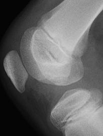
5 Bones: Bipartite patella Patellar ossification primary center: 4-6 yrs secondary centers: 8-12 yrs failure of fusion 4 yr old male Bipartite 2-3% Bilateral 50% Types (Saupe) 1 inferior pole (5%) 2 lateral margin (20%) 3 superolateral (75%)

 1 inferior pole (5%) 2 lateral margin (20%) 3")
6 Bones: Bipartite patella Patellar ossification primary center: 4-6 yrs secondary centers: 8-12 yrs failure of fusion 4 yr old male Bipartite 2-3% Bilateral 50% Types (Saupe) 1 inferior pole (5%) 2 lateral margin (20%) 3 superolateral (75%)

7 Bones: bipartite patella Symptomatic acute / chronic trauma MRI fracture / avulsion may be overlooked as etiology edema along margins Kavanagh, Skeletal Radiol pts knee pain only MRI finding: edema along bipartite patella

8 Bones: dorsal defect of the patella Unknown etiology Incidence 0.3 1% / bilat - up to 30% may be seen with bipartite Appearance well circumscribed round, lytic lesion superolateral patella MRI lack of edema evaluate overlying cartilage

9 Bones: cortical desmoid AKA distal femoral cortical irregularity avulsive cortical irregularity periosteal / juxtacortical desmoid Avulsive / tug etiology reactive, fibro-osseous lesion Medial supracondylar femur lytic concave medial head of gastroc proliferative adductor magnus

10 Bones: cortical desmoid Radiographic DDx: MRI FCD, distal femoral stripe Neoplasm Infection T1 - SI T2 - SI low SI rim classic location

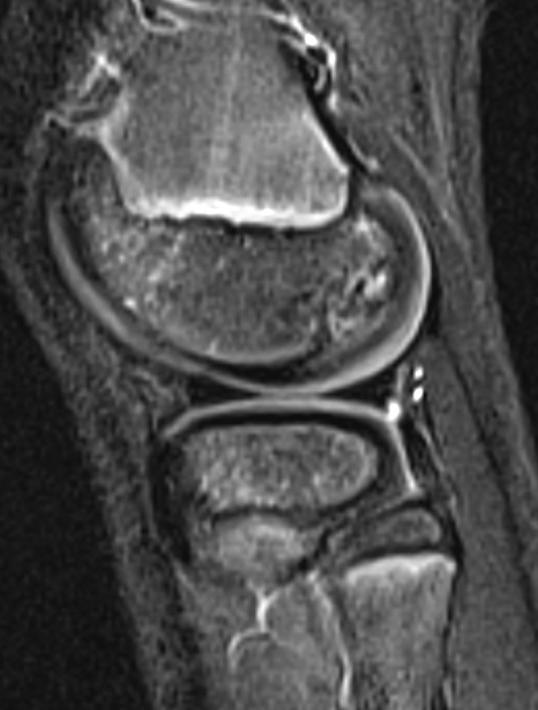
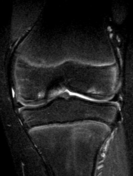
11 Bones: distal femoral irregularity Normal variation vs. OCD Uneven mineralization 3 13 yrs old related to rapid growth usually posterior LFC Appearance spiculation, puzzle piece overlying cartilage intact lack of marrow edema

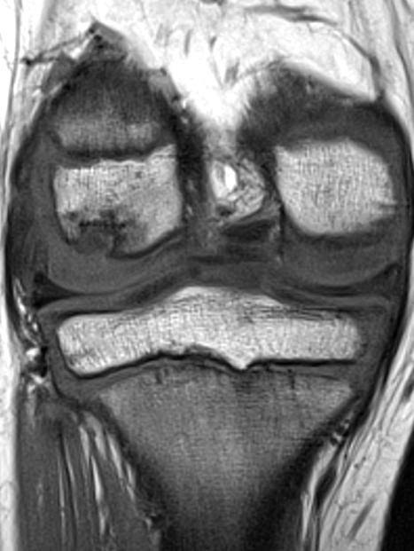
12 11 yr old male
 more commonly bilateral + LFC MRI signs of fragment instability less predictive than in adult Kijowski, Radiology")
13 Bones: Juvenile OCD Juvenile OCD open physes mean age: yrs central 1/3 + intercondylar adjacent edema common Vs. Adult better prognosis (80% resolve) more commonly bilateral + LFC MRI signs of fragment instability less predictive than in adult Kijowski, Radiology 2008 Gebarski, Pediatr Radiol 2005


14 10 yr old male Med Med Lat Lat


15 4 years later (14 yo) Lat Med Med
16 Bones: normal vs. OCD Normal Ossification OCD Age 3-13 yrs (not seen F>10 M>13) Avg age: yrs (not seen < 8 yrs) Location Posterior 1/3 MFC = LFC Middle 1/3 Intercondylar Lesion angle Deeper More steep Elongated More shallow Bilateral 25% 11% Marrow Edema Uncommon Common Jans, Radiology 2010
 SI wgt bearing surface Later (3-5 yrs) increased SI stippled well defined Varich, Radiology 2000 Laor, Radiology")
17 Epiphyseal Cartilage: signal intensity Age related changes 24 6 yr old female Early (< 1 yr) homogeneous Wgt-bearing (1-3 yrs) SI wgt bearing surface Later (3-5 yrs) increased SI stippled well defined Varich, Radiology 2000 Laor, Radiology 2009

18 Cartilage: lateral sulcus thinning Terminal sulcus lateral femoral condyle separates trrochlear from wgt-bearing cartilage cartilage appears thinned (esp on sagittal images)


19 Cartilage: upper trochlear defect Axial scans Fat saturation Above articular cartilage Cross ref with sagittal


20 Cartilage: upper trochlear defect Axial scans Fat saturation Cross-reference sagittal above articular cartilage Asymmetric cartilage lateral extends more proximally MED LAT
 MEDIAL")
21 Synovium: Plicae Embryologic remnants peripheral cavitations fail to coalesce synovial folds three compartments Types infrapatellar suprapatellar mediopateallar lateral (rare) MEDIAL INFRAPAT

22 Medial plica

23 Lateral plica


24 Synovium: Plicae Plica Syndrome? mediopatellar thickens impinges on femur/patella cartilage impingement lesion MR Findings appearance does not correlate with symptoms Boles, JCAT 2004 Weckstrom, The Knee 2010 Demirag, Knee Surg Sports Traumatol Arthrosc 2006
25 The Knee: normal variants Bone Bipartite patella Dorsal defect of the patella Cortical desmoid Irregular ossification vs. juvenile OCD Posterior stripe Menisci Meniscal roots Transverse ligament Meniscofemoral ligaments Semimembranosus insertion Lateral inferior geniculate vessels Meniscal ossicle Cartilage Juvenile cartilage signal intensity Terminal sulcus cartilage thinning Upper trochlear defect Plicae Medial patellar Suprapatellar Infrapatellar
MRI of the Knee: Part 2 - menisci. Mark Anderson, M.D. University of Virginia Health System
 MRI of the Knee: Part 2 - menisci Mark Anderson, M.D. University of Virginia Health System Learning Objectives At the end of the presentation, each participant should be able to: describe the normal anatomy
The Knee. Prof. Oluwadiya Kehinde
 The Knee Prof. Oluwadiya Kehinde www.oluwadiya.sitesled.com The Knee: Introduction 3 bones: femur, tibia and patella 2 separate joints: tibiofemoral and patellofemoral. Function: i. Primarily a hinge joint,
Joints of the Lower Limb II
 Joints of the Lower Limb II Lecture Objectives Describe the components of the knee and ankle joint. List the ligaments associated with these joints and their attachments. List the muscles acting on these
Bone and soft tissue variants of knee with Magnetic Resonance.
 Bone and soft tissue variants of knee with Magnetic Resonance. Poster No.: C-2420 Congress: ECR 2013 Type: Educational Exhibit Authors: M. C. Ruibal Villanueva, P. Sucasas-Hermida, C. Saborido Avila, M.
Imaging the Athlete s Knee. Peter Lowry, MD Musculoskeletal Radiology University of Colorado
 Imaging the Athlete s Knee Peter Lowry, MD Musculoskeletal Radiology University of Colorado None Disclosures Knee Imaging: Radiographs Can be performed weight-bearing or non-weight-bearing View options
The Knee. Tibio-Femoral
 The Knee Tibio-Femoral Osteology Distal Femur with Proximal Tibia Largest Joint Cavity in the Body A modified hinge joint with significant passive rotation Technically, one degree of freedom (Flexion/Extension)
MY PATIENT HAS KNEE PAIN. David Levi, MD Chief, Division of Musculoskeletal l limaging Atlantic Medical Imaging
 MY PATIENT HAS KNEE PAIN David Levi, MD Chief, Division of Musculoskeletal l limaging Atlantic Medical Imaging Causes of knee pain Non traumatic Trauma Osteoarthritis Patellofemoral pain Menisci or ligaments
The Knee Joint By Prof. Dr. Muhammad Imran Qureshi
 The Knee Joint By Prof. Dr. Muhammad Imran Qureshi Structurally, it is the Largest and the most complex joint in the body because of the functions that it performs: Allows mobility (flexion/extension)
Knee Contusions and Stress Injuries. Laura W. Bancroft, M.D.
 Knee Contusions and Stress Injuries Laura W. Bancroft, M.D. Objectives Review 5 types of contusion patterns Pivot shift Dashboard Hyperextension Clip Lateral patellar dislocation Demonstrate various stress
Knee MRI Update Case Review 2009 Russell C. Fritz, M.D. National Orthopedic Imaging Associates San Francisco, CA
 Knee MRI Update Case Review 2009 Russell C. Fritz, M.D. National Orthopedic Imaging Associates San Francisco, CA Meniscal Tears -linear increased signal extending to an articular surface is the hallmark
To describe he knee joint, ligaments, structure & To list the main features of other lower limb joints
 To describe he knee joint, ligaments, structure & neurovascular supply To demonstrate the ankle joint anatomy To list the main features of other lower limb joints To list main groups of lymph nodes in
The Knee. Two Joints: Tibiofemoral. Patellofemoral
 Evaluating the Knee The Knee Two Joints: Tibiofemoral Patellofemoral HISTORY Remember the questions from lecture #2? Girth OBSERVATION TibioFemoral Alignment What are the consequences of faulty alignment?
BASELINE QUESTIONNAIRE (SURGEON)
") SECTION A: STUDY INFORMATION Subject ID: - - Study Visit: Baseline Site Number: Date: / / Surgeon ID: SECTION B: INITIAL SURGEON HISTORY B1. Previous Knee Surgery: Yes No Not recorded B2. Number of Previous
SECTION A: STUDY INFORMATION Subject ID: - - Study Visit: Baseline Site Number: Date: / / Surgeon ID: SECTION B: INITIAL SURGEON HISTORY B1. Previous Knee Surgery: Yes No Not recorded B2. Number of Previous
Stage-I osteochondritis dissecans versus normal variants of ossification in the knee in children
 Pediatr Radiol (2005) 35: 880 886 DOI 10.1007/s00247-005-1507-6 ORIGINAL ARTICLE Kathleen Gebarski Ramiro J. Hernandez Stage-I osteochondritis dissecans versus normal variants of ossification in the knee
MENISCAL INJURY. Meniscus. Anterior Roots. Medial Meniscus. Lateral Meniscus. Posterior Roots. MRI and Arthroscopic Findings
 Meniscus Anterior Roots MENISCAL INJURY MRI and Arthroscopic Findings Medial Meniscus AH PH PH AH Lateral Meniscus Rawiwan Pattaweerakul Naresuan University Hospital Posterior Roots Meniscus Normal Meniscus
Standardised. knee. scanning of the. Basic pathology. Nemanja Damjanov. University of Belgrade Institute of Rheumatology
 Standardised scanning of the Nemanja Damjanov University of Belgrade Institute of Rheumatology knee Basic pathology Disclosure Lecturer: Pfizer, Abbvie, Roche, MSD, Boehringer-Ingelheim, Gedeon Richter,
MRI KNEE WHAT TO SEE. Dr. SHEKHAR SRIVASTAV. Sr.Consultant KNEE & SHOULDER ARTHROSCOPY
 MRI KNEE WHAT TO SEE Dr. SHEKHAR SRIVASTAV Sr.Consultant KNEE & SHOULDER ARTHROSCOPY MRI KNEE - WHAT TO SEE MRI is the most accurate and frequently used diagnostic tool for evaluation of internal derangement
CLASSIFICATION OF JOINTS STRUCTURAL VS FUNCTIONAL
 CHAPTER 8 JOINTS CLASSIFICATION OF JOINTS STRUCTURAL VS FUNCTIONAL The most moveable type of joint is a 1) Synarthrosis 2) Amphiarthrosis 3) Diarthrosis FIBROUS JOINTS Figure 8.1 Fibrous joints. (a) Suture
and K n e e J o i n t Is the most complicated joint in the body!!!!
 K n e e J o i n t K n e e J o i n t Is the most complicated joint in the body!!!! 1-Consists of two condylar joints between: A-The medial and lateral condyles of the femur and The condyles of the tibia
Original Report. The Reverse Segond Fracture: Association with a Tear of the Posterior Cruciate Ligament and Medial Meniscus
 Eva M. Escobedo 1 William J. Mills 2 John. Hunter 1 Received July 10, 2001; accepted after revision October 1, 2001. 1 Department of Radiology, University of Washington Harborview Medical enter, 325 Ninth
Imaging of the Athle/c Knee: injuries associated with ACL disrup/on
 Imaging of the Athle/c Knee: injuries associated with ACL disrup/on Brian Petersen, MD Associate Professor of Radiology and Orthopaedics Chief of MSK Radiology University of Colorado CU Sports Medicine
The posterolateral corner of the knee: the normal and the pathological
 The posterolateral corner of the knee: the normal and the pathological Poster No.: P-0104 Congress: ESSR 2014 Type: Educational Poster Authors: M. Bartocci 1, C. Dell'atti 2, E. Federici 1, V. Martinelli
ADVANCED IMAGING OF THE KNEE
 MENISCAL ANATOMY ADVANCED IMAGING OF THE KNEE MENISCAL ABNORMALITIES MENISCAL FUNCTION MENISCAL FUNCTION load transmission shock absorption stability The menisci DO NOT function as primary stabilizers
MRI of distended bursa around the knee
 MRI of distended bursa around the knee Poster No.: C-2240 Congress: ECR 2010 Type: Educational Exhibit Topic: Musculoskeletal Authors: P. Papadopoulou, I. Kalaitzoglou, N. Michailidis, I. Tsifoundoudis,
40 th Annual Symposium on Sports Medicine. Knee Injuries In The Pediatric Athlete. Disclosure
 40 th Annual Symposium on Sports Medicine Travis Murray, MD Assistant Professor University of Texas Health Science Center San Antonio Knee Injuries In The Pediatric Athlete Disclosure Dr. Travis Murray
42 nd Annual Symposium on Sports Medicine. Knee Injuries In The Pediatric Athlete. Disclosure
 42 nd Annual Symposium on Sports Medicine Travis Murray, MD Assistant Professor University of Texas Health Science Center San Antonio January 23, 2015 Knee Injuries In The Pediatric Athlete Disclosure
Imaging of Ankle and Foot pain
 Imaging of Ankle and Foot pain Pramot Tanutit, M.D. Department of Radiology Faculty of Medicine, Prince of Songkla University 1 Outlines Plain film: anatomy Common causes of ankle and foot pain Exclude:
MR Imaging in Athlete s Hip/Pelvis
 MR Imaging in Athlete s Hip/Pelvis Tara Lawrimore, MD FRCPC Department of Radiology Musculoskeletal Division Massachusetts General Hospital Harvard Medical School No disclosures MR and Hip Pain in the
Uncommon normal variants on the knee MRI, mimicking pathologic conditions.
 Uncommon normal variants on the knee MRI, mimicking pathologic conditions. Poster No.: C-1012 Congress: ECR 2016 Type: Educational Exhibit Authors: D. J. Kang, I. S. Moon, Y. N. Seo, S. J. Lee; Busan/KR
The Lower Limb II. Anatomy RHS 241 Lecture 3 Dr. Einas Al-Eisa
 The Lower Limb II Anatomy RHS 241 Lecture 3 Dr. Einas Al-Eisa Tibia The larger & medial bone of the leg Functions: Attachment of muscles Transfer of weight from femur to skeleton of the foot Articulations
UNIT 7 JOINTS. Knee and Ankle Joints DR. ABDEL-MONEM A. HEGAZY
 UNIT 7 JOINTS Knee and Ankle Joints BY DR. ABDEL-MONEM A. HEGAZY (Degree in Bachelor of Medicine and Surgery with honor 1983, Dipl."Gynaecology and Obstetrics "1989, Master "Anatomy and Embryology "1994,
Sonography of Knee and Calf Pain: the differential considerations
 Sonography of Knee and Calf Pain: the differential considerations Dr. Lisa L. S.Wong Consultant Radiologist St Paul s Hospital Outline Ultrasound techniques Common pathologies in calf and posterior knee
The Hip (Iliofemoral) Joint. Presented by: Rob, Rachel, Alina and Lisa
 Joint. Presented by: Rob, Rachel, Alina and Lisa") The Hip (Iliofemoral) Joint Presented by: Rob, Rachel, Alina and Lisa Surface Anatomy: Posterior Surface Anatomy: Anterior Bones: Os Coxae Consists of 3 Portions: Ilium Ischium Pubis Bones: Pubis Portion
The Hip (Iliofemoral) Joint Presented by: Rob, Rachel, Alina and Lisa Surface Anatomy: Posterior Surface Anatomy: Anterior Bones: Os Coxae Consists of 3 Portions: Ilium Ischium Pubis Bones: Pubis Portion
Do Not Fall on Your Knees - Recognizing Common and Uncommon Pitfalls that May Simulate Meniscal Tears
 Do Not Fall on Your Knees - Recognizing Common and Uncommon Pitfalls that May Simulate Meniscal Tears Poster No.: C-1146 Congress: ECR 2016 Type: Educational Exhibit Authors: P. Musa Aguiar, J. Goncalves,
Ultrasound of the Knee Joint. Jun Sung Park,M.D. Bundang General Hospital Dept. of Rehabilitation Medicine
 Ultrasound of the Knee Joint Jun Sung Park,M.D. Bundang General Hospital Dept. of Rehabilitation Medicine Clinical History and P/E Chronic or Acute Symptoms Chronic Sx. : possible of systemic articular
Knee Joint Anatomy 101
 Knee Joint Anatomy 101 Bone Basics There are three bones at the knee joint femur, tibia and patella commonly referred to as the thighbone, shinbone and kneecap. The fibula is not typically associated with
MRI Features of Cortical Desmoid in Acute Knee Trauma
 Musculoskeletal Imaging linical Perspective La Rocca Vieira et al. MRI of ortical Desmoid Musculoskeletal Imaging linical Perspective Downloaded from www.ajronline.org by 37.44.206.187 on 12/15/17 from
What s your diagnosis?
 Case Study 58 A 61-year-old truck driver man presented with a valgus injury to the left knee joint when involved in a truck accident. What s your diagnosis? Diagnosis : Avulsion of Deep MCL The medial
In the name of god. Knee. By: Tofigh Bahraminia Graduate Student of the Pathology Sports and corrective actions. Heat: Dr. Babakhani. Nov.
 In the name of god Knee By: Tofigh Bahraminia Graduate Student of the Pathology Sports and corrective actions Heat: Dr. Babakhani Nov. 2014 1 Anatomy-Bones Bones Femur Medial/lateral femoral condyles articulate
Running Injuries. Lower Extremity
 Running Injuries Lower Extremity Causes of Overuse Injuries Repetition Surface/Environment Postural Alignment Improper Biomechanics Poor Footwear/equipment Over-training/improper training Stress Injuries
Exercise Science Section 4: Joint Mechanics and Joint Injuries
 Exercise Science Section 4: Joint Mechanics and Joint Injuries An Introduction to Health and Physical Education Ted Temertzoglou Paul Challen ISBN 1-55077-132-9 Types of Joints Fibrous joint Cartilaginous
Personal use only. MRI of the extensor mechanism of the knee. 5 th Musculoskeletal MRI meeting. Falkowski, MD, MHBA
 MRI of the extensor mechanism of the knee 5 th Musculoskeletal MRI meeting Falkowski, MD, MHBA Outline extensor mechanism - anatomy - pathology - controversies anterior knee pain biomechanics 05.05.2018
Osteochondritis Dissecans of the Knee. M Lucas Murnaghan MD, MEd, FRCSC
 Osteochondritis Dissecans of the Knee M Lucas Murnaghan MD, MEd, FRCSC Outline 1. Clinical Presentation 2. Investigations 3. Classification 4. Non-operative Treatment 5. Operative Treatment 6. Treatment
Knee Joint Assessment and General View
 Knee Joint Assessment and General View Done by; Mshari S. Alghadier BSc Physical Therapy RHPT 366 m.alghadier@sau.edu.sa http://faculty.sau.edu.sa/m.alghadier/ Functional anatomy The knee is the largest
Lecture 09. Popliteal Fossa. BY Dr Farooq Khan Aurakzai
 Lecture 09 Popliteal Fossa BY Dr Farooq Khan Aurakzai Dated: 14.02.2018 What is popliteus? Introduction Anything relating to, or near the part of the leg behind the knee. From New Latin popliteus the muscle
Current Thinking of the Osteochondroses. Diego Jaramillo, M.D., M.P.H. Department of Radiology Stanford Children s Hospital
 Current Thinking of the Osteochondroses Diego Jaramillo, M.D., M.P.H. Department of Radiology Stanford Children s Hospital What is an osteochondrosis? Abnormal endochondral ossification and epiphyseal
Learning IRM. The Knee: lateral ligaments and anatomical quadrants.
 Learning IRM. The Knee: lateral ligaments and anatomical quadrants. Poster No.: C-1733 Congress: ECR 2014 Type: Educational Exhibit Authors: A. Amador Gil, M. D. C. Jurado Gómez, V. de Lara Bendahan ;
This presentation is the intellectual property of the author. Contact them for permission to reprint and/or distribute.
 MRI of the Knee Jennifer Swart, M.D. Musculoskeletal Radiology South Texas Radiology Group Outline Coils, Patient Positioning Acquisition Parameters, Planes and Pulse Sequences Knee Arthrography Normal
Unlocking the locked Knee
 Unlocking the locked Knee Poster No.: P-0027 Congress: ESSR 2013 Type: Scientific Exhibit Authors: J. P. SINGH, S. Srivastava, S. S. BAIJAL ; Gurgaon, Delhi 1 1 2 1 2 NCR/IN, LUCKNOW, UTTAR PRADESH/IN
Case Report Painful Os Peroneum Syndrome: Underdiagnosed Condition in the Lateral Midfoot Pain
 Case Reports in Radiology Volume 2016, Article ID 8739362, 4 pages http://dx.doi.org/10.1155/2016/8739362 Case Report Painful Os Peroneum Syndrome: Underdiagnosed Condition in the Lateral Midfoot Pain
Stability of Post Traumatic Osteochondritis Dissecans of the Knee: MR Imaging Findings
 Chin J Radiol 2005; 30: 199-204 199 Stability of Post Traumatic Osteochondritis Dissecans of the Knee: MR Imaging Findings YU-CHUNG HUNG 1 JON-KWAY HUANG 1,2 Department of Radiology 1, Mackay Memorial
Original Report MR Imaging of Infrapatellar Plica Injury OBJECTIVE. CONCLUSION. Materials and Methods
 R. Lee Cothran 1 Philip M. McGuire 1,2 Clyde A. Helms 1 Nancy M. Major 1 David E. Attarian 3 Received October 25, 2001; accepted after revision October 11, 2002. 1 Department of Radiology, Box 3808, Duke
MRI grading of postero-lateral corner and anterior cruciate ligament injuries
 MRI grading of postero-lateral corner and anterior cruciate ligament injuries Poster No.: C-2533 Congress: ECR 2012 Type: Educational Exhibit Authors: J. Lopes Dias, J. A. Sousa Pereira, L. Fernandes,
The thigh. Prof. Oluwadiya KS
 The thigh Prof. Oluwadiya KS www.oluwadiya.com The Thigh: Boundaries The thigh is the region of the lower limb that is approximately between the hip and knee joints Anteriorly, it is separated from the
This presentation is the intellectual property of the author. Contact them at for permission to reprint and/or distribute.
 MRI of the Knee Jennifer Swart, M.D. Musculoskeletal Radiology South Texas Radiology Group Financial Disclosure Dr. Jennifer Swart has no relevant financial relationships with commercial interests to disclose.
Knee: Cruciate Ligaments
 72 Knee: Cruciate Ligaments R. Kent Sanders Sagittal oblique 2.5-mm sequences along the plane of the anterior cruciate ligament (ACL) typically yield three to four images of the ACL, with the first medial
PRE & POST OPERATIVE RADIOLOGICAL ASSESSMENT IN TOTAL KNEE REPLACEMENT. Dr. Divya Rani K 2 nd Year Resident Dept. of Radiology
 PRE & POST OPERATIVE RADIOLOGICAL ASSESSMENT IN TOTAL KNEE REPLACEMENT Dr. Divya Rani K 2 nd Year Resident Dept. of Radiology PRE OPERATIVE ASSESSMENT RADIOGRAPHS Radiographs are used for assessment and
Learning Objectives. Lecture Outline. Knee Stability. Cruciate Ligaments. Knee Stability. MRI of the Knee: Part 3: ligaments
 Learning Objectives MRI of the Knee: Part 3: ligaments Mark Anderson, M.D. University of Virginia Health System At the end of the presentation, each participant should be able to: describe the anatomy
Meniscal tears on 3T MR: Patterns, pearls and pitfalls
 Meniscal tears on 3T MR: Patterns, pearls and pitfalls Poster No.: C-2221 Congress: ECR 2010 Type: Educational Exhibit Topic: Musculoskeletal Authors: J. C. Kandathil; Singapore/SG Keywords: Knee injuries,
Multi-ligamentous knee injuries - MRI injury patterns at a glance
 Multi-ligamentous knee injuries - MRI injury patterns at a glance Poster No.: P-0068 Congress: ESSR 2015 Type: Educational Poster Authors: A. Rastogi, D. Whelan, R. Martin, W. Mak, D. Pearce ; 1 1 1 2
Post-injury painful and locked knee
 H R J Post-injury painful and locked knee, p. 54-59 Clinical Case - Test Yourself Musculoskeletal Imaging Post-injury painful and locked knee Ioannis I. Daskalakis 1, 2, Apostolos H. Karantanas 1, 2 1
Imaging the Knee 17/10/2017. Friction syndrome Common in runners or cyclists Fluid between ITB and Lateral femoral condyle
 17/10/2017 Imaging the Knee Alicia M. Yochum RN, DC, DACBR, RMSK Iliotibial Band Syndrome Ligamentous Tears (ACL, PCL, MCL, LCL) Meniscal Tears Cartilage Degeneration Quadriceps/Patellar tendinosis Osteochondral
Differential Diagnosis
 Case 31yo M who sustained an injury to L knee while playing Basketball approximately 2 weeks ago. He describes pivoting and hyperextending his knee, which swelled over the next few days. He now presents
Anatomage Table Instructors Guide- Lower Limb
 The Lower Limb Anatomage Table Instructors Guide- Lower Limb Table of Contents Lower Limb 1- The Skeletal System...3 1: Hip Bone...3 2: Hip Joint and Femur...4 3: Patella and Knee Joint...7 4: Tibia, Fibula,
Case study #11 Rt. knee
 The patient is a 55 year old female who presents with bilateral knee pain. Patient is a collegiate softball coach and has a very active lifestyle and career that is hampered by her chronic knee pain. She
Ligamentous and Meniscal Injuries: Diagnosis and Management
 Ligamentous and Meniscal Injuries: Diagnosis and Management Daniel K Williams, MD Franciscan Physician Network Orthopedic Specialists September 29, 2017 No Financial Disclosures INTRODUCTION Overview of
Anterolateral Ligament. Bradd G. Burkhart, MD Orlando Orthopaedic Center Sports Medicine
 Anterolateral Ligament Bradd G. Burkhart, MD Orlando Orthopaedic Center Sports Medicine What in the world? TIME magazine in November 2013 stated: In an age filled with advanced medical techniques like
RN(EC) ENC(C) GNC(C) MN ACNP *** MECHANISM OF INJURY.. MOST IMPORTANT *** - Useful in determining mechanism of injury / overuse
 ENC(C) GNC(C) MN ACNP *** MECHANISM OF INJURY.. MOST IMPORTANT *** - Useful in determining mechanism of injury / overuse") HISTORY *** MECHANISM OF INJURY.. MOST IMPORTANT *** Age of patient Sport / Occupation - Certain conditions are more prevalent in particular age groups (Osgood Schlaters in youth / Degenerative Joint Disease
HISTORY *** MECHANISM OF INJURY.. MOST IMPORTANT *** Age of patient Sport / Occupation - Certain conditions are more prevalent in particular age groups (Osgood Schlaters in youth / Degenerative Joint Disease
Arthroscopy / MRI Correlation Conference. Department of Radiology, Section of MSK Imaging Department of Orthopedic Surgery 7/19/16
 Arthroscopy / MRI Correlation Conference Department of Radiology, Section of MSK Imaging Department of Orthopedic Surgery 7/19/16 Case 1: 29 YOM with recurrent shoulder dislocations Glenoid Axial T1FS
Downloaded from by on 12/22/17 from IP address Copyright ARRS. For personal use only; all rights reserved
 Downloaded from www.ajronline.org by 46.3.205.8 on 12/22/17 from IP address 46.3.205.8. opyright RRS. For personal use only; all rights reserved Pictorial Essay MR Imaging of the natomy of and Injuries
What is the most effective MRI specific findings for lateral meniscus posterior root tear in ACL injuries
 What is the most effective MRI specific findings for lateral meniscus posterior root tear in ACL injuries Kazuki Asai 1), Junsuke Nakase 1), Kengo Shimozaki 1), Kazu Toyooka 1), Hiroyuki Tsuchiya 1) 1)
MCL Injuries: When and How to Repair Scott D. Mair, MD
 MCL Injuries: When and How to Repair Scott D. Mair, MD Professor and Team Physician: Orthopaedic Surgery University of Kentucky School of Medicine Disclosure Institution: Research/Education Smith-Nephew
The Knee. Clarification of Terms. Osteology of the Knee 7/28/2013. The knee consists of: The tibiofemoral joint Patellofemoral joint
 The Knee Clarification of Terms The knee consists of: The tibiofemoral joint Patellofemoral joint Mansfield, p273 Osteology of the Knee Distal Femur Proximal tibia and fibula Patella 1 Osteology of the
BAD RESULTS OF CONSERVATIVE TREATMENT OF ACL TEARS IN CHILDREN. Guy BELLIER PARIS France
 BAD RESULTS OF CONSERVATIVE TREATMENT OF ACL TEARS IN CHILDREN Guy BELLIER PARIS France TREATMENT OF ACL TEARS IN CHILDREN CONTROVERSIAL DIAGNOSIS clinical exam X-rays (stress) M.R.I. arthroscopy ACL TEARS
Muscle Testing of Knee Extensors. Yasser Moh. Aneis, PhD, MSc., PT. Lecturer of Physical Therapy Basic Sciences Department
 Muscle Testing of Knee Extensors Yasser Moh. Aneis, PhD, MSc., PT. Lecturer of Physical Therapy Basic Sciences Department Muscle Testing of Knee Extensors othe Primary muscle Quadriceps Femoris -Rectus
HOW DO WE DIAGNOSE LAMENESS IN YOUR HORSE?
 HOW DO WE DIAGNOSE LAMENESS IN YOUR HORSE? To help horse owners better understand the tools we routinely use at VetweRx to evaluate their horse s soundness, the following section of this website reviews
Copyright 2003 Pearson Education, Inc. publishing as Benjamin Cummings. Dr. Nabil Khouri MD, MSc, Ph.D
 Dr. Nabil Khouri MD, MSc, Ph.D Pelvic Girdle (Hip) Organization of the Lower Limb It is divided into: The Gluteal region The thigh The knee The leg The ankle The foot The thigh and the leg have compartments
Knee Injury Assessment
 Knee Injury Assessment Clinical Anatomy p. 186 Femur Medial condyle Lateral condyle Femoral trochlea Tibia Intercondylar notch Tibial tuberosity Tibial plateau Fibula Fibular head Patella Clinical Anatomy
Knee Ultrasonography step by step
 Knee Ultrasonography step by step Poster No.: C-2809 Congress: ECR 2018 Type: Educational Exhibit Authors: J. A. Torres de Abreu Macedo, N. Pereira da Silva, A. I. Aguiar, F. Alves, F. Caseiro Alves; Coimbra/PT
Periosteal stripping of the MCL
 Periosteal stripping of the MCL Poster o.: P-0014 Congress: ESSR 2016 Type: Educational Poster Authors: R. Pedersen; Toensberg/O Keywords: Musculoskeletal soft tissue, Musculoskeletal joint, Anatomy, MR,
Where to Draw the Line:
 Where to Draw the Line: Anatomical Measurements Used to Evaluate Patellofemoral Instability Murray Grissom, MD 1 Bao Do, MD 2 Kathryn Stevens, MD 2 1 Santa Clara Valley Medical Center, San Jose, CA 2 Stanford
Ultrasound of the Knee
 Ultrasound of the Knee Jon A. Jacobson, M.D. Professor of Radiology Director, Division of Musculoskeletal Radiology University of Michigan Disclosures: Consultant: Bioclinica Book Royalties: Elsevier Advisory
ORIGINAL ARTICLE. ROLE OF MRI IN EVALUATION OF TRAUMATIC KNEE INJURIES Saurabh Chaudhuri, Priscilla Joshi, Mohit Goel
 ROLE OF MRI IN EVALUATION OF TRAUMATIC KNEE INJURIES Saurabh Chaudhuri, Priscilla Joshi, Mohit Goel 1. Associate Professor, Department of Radiodiagnosis & imaging, Bharati Vidyapeeth Medical College and
Knee: Meniscus Back to Basics
 Knee: Meniscus Back to Basics Kyung Jin Suh kyungjin.suh@gmail.com Doctor Radiology, Daegu, KOREA Medial Lateral 7.7 10.2 11.6 9.6 10.6 mm Posterior > Anterior horn 10.6 mm Posterior = Anterior horn Medial
Role of Magnetic Resonance Imaging in Patients with Knee Trauma
 Original Research Article Role of Magnetic Resonance Imaging in Patients with Knee Trauma Bhautik Kapadia 1, Bhumika Suthar 2* 1 Associate Professor, 2 Assistant Professor, Department of Radiodiagnosis,
Case study #12 Left knee
 The patient is a 55 year old female who presents with bilateral knee pain. Patient is a collegiate softball coach and has a very active lifestyle and career that is hampered by her chronic knee pain. She
HUMAN BODY COURSE LOWER LIMB NERVES AND VESSELS
 HUMAN BODY COURSE LOWER LIMB NERVES AND VESSELS October 22, 2010 D. LOWER LIMB MUSCLES 2. Lower limb compartments ANTERIOR THIGH COMPARTMENT General lfunction: Hip flexion, knee extension, other motions
Impingement Syndromes of the Ankle. Noaman W Siddiqi MD 5/4/2006
 Impingement Syndromes of the Ankle Noaman W Siddiqi MD 5/4/2006 Ankle Impingement Overview Clinical DX Increasingly recognized cause of chronic ankle pain Etiology can be soft tissue or osseous Professional
KNEE MRI. MENISCI (PD w/ FATSAT): 2 bowtie for body of meniscus on sag view (4-5mm slices)
: 2 bowtie for body of meniscus on sag view (4-5mm slices)") KNEE MRI MENISCI (PD w/ FATSAT): 2 bowtie for body of meniscus on sag view (4-5mm slices) -MEDIAL MENISCUS (post horn > ant horn) -LATERAL MENISCUS (discoid 3 bowties on sag and extends into IC notch on
KNEE MRI MENISCI (PD w/ FATSAT): 2 bowtie for body of meniscus on sag view (4-5mm slices) -MEDIAL MENISCUS (post horn > ant horn) -LATERAL MENISCUS (discoid 3 bowties on sag and extends into IC notch on
Ultrasound Evaluation of Masses
 Ultrasound Evaluation of Masses Jon A. Jacobson, M.D. Professor of Radiology Director, Division of Musculoskeletal Radiology University of Michigan Disclosures: Consultant: Bioclinica Advisory Panel: GE,
Functional Orthopedic Imaging Capturing Motion, Flow and Perfusion. Case Study Brochure Centre University Hospital Nancy.
 Capturing Motion, Flow and Perfusion dynamic volume CT Case Study Brochure Centre University Hospital Nancy http://www.toshibamedicalsystems.com Toshiba Medical Systems Corporation 2013. All rights reserved.
Ricki Shah, M.D., Nirav Shelat, D.O., Georges Y. El-Khoury, M.D., D. Lee Bennett, M.A., M.B.A., M.D.
 vulsion Injuries of the Pelvis Ricki Shah, M.D., Nirav Shelat, D.O., Georges Y. El-Khoury, M.D., D. Lee ennett, M.., M..., M.D. Division of Musculoskeletal Radiology, University of Iowa Hospitals & linics,
Will She Still Make the WNBA? Sports Injuries & Fractures
 Will She Still Make the WNBA? Sports Injuries & Fractures Aharon Z. Gladstein MD Pediatric Orthopaedic Surgery Pediatric Sports Medicine Sports Injuries Chronic (overuse) Acute Who can be treated in PCP
MRI of ligaments. Ligament biomechanics Spine Shoulder Elbow Hand/wrist Pelvis/hip Knee Foot/ankle
 MRI of ligaments Chang Ho Kang M.D. Korea University Anam Hospital Spine Shoulder Elbow Hand/wrist Pelvis/hip Knee Foot/ankle Introduction Ligament Fibrous connective tissue Attaches bone to bone Holds
DISSECTION SCHEDULE. Session I - Hip (Front) & Thigh (Superficial)
 & Thigh (Superficial)") DISSECTION SCHEDULE Session I - Hip (Front) & Thigh (Superficial) Surface anatomy Inguinal region Gluteal region Thigh Leg Foot bones Hip bone Femur Superficial fascia Great saphenous vein Superficial
DISSECTION SCHEDULE Session I - Hip (Front) & Thigh (Superficial) Surface anatomy Inguinal region Gluteal region Thigh Leg Foot bones Hip bone Femur Superficial fascia Great saphenous vein Superficial
Joints of the lower limb
 Joints of the lower limb 1-Type: Hip joint Synovial ball-and-socket joint 2-Articular surfaces: a- head of femur b- lunate surface of acetabulum Which is deepened by the fibrocartilaginous labrum acetabulare
Musculoskeletal Applications for CT. Tal Laor, MD Cincinnati Children s Hospital University of Cincinnati College of Medicine
 Musculoskeletal Applications for CT Tal Laor, MD Cincinnati Children s Hospital University of Cincinnati College of Medicine I have no commercial disclosures. Why CT? Complimentary to other modalities
Medial Knee Osteoarthritis Precedes Medial Meniscal Posterior Root Tear with an Event of Painful Popping
 Medial Knee Osteoarthritis Precedes Medial Meniscal Posterior Root Tear with an Event of Painful Popping Dhong Won Lee, M.D, Ji Nam Kim, M.D., Jin Goo Kim, M.D., Ph.D. KonKuk University Medical Center
Osteochondritis dissecans: Definition, etiology, epidemiology
 5 th course of advanced surgery of the knee Val d Isère, 02-2014 Osteochondritis dissecans: Definition, etiology, epidemiology Prof. Romain Seil, MD, PhD Orthopaedic Surgery Sports Medicine Research Laboratory
Focal Periphyseal Edema (FOPE) Zone on MRI of the Adolescent Knee: A Potentially Painful Manifestation of Physiologic Physeal Fusion?
 Zone on MRI of the Adolescent Knee: A Potentially Painful Manifestation of Physiologic Physeal Fusion?") Pediatric Imaging Original Research Zbojniewicz and Laor MRI of the Adolescent Knee Pediatric Imaging Original Research Andrew M. Zbojniewicz 1 Tal Laor Zbojniewicz AM, Laor T Keywords: children, knee,
Pediatric Imaging Original Research Zbojniewicz and Laor MRI of the Adolescent Knee Pediatric Imaging Original Research Andrew M. Zbojniewicz 1 Tal Laor Zbojniewicz AM, Laor T Keywords: children, knee,