Congenital Pseudarthrosis of the Tibia with Complex Deformity and Multiple Previous Surgeries Treated by Taylor Spatial Frame at Age of 16 Years
|
|
|
- Madlyn Ward
- 6 years ago
- Views:
Transcription
1 JKAU: Med. Sci., Vol. 17 No. 1, pp: (2010 A.D. / 1431 A.H.) DOI: /Med Congenital Pseudarthrosis of the Tibia with Complex Deformity and Multiple Previous Surgeries Treated by Taylor Spatial Frame at Age of 16 Years Luft A. Abumunaser, MD, FACHARZT and Mohammed J. Alsayyad, MD, FRCS(C) Department of Orthopedic Surgery, Faculty of Medicine, King Abdulaziz University, Jeddah, Saudi Arabia labumunaser@yahoo.co.uk Abstract. Congenital pseudarthrosis of the tibia remains one of the most difficult conditions in pediatric orthopedic surgery. The numerous treatment options reflect this difficulty. The aim of successful treatment is to achieve union, length and deformity correction. This reports a 16-years-old male patient with congenital pseudarthrosis of the tibia diagnosed at age of two years, who previously underwent 14 different operations. The patient was referred to King Abdulaziz University Hospital, where he was treated with Taylor Spatial Frame, excision of pseudarthrosis, autogenus bone grafts and Demineralized Bone Matrix was performed, and union was achieved. Stability and deformity correction permitted by Taylor Spatial Frame, in addition to stimulation of bone healing through proper grafting provided a successful option to treat this complex condition. Keywords: Congenital pseudarthrosis of the tibia, Taylor spatial frame, Demineralized bone matrix, Bone graft. Introduction Congenital Pseudarthrosis of tibia (CPT) is one of the most challenging problems in pediatric orthopedics [1]. It is an uncommon entity with a reported incidence of 1:140,000 to 1:250,000 neonates. Bilateral Correspondence & reprint request to: Dr. Luft A. Abumunaser P.O. Box 80215, Jeddah 21589, Saudi Arabia Accepted for publication: 30 December Received: 26 October
2 68 L.A. Abumunaser and M.J. Alsayyad occurrence is rare. Usually the disease becomes evident within the first two years of life, but may be undetected till the age of 12 years [2,3]. In 40-80% of patients, it is associated with neurofibromatosis Type 1 (NF- 1) which generally does not influence the final outcome [4]. Approximately 10% of patients with NF-1 develop pseudarthrosis of tibia [3]. The natural history is persistent instability and progressive deformity [5]. The condition exhibits a wide range of severity, and the response to the treatment is unpredictable [1]. The classification of CPT includes that of Anderson et al. [6], which differentiates the morphology of pseudarthrosis as dysplastic, cystic or sclerotic [6]. Crawford et al. [7], described four types of CPT, all have in common an anterolateral bowing of the affected tibia. A limitation of all classifications is the change of the disease morphology caused by children s growth. However, determining the type of the disease at initial imaging is the most important for the prognosis [2]. Paley et al. [12], classified CPT into three types, where type I is characterized by atrophic, narrow bone ends, mobile and with no previous surgery. In Type II, however, there is atrophic, narrow bone ends, also mobile, but with previous surgery. Type III have hypertrophic, wide bone ends, stiff, and with or without previous surgery [8]. Treatment options have varied greatly and have included both operative and non-operative approaches. Although, no single method has proven ideal, the highest rate of union have been reported with surgery. The surgical procedures mostly used are intramedullary nail (IMN) with bone graft, vascularized fibular graft (VFG) and Ilizarov circular external fixator (CEF) technique [1]. Bone morphogenetic proteins (BMPs) have been used successfully in adult patients for treating tibial non-union and spinal fusion. BMP s also facilitates rapid bone regeneration in CPT cases and it is suggested that in addition to standard surgical procedures, local application of bone morphogenetic protein-7 (BMP-7) could safely stimulate bone union [3-9]. In this report, it describes a neglected case of CPT which failed a large number of surgical interventions plus developed severe deformity and shortening; had this deformity corrected achieving union with the six axis deformity correction capability of the ring fixator, the Taylor Spatial Frame (TSF) using the osteoinductive and conductive capabilities of Demineralized Bone Matrix (DBM).


.")
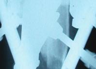
3 Congenital Pseudarthrosis of the Tibia 69 Case Report A sixteen-year-old adolescent was diagnosed at age of one and halfyear with CPT. Fourteen-surgical procedures in attempt to achieve union were performed, including two attempts of open reduction and plating of the tibia with bone grafting. Followed by failure and removal of plates and screw; multiple trials of separate bone grafting procedures with frequent cast changes were done, and finally trial of intramedullary nail with grafting which failed and the hardware was then removed. Due to severe deformity, shortening, pain and stiff ankle with valgus deformity, the patient was referred to the authors. Examination revealed clear stigmata of neurofibromatosis, with severe angulations of the tibia apex anterior 90 and medial 70 with leg length discrepancy (LLD) of 17 cm (Fig. 1A, B, C). Radiographs showed atrophic nonunion of distal tibia with tapered sclerotic ends and obliterated medullary canal, in addition to the above mentioned deformity (Fig. 2A, B). The above description indicates that our case is Type II according to Paley s classification. The decision was to deal with this patient in two stages; first to achieve union with plans to correct residual leg length discrepancy at later stage, as per the patient s request. A B C Fig. 1. Preoperative pictures of our patient with CPT demonstrate the obvious deformity. (A) Lateral view. (B) Anterior with weight bearing and (C) Anterior without weight bearing. The patient was taken to operating room where standard ring first TSF technique was applied proximally and distally on the tibial segments[10]. Then an 8 cm long incision was made at non-union site, where excision of pseudarthrosis was done. It was ensured that all dysplastic bone has been removed from both sides of the pseudarthrosis site until healthy bleeding bone, though keeping the resection to the
.")

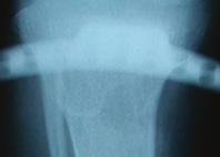
4 70 L.A. Abumunaser and M.J. Alsayyad minimum to avoid unnecessary bone loss. Removal of thickened fibrous tissue of previous surgeries was also performed. However, the deformity was not corrected acutely because of the soft tissue contracture. The proximal fixation included four-half pins of 6 mm in diameter (hydroxyapatite coated). Distally it was utilized 1.8 mm Ilizarov wires X 5 (Smith & Nephew, Memphis, TN USA) holding the short distal fragment. Grafting with periosteum from the iliac crest was performed as follows; periosteum was harvested from the inner table of the iliac crest and was meshed like the split thickness skin graft, and was applied on top of the cleaned pseudarthrosis site. Iliac crest bone graft and 10 cc of DBM (Grafton, Osteotech, Inc., Eatontown, NJ, USA) were performed, then closure in layers (Fig. 3A, B). Fig. 2. A B Preoperative X-ray is showing the anteromedial angular deformity of Rt tibia. (A) Anteroposterior. (B) Lateral view.
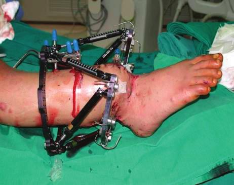
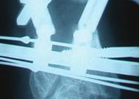
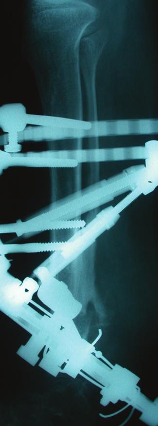
5 Congenital Pseudarthrosis of the Tibia A 71 B Fig. 3. Immediate post-operative view with TSF: (A) Anterior, (B) Lateral. A B Fig. 4. Gradual correction of the deformity with TSF: (A) Anteroposterior, (B) Lateral view.


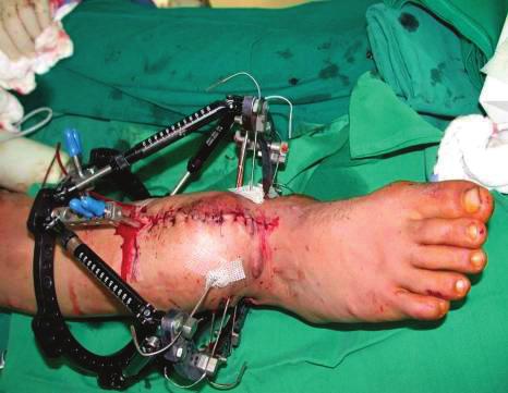
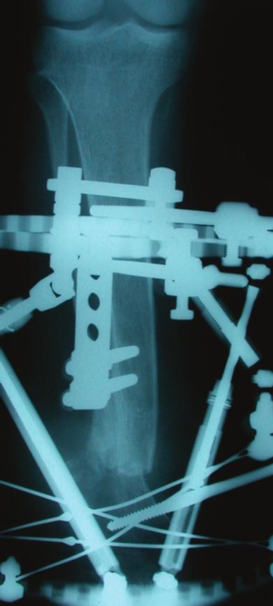
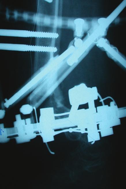
6 72 L.A. Abumunaser and M.J. Alsayyad Postoperatively gradual correction of deformity was done utilizing TSF online software program (Smith & Nephew, Memphis, TN USA) (Figs. 4A, B). Full correction was achieved within 74 days. The patient was mobilized with full weight bearing as tolerated. Union was reached after six months of treatment. This was followed by removal of the frame one month later, after frame dynamization (Figs. 5A, B). The patient continued to utilize braces for four months postoperatively, with 11cm leg length discrepancy remaining (Figs. 6A, B, C) and was able to walk for the first time in his life without crutches utilizing a shoe raise of 7 cm. The patient has been followed for 4-years now, and currently is undergoing lengthening using a second TSF. The patient had an excellent range of motion after surgery of 10 planter-flexion and 40 dorsi-flexion, and the range remained unchanged with no further improvement in plantar flexion during the follow-up period. A B Fig. 5. Solid union and deformity correction: (A) Anteroposterior, (B) Lateral view.
 From posterior, standing with FWB.")


7 Congenital Pseudarthrosis of the Tibia 73 A Fig. 6. B C Full correction of the deformity: (A) Lateral, (B) Anterior view. (C) From posterior, standing with FWB. Discussion Congenital pseudarthrosis of the tibia represents a great challenge to treat in a pediatric orthopedic practice. Treatment measures and in literatures addressing this condition are numerous, reflecting the difficulty in management. The aim of successful treatment is to achieve union, length and normal axis of the affected leg. This success could be possible surgically by achieving the following principles: Correcting the axis of the limb; resection of pseudarthrosis and opening of the medullary canal; lengthening of tibia with proximal osteotomy and concurrent compression of the fracture site [11]. The age is an important factor with regard to bone consolidation. According to European Pediatric
8 74 L.A. Abumunaser and M.J. Alsayyad Orthopedic Society (EPOS) multicenter study a clear correlation was shown between age at surgery and final outcome. Therefore, it has been proposed that surgery should not be performed on patients younger than the age of 3 years and recommended to be postponed until the age of 5 years [4]. Our patient did have multiple surgeries at a very young age without much success. The surgical technique of vascularized bone grafting (VBG), intramedullary stabilization with allograft, circular external fixation have been reported to be relatively successful in the treatment of CPT [4]. The superiority of any specific procedure is difficult to determine as that depends on different factors, e.g., type of CPT, the effect of previous operation, age at time of surgery, the need for follow-up until skeletal maturity and definition of what is a successful outcome. All these factors make it difficult to compare the outcome of the different procedures [1]. In series of VFG for treatment of CPT, bone consolidation was reported to be obtained in about 94% of cases, nevertheless complications of refracture, non-union, tibial angular deformities may occur, as well as severe ankle valgus deformity with ankle pain [1-4]. Numerous authors have reported on the use of Ilizarov circular external fixator in the management of CPT [1]. It is a comprehensive approach to all aspects of CPT including resection of pseudarthrosis, deformity correction, shortening the defect, improving joint function and weight bearing. However, its disadvantage is relative complexity, duration of treatment, pin tract infection, refracture and ankle valgus. Paley et al. [12] reported on 16 patients with union rate of 94% with one treatment and100% with two treatments. Grill et al. in a multicentre study analyzed the different therapeutic methods used by the EPOS [13]. The treatment data of 340 patients who underwent 1287 procedures for CPT were analyzed. The therapeutic modalities, which were reviewed, included the McFarland bypass graft, plating, rodding, and grafting, the Ilizarov fixator as well as conservative measures. The findings of that study demonstrated that plating and rodding seemed to afford inadequate stability as to allow the pseudarthrosis to heal. Those surgeons who used that kind of fixation resected too little of the pseudarthrotic bone in an attempt to avoid shortening. The results of that study showed that the Ilizarov technique was the method of choice in the treatment of CPT. In addition to success
9 Congenital Pseudarthrosis of the Tibia 75 in correction of the other deformities, this method achieved the highest rate of union of 75.5%. The Ilizarov technique is useful in many cases of CPT in which union failed to occur in spite of many previous surgeries. The use of this method does not preclude the use of other procedures, but the Ilizarov method takes considerable time and effort to obtain good results [13]. There have been previous reports in the literature of successful outcomes after the use of TSF in the correction of pediatric lower limb deformities, and in the acute sitting [14-16]. Sluga et al. reported the use of TSF in a series of 5 children with shortened and deformed lower extremities, of which 4 due to fracture, osteomyelitis, and a congenitally short femur, and 1 patient had a pseudoarthrosis of the tibia. The mean elongation achieved was 5.9 cm and a mean valgus correction of 10.6 and a mean lateral translation of 12.6 mm. In all patients, bones fused without major complications or residual deformity [15]. Eidelman et al. reported their experience with the use of the TSF in pediatric deformities, achieving an anatomical correction in 30 of the 31 children treated [16]. Al-Sayyad recently described his encouraging results in the use of TSF in treating 10 unstable tibial fractures, including 5 open fractures, showing a mean fracture healing time of 18 weeks [17]. Recently, the TSF has been compared with the Ilizarov external fixator for tibial lengthening. Although no difference was found in the mean lengthening time or complication rate, the authors noted that multiplanar deformity correction was easier to perform with TSF [18]. Naqui et al. [19] treated 3 neurofibromatosis pseudoarthrosis. One is a teenage patient with a history of psychiatric problems, who had a stacked frame for pseudarthrosis of the tibia, refused to weight bear and demanded the frame to be removed before full regenerate formation. At this time, there was no residual deformity. He eventually returned and requested further treatment. A second frame was applied 14 months later addressing the subsequent fibrous malunion, and residual deformity of 10-degree valgus and 10-mm shortening. The second treatment was without complication; he had no residual deformity and a full range of movement at the knee and ankle [19]. The result in this patient was obtained after prolonged use of TSF. While in our patient, the union was achieved after only 6 months and in one surgical procedure.
10 76 L.A. Abumunaser and M.J. Alsayyad This case demonstrated a success of TSF in obtaining easy correction of a severe deformity with improvement of contact area, which in return promotes healing, in addition to permitting excellent stability and ability to an early weight bear. The addition of DBM improves osteoinduction and conduction. References [1] Rose RE, Wright DE. Treatment of congenital pseudarthrosis of the tibia with the Ilizarov technique. Case report and review of the literature. West Indian Med J 2007; 56(3): [2] Mahnken AH, Staatz G, Hermanns B, Gunter RW, Weber M. Congenital pseudarthrosis of the tibia in pediatric patients: MR imaging. AJR Am J Roentgenol 2001; 177(5): [3] Fabeck L, Ghafil D. Gerroudj M. Bailon R, Delince PH. Bone morphogenetic protein 7 in the treatment of congenital pseudarthrosis of the tibia. J Bone Joint Surg Br 2006; 88(1): [4] Morissy RT. Cogenital pseudarthrosis of the tibia. Factors that affect results. Clin Orthop Relat Res 1982; 166: [5] Sakamoto A, Yoshida T, Uchida Y, Kojima T, Kubota H, Iwamoto Y. Long-term follow-up on the use of vascularized fibular graft for the treatment of congenital pseudarthrosis of the tibia. J Orthop Surg Res 2008; 3: 13. [6] Anderson KS. Radiological classification of congenital pseudarthrosis of the tibia. Acta Orthop Scand 1973; 44(6): [7] Crawford AH. Neurofibromatosis in children. Acta Orthop Scand Suppl 1986; 218: [8] El-Rosasy MA, Paley D, Herzenberg JE. Congenital pseudarthrosis of the tibia. In: Rozbruch RS, Ilizarov S, (eds.) Limb Lengthening and Reconstruction Surgery. New York: Informa Healthcare, Chp 34, [9] Anticevic D, Jelic M, Vukicevic S. Treatment of a congenital pseudarthrosis of the tibia by osteogenic protein-1 (bone morphogenetic protein-7): a case report. J Pediatr Orthop B 2006; 15(3): [10] Binski J. Taylor Spatial Frame in Acute Fracture Care. Techniques in Orthopedics 2002; 17(2): [11] Odeski Y. The surgical management of congenital pseudarthrosis of the tibia. J Bone Joint Surg Br 2002; 84-B (SUPP III): [12] Paley D, Catagni M, Argnani F, Prevot J, Bell D, Armstrong P. Treatment of congenital pseudarthrosis of the tibia using the Ilizarov technique. Clin Orthop Relat Res 1992; 280: [13] Grill F, Bollini G, Dungl P, Fixsen J, Hefti F, Ippolito E, Romanus B, Tudisco C, Wientroub S. Treatment approaches for congenital pseudarthrosis of tibia: results of the EPOS multicenter study. European Paediatric Orthopaedic Society (EPOS). J Pediatr Orthop B 2000; 9(2): [14] Fadel M, Hosny G. The Taylor spatial frame for deformity correction in the lower limbs. Int Orthop 2005; 29(2):
11 Congenital Pseudarthrosis of the Tibia 77 [15] Sluga M, Pfeiffer M, Kotz R, Nehrer S. Lower limb deformities in children: two-stage correction using the Taylor spatial frame. J Pediatr Orthop B 2003; 12(2): [16] Eidelman M, Bialik V, Katzman A. Correction of deformities in children using the Taylor spatial frame. J Pediater Orthop B 2006; 15(6): [17] Al-Sayyad MJ. Taylor spatial frame in the treatment of pediatric and adolescent tibial shaft fractures. J Pediater Orthop 2006; 26(2): [18] Kristiansen LP, Steen H, Reikerås O. No difference in tibial lengthening index by use of Taylor spatial frame or Ilizarov external fixator. Acta Orthop 2006; 77(5): [19] Naqui SZ, Thiryayi W, Foster A, Tselentakis G, Evans M, Day JB. Correction of simple and complex pediatric deformities using the Taylor-Spatial Frame. J Pediatr Orthop 2006; 28(6):
12 78 L.A. Abumunaser and M.J. Alsayyad "Taylor Spatial Frame" "Taylor Spatial Frame" (Autogenus bone graft) (Demineralized bone matrix).
Correction of Traumatic Ankle Valgus and Procurvatum using the Taylor Spatial Frame: A Case Report
 The Foot and Ankle Online Journal Official publication of the International Foot & Ankle Foundation Correction of Traumatic Ankle Valgus and Procurvatum using the Taylor Spatial Frame: A Case Report by
Congenital Pseudarthrosis of the Tibia
 Congenital Pseudarthrosis of the Tibia 5 th Annual SLAOTI Meeting Sao Paolo, Brazil October 12-14, 2017 Richard M Schwend MD Professor Orthopaedics and Pediatrics Director of Research Children s Mercy
Prophylactic surgical correction of Crawford s type II anterolateral bowing of the tibia using Ilizarov s method
 Acta Orthop. Belg., 2005, 71, 577-581 ORIGINAL STUDY Prophylactic surgical correction of Crawford s type II anterolateral bowing of the tibia using Ilizarov s method Hani EL-MOWAFI, Mazen ABULSAAD, Wael
ILIZAROV TECHNIQUE IN CORRECTING LIMBS DEFORMITIES: PRELIMINARY RESULTS
 Bahrain Medical Bulletin, Volume 17, Number 2, June 1995 Original ILIZAROV TECHNIQUE IN CORRECTING LIMBS DEFORMITIES: PRELIMINARY RESULTS Saleh W. Al-Harby, FRCS(Glasg)* This is a prospective study of
Case Report. Antegrade Femur Lengthening with the PRECICE Limb Lengthening Technology
 Case Report Antegrade Femur Lengthening with the PRECICE Limb Lengthening Technology S. Robert Rozbruch, MD Hospital for Special Surgery New York, NY, USA ABSTRACT This is a case illustrating a 4.5 cm
Isolated congenital anterolateral bowing of the fibula : A case report with 24 years follow-up
 Acta Orthop. Belg., 2009, 75, 842-846 CASE REPORT Isolated congenital anterolateral bowing of the fibula : A case report with 24 years follow-up Karolien LELIEFELD, Hans VAN DER SLUIJS, Ibo VAN DER HAVEN
Tibial deformity correction by Ilizarov method
 International Journal of Research in Orthopaedics http://www.ijoro.org Case Report DOI: http://dx.doi.org/10.18203/issn.2455-4510.intjresorthop20180422 Tibial deformity correction by Ilizarov method Robert
Small-wire circular fixators and hybrid external fixation
 ORIGINAL ARTICLE Correction of Tibial Malunion and Nonunion With Six-Axis Analysis Deformity Correction Using the Taylor Spatial Frame David S. Feldman, MD, Steven S. Shin, MD, Sanjeev Madan, MD, and Kenneth
Is Distraction Histiogenesis a Reliable Method to Reconstruct Segmental Bone and Acquired Leg Length Discrepancy in Tibia Fractures and Non Unions?
 Is Distraction Histiogenesis a Reliable Method to Reconstruct Segmental Bone and Acquired Leg Length Discrepancy in Tibia Fractures and Non Unions? James J Hutson Jr MD Professor Orthopedic Trauma Ryder
CASE REPORT. Antegrade tibia lengthening with the PRECICE Limb Lengthening technology
 CASE REPORT Antegrade tibia lengthening with the PRECICE Limb Lengthening technology Austin T. Fragomen, M.D. Hospital for Special Surgery New York, NY 1 1 PR O D U CTS CONDITION Nonunion of an attempted
Large segmental defects of the tibia caused by highenergy. Ten Year Experience with Use of Ilizarov Bone Transport for Tibial Defects
 Bulletin Hospital for Joint Diseases Volume 61, Numbers 3 & 4 2003-2004 101 Ten Year Experience with Use of Ilizarov Bone Transport for Tibial Defects Gene D. Bobroff, M.D., Stuart Gold, M.D., and Daniel
Adult Posttraumatic Reconstruction Using a Magnetic Internal Lengthening Nail
 SUPPLEMENT ARTICLE Adult Posttraumatic Reconstruction Using a Magnetic Internal Lengthening Nail S. Robert Rozbruch, MD Summary: A new generation of internal lengthening nail is now available that has
Neurologic Damage. The most common neurologic injury following intramedullary tibial nailing is injury to the peroneal nerve.
 COMPLICATIONS Knee Pain Anterior knee pain was present in 55% Affects younger more than older patients A significant number of the patients with anterior knee pain had pain with kneeling [90%] They also
.org. Tibia (Shinbone) Shaft Fractures. Anatomy. Types of Tibial Shaft Fractures
 Shaft Fractures. Anatomy. Types of Tibial Shaft Fractures") Tibia (Shinbone) Shaft Fractures Page ( 1 ) The tibia, or shinbone, is the most common fractured long bone in your body. The long bones include the femur, humerus, tibia, and fibula. A tibial shaft fracture
Tibia (Shinbone) Shaft Fractures Page ( 1 ) The tibia, or shinbone, is the most common fractured long bone in your body. The long bones include the femur, humerus, tibia, and fibula. A tibial shaft fracture
Lengthening & Deformity correction with. Fixator Assisted Nailing
 Lengthening & Deformity correction with Fixator Assisted Nailing External Fixation Used as *Intra-Op Alignment tool * for lengthening with the main intention of reducing External fixation time! Advantages
Guang-hui Zhu, Hai-bo Mei *, Rong-guo He, Yao-xi Liu, Kun Liu, Jin Tang and Jiang-yan Wu
 Zhu et al. BMC Musculoskeletal Disorders (2016) 17:443 DOI 10.1186/s12891-016-1295-1 RESEARCH ARTICLE Open Access Combination of intramedullary rod, wrapping bone grafting and Ilizarov s fixator for the
Treatment of delayed union or non-union of the tibial shaft with partial fibulectomy and an Ilizarov frame
 Acta Orthop. Belg., 2007, 73, 630-634 ORIGINAL STUDY Treatment of delayed union or non-union of the tibial shaft with partial fibulectomy and an Ilizarov frame Jo DUJARDYN, Johan LAMMENS From the University
The role of Taylor Spatial Frame for the treatment of acquired and congenital tibial deformities in children
 Acta Orthop. Belg., 2014, 80, 419-425 ORIGINAL STUDY The role of Taylor Spatial Frame for the treatment of acquired and congenital tibial deformities in children Haridimos Tsibidakis, Anastasios D. Kanellopoulos,
Fibula Lengthening Using a Modified Ilizarov Method S. Robert Rozbruch, MD; Matthew DiPaola, BA; Arkady Blyakher,MD
 Fibula Lengthening Using a Modified Ilizarov Method S. Robert Rozbruch, MD; Matthew DiPaola, BA; Arkady Blyakher,MD Limb Lengthening Service Hospital for Special Surgery Abstract A unique combination of
CASE REPORT. Bone transport utilizing the PRECICE Intramedullary Nail for an infected nonunion in the distal femur
 PRODUCTS CASE REPORT Bone transport utilizing the PRECICE Intramedullary Nail for an infected nonunion in the distal femur Robert D. Fitch, M.D. Duke University Health System 1 1 CONDITION Infected nonunion
Delayed presentation of congenital tibial pseudarthrosis and neurofibromatosis: a difficult union
 Asian Biomedicine Vol. 8 No. 1 February 2014; 111-118 Clinical report DOI: 10.5372/1905-7415.0801.269 Delayed presentation of congenital tibial pseudarthrosis and neurofibromatosis: a difficult union Maxim
SCIENTIFIC POSTER #28 Tibia OTA 2016
 SCIENTIFIC POSTER #28 Tibia OTA 2016 Effectiveness of Complex Combined Nonunion/Malunion Correction, Utilizing a Hexapod External Fixator John Arvesen, MD 1 ; J. Tracy Watson, MD 2 ; Heidi Israel, PhD,
Free vascularized fibular graft for tibial pseudarthrosis in neurofibromatosis
 Acta Orthop Scand 1988;59(4):425-429 Free vascularized fibular graft for tibial pseudarthrosis in neurofibromatosis 03 17878 1 luli lrl Herman H. de Boer', Abraham J. Verbout', Hans K. L. Nielsen2 and
The space shuttle docking at the international space station
 Docking Tips The space shuttle docking at the international space station Bone gap after component fracture tibia A standard distal to proximal bone transport Apparently simple pictures of the bone transport
Kocaoglu, Mehmet MD; Eralp, Levent MD; Sen, Cengiz MD; Cakmak, Mehmet MD; Dincyürek, Hakan MD; Göksan, S. Bora MD
 2003 Lippincott Williams & Wilkins, Inc. Volume 17(8), September 2003, pp 543-548 Management of Stiff Hypertrophic Nonunions by Distraction Osteogenesis: A Report of 16 Cases [Original Articles] Kocaoglu,
Fractures of the tibia shaft treated with locked intramedullary nail Retrospective clinical and radiographic assesment
 ARS Medica Tomitana - 2013; 4(75): 197-201 DOI: 10.2478/arsm-2013-0035 Șerban Al., Botnaru V., Turcu R., Obadă B., Anderlik St. Fractures of the tibia shaft treated with locked intramedullary nail Retrospective
Assessment of Regenerate in Limbs by Ilizarov External Fixation
 Original Article Print ISSN: 2321-6379 Online ISSN: 2321-595X DOI: 10.17354/ijss/2016/383 Assessment of Regenerate in Limbs by Ilizarov External Fixation T Suresh Kumar 1, Swagat Mahapatra 2 1 Assistant
Stress Fracture Of The Supracondylar Region Of The Femur Induced By The Weight Of The Tibial Ring Fixator
 ISPUB.COM The Internet Journal of Orthopedic Surgery Volume 4 Number 1 Stress Fracture Of The Supracondylar Region Of The Femur Induced By The Weight Of The Tibial Ring S Dhar, M Mir Citation S Dhar, M
Evaluation of the functional outcome in open tibial fractures managed with an Ilizarov fixator as a primary and definitive treatment modality
 2017; 3(2): 436-440 ISSN: 2395-1958 IJOS 2017; 3(2): 436-440 2017 IJOS www.orthopaper.com Received: 05-02-2017 Accepted: 06-03-2017 Dr. SK Irfan Ali Assistant Professor, Dr. Sujai S Associate Professor,
Knee spanning solutions
 Knee spanning solutions System features Indications Intended to be used on adults or pediatric patients as required for fracture fixation (open or closed); post-traumatic joint contracture which has resulted
Of approximately 2 million long bone fractures
 Proceedings S.Z.P.G.M.I vol: 13(1-2) 1999, pp. 71-75. Treatment of Tibial Non-Union with the Ilizarov Method Pervaiz Iqbal, Muhammad Maq, Hamid Qayum Department of Orthopaedics, Shaikh Zayed Hospital,.
Deformity correction during growth after partial physeal arrest
 Acta Orthop. Belg., 2009, 75, 219-224 ORIGINAL STUDY Deformity correction during growth after partial physeal arrest Joachim HORN, Leif Pål KRISTIANSEN, Harald STEEN From the Department of Orthopaedics,
Orthopedic & Sports Medicine, Bay Care Clinic, 501 N. 10th Street, Manitowoc, WI Procedure. Subtalar arthrodesis
 OSTEOAMP Allogeneic Morphogenetic Proteins Subtalar Nonunions OSTEOAMP Case Report SUBTALAR NONUNIONS Dr. Jason George DeVries and Dr. Brandon M. Scharer Orthopedic & Sports Medicine, Bay Care Clinic,
Management of Nonunion of Tibia by Ilizarov Technique Haque MA 1, Islam SM 2, Chowdhury MR 3
 ...Original Article Management of Nonunion of Tibia by Ilizarov Technique Haque MA 1, Islam SM 2, Chowdhury MR 3 Abstract This prospective study was carried out during the period of January 2007 to December
Temporary Intentional Leg Shortening and Deformation to Facilitate Wound Closure Using the Ilizarov/Taylor Spatial Frame
 TECHNICAL TRICK Temporary Intentional Leg Shortening and Deformation to Facilitate Wound Closure Using the Ilizarov/Taylor Spatial Frame Shane J. Nho, MD, David L. Helfet, MD, and S. Robert Rozbruch, MD
Accuracy of complex lower-limb deformity correction with external fixation: a comparison of the Taylor Spatial Frame with the Ilizarov Ringfixator
 J Child Orthop (2007) 1:55 61 DOI 10.1007/s11832-006-0005-1 ORIGINAL CLINICAL ARTICLE Accuracy of complex lower-limb deformity correction with external fixation: a comparison of the Taylor Spatial Frame
Multiple Exostoses / Multiple Osteochondroma of the Lower Limb Guide By Dror Paley M.D.,
 Multiple Exostoses / Multiple Osteochondroma of the Lower Limb Guide By Dror Paley M.D., Director of the Paley Advanced Limb Lengthening Institute at St. Mary s Hospital in West Palm Beach, Florida. Located
Metatarsal Lengthening By Callus Distraction For Brachymetatarsia: Case Report and Review of the Literature
 ISPUB.COM The Internet Journal of Third World Medicine Volume 1 Number 2 Metatarsal Lengthening By Callus Distraction For Brachymetatarsia: Case Report and Review of the R Rose Citation R Rose. Metatarsal
EXPERT TIBIAL NAIL PROTECT
 EXPERT TIBIAL NAIL PROTECT Enhance your first line of defense This publication is not intended for distribution in the USA. CLINICAL EVIDENCE CONTENT AUTHOR TITLE OF CHAPTER PAGE ETN PROtect clinical evidence
Surgical treatment of aseptic nonunion in long bones: review of 193 cases
 J Orthopaed Traumatol (2007) 8:11 15 DOI 10.1007/s10195-007-0155-z ORIGINAL A. Megaro S. Marchesi U.E. Pazzaglia Surgical treatment of aseptic nonunion in long bones: review of 193 cases Received: 5 September
A Clinical Study For Evaluation Of Results Of Closed Interlocking Nailing Of Fractures Of The Shaft Of The Tibia
 ISPUB.COM The Internet Journal of Orthopedic Surgery Volume 17 Number 2 A Clinical Study For Evaluation Of Results Of Closed Interlocking Nailing Of Fractures Of The Shaft Of R Gupta, T Motten, N Kalsotra,
The Minimally Invasive Plate Osteosynthesis (MIPO) Technique with a Locking Compression Plate for Femoral Lengthening
 Technique with a Locking Compression Plate for Femoral Lengthening") 2008 62 5 333 339 The Minimally Invasive Plate Osteosynthesis (MIPO) Technique with a Locking Compression Plate for Femoral Lengthening a* b a a a a a a b 334 62 5 5 1 16 9 0 100 11 31 131 14 2 16 9 0
2008 62 5 333 339 The Minimally Invasive Plate Osteosynthesis (MIPO) Technique with a Locking Compression Plate for Femoral Lengthening a* b a a a a a a b 334 62 5 5 1 16 9 0 100 11 31 131 14 2 16 9 0
Clinical. Solutions. Synthes Solutions. Foot and Ankle.
 Clinical Solutions Foot and Ankle. Foot and Ankle. Fractures of the tibial shaft Fractures of the distal fibula Fractures of the distal tibia Fractures and osteotomies of the calcaneus Arthrodesis Fractures,
Comparison of acute compression distraction and segmental bone transport techniques in the treatment of tibia osteomyelitis
 Comparison of acute compression distraction and segmental bone transport techniques in the treatment of tibia osteomyelitis Eralp Levent 1, Sen Cengiz 1, Dikmen Goksel 1, Tomak Yılmaz 2, Gulsen Mahir 3,
Miami combined ILLRS LLRS and ASAMI-BR Conference Presentations by Dr. M M Bari
 Orthopedics and Rheumatology Open Access Journal Conference Proceedings Volume 1 Issue 5 - December 2015 Ortho & Rheum Open Access J Copyright All rights are reserved by Ashraf Elazab Miami combined ILLRS
Surgical interventions in chronic osteomyelitis
 Kathmandu University Medical Journal (2005) Vol. 3, No. 1, Issue 9, 50-54 Surgical interventions in chronic osteomyelitis Shrestha BK 1, Rajbhandary T 2, Bijukachhe B 2, Banskota AK 3 1 Associate Professor,
2017 Resident Advanced Trauma Techniques Course COMPLICATIONS / CHALLENGES MALUNIONS/DEFORMITY
 2017 Resident Advanced Trauma Techniques Course COMPLICATIONS / CHALLENGES MALUNIONS/DEFORMITY What is a Malunion? Definition: a fracture that has healed in a nonanatomic (i.e. deformed) position Must
S. Robert Rozbruch, MD. Chief, Limb Lengthening & Complex Reconstruction Service Professor of Clinical Orthopedic Surgery
 S. Robert Rozbruch, MD Chief, Limb Lengthening & Complex Reconstruction Service Professor of Clinical Orthopedic Surgery Small Bone Innovations: consultant and royalties Smith and Nephew: consultant External
HUMERAL SHAFT FRACTURES: ORIF, IMN, NONOP What to do?
 HUMERAL SHAFT FRACTURES: ORIF, IMN, NONOP What to do? TRAUMA 101 2018 FRACTURE CARE FOR THE COMMUNITY ORTHOPEDIST William W. Cross III, MD Assistant Professor Division of Orthopaedic Trauma Chair, Division
Galal Zaki Said 1, *, Osama Ahmed Farouk 1, Hatem Galal Said 1
 KOWSAR Two-Stage Surgical Treatment for Non-Union of a Shortened Osteoporotic Femur Galal Zaki Said 1, *, Osama Ahmed Farouk 1, Hatem Galal Said 1 1 Department of Orthopedic Surgery, Faculty of Medicine,
S-osteotomy with lengthening and then nailing compared with traditional Ilizarov method
 International Orthopaedics (SICOT) (2013) 37:1995 2000 DOI 10.1007/s00264-013-1962-x ORIGINAL PAPER S-osteotomy with lengthening and then nailing compared with traditional Ilizarov method Xia Lan & Lihai
We present a series of ten hypertrophic nonunions
 MANAGEMENT OF NONUNION OF FRACTURES BY DISTRACTION WITH CORRECTION OF ANGULATION AND SHORTENING MICHAEL SALEH, SIMON ROYSTON From the Northern General Hospital, Sheffield, England We present a series of
Fixator-assisted nailing and consecutive lengthening over an intramedullary nail for the correction of tibial deformity
 Fixator-assisted nailing and consecutive lengthening over an intramedullary nail for the correction of tibial deformity F. E. Bilen, M. Kocaoglu, L. Eralp, H. I. Balci From Istanbul Medical School, Istanbul
Metatarsal Lengthening By Callus Distraction For Brachymetatarsia. Case Report and Review of the Literature
 The Internet Journal of Third World Medicine TM ISSN: 1539-4646 Home Current Issue Archives Instructions for Authors Disclaimer Printable Version Metatarsal Lengthening By Callus Distraction For Brachymetatarsia:
Results of Surgical Treatment of Coxa Vara in Children: Valgus Osteotomy with Angle Blade Plate Fixation
 Results of Surgical Treatment of Coxa Vara in Children: Valgus Osteotomy with Angle Blade Plate Fixation Chatupon Chotigavanichaya MD*, Duangjai Leeprakobboon MD*, Perajit Eamsobhana MD*, Kamolporn Kaewpornsawan
ANTERIOR MEDIAL AND POSTERIOR MEDIAL DEFORMITY OF THE TIBIA
 ANTERIOR MEDIAL AND POSTERIOR MEDIAL DEFORMITY OF THE TIBIA 5 TH ANNUAL SLAOTI MEETING SAO PAOLO, BRAZIL OCTOBER 12-14, 2017 Richard M Schwend MD Professor Orthopaedics and Pediatrics Director of Research
Use of an intramedullary rod for treatment of congenital pseudarthrosis of the tibia: A long-term follow-up study
 Washington University School of Medicine Digital Commons@Becker Open Access Publications 6-1-2004 Use of an intramedullary rod for treatment of congenital pseudarthrosis of the tibia: A long-term follow-up
Multiapical Deformities p. 97 Osteotomy Concepts and Frontal Plane Realignment p. 99 Angulation Correction Axis (ACA) p. 99 Bisector Lines p.
 p. 99 Bisector Lines p.") Normal Lower Limb Alignment and Joint Orientation p. 1 Mechanical and Anatomic Bone Axes p. 1 Joint Center Points p. 5 Joint Orientation Lines p. 5 Ankle p. 5 Knee p. 5 Hip p. 8 Joint Orientation Angles
Normal Lower Limb Alignment and Joint Orientation p. 1 Mechanical and Anatomic Bone Axes p. 1 Joint Center Points p. 5 Joint Orientation Lines p. 5 Ankle p. 5 Knee p. 5 Hip p. 8 Joint Orientation Angles
Patient Guide. Intramedullary Skeletal Kinetic Distractor For Tibial and Femoral Lengthening
 Patient Guide Intramedullary Skeletal Kinetic Distractor For Tibial and Femoral Lengthening Introduction You have decided to have a limb lengthening operation. The surgery you have chosen uses a device
Bone Lengthening for Management of Type 1a Proximal Femoral Deficiency with or without Varus Deformity
 American Research Journal of Orthopedics and Traumatology (ARJOT) Volume 2016, 8 Pages Research Article Keywords: Bone lengthening, proximal, femoral deficiency, deformity. Introduction Open Access Bone
Choice of spacer material for HTO! P. Landreau, MD Chief of Surgery Aspetar, Orthopaedic and Sports Medicine Hospital Doha, Qatar
 Choice of spacer material for HTO! P. Landreau, MD Chief of Surgery Aspetar, Orthopaedic and Sports Medicine Hospital Doha, Qatar High Tibial Osteotomy: HTO! Valgisation HTO Intended to transfer the mechanical
Tibial Shaft Fractures
 Tibial Shaft Fractures Mr Krishna Vemulapalli Consultant Orthopaedics Surgeon Queens & King George Hospitals Queens Hospital 14/03/2018 Google Maps Map data 2018 Google 10 km Orthopaedics Department Covers
Management of complex and complicated orthopedic conditions of leg with Taylor frame
 Original Article Management of complex and complicated orthopedic conditions of leg with Taylor spatial frame Bhushan Patil 1*, Sandeep Shrivastava 2, Sanjay V. Deshpande 3, Sparsh Naik 4, Moin Mehmood
Outcome of Treatment of Nonunion Tibial Shaft Fracture by Intramedullary Interlocking Nail augmentated with Autogenous Cancellous Bone Graft.
 Original Article Nepal Med Coll J 2014; 16(1): 58-62 Outcome of Treatment of Nonunion Tibial Shaft Fracture by Intramedullary Interlocking Nail augmentated with Autogenous Cancellous Bone Graft. Shah SB,
The Surgical Management of Rickets & Osteogenesis Imperfecta
 The Surgical Management of Rickets & Osteogenesis Imperfecta Dr Greg Firth Chris Hani Baragwanath Academic Hospital Department of Orthopaedics University of the Witwatersrand Rickets Inadequate mineralization
Femoral Fractures in Adolescents: A Comparison of Four Methods of Fixation
 Femoral Fractures in Adolescents: A Comparison of Four Methods of Fixation By Leonhard E. Ramseier, MD, Joseph A. Janicki, MD, Shannon Weir, BSc, and Unni G. Narayanan, MBBS, MSc, FRCSC Investigation performed
LIMB LENGTH DISCREPANCIES
 LIMB LENGTH DISCREPANCIES Jill C Flanagan, MD OBJECTIVES Evaluate the patient with a possible limb length difference (LLD) Understand general treatment principles when managing limb length differences
Assessment of percutaneous V osteotomy of the calcaneus with Ilizarov application for correction of complex foot deformities
 Acta Orthop. Belg., 2004, 70, 586-590 ORIGINAL STUDY Assessment of percutaneous V osteotomy of the calcaneus with Ilizarov application for correction of complex foot deformities Hani EL-MOWAFI From Mansoura
Vasu Pai FRACS, MCh, MS, Nat Board Ortho Surgeon Gisborne
 Vasu Pai FRACS, MCh, MS, Nat Board Ortho Surgeon Gisborne FRACTURE MANAGEMENT I Simple closed fracture : Complete or Incomplete Stable or unstable II Open fracture III Multiple fracture IV Polytrauma Fractures
Segmental tibial fractures treated with unreamed interlocking nail A prospective study
 2017; 3(2): 714-719 ISSN: 2395-1958 IJOS 2017; 3(2): 714-719 2017 IJOS www.orthopaper.com Received: 13-02-2017 Accepted: 14-03-2017 Ashok Singhvi Hemant Jain Siddharth Jauhar Kishore Raichandani Segmental
Other Congenital & Developmental Knee & Leg Disease. Jong Sup Shim,M.D. Department of Orthopedic Surgery Samsung Medical Center
 Other Congenital & Developmental Knee & Leg Disease Jong Sup Shim,M.D. Department of Orthopedic Surgery Samsung Medical Center Sungkyunkwan University School of Medicine Torsional Deformity (Rotational
Closed reduction and internal fixation of fractures of the shaft of the femur by the Titanium Elastic Nailing System in children.
 ISPUB.COM The Internet Journal of Orthopedic Surgery Volume 17 Number 1 Closed reduction and internal fixation of fractures of the shaft of the femur by the Titanium Elastic Nailing System in children.
Fibula bone grafting in infected gap non union: A prospective case series
 2019; 3(1): 06-10 ISSN (P): 2521-3466 ISSN (E): 2521-3474 Clinical Orthopaedics www.orthoresearchjournal.com 2019; 3(1): 06-10 Received: 03-11-2018 Accepted: 06-12-2018 Dr. Mohammed Nazim M.S (Ortho),
USE OF THE IPSILATERAL VASCULARISED FIBULA FOR
 USE OF THE IPSILATERAL VASCULARISED FIBULA FOR TIBIAL RECONSTRUCTION R. HERTEL, M. PISAN, R. P. JAKOB From the University of Berne, Switzerland Between 1989 and 1994 we used a vascularised ipsilateral
Other Congenital & Developmental Knee & Leg Disease. Jong Sup Shim,M.D. Department of Orthopedic Surgery Samsung Medical Center
 Other Congenital & Developmental Knee & Leg Disease Jong Sup Shim,M.D. Department of Orthopedic Surgery Samsung Medical Center Sungkyunkwan University School of Medicine Rotational Deformity Intoeing Outtoeing
Computer-Assisted Correction of Bone Deformities Using a 6-DOF Parallel Spatial Mechanism
 Computer-Assisted Correction of Bone Deformities Using a 6-DOF Parallel Spatial Mechanism O. Iyun 1, D.P. Borschneck 3, and R.E. Ellis 1,2,3 1 School of Computing 2 Mechanical Engineering Queen s University
ILIZAROV METHOD IN TREATMENT OF TIBIAL AND FEMORAL INFECTED NON-UNIONS IN PATEITNES WITH HIGH-ENERGY TRAUMA AND BATTLE-FIELD WOUNDS
 ILIZAROV METHOD IN TREATMENT OF TIBIAL AND FEMORAL INFECTED NON-UNIONS IN PATEITNES WITH HIGH-ENERGY TRAUMA AND BATTLE-FIELD WOUNDS M. N. Tahmasebi * and Sh. Jalali Mazlouman Department of Orthopedic Surgery,
Pediatric Tibia Fractures Key Points. Christopher Iobst, MD
 Pediatric Tibia Fractures Key Points Christopher Iobst, MD Goals Bone to heal Return to full weight bearing Acceptable alignment rule of 10s 10 degrees of varus 8 degrees of valgus 12 degrees of procurvatum
The use of the Taylor spatial frame in adolescent Blount s disease: is fibular osteotomy necessary?
 J Child Orthop (2008) 2:199 204 DOI 10.1007/s11832-008-0099-8 ORIGINAL CLINICAL ARTICLE The use of the Taylor spatial frame in adolescent Blount s disease: is fibular osteotomy necessary? Mark Eidelman
Equalization of Limb Length Discrepancy using Growth Arrest vs Intramedullary Lengthening
 Equalization of Limb Length Discrepancy using Growth Arrest vs Intramedullary Lengthening Ahmed Hammouda, MD Vivian Szymczuk, MD Martin G. Gesheff, BS Shawn Standard, MD John E. Herzenberg, MD International
Gentle Guided Growth to Correct Knock Knees and Bowed Legs in Children
 PATIENT INFORMATION Gentle Guided Growth to Correct Knock Knees and Bowed Legs in Children The Guided Growth System eight-plate quad-plate INTRODUCTION Children need gentle guidance and correction in many
Study of Ender s Nailing in Paediatric Tibial Shaft Fractures
 Study of Ender s Nailing in Paediatric Tibial Shaft Fractures Dr. Himanshu G. Ladani 1* 1 Ex. Assistant Professor of Orthopaedics, M.P.Shah Medical College, Jamnagar, Gujarat. ABSTRACT Background: Closed
Fibula-related complications during bilateral tibial lengthening
 Acta Orthopaedica 2012; 83 (3): 271 275 271 Fibula-related complications during bilateral tibial lengthening 60 patients followed for mean 5 years Seung-Ju Kim, Mandar Vikas Agashe, Sang-Heon Song, and
Guidance for the Physiotherapy Management of Patients Undergoing Limb Reconstruction with a Circular Frame External Fixator.
 Guidance for the Physiotherapy Management of Patients Undergoing Limb Reconstruction with a Circular Frame External Fixator. Table of Contents INTRODUCTION... 2 TARGET AUDIENCE... 2 KEY CHANGES FROM PREVIOUS
Pocket Guide. Version 4.1: Fracture reduction and deformity correction software
 Pocket Guide www.spatialframe.com Version 4.1: Fracture reduction and deformity correction software Shoulder bolt Master tab Strut 5 Strut 1 Strut 6 Strut 4 Strut 2 Strut 3 ID band Figure 1 Frame assembly
The Effect of Bone Marrow Aspirate Concentrate (BMAC) and Platelet-Rich Plasma (PRP) during Distraction Osteogenesis of the Tibia
 and Platelet-Rich Plasma (PRP) during Distraction Osteogenesis of the Tibia") The Effect of Bone Marrow Aspirate Concentrate (BMAC) and Platelet-Rich Plasma (PRP) during Distraction Osteogenesis of the Tibia Dong Hoon Lee, MD, Ph.D, Keun Jung Ryu MD Limb Lengthening and Deformity
The Effect of Bone Marrow Aspirate Concentrate (BMAC) and Platelet-Rich Plasma (PRP) during Distraction Osteogenesis of the Tibia Dong Hoon Lee, MD, Ph.D, Keun Jung Ryu MD Limb Lengthening and Deformity
Circumferential skin defect - Ilizarov technique in plastic surgery
 Brief Communication Circumferential skin defect - Ilizarov technique in plastic surgery Vrisha Madhuri, Shankar R. Kurpad, Manasseh Nithyananth, Thilak S Jepegnanam, V. T. K. Titus, Prema Dhanraj Department
Introduction to the Taylor Spatial Frame Hardware. Trademark of Smith & Nephew. Certain marks Reg. US Pat. & TM Off.
 Introduction to the Taylor Spatial Frame Hardware Trademark of Smith & Nephew. Certain marks Reg. US Pat. & TM Off. What is the Taylor Spatial Frame? Next generation circular fixator capable of 6 axes
Ankle Valgus in Cerebral Palsy
 Ankle Valgus in Cerebral Palsy Freeman Miller Contents Introduction... 2 Natural History... 2 Treatment... 3 Diagnostic Evaluations... 3 Indications for Intervention... 3 Outcome of Treatment... 5 Complications
Ankle and subtalar arthrodesis
 OSTEOAMP Allogeneic Morphogenetic Proteins Ankle Nonunions OSTEOAMP Case Report ANKLE NONUNIONS Dr. Jason George DeVries Orthopedic & Sports Medicine, Bay Care Clinic, 501 N. 10th Street, Manitowoc, WI
V osteotomy and Ilizarov technique for residual idiopathic or neurogenic clubfeet
 Journal of Orthopaedic Surgery 2008;16(2):215-9 V osteotomy and Ilizarov technique for residual idiopathic or neurogenic clubfeet E Segev, E Ezra, M Yaniv, S Wientroub, Y Hemo Department of Pediatric Orthopaedics,
The role of external fixators in paediatric trauma
 Acta Orthop. Belg., 2015, 81, 363-367 ORIGINAL STUDY The role of external fixators in paediatric trauma Joel A. Humphrey, Syed Gillani, Matthew J. Barry From the Royal London Hospital, Whitechapel Road,
Application of a Constrained External Fixator Frame for Treatment of a Fixed Equinus Contracture
 Application of a Constrained External Fixator Frame for Treatment of a Fixed Equinus Contracture Robert W. Mendicino, DPM, FACFAS, 1 Lara J. Murphy, DPM, 2 Michael P. Maskill, DPM, 3 Alan R. Catanzariti,
BRIDGE PLATING OF COMMINUTED SHAFT OF FEMUR FRACTURES
 BRIDGE PLATING OF COMMINUTED SHAFT OF FEMUR FRACTURES Mohammad Abul kalam, Pradeep Kumar, Mohammad Afzal Hussain and Iqbal Ahmad Abstract A prospective study of forty comminuted femoral shaft fractures,
Computer Assisted Deformity Correction Using the Ilizarov Method
 Computer Assisted Deformity Correction Using the Ilizarov Method A.L. Simpson 1,B.Ma 1,D.P.Borschneck 2, and R.E. Ellis 1,2,3 1 School of Computing 2 Department of Surgery 3 Department of Mechanical Engineering,
Limb lengthening and correction of deformity in the lower limbs of children with osteogenesis imperfecta
 Limb lengthening and correction of deformity in the lower limbs of children with osteogenesis imperfecta K. A. N. Saldanha, M. Saleh, M. J. Bell, J. A. Fernandes From Sheffield Children s Hospital, Sheffield,
Use of Unlocked Intramedullary Nailing in Winquist Type I and II Femoral Isthmus Fracture
 Use of Unlocked Intramedullary Nailing in Winquist Type I and II Femoral Isthmus Fracture HT Ling, MBBS (UM), WM Ng, MS Ortho (UM), MK Kwan, MS Ortho (UM), LK Fathi Aizuddeen, MBBS (UM), PCM Tay, MBBS
External Fixator Brochure
 External Fixator Brochure Response Ortho is a global orthopaedic trauma solutions manufacturer offering premium products created under its founding principles of innovation, excellence by design and functional
Bone transport for the management of severely comminuted fractures without bone loss
 Strat Traum Limb Recon (2016) 11:19 24 DOI 10.1007/s11751-016-0241-y ORIGINAL ARTICLE Bone transport for the management of severely comminuted fractures without bone loss Mootaz F. Thakeb 1 Mahmoud A.