Bone tumors. RMG: jan
|
|
|
- Stuart Robertson
- 6 years ago
- Views:
Transcription
1 Bone tumors RMG: jan KIZZA JOHN KIJOHS

2 Diseases arising in bone Lipoma Fibrous cortical defects Non-ossifying fibroma Bone island Benign simple cysts Enchondroma Osteochondroma Osteoid osteoma Giant cell tumor Chondromyoxoid fibroma Chondromyxoid fibroma Chondrosarcoma Ewing s sarcoma Osteosarcoma Bone metastases *Osteomyelitis Metastasis, prostate cancer with mixed lytic and sclerotic pattern

3 Bone tumor description Age of patient Number of lesions Size Location Edge of the lesion Bone destruction Internal density or structure Periosteal reaction
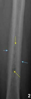
4 Benign bone lesion Solitary 4cm lesion in distal shaft of tibia Narrow zone of transition Well defined, sclerotic margin Some internal septation No matric calcification No periosteal reaction Dx: Non-ossifying fibroma

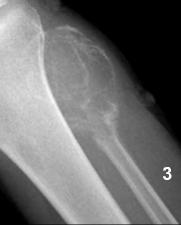
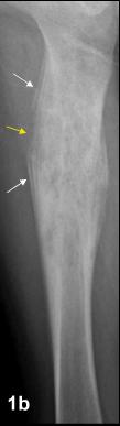
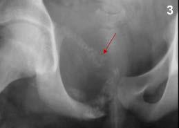
5 Malignant bone lesion Solitary area of ill-defined bone destruction in upper fibular shaft of a 19 year old girl Permeative bone destruction with well-defined edge Wide zone of transition Soft tissue calcification adjacent to the bone lesion, with elevation of the periosteum inferiorly Dx: Ewing s sarcoma

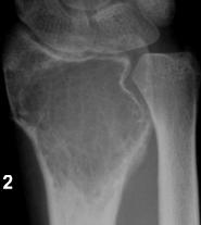
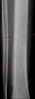
6 Indeterminate bone lesion Solitary area of ill-defined bone destruction in proximal femur in a 55 year old man Edge of lesion not well defined. Some medial sclerosis Zone of transition narrow No visible internal structure No calcifications No visible periosteal reaction Dx: Chondrosarcoma

7 Lipoma of bone Unusual Specifically occur in calcaneus Well defined sclerotic edge Narrow zone of transition No periosteal reaction Central calcification due to necrosis of the fat in the middle of the mass Peak age: can occur at any age, mostly 30-60


 May enlarge to form a")
8 Fibrous cortical defects Small areas of fibrous tissue extending into the cortex of the bone usually in the metaphyses of a child 2-10yrs, peak 7-8 Disappear but can be seen in adults Round or oval with all features of benign lesion (leave me alone lesion) May enlarge to form a non-ossifying fibroma
 More in the shaft than metaphysis probably")

9 Non-ossifying fibroma Larger version of a fibrous cortical defect 8-20yrs (Peak: 10-20) More in the shaft than metaphysis probably reflecting growth of the fibrous cortical defect which started in the metaphysis Benign lesion, most of which disappear by themselves


10 Fibro-osseous lesion Non-ossifying fibroma Range of appearance AP: smooth hazy appearance of the tissue inside it which was not evident on the lateral Could be a focus of fibrous dysplasia and could just be called a fibro-osseous lesion (bone tissue has too much fibrous tissue in it)


 Can be")

11 Bone island Common lesions in adults of any age and in children to a lesser extent Consists of almost normal bone are benign Well defined dense areas often with small dense lines running into adjacent bone (brush boarder) Can be mistaken for sclerotic mets, but these tends to be large and less regular


12 Benign (simple) bone cysts Fluid filled benign lesions Start in metaphysis and may migrate in diaphysis with growth Expansion of bone with thinning of the cortex Commonly cause pathologic # # thru bone cyst may heal with complex septations. 3-20yrs for 80% of cases.

 Bone often expanded.")
13 Fibrous dysplasia Areas of bone don t develop in normal bone forming cells, where there should be bone, is calcified fibrous tissue Has several forms. Can affect in just one small area (90% cases) or multiple areas Focal or wide spread areas with no bone trabeculae, instead, a smooth homogenous density (ground glass appearance) Bone often expanded. Cortical thinning may occur 3-15yrs Pathologic # +/-


14 Fibrous dysplasia, polyostotic (multiple bones) Uncommon form in wc multiple bones are affected Diffuse expansion of bone Evidence of previous pathologic #


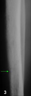
15 Enchondroma Benign tumor of cartilage growing inside a bone Found incidentally or after pathologic # Occur anyway except the skull, commonly in small bones of hand feet Internal calcification, a feature of these tumors, may be the only part visible Usually left alone, but can change into chondrosarcoma, causing pain and swelling around previously asymptomatic lesion
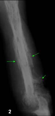
16 Osteochondroma Benign cartilage tumor Grows outside the bone as a stalk of bone with a cartilage cup on top of it (imgae 1 next slide) or as a broad-base bone mass covered by cartilage (image 2 next slide) Lesions are usually bigger than they appear on x-ray cartilage partially calcified May be solitary or multiple (in a condition diaphyseal aclasis) Benign but can become malignant chondrosarcoma Should be suspected if known lesion start to grow rapidly and become bainful
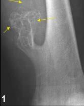
17 Osteochondroma
, with a small central lucency (called nidus")


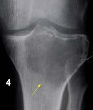
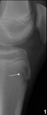
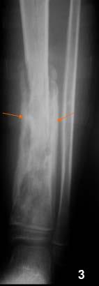
18 Osteoid osteoma Unusual tumor which forms a dense sclerotic area of bone (white arrow), with a small central lucency (called nidus yellow arrow) The nidus may be small or hidden by an overlying dense bone. Cortical lesion may cause a benign periosteal reaction. These lesions are painful and tender Young adults, age range of 5-25 This is a benign condition which is cured by removing the nidus
19 Giant cell tumor Almost all affect the bone after the epiphysis has fused Mostly affect long bones and typically form low density areas which extent right up to the joint surface There may be internal septations as in cases 2 and 3 (next slide) Mostly age: 20-40yrs They cause pain and tenderness and have a risk of pathologic # 15% will become malignant with lung metastases Narrow zone of transition indicate benign, wider zone of transition suggests a malignant lesion. Biopsy and pathologic examination is required to make that dx. Image 3. next slide: expanding lesion of the upper end of the fibula looks like a simple cyst, perhaps with a few septations. But the age of the patient changes the likely dx as well as the other ddx A simple bone cyst is rare after the epiphysis

20 Giant cell tumor

, and the internal septa (yellow arrow) Peak age.")
21 Chondromyxoid fibroma Rare tumor affecting the shaft of the bone, and arising from cartilage It has benign features Note the well defined margin of the compartment within bone (white arrow), and the internal septa (yellow arrow) Peak age yrs
 Both these examples show extensive")

22 Chondrosarcoma Malignant tumor of cartilage It may go by itself as in patient 1 or arise in a preexisting benign tumor as in patient 2 (diaphyseal aclasis) Both these examples show extensive tumor calcification May be larger than visible coz of uncalcified cartilage The pelvis is a common site but can also occur in long bones Peak age range: 40-6oyrs and also occasionally in children
23 Ewing s sarcoma Commonest malignant bone tumor in children Peak age: 15yrs but has a wide age range Can behave in many ways like infection, with intense local pain, fever and raised WBC count Patient 1(next slide): typical features; Location in the diaphysis of the bone, Multiple bone destruction, Interruption yellow arrow feature of aggressive tumor and makes infection less likely Prominent periosteal reaction Periosteal rxn formed in layers at some points white arrow (lamellated / onion skin periosteal reaction) Patients 2 and 3(next slide): two variations in the pelvis Sclerotic in patient 2 with involvement of the whole lt ileum, note the associated soft tissue mass displacing the contrast filled UBL blue arrow, Lytic and expanding in patient 3


24 Ewing s sarcoma


25 Osteosarcoma (osteogenic sarcoma) Commonest primary malignant bone tumor in young adults Age peak: 10-25yrs Can occur in older patients especially radiation therapy or those with Paget's disease May present with pathologic # or local pain and swelling, fever Commonly occurs in metaphysis of long bone with knee region commonest site Bone destruction with ill defined margins, and wide zone of transition, irregular and interrupted periosteal reactiona nd new bone formation


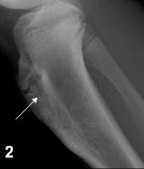
26 Adductor magnus insertion, a normal variation May be concerned after seeing previous periosteal reaction Normal and medial metaphysis of the distal femur A large muscle, adductor magnus attaches in this area and can sometimes cause this irregularity (white arrow) in adolescents


27 Osteosarcoma Comes in many variations Pt 1. periosteal mass, destruction of underlying bone and a Codman triangle yellow arrow Pt 2. has a periosteal type which is which is seen in older patients and forms a dense mass completely outside the bone Pt 3. teenager with a pathologic # thru a Telangiectatic osteosarcoma, a very aggressive and highly vascular form
 and site")


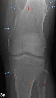
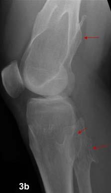
28 Three tumors Three female patients all with a similar lytic tumor in the distal femur. With slight variations of lesion margins, zone of transition, periosteal reaction (red arrow) and site Look similar apart from the patho # in pt 1 Pt 1: osteosarcoma, Pt 2: Giant cell tumor. Pt 3: condrosarcoma Point here; while the X-ray is a very important part of a dx tic process, there is enough of a range for each tumor that unless you see the classic features you will risk giving a misleading dx. Final dx of these malignant or possible malignant will always depend on combition of clinical hx, radiology and pathology


29 Bone metastases - sclerotic Commonest malignant bone tumor by huge margin Can be sclerotic in which new bone is made bcz of tumor cells, lytic in wc the tumor cells grow and destroy bone without any bone reaction, or mixed Sclerotic metastases in a man are more likely to be from a prostate carcinoma, and a woman from breast carcinoma, Other sites can be the cause Pt 1. bladder cancers with focal areas of increase density Pt 2. breast cancer with denser and more well defined lesions


30 Bone metastases - sclerotic Sclerotic mets can be very painful, but are often asymptomatic, and extensive Img1: sclerotic mets in a pt with bladder cancer seen in slide above. Generalized patchy increase in bone density throughout the tibia Img2: Prostate carcinoma mets causing generalized increase in the density of the ribs. There are some focal areas of sparing


31 Bone metastases - lytic Lytic mets carry a risk of pathologic # Common primary sites include; breast in women, and lung in men, with many others Pt1: lytic lesion in the humerus with malignant features already discussed. Though solitary, in a pt aged 60, metastases likely dx Img1c. Same lesion at an earlier stage, just beginning to erode the cortex (green arrow). Patient has lung cancer Pt2. has breast cancer. Ill defined areas of decreased bone density

, lytic lesion with periosteal reaction lt")

, acetabular floor (green arrows) Img 2b;")
32 Bone metastases - lytic 2 egs of mets in pelvic region Pt1: has thyroid cancer. 1a, lytic lesion above the acetabular roof (*), destructive lesion in the medial cortex ( ), lytic lesion with periosteal reaction lt anterior inferior iliac spine ( ). Img1b; 3 months later, lytic areas have joined together and expanded inferiorly Pt2: colon cancer with lytic areas in the femoral head (yellow star), acetabular floor (green arrows) Img 2b; growth of the yellow star lesion and new lesions formed in the proximal femur shown by orange arrows


33 Rib metastases Breast cancer with multiple lytic metastases in the ribs (white / yellow arrows) Yellow arrow lesions: diffuse bone destruction and associated extrapleural mass Blue arrow: expanding lytic lesion c a pathologic # An expanding lesion is most often associated with primary tumors of breast, kidney or thyroid. These shd be the organs to examine if a patient presents with an expanding bone metastes


 Pt2: metastatic nodule in the periosteum")

34 Bone metastases - atypical Most bone metastases start centrally in the bone in the medullary cavity where the blood supply in richer and the bone is softer and easier to destroy. Pt1: metastasis in the cortex of the femur (white arrow) Pt2: metastatic nodule in the periosteum (yellow arrow). Both had lung cancer Pt3: expanding metastasis in the ischium (blue arrows) from bladder cancer. There are lytic areas as well ( red arrow)


35 Pathologic # thru metastasis Aggressive looking lesion, a metastasis from a sarcoma Ill defined bone destruction extends all the way between the white arrows There are small lytic areas further inferiorly, yellow arrows There is a crack # btn the blue arrows as the cause for the current pain. This was missed at the time, but very obvious when the patient returned 3days later


36 Assessing the risk of a pathologic # More likely in the lower limb than the upper, lytic lesions rather than sclerotic, and bigger rather than small. Pain is also a warning sign Lesion in a woman with breast cancer has a moderately aggressive appearance. Zone of transition is wide, on lateral; full thickness erosion of the anterior cortex c interrupted periosteal reaction (yellow arrow), dorsal cortex beginning to be eroded (blue). Pt at high risk of path #
37 Acute osteomyelitis, blood-borne Infection in bone Fever and local pain Periosteal reaction usually continuous, may be smooth or wavey. Images on next slide; findings are; Bone destruction (white arrow in pt, yellow arrow in pt2) Periosteal reactionblue arrows Soft tissue swelling Pt 3. thick periosteal reaction (red arrows) suggesting a slower process, lesser aggressive bacteria than patient 2. has bone destruction in anterior cortex (green arrow) rather than medullary cavity, as in pt2
38 Acute osteomyelitis, blood-borne
39 Chronic osteomyelitis If detected and treated, OM will resolve, if an treated will become cronic. Periosteum gets lifted off the cortex by pus which comes out of the medullary cavity along channels for blood vessels Periosteum thickens and makes new bone Bone itself has high pressure pus inside its medullary cavity, wc interferes with its blood supply Cortex dies. Thus new bone like structure outside the original bone, and a dead piece of bone inside, surrounded by pus and bacteria. More often condition becomes chronic Pus eventually breaks out thru a hole in the new bone and cause a drainage hole to form in the overlying skin (sinus).healing can not occur in this situation coz the dead bone surrounded by bacteria has no blood supply ABCs cant reach it to kill the bacterior
40 Chronic osteomyelitis


41 Chronic osteomyelitis the sequestrum Piece of dead bone floating in pus in the medullary cavity of the infected bone Treatment is to remove the sequestrum surgically Dense piece of a bone with a lucent area right around it. Its not attached to anything


42 Brodie s abscess If the battle btn the bacteria and the bone is not quiet so an even, a chronic abscess may occur inside the bone, without the death of cortex as seen above This is called a Brodie s abscess chronic low grade infection in the bone More common in children, in metaphyses as a well defined lytic lesion with benign features Pt 1 & 2 typical features
Bone Tumors Clues and Cues
 William Herring, M.D. 2002 Bone Tumors Clues and Cues In Slide Show mode, advance the slides by pressing the spacebar All Photos Retain the Copyright of their Authors Clues by Appearance of Lesion Patterns
The Radiology Assistant : Bone tumor - ill defined osteolytic tumors and tumor-like lesions
 Bone tumor - ill defined osteolytic tumors and tumor-like lesions Henk Jan van der Woude and Robin Smithuis Radiology department of the Onze Lieve Vrouwe Gasthuis, Amsterdam and the Rijnland hospital,
Primary bone tumors > metastases from other sites Primary bone tumors widely range -from benign to malignant. Classified according to the normal cell
 Primary bone tumors > metastases from other sites Primary bone tumors widely range -from benign to malignant. Classified according to the normal cell counterpart and line of differentiation. Among the
The Radiology Assistant : Bone tumor - well-defined osteolytic tumors and tumor-like lesions
 Bone tumor - well-defined osteolytic tumors and tumor-like lesions Henk Jan van der Woude and Robin Smithuis Radiology department of the Onze Lieve Vrouwe Gasthuis, Amsterdam and the Rijnland hospital,
APMA 2018 Radiology Track Bone Tumors When to say Gulp!
 APMA 2018 Radiology Track Bone Tumors When to say Gulp! DANIEL P. EVANS, DPM, FACFAOM Professor, Department of Podiatric Medicine and Radiology Dr. Wm. Scholl College of Podiatric Medicine Conflict of
MRI XR, CT, NM. Principal Modality (2): Case Report # 2. Date accepted: 15 March 2013
: Case Report # 2. Date accepted: 15 March 2013") Radiological Category: Musculoskeletal Principal Modality (1): Principal Modality (2): MRI XR, CT, NM Case Report # 2 Submitted by: Hannah Safia Elamir, D.O. Faculty reviewer: Naga R. Chinapuvvula, M.D.
Radiological Category: Musculoskeletal Principal Modality (1): Principal Modality (2): MRI XR, CT, NM Case Report # 2 Submitted by: Hannah Safia Elamir, D.O. Faculty reviewer: Naga R. Chinapuvvula, M.D.
MARK D. MURPHEY MD, FACR. Physician-in-Chief, AIRP. Chief, Musculoskeletal Imaging
 ALPHABET SOUP AND CYSTIC LESIONS OF THE BONE MARK D. MURPHEY MD, FACR Physician-in-Chief, AIRP Chief, Musculoskeletal Imaging ALPHABET SOUP AND CYSTIC LESIONS OF THE BONE Giant cell tumor (GCT) Unicameral
Malignant bone tumors. Incidence Myeloma 45% Osteosarcoma 24% Chondrosarcoma 12% Lyphoma 8% Ewing s Sarcoma 7%
 Malignant bone tumors Incidence Myeloma 45% Osteosarcoma 24% Chondrosarcoma 12% Lyphoma 8% Ewing s Sarcoma 7% Commonest primary bone sarcoma is osteosarcoma X ray Questions to ask 1. Solitary or Multiple
Typical skeletal location and differential diagnosis of bone tumors.
 Typical skeletal location and differential diagnosis of bone tumors. Poster No.: C-2418 Congress: ECR 2015 Type: Educational Exhibit Authors: M. Barros, L. A. Ferreira, Y. Costa, P. J. V. Coelho, F. Caseiro
COPYRIGHT 2004 BY THE JOURNAL OF BONE AND JOINT SURGERY, INCORPORATED
 84 COPYRIGHT 2004 BY THE JOURNAL BONE AND JOINT SURGERY, INCORPORATED Radiographic Evaluation of Pathological Bone Lesions: Current Spectrum of Disease and Approach to Diagnosis BY BENJAMIN G. DOMB, MD,
Bone Tumours - a synopsis. Dr Zena Slim SpR in Histopathology QAH 2009
 Bone Tumours - a synopsis Dr Zena Slim SpR in Histopathology QAH 2009 Aims General approach to diagnosis Common entities.and not so common ones. Mini quiz Challenge of bone tumour diagnosis Bone tumours
Bubbly Lesions of Bone
 Residents Section Pattern of the Month w79 08.18.09 Eisenberg Residents Section Pattern of the Month Residents inradiology Ronald L. Eisenberg 1 Eisenberg RL Keywords: bubbly lesions, fegnomashic, skeletal
Skeletal metastases are the most common variety of bone tumors and should always be considered in the differential diagnosis, particularly in older
 Dr Brajesh Nandan Skeletal metastases are the most common variety of bone tumors and should always be considered in the differential diagnosis, particularly in older patients. Cancers of the breast, prostate,
Radiography in the Initial Diagnosis of Primary Bone Tumors
 Residents Section Structured Review Costelloe and Madewell Radiography of Primary Bone Tumors Residents Section Structured Review Colleen M. Costelloe 1 John E. Madewell Costelloe CM, Madewell JE Keywords:
SMALL ROUND BLUE CELL LESION OF BONE
 DISCLOSURE SMALL ROUND BLUE CELL LESION OF BONE Dr. Alistair Jordan University of South Alabama No financial support or endorsement OBJECTIVES Describe the more common small round cell lesions of bone
Bone and Joint Part 2. Leslie G Dodd, MD
 Bone and Joint Part 2 Leslie G Dodd, MD Relative rates of cancer Sarcomas are relatively uncommon tumors New cancer cases 2007 All sites 1.4 million prostate 218,890 lung 213,380 breast 180,510 Soft tissue
Radiologic approach to pediatric lytic bone lesions
 Radiologic approach to pediatric lytic bone lesions Poster No.: C-1177 Congress: ECR 2016 Type: Educational Exhibit Authors: J. L. LERMA GALLARDO, I. de la Pedraja, A. Lancharro 1 1 1 2 1 1 Zapata, J.
Imaging Findings Of Bone Tumors: A Pictorial Review
 Imaging Findings Of Bone Tumors: A Pictorial Review Poster No.: C-2511 Congress: ECR 2015 Type: Educational Exhibit Authors: M. Limeme, N. Benzina, A. BelKhiria, H. Zaghouani, S. Majdoub, N. Mallat, H.
Primary Tumors of Ribs
 Primary Tumors of Ribs Frank E. Schmidt, M.D., and Max J. Trummer, Capt, MC, USN ABSTRACT An analysis of 50 consecutive patients with primary rib tumors operated on at the U.S. Naval Hospital, San Diego,
The Skeletal System:Bone Tissue
 The Skeletal System:Bone Tissue Dynamic and ever-changing throughout life Skeleton composed of many different tissues cartilage, bone tissue, epithelium, nerve, blood forming tissue, adipose, and dense
Disclosures. Giant Cell Rich Tumors of Bone. Outline. The osteoclast. Giant cell rich tumors 5/21/11
 Disclosures Giant Cell Rich Tumors of Bone Andrew Horvai, MD, PhD Associate Clinical Professor, Pathology This lecture discusses "off label" uses of a number of pharmaceutical agents. The speaker is describing
VALORACIÒN RADIOLÓGICA DE LA LESIÒN ÒSEA SOLITARIA IMAGENOLOGIA MEDICA UNIVERSIDAD HISPANOAMERICANA
 VALORACIÒN RADIOLÓGICA DE LA LESIÒN ÒSEA SOLITARIA IMAGENOLOGIA MEDICA UNIVERSIDAD HISPANOAMERICANA TUMORES ÓSEOS SE PRESENTAN POR RANGOS DE EDAD, PRINCIPALMENTE: MENORES DE 20 AÑOS 20 A 40 AÑOS MAYORES
Fluid-fluid levels in bone tumors: A pictorial review
 Fluid-fluid levels in bone tumors: A pictorial review Poster No.: C-578 Congress: ECR 2009 Type: Educational Exhibit Topic: Musculoskeletal Authors: L. Figueroa Nasra, C. Martín Hervás, M. Tapia-Viñé,
Functions of the Skeletal System. Chapter 6: Osseous Tissue and Bone Structure. Classification of Bones. Bone Shapes
 Chapter 6: Osseous Tissue and Bone Structure Functions of the Skeletal System 1. Support 2. Storage of minerals (calcium) 3. Storage of lipids (yellow marrow) 4. Blood cell production (red marrow) 5. Protection
Primary bone tumors according to the WHO classification: a review of 13 years with illustrative examples
 Primary bone tumors according to the WHO classification: a review of 13 years with illustrative examples Poster No.: C-1741 Congress: ECR 2015 Type: Educational Exhibit Authors: J. Silva, M. A. Ramírez
FORMATION OF BONE. Intramembranous Ossification. Bone-Lec-10-Prof.Dr.Adnan Albideri
 FORMATION OF BONE All bones are of mesodermal origin. The process of bone formation is called ossification. We have seen that formation of most bones is preceded by the formation of a cartilaginous model,
Bread and Butter Bone Pathology
 Bread and Butter Bone Pathology NICOLE D. RIDDLE, MD RUFFOLO, HOOPER, AND ASSOC. / UNIVERSITY OF SOUTH FLORIDA Goals: Fundamentals of neoplastic bone pathology Bone Producing Cartilage Producing Miscellaneous
GIANT CELL-RICH OSTEOSARCOMA: A CASE REPORT
 Nagoya J. Med. Sci. 59. 151-157, 1996 CASE REPORTS GIANT CELL-RICH OSTEOSARCOMA: A CASE REPORT KEIJI SATO!, SHIGEKI YAMAMURA!, HISASHI IWATA!, HIDESHI SUGIURA 2, NOBUO NAKASHIMA 3 and TETSURO NAGASAKA
Review Course «Musculoskeletal Oncology» October 6, 2011 UNIKLINIK BALGRIST. Imaging of Bone and Soft Tissue. Tumors
 Imaging of Bone and Soft Tissue Tumors Approach from a radiologist s point of view Florian Buck Radiology Radio- Radio- Oncologist Oncologist Orthopedist Orthopedist Patient Management Oncologist Oncologist
Musculoskeletal Sarcomas
 Musculoskeletal Sarcomas Robert C. Orth, M.D., Ph.D. Edward B. Singleton Department of Pediatric Radiology Texas Children s Hospital Page 0 xxx00.#####.ppt 9/23/2012 9:01:18 AM No disclosures Page 1 xxx00.#####.ppt
4/28/2010. Fractures. Normal Bone and Normal Ossification Bone Terms. Epiphysis Epiphyseal Plate (physis) Metaphysis
 Metaphysis") Fractures Normal Bone and Normal Ossification Bone Terms Epiphysis Epiphyseal Plate (physis) Metaphysis Diaphysis 1 Fracture Classifications A. Longitudinal B. Transverse C. Oblique D. Spiral E. Incomplete
Fractures Normal Bone and Normal Ossification Bone Terms Epiphysis Epiphyseal Plate (physis) Metaphysis Diaphysis 1 Fracture Classifications A. Longitudinal B. Transverse C. Oblique D. Spiral E. Incomplete
General Approach to Lytic Bone Lesions D. Lee Bennett, MD, MA, Georges Y. El Khoury, MD Appl Radiol. 2004;33(5)
") General Approach to Lytic Bone Lesions D. Lee Bennett, MD, MA, Georges Y. El Khoury, MD Appl Radiol. 2004;33(5) www.medscape.com Abstract and Introduction Abstract When interpreting musculoskeletal radiographs,
General Approach to Lytic Bone Lesions D. Lee Bennett, MD, MA, Georges Y. El Khoury, MD Appl Radiol. 2004;33(5) www.medscape.com Abstract and Introduction Abstract When interpreting musculoskeletal radiographs,
FIBROUS CORTICAL DEFECT AND NON-OSSIFYING FIBROMA
 POSTGRAD. MED. J. (1965), 41, 672. FIBROUS CORTICAL DEFECT AND NON-OSSIFYING FIBROMA PETER G. BULLOUGH, M.B., Ch.B. JON WALIFY, F.R.C.S. Nuffield Department of Orthopaedic University of Oxford, Surgery,
Case Report Intramedullary Chondrosarcoma of Proximal Humerus
 Hindawi Publishing Corporation Case Reports in Radiology Volume 2012, Article ID 642062, 7 pages doi:10.1155/2012/642062 Case Report Intramedullary Chondrosarcoma of Proximal Humerus Pratiksha Yadav, Dolly
Multifocal fibrous Dysplasia with enchondroma-like areas: Fibrocartilaginous Dysplasia
 ISPUB.COM The Internet Journal of Pathology Volume 7 Number 2 Multifocal fibrous Dysplasia with enchondroma-like areas: Fibrocartilaginous Dysplasia V Monappa, R Kudva Citation V Monappa, R Kudva. Multifocal
General osteology. General anatomy of the human skeleton. Development and classification of bones. The bone as a multifunctional organ.
 General osteology. General anatomy of the human skeleton. Development and classification of bones. The bone as a multifunctional organ. Composed by Natalia Leonidovna Svintsitskaya, Associate professor
Grading of Bone Tumors
 Grading of Bone Tumors Joon Hyuk Choi, M.D. Department of Pathology College of Medicine, Yeungnam University Introduction to grading system of bone tumor used at Mayo Clinic WHO Histologic Classification
Common Primary Tumors of Bone
 Special Report Common Primary Tumors of Bone Primary bone tumors are a relatively rare occurrence, however, they can have serious deleterious consequences. Many possess the ability to degenerate into malignant
Skeletal Radiology. Solitary (unicameral) bone cyst. The fallen fragment sign revisited
 bone cyst. The fallen fragment sign revisited") Skeletal Radiol (1989) 18:261-265 Skeletal Radiology Solitary (unicameral) bone cyst The fallen fragment sign revisited S. Struhl, M.D., C. Edelson, M.D., H. Pritzker, M.D., L.P. Seimon, M.D., and H.D.
Skeletal Radiol (1989) 18:261-265 Skeletal Radiology Solitary (unicameral) bone cyst The fallen fragment sign revisited S. Struhl, M.D., C. Edelson, M.D., H. Pritzker, M.D., L.P. Seimon, M.D., and H.D.
Fluid fluid levels in bone tumors and tumoral lesions - Pictorial essay
 Review Fluid fluid levels in bone tumors and tumoral lesions - Pictorial essay Subbarao Kakarla 1,* 1 KIMS Foundation and Research Centre, Minister Road, Secunderabad - 500003, Telangana, India Abstract
Malignant Bone Tumors - Part I: a brief revision of diagnostic aspects with conventional radiology
 Malignant Bone Tumors - Part I: a brief revision of diagnostic aspects with conventional radiology Poster No.: C-2473 Congress: ECR 2013 Type: Educational Exhibit Authors: I. Candelaria, L. B. Barbosa,
Bone (2) Chapter 8. The bone is surrounded by the periosteum, the periosteum consists of two layers: a fibrous outer layer and an innercellular layer.
 Chapter 8. The bone is surrounded by the periosteum, the periosteum consists of two layers: a fibrous outer layer and an innercellular layer.") Bone (2) Chapter 8 The bone is surrounded by the periosteum, the periosteum consists of two layers: a fibrous outer layer and an innercellular layer. The innercellular layer contains osteoprogenitor cells,
Bone (2) Chapter 8 The bone is surrounded by the periosteum, the periosteum consists of two layers: a fibrous outer layer and an innercellular layer. The innercellular layer contains osteoprogenitor cells,
The Radiology Assistant : Bone tumor A-G
 Bone tumor A-G Bone tumors and tumor-like lesions in alphabethic order Henk Jan van de Woude and Robin Smithuis Radiology department of the Onze Lieve Vrouwe Gasthuis, Amsterdam and the Rijnland hospital,
Radiologic Pathologic Correlation of Intraosseous Lipomas. Tim Propeck 1, Mary Anne Bullard 1, John Lin 1, Kei Doi 2, William Martel 1
 Downloaded from www.ajronline.org by 148.251.232.83 on 04/10/18 from IP address 148.251.232.83. opyright RRS. For personal use only; all rights reserved Radiologic Pathologic orrelation of Intraosseous
Heterogeneous osteoblastic activity in the right ischium of unclear etiology seen on NaF18-PET/CT
 CASE REPORT Heterogeneous osteoblastic activity in the right ischium of unclear etiology seen on NaF18-PET/CT Aung Zaw Win, Carina Mari Aparici Dept. Radiology, Nuclear Medicine section, San Francisco
An Introduction to the Skeletal System Skeletal system includes Bones of the skeleton Cartilages, ligaments, and connective tissues
 An Introduction to the Skeletal System Skeletal system includes Bones of the skeleton Cartilages, ligaments, and connective tissues Functions of the Skeletal System Support Storage of minerals (calcium)
FRACTURE CALLUS ASSOCIATED WITH BENIGN AND MALIGNANT BONE LESIONS AND MIMICKING OSTEOSARCOMA
 THE AMERICAN JOURNAL OF CLINICAL PATHOLOGY Vol. 52, No. 1 Copyright 1969 by The Williams & Wilkins Co. Printed in U.S.A. FRACTURE CALLUS ASSOCIATED WITH BENIGN AND MALIGNANT BONE LESIONS AND MIMICKING
BIOH111. o Cell Module o Tissue Module o Integumentary system o Skeletal system o Muscle system o Nervous system o Endocrine system
 BIOH111 o Cell Module o Tissue Module o Integumentary system o Skeletal system o Muscle system o Nervous system o Endocrine system Endeavour College of Natural Health endeavour.edu.au 1 TEXTBOOK AND REQUIRED/RECOMMENDED
Osteosarcoma (Canine)
") Osteosarcoma (Canine) Answering Your Questions About Osteosarcoma In Dogs What Is Osteosarcoma? Usual Sites for Osteosarcoma Development Osteosarcoma is by far the most common bone tumor of the dog, usually
Osteosarcoma (Canine) Answering Your Questions About Osteosarcoma In Dogs What Is Osteosarcoma? Usual Sites for Osteosarcoma Development Osteosarcoma is by far the most common bone tumor of the dog, usually
Due in Lab. Due next week in lab - Scientific America Article Select one article to read and complete article summary
 Due in Lab 1. Skeletal System 33-34 2. Skeletal System 26 3. PreLab 6 Due next week in lab - Scientific America Article Select one article to read and complete article summary Cell Defenses and the Sunshine
Incidental bone tumors are asymptomatic lesions that are. Incidental Bone Lesions. When to Refer to the Tumor Specialist
 Bulletin of the NYU Hospital for Joint Diseases 2012;70(4):235-40 235 Incidental Bone Lesions When to Refer to the Tumor Specialist LT Suezie Kim, M.D., M.C., U.S.N., Catherine N. Laible, M.D., Leon D.
Osseous Tissue and Bone Structure
 C h a p t e r 6 Osseous Tissue and Bone Structure PowerPoint Lecture Slides prepared by Jason LaPres Lone Star College - North Harris Copyright 2009 Pearson Education, Inc., publishing as Pearson Benjamin
The Skeletal System. Mosby items and derived items 2010, 2006, 2002, 1997, 1992 by Mosby, Inc., an affiliate of Elsevier Inc.
 The Skeletal System Functions of Skeletal System Provides internal framework that supports the body Protects internal organs Helps fight disease by producing white blood cells 2 Functions of Skeletal System
SKELETAL TISSUES CHAPTER 7 INTRODUCTION TO THE SKELETAL SYSTEM TYPES OF BONES
 SKELETAL TISSUES CHAPTER 7 By John McGill Supplement Outlines: Beth Wyatt Original PowerPoint: Jack Bagwell INTRODUCTION TO THE SKELETAL SYSTEM STRUCTURE Organs: Bones Related Tissues: Cartilage and Ligaments
USCAP 2014 Common problems in bone and soft tissue pathology: Cartilage tumors
 USCAP 2014 Common problems in bone and soft tissue pathology: Cartilage tumors Andrew Horvai MD PhD Clinical Professor, Pathology UCSF, San Francisco, CA Outline Common intramedullary tumors Enchondroma
A 76 year old male presented with sudden increase of dyspnoea on 15 November 2014, following a biopsy. A previous CXR was reviewed.
 Question 1 A 76 year old male presented with sudden increase of dyspnoea on 15 November 2014, following a biopsy. A previous CXR was reviewed. Imaging A CXR was performed on 28 May 2014. A CT of the chest
Bone/Osteoid Producing Lesions
 Chapter 2 Bone/Osteoid Producing Lesions Introduction There are many lesions that are associated with reactive new bone formation; this chapter predominantly covers those in which deposition of osteoid/bone
Pictorial Essay Benign and Malignant Bone Tumors: Radiological Diagnosis and Imaging Features
 Clinical Orthopedic Imaging Pictorial Essay Benign and Malignant Bone Tumors: Radiological Diagnosis and Imaging Features Katharina Grünberg, M.D.; Christoph Rehnitz, M.D.; Marc-André Weber, M.D., M.Sc.
Osteomyelitis in infancy and childhood: A clinical and diagnostic overview M. Mearadji
 Osteomyelitis in infancy and childhood: A clinical and diagnostic overview M. Mearadji International Foundation for Pediatric Imaging Aid Introduction Osteomyelitis is a relative common disease in infancy
Introduction to Musculoskeletal Tumors. James C. Wittig, MD Orthopedic Oncologist Sarcoma Surgeon
 Introduction to Musculoskeletal Tumors James C. Wittig, MD Orthopedic Oncologist Sarcoma Surgeon www.tumorsurgery.org Definitions Primary Bone / Soft tissue tumors Mesenchymally derived tumors (Mesodermal)
Chapter 6 & 7 The Skeleton
 Chapter 6 & 7 The Skeleton Try this Make clockwise circles with your RIGHT foot, while doing this, draw the number 6 in the air with you RIGHT hand what happens to your foot???? Bony Background Adult body
A Modified Lodwick-Madewell Grading System for the Evaluation of Lytic Bone Lesions
 Musculoskeletal Imaging Original Research Caracciolo et al. Evaluation of Lytic one Lesions Musculoskeletal Imaging Original Research Jamie T. Caracciolo 1 H. Thomas Temple 2 G. Douglas Letson 3 Mark J.
Benign Fibro-osseous Lesions
 Benign Fibro-osseous Lesions Plus Vision is the art of seeing things invisible. Jonathan Swift 1667-1745 Steven R. Singer, DDS srs2@columbia.edu 212.305.5674 Benign Fibro-osseous Lesions A group of lesions
History. 33 y/o F with hx of palpable anterior tibial mass x 2 years, only painful with palpation
 History 33 y/o F with hx of palpable anterior tibial mass x 2 years, only painful with palpation Imaging Photo Album Patient also had a smaller lesion 1 cm proximal to this lesion, not seen radiographically.
FEGNOMASHIC: from x-ray to MRI
 FEGNOMASHIC: from x-ray to MRI Poster No.: C-2441 Congress: ECR 2015 Type: Educational Exhibit Authors: S. Fouassier, A. L. C. Duarte, C. Ruivo, J. Velez ; Évora/PT, 1 2 1 2 3 1 3 Coimbra/PT, PT Keywords:
Advertisement. Osteochondroma
 Advertisement Osteochondroma An osteochondroma is a benign (noncancerous) tumor that develops during childhood or adolescence. It is an abnormal growth that forms on the surface of a bone near the growth
MRI of the Knee: Part 4 - normal variants that may simulate disease. Mark Anderson, M.D. University of Virginia
 MRI of the Knee: Part 4 - normal variants that may simulate disease Mark Anderson, M.D. University of Virginia discuss the most common normal variants in the pediatric knee that may simulate pathology
Benign Tumors of Bone
 REVIEW ARTICLE Benign Tumors of Bone Subbarao K Padmshri Prof. Dr. Kakarla Subbara, Hyderabad, India. Benign tumors of bone are common while malignant tumors are rare. Benign tumors constitute about 75%
Bone Tumors: In 1 Simple Chart
 Bone Tumors with PowerPoint Interactivity Download this entire slideshow from When running this on your own computer you can jump from slide to slide using these buttons at bottom of each slide: Last slide
Chapter 5 The Skeletal System
 Chapter 5 The Skeletal System The Skeletal System Parts of the skeletal system Bones (skeleton) Joints Cartilages Ligaments (bone to bone)(tendon=bone to muscle) Divided into two divisions Axial skeleton:
BONES & JOINTS INFECTION BONE TUMOURS
 BONES & JOINTS INFECTION BONE TUMOURS IMPORTANT SERIOUS CONSEQUENCE PLEASE DON T MISS!! EARLY DIAGNOSIS & PROPER TREATMENT HOW?? AWARE of THEIR EXISTENCE (Knowledge) PREPARE for THEIR OCCURRENCE A HIGH
The Skeletal System PART A. PowerPoint Lecture Slide Presentation by Patty Bostwick-Taylor, Florence-Darlington Technical College
 PowerPoint Lecture Slide Presentation by Patty Bostwick-Taylor, Florence-Darlington Technical College The Skeletal System 5 PART A The Skeletal System Parts of the skeletal system Bones (skeleton) Joints
Friday Teaching. Bones
 Friday Teaching Bones Regarding slipped femoral capital epiphysis It represents Salter Harris type V injury 20% are bilateral There is slight widening of the joint space Slip is typically posteromedial
Radiology Pathology Conference
 Radiology Pathology Conference Sharlin Johnykutty,, MD, Cytopathology Fellow Sara Majewski, MD, Radiology Resident Friday, August 28, 2009 Presentation material is for education purposes only. All rights
Aneurysmal Bone Cyst of the Pelvis: A Challenge in Treatment: Review of the Literature
 ISPUB.COM The Internet Journal of Orthopedic Surgery Volume 8 Number 1 Aneurysmal Bone Cyst of the Pelvis: A Challenge in Treatment: Review of the Literature S Bajracharya, G Khanal, A Sundas, S Pandey,
ISSN: DISTRIBUTION OF BONE AND CARTILAGINOUS TUMORS IN PEDIATRIC AGE GROUP IN WESTERN UTTAR-PRADESH: AN EVALUATIVE STUDY
 : 289-295 ISSN: 2277 4998 DISTRIBUTION OF BONE AND CARTILAGINOUS TUMORS IN PEDIATRIC AGE GROUP IN WESTERN UTTAR-PRADESH: AN EVALUATIVE STUDY QADRI S, HASAN M, AKHTAR K * AND SHERWANI RK The Departments
IMAGING OF THE SKELETAL SYSTEM
 IMAGING OF THE SKELETAL SYSTEM 1 DISEASES AFFECTING THE SKELETAL SYSTEM CAN BE BROADLY DIVIDED INTO: 1. Congenital or developmental 2. Traumatic 3. Infective 4. Bone tumours 5. Metabolic/Systemic diseases
Residents Section Pattern of the Month
 Residents Section Pattern of the Month Rana et al. Periosteal Reaction Residents Section Pattern of the Month Residents inradiology Rich S. Rana 1 Jim S. Wu Ronald L. Eisenberg Rana RS, Wu JS, Eisenberg
Radiology-Pathology Conference
 July 31, 2009 Radiology-Pathology Conference Daniel T Ginat, M.D., M.S. Sharlin Johnykutty,, M.D. Presentation material is for education purposes only. All rights reserved. 2009 URMC Radiology Page 1 of
DIAGNOSING EWING S SARCOMA OF THE RIB IN CHILDREN IS IT QUITE CHALLENGING??
 Indian Journal of Medical s ISSN: 2319 3832(Online) DIAGNOSING EWING S SARCOMA OF THE RIB IN CHILDREN IS IT QUITE CHALLENGING?? *Arvind Kumar S.M. and Shyam Sundar V, Dinakar B.K. and SandhyaV. and Kailash
Malignant Bone Tumours. PathoBasic, Daniel Baumhoer
 Malignant Bone Tumours PathoBasic, 20.03.18 Daniel Baumhoer FNCLCC Grading The differentiation score is defined as the extent to which a tumor resembles adult mesenchymal tissue (score 1), the extent to
Unusual location of bone sarcoma in children
 Unusual location of bone sarcoma in children Poster No.: C-1517 Congress: ECR 2014 Type: Educational Exhibit Authors: S. JERBI, A. Khalfalli, G. Abid, O. Bradai, N. chouchane, H. HAMZA; Mahdia/TN Keywords:
"X marks the spot": The skeletal manifestations of Langerhans cell histiocytosis in the paediatric age group
 "X marks the spot": The skeletal manifestations of Langerhans cell histiocytosis in the paediatric age group Poster No.: C-2847 Congress: ECR 2010 Type: Educational Exhibit Topic: Pediatric Authors: A.
Skeletal System. Chapter 6.1 Human Anatomy & Physiology
 Skeletal System Chapter 6.1 Human Anatomy & Physiology Overview of Skeletal System Bones Joints Skeletal System Cartilage Tendons (bone to muscle) Ligaments (bone to bone) Function of the Skeletal System
SKELETAL SYSTEM. Introduction Notes (pt 1)
") SKELETAL SYSTEM Introduction Notes (pt 1) I. INTRODUCTION 1. Bones include active, living tissues: bone tissue, cartilage, dense connective tissue, blood, and nervous tissue. 2. Bones: support and protect
SKELETAL SYSTEM Introduction Notes (pt 1) I. INTRODUCTION 1. Bones include active, living tissues: bone tissue, cartilage, dense connective tissue, blood, and nervous tissue. 2. Bones: support and protect
Human Skeletal System Glossary
 Acromegaly Apatite Acromegaly - is a condition which involves excessive growth of the jaw, hands, and feet. It results from overproduction of somatotropin in adults (after fusion of the ossification centres
MUSCULOSKELETAL RADIOLOGY
 MUSCULOSKELETAL RADOLOGY SECTON www.cambridge.org Achilles tendonopathy/rupture Characteristics Describes pathology of the combined tendon of the gastro-soleus complex, which inserts onto the calcaneum.
D URING the course of our routine work
 THE ROENTGENOGRAPHIC APPEARANCE OF RENAL CANCER METASTASIS IN BONE ROBERT S. SHERMAN, M.D., and T. ARTHUR PEARSON, M.D.* D URING the course of our routine work it occurred to us that renal cancer metastatic
Prof Oluwadiya KS FMCS (Orthop) Consultant Orthopaedic Surgeon / Associate Professor Division of Orthopaedics and Traumatology Department of Surgery
 Consultant Orthopaedic Surgeon / Associate Professor Division of Orthopaedics and Traumatology Department of Surgery") Prof Oluwadiya KS FMCS (Orthop) Consultant Orthopaedic Surgeon / Associate Professor Division of Orthopaedics and Traumatology Department of Surgery College of Health Sciences Ladoke Akintola University
Prof Oluwadiya KS FMCS (Orthop) Consultant Orthopaedic Surgeon / Associate Professor Division of Orthopaedics and Traumatology Department of Surgery College of Health Sciences Ladoke Akintola University
Copyright 2003 Pearson Education, Inc. publishing as Benjamin Cummings. Dr. Nabil Khouri MD, MSc, Ph.D
 Dr. Nabil Khouri MD, MSc, Ph.D Pelvic Girdle (Hip) Organization of the Lower Limb It is divided into: The Gluteal region The thigh The knee The leg The ankle The foot The thigh and the leg have compartments
Giant cell tumour of the sternum-two cases
 Giant cell tumour of the sternum-two cases Nishaa.P 1, Raghuram.P 2, Navin patil 3, Jaipal B.R 4 Akkamahadevi patel 5 Assistant Professor ESIC medical college and PGIMSR 1 Professor and HOD, 2 Professor
Effective local and systemic therapy is necessary for the cure of Ewing tumor Most chemotherapy regimens are a combination of cyclophosphamide,
 Ewing Tumor Perez Ewing tumor is the second most common primary tumor of bone in childhood, and also occurs in soft tissues Ewing tumor is uncommon before 8 years of age and after 25 years of age In the
Essential Dermatopathology. Jinah Kim, MD, PhD Department of Pathology and Dermatology Stanford University Medical Center
 Essential Dermatopathology Jinah Kim, MD, PhD Department of Pathology and Dermatology Stanford University Medical Center OBJECTIVES Review clinical, pathologic and molecular aspects of bone and fat tumors
Radiology Corner. Osteoid Osteoma
 Radiology Corner Osteoid Osteoma Guarantor: COL Timothy G. Sanders, MC, USAF (Ret.) Contributors: COL Timothy G. Sanders, USAF, MC, (Ret.); CAPT John P. Lichtenberger, USAF, MC; COL Les Folio, USAF, MC,
Topics. Musculoskeletal Infection Extremities. Detection of Infection. Role of Imaging in Extremity Infection. Detection of Infection
 Topics Musculoskeletal Infection Extremities Nuttaya Pattamapaspong M.D. Department of Radiology, Faculty of Medicine, Chiang Mai University, Chiang Mai, Thailand Role of imaging in extremity infection
The Skeletal System PART A
 5 The Skeletal System PART A PowerPoint Lecture Slide Presentation by Jerry L. Cook, Sam Houston University ESSENTIALS OF HUMAN ANATOMY & PHYSIOLOGY EIGHTH EDITION ELAINE N. MARIEB The Skeletal System
Paediatric post-traumatic osseous cystic lesion following a distal radial fracture
 Paediatric post-traumatic osseous cystic lesion following a distal radial fracture Joey Chan Yiing Beh 1*, Ehab Shaban Mahmoud Hamouda 1 1. Department of Diagnostic Imaging, KK Women's and Children's Hospital,
Parts of the skeletal system. Bones (skeleton) Joints Cartilages Ligaments (bone to bone)(tendon=bone to muscle)
 Joints Cartilages Ligaments (bone to bone)(tendon=bone to muscle)") The Skeletal System The Skeletal System Parts of the skeletal system Bones (skeleton) Joints Cartilages Ligaments (bone to bone)(tendon=bone to muscle) Divided into two divisions Axial skeleton Appendicular
The Skeletal System The Skeletal System Parts of the skeletal system Bones (skeleton) Joints Cartilages Ligaments (bone to bone)(tendon=bone to muscle) Divided into two divisions Axial skeleton Appendicular
Imaging in breast cancer. Mammography and Ultrasound Donya Farrokh.MD Radiologist Mashhad University of Medical Since
 Imaging in breast cancer Mammography and Ultrasound Donya Farrokh.MD Radiologist Mashhad University of Medical Since A mammogram report is a key component of the breast cancer diagnostic process. A mammogram