MARK D. MURPHEY MD, FACR. Physician-in-Chief, AIRP. Chief, Musculoskeletal Imaging
|
|
|
- Camron Phelps
- 5 years ago
- Views:
Transcription
1 ALPHABET SOUP AND CYSTIC LESIONS OF THE BONE MARK D. MURPHEY MD, FACR Physician-in-Chief, AIRP Chief, Musculoskeletal Imaging
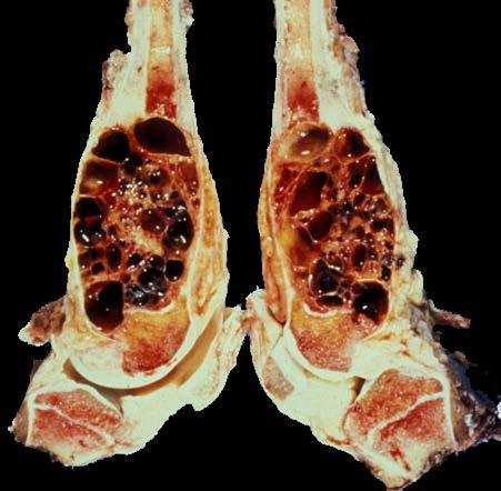
2 ALPHABET SOUP AND CYSTIC LESIONS OF THE BONE Giant cell tumor (GCT) Unicameral bone cyst (UBC) Aneurysmal bone cyst (ABC) Epidermoid inclusion cyst Subchondral cyst Intraosseous ganglion Post-traumatic cyst
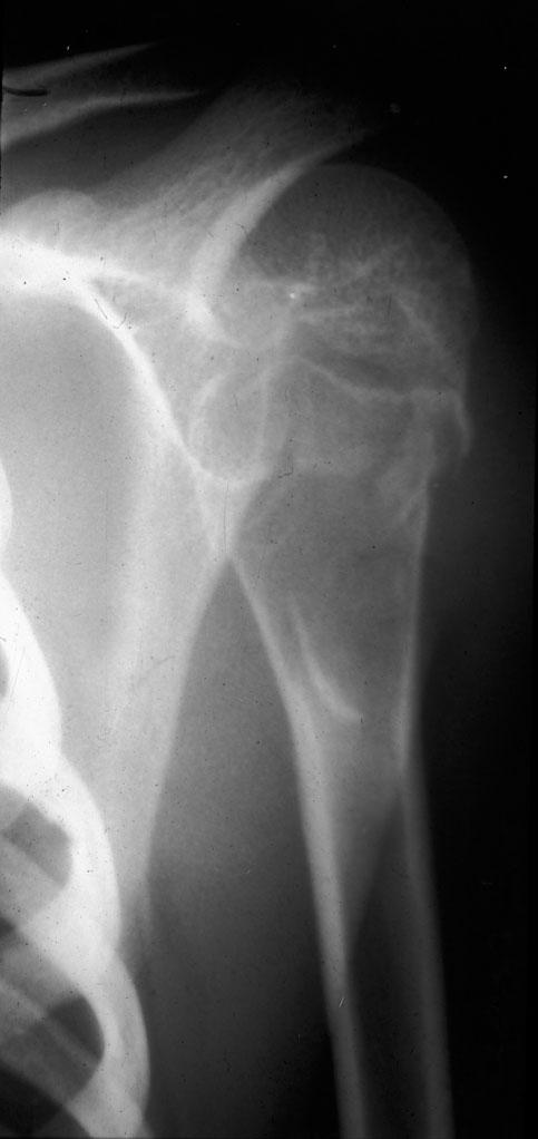
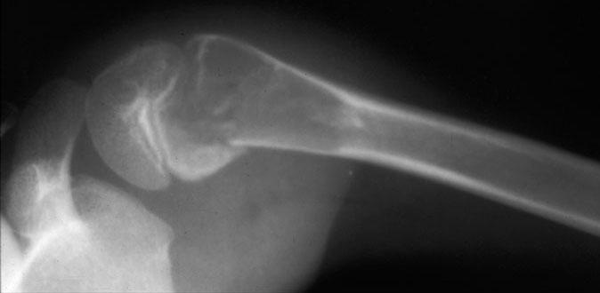
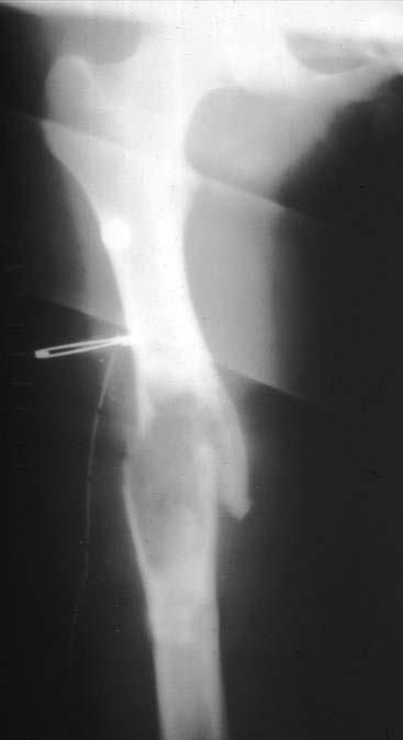
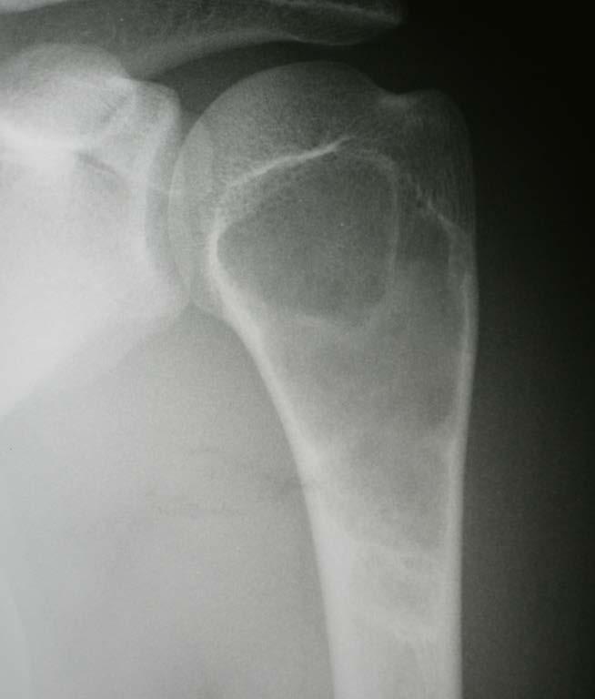
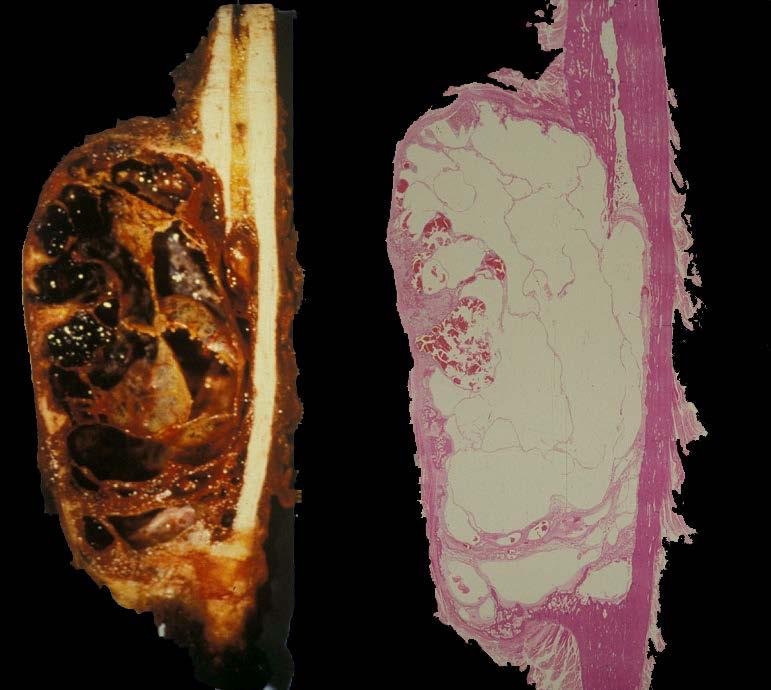
3 GIANT CELL TUMOR (GCT) CLINICAL FEATUES Approximately 5%-10% of all biopsied primary bone tumors; 18%-23% of benign bone neoplasms Symptoms-pain and swelling often relieved by decreased activity Pathologic fracture 10% - 35%
4 GIANT CELL TUMOR CLINICAL FEATUES Adults- 80% between years More common in Chinese (20% of primary bone tumors) Rare in children 1% - 3% (<14 years) Approximately equal sex distribution F - M ratio 3:2 benign GCT M - F ratio 3:1 aggressive GCT
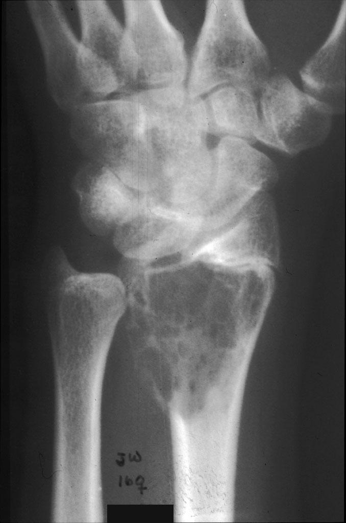
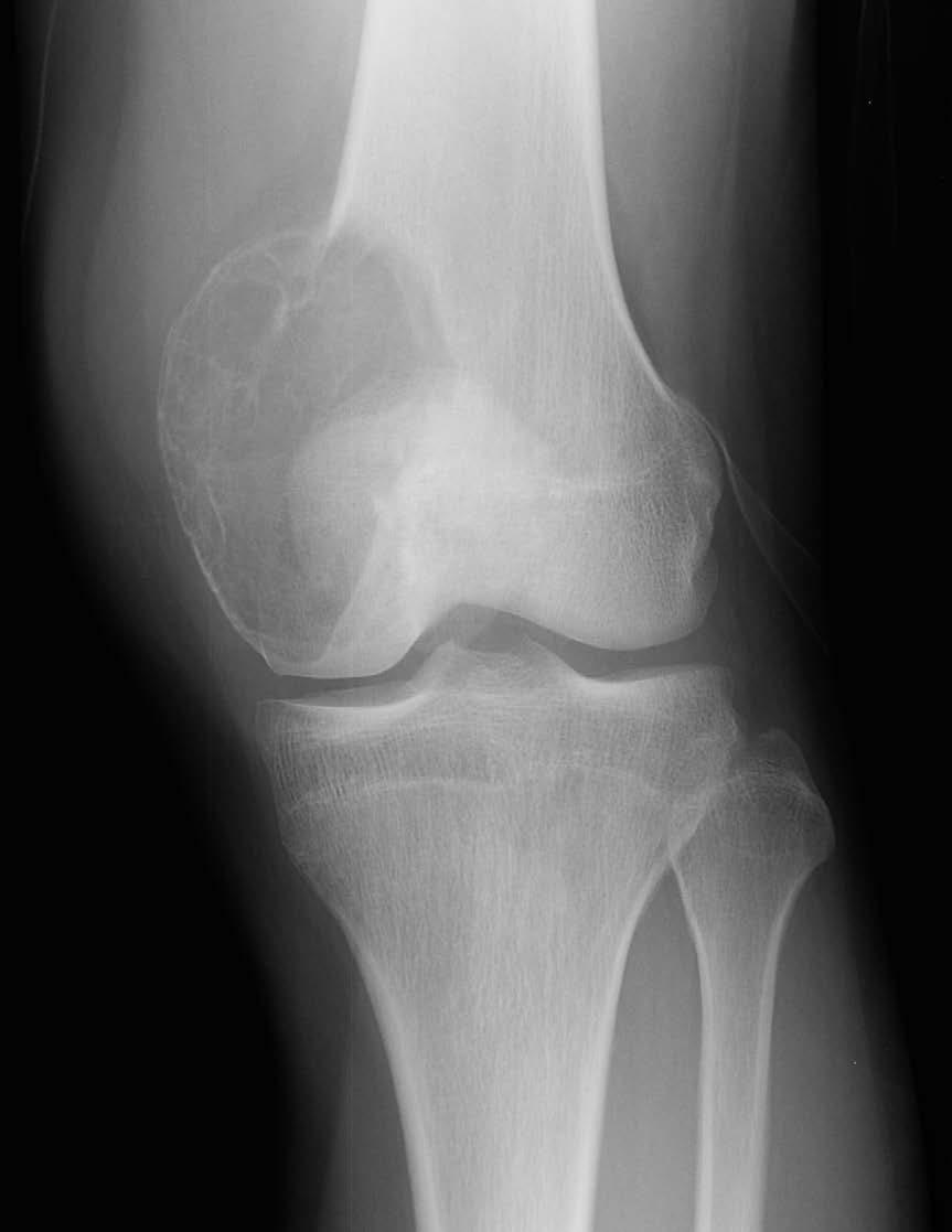
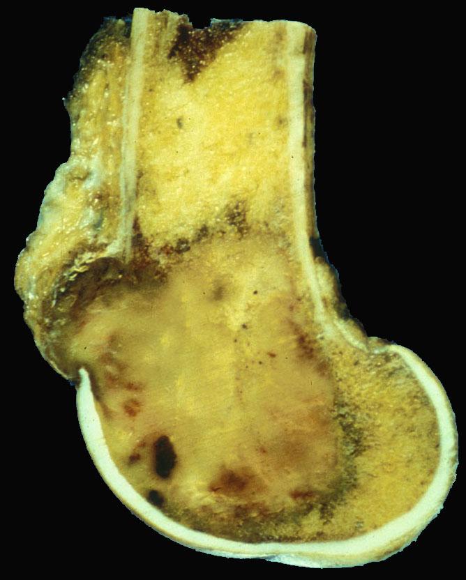
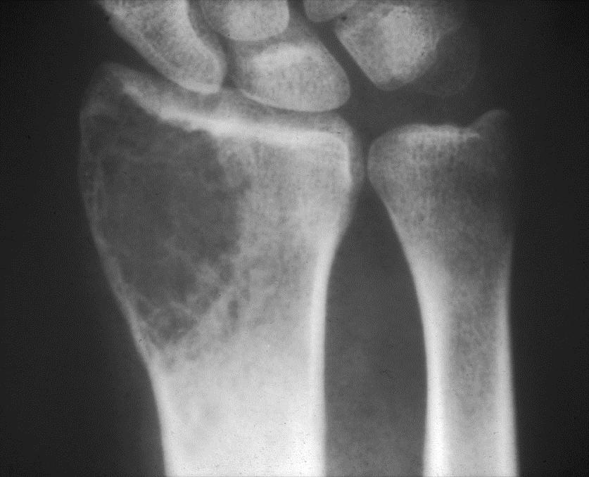
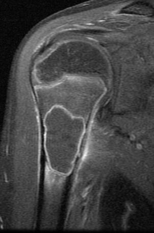
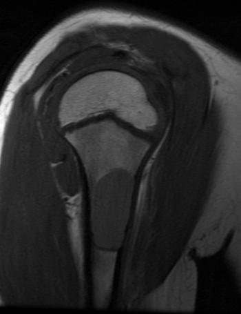
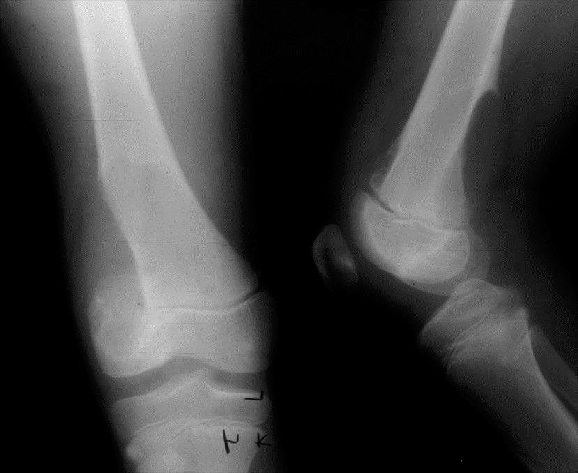
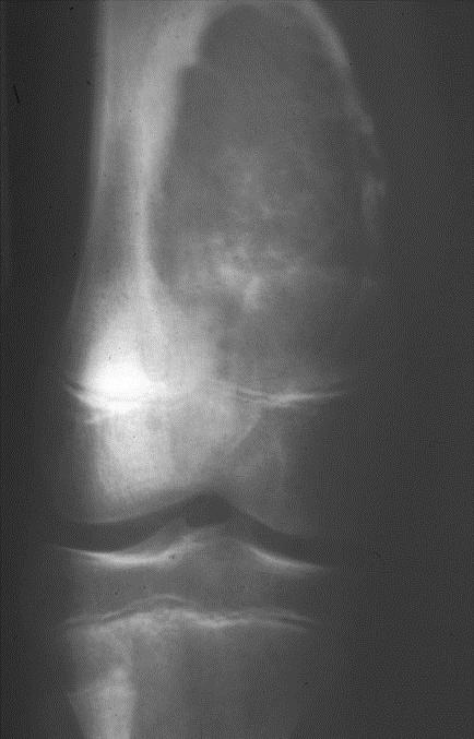
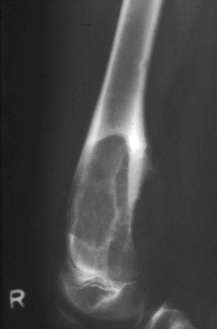
5 GIANT CELL TUMOR LOCATION Originate metaphyseal side of long bone growth plate and grow to subchondral bone (84% - 99%) Long tubular bones 75% - 90% About knee 50% - 65%; distal femur 23%-30%; proximal tibia 20%-25% Radius (10%-12%); humerus (4%-8%)
6
7
8
9
10

11
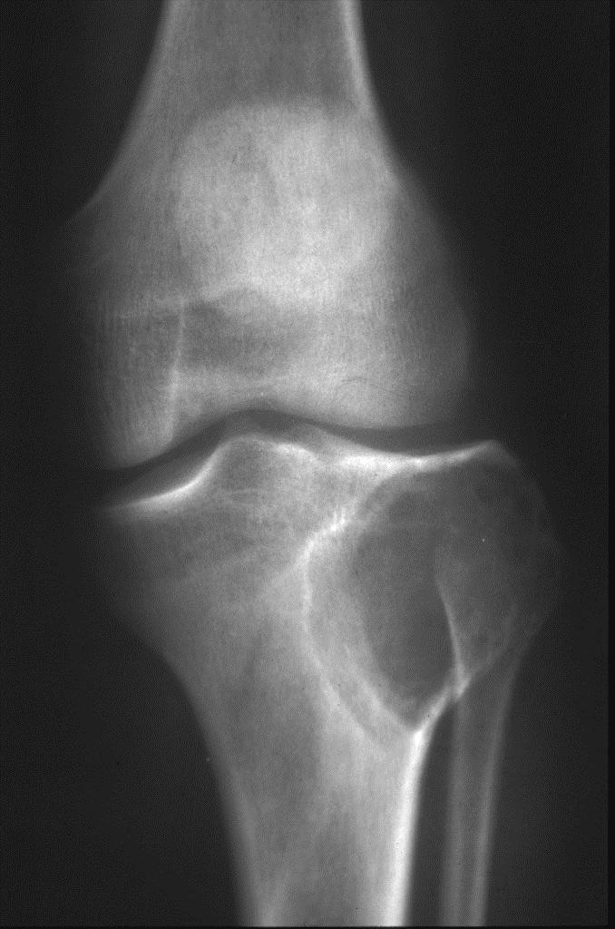
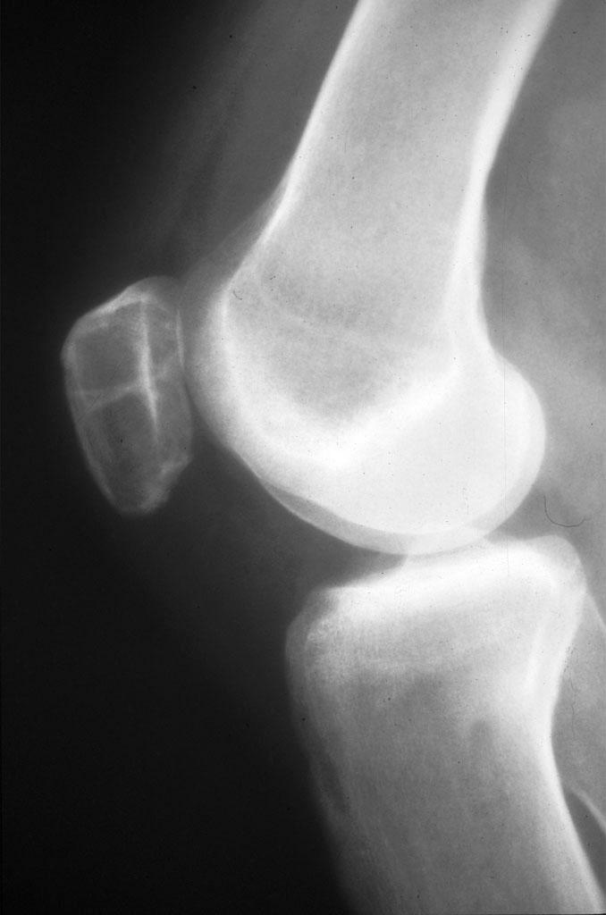
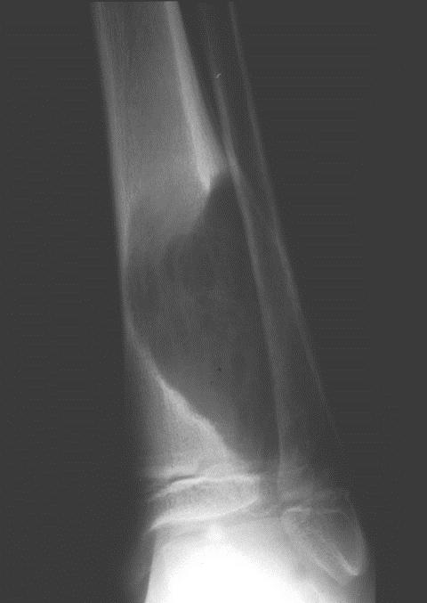
; humerus (4%-8%)")
12 GIANT CELL TUMOR LOCATION Originate metaphyseal side of long bone growth plate and grow to subchondral bone (84% - 99%) Long tubular bones 75% - 90% About knee 50% - 65%; distal femur 23%-30%; proximal tibia 20%-25% Radius (10%-12%); humerus (4%-8%)
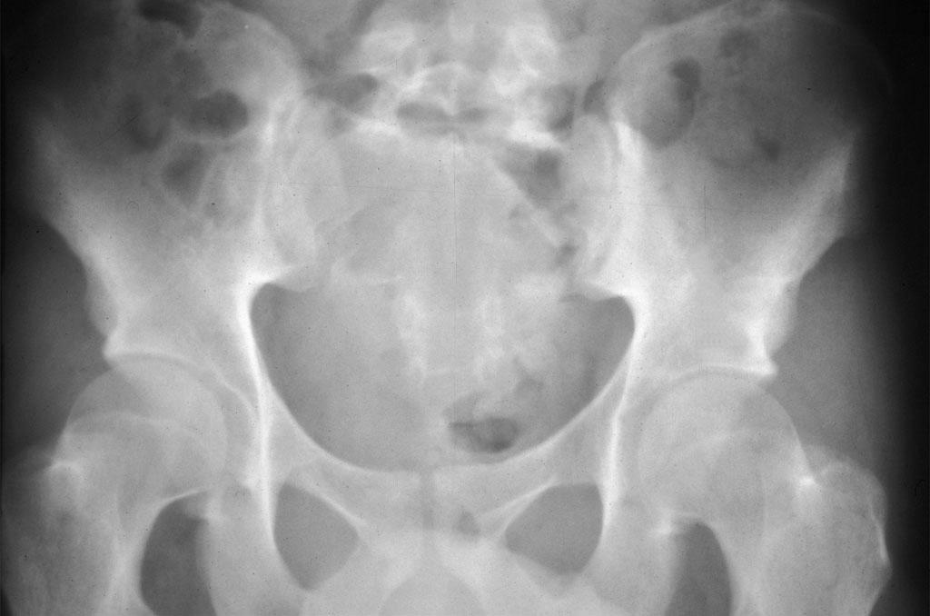
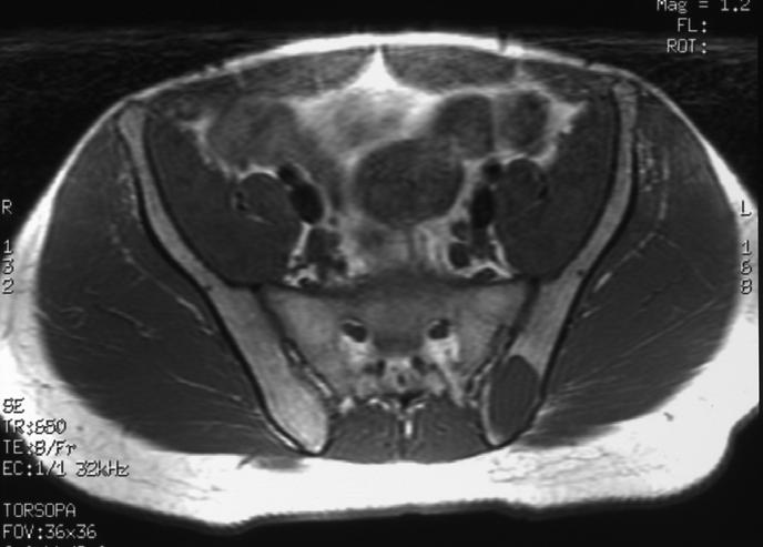
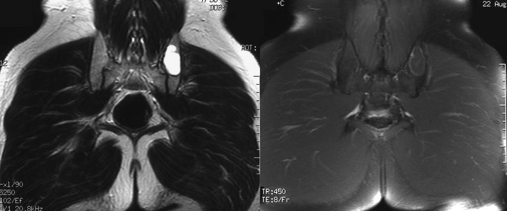
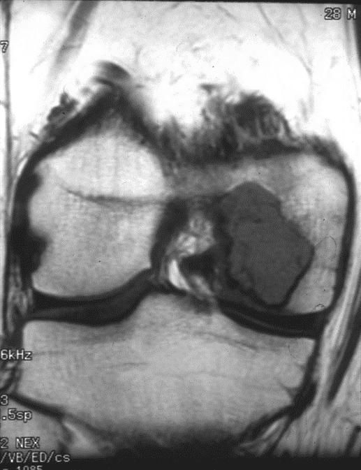
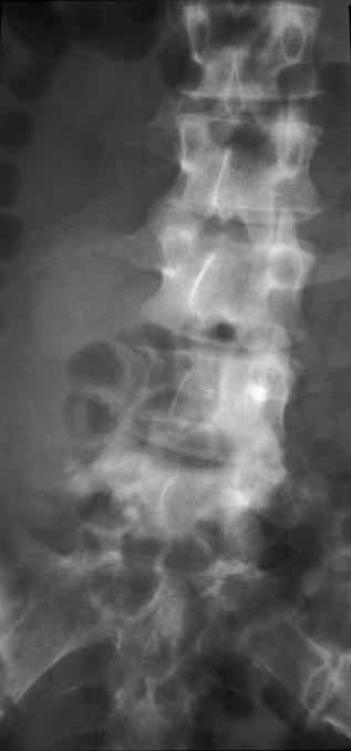
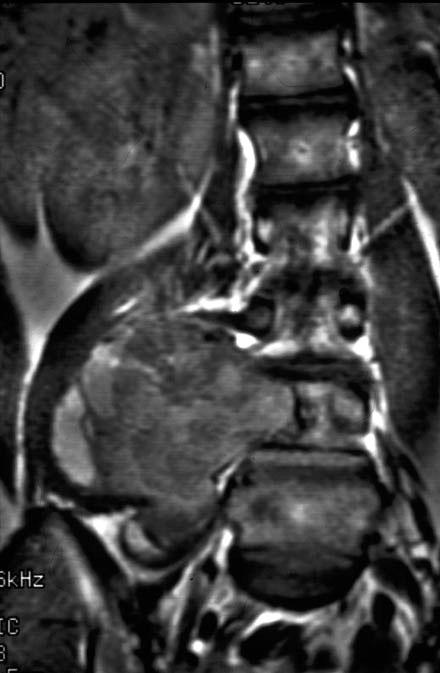
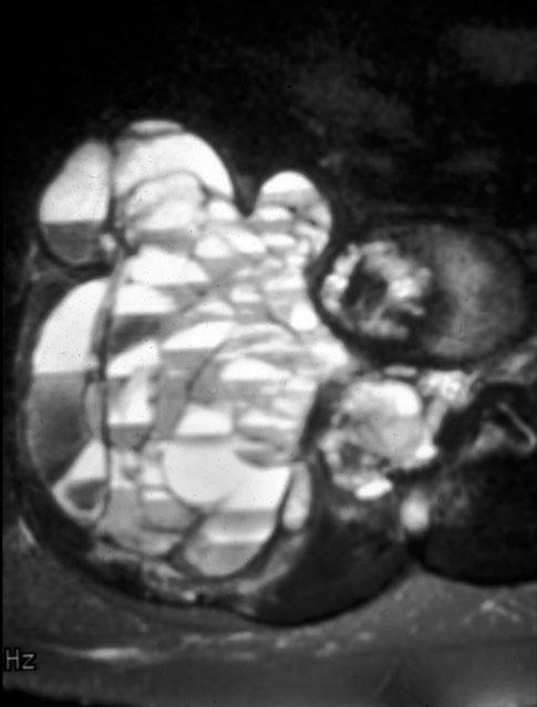
13 GIANT CELL TUMOR LOCATION Spine (7%-15%) - vertebral body sacrum-thoracic-cervical-lumbar Pelvis 4%; Hands/feet - 5% Multifocal (0.5% - 1%) - skull and face (Paget disease), Goltz syndrome
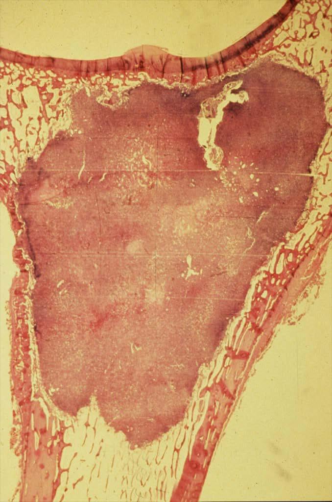
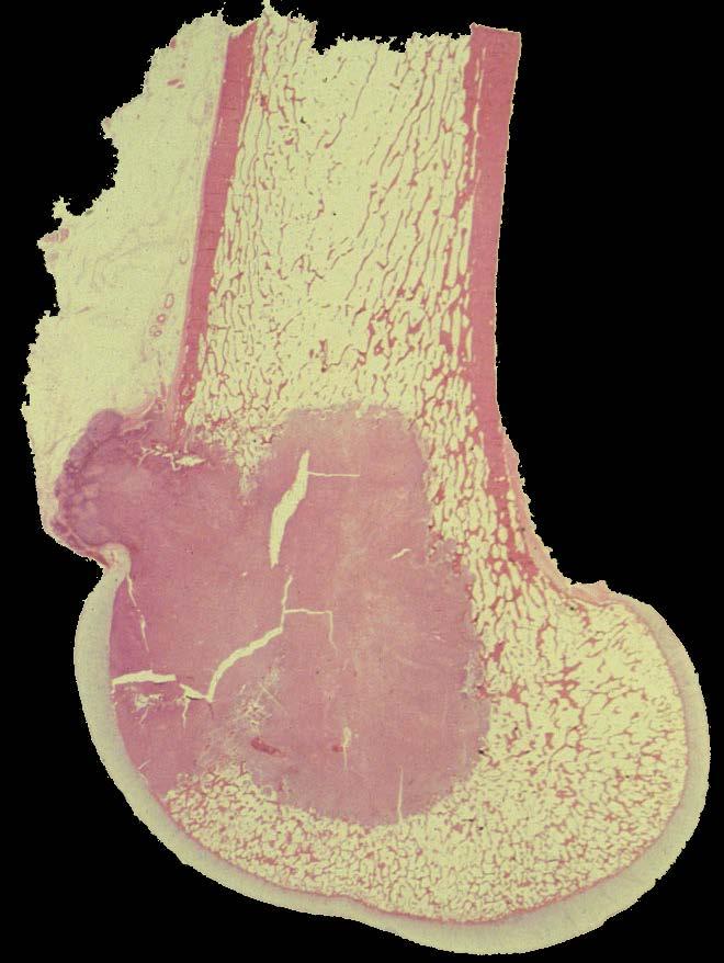
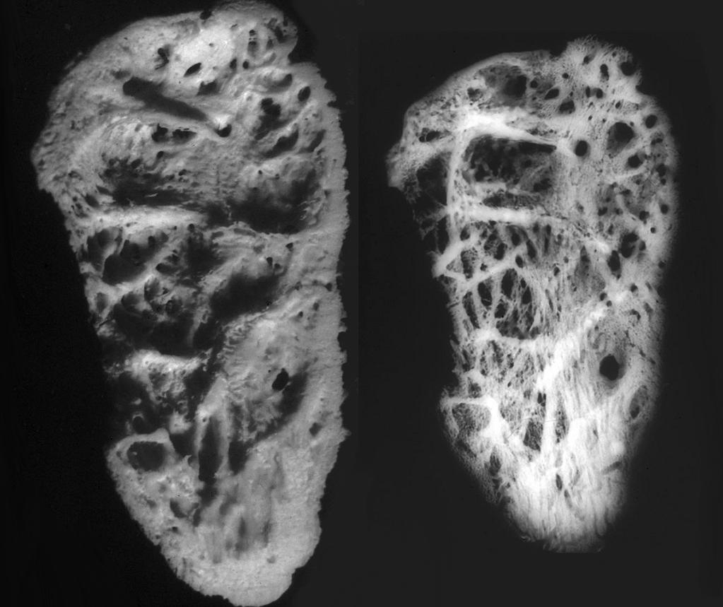
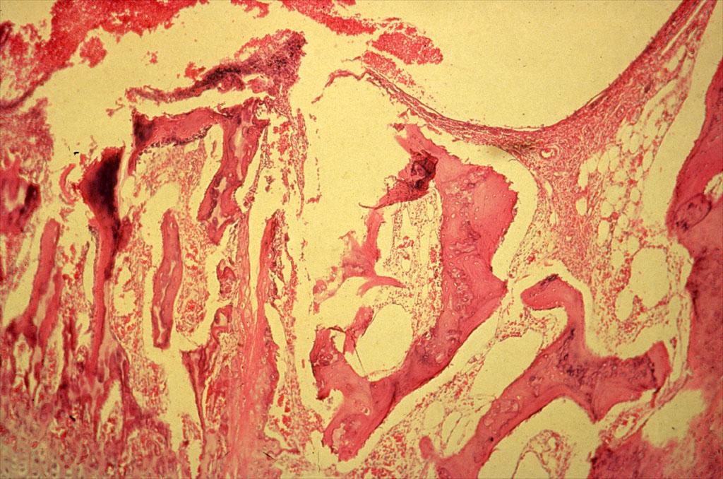
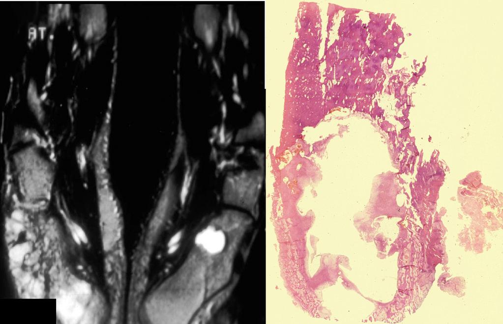
14 GIANT CELL TUMOR PATHOLOGY Osteoclast like giant cells (90%) Mononuclear spindle cell stromal component Hemorrhage, necrosis and hemosiderin ABC like areas 10% - 15% Cytogenetic aberration H3F3A (92%)
15
16
17
18 OSSEOUS LESIONS CONTAINING GIANT CELLS GCT/ABC/UBC NOF/CMF/OGS Brown tumor HPT/chondroblastoma Fibrous dysplasia and variants Osteoblastoma Giant cell reparative granuloma
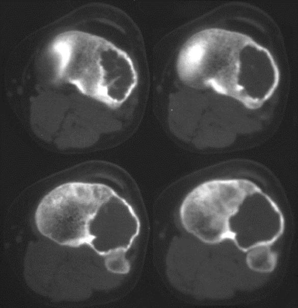
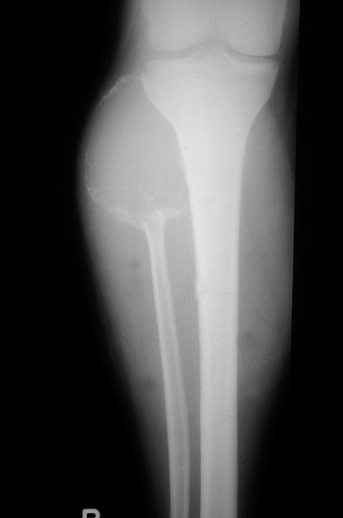
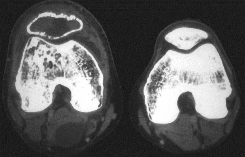
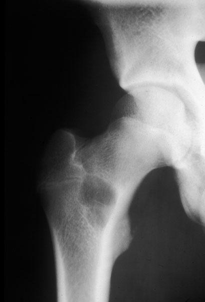
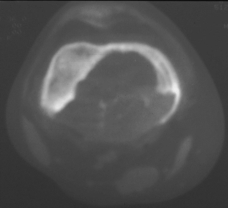
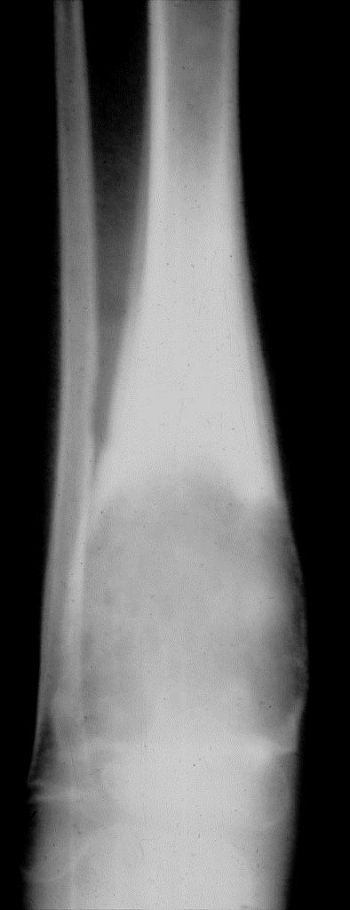
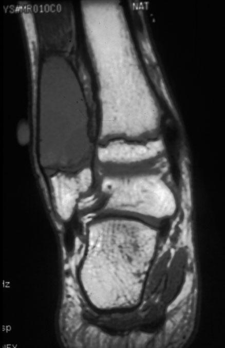
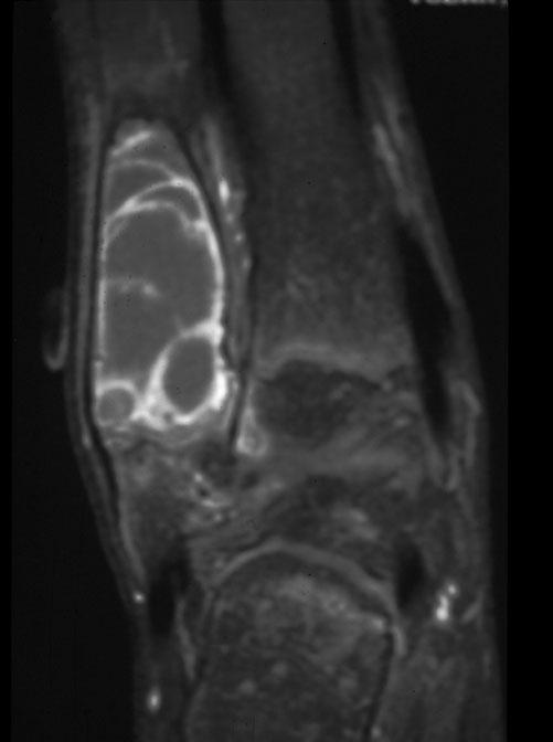
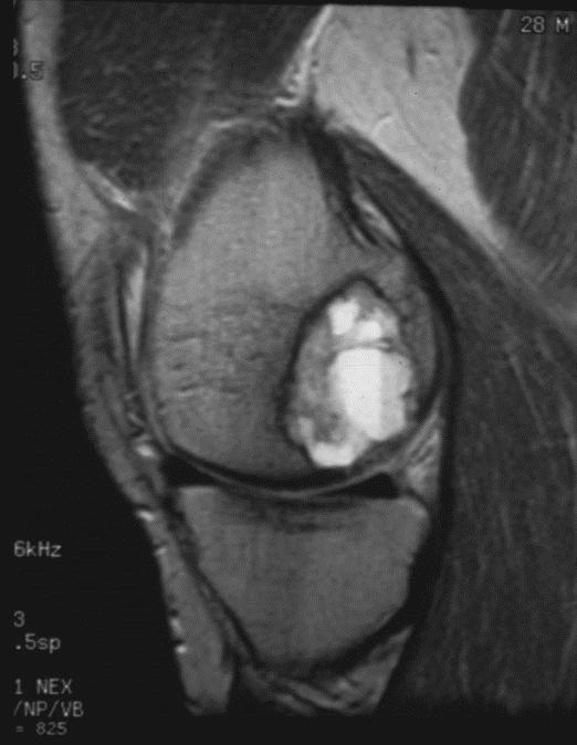
19 GIANT CELL TUMOR RADIOLOGY Solitary eccentric geographic lytic lesion extending into subchondral bone Center of lesion-metaepiphysis Margin IB (80%-85%), IC (10%-20%), IA (1%-2% but up to 20% by CT) No mineralized matrix
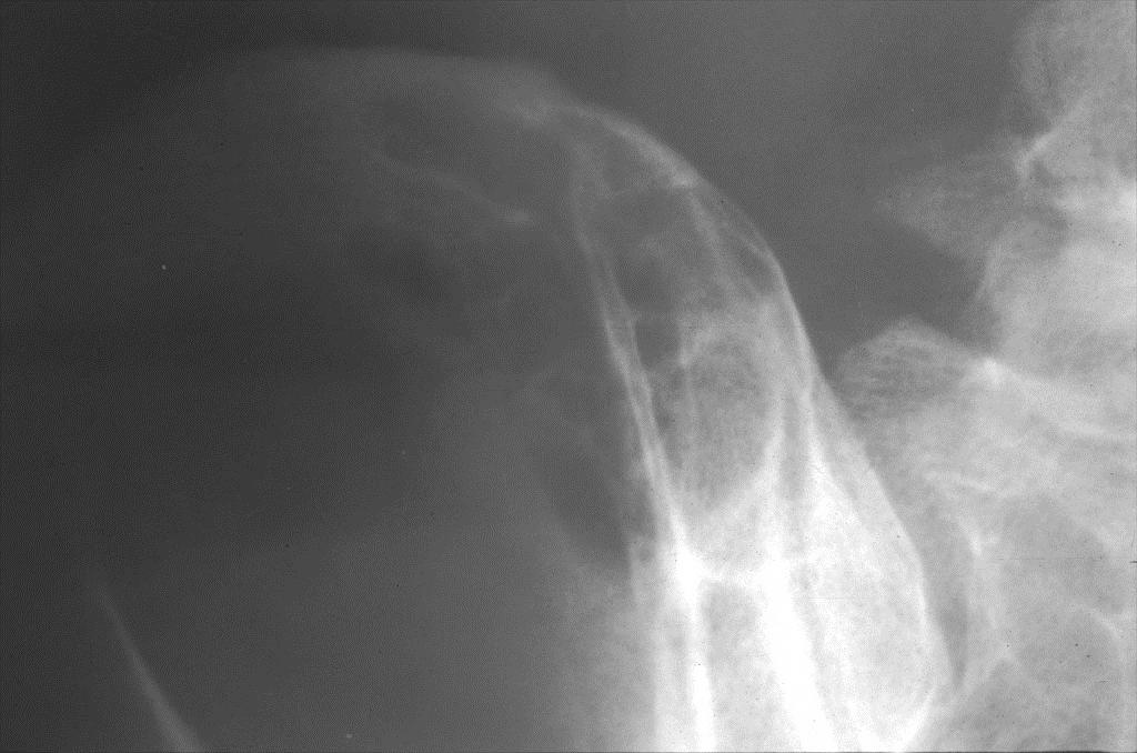
20 GIANT CELL TUMOR RADIOLOGY Expansile remodeling (47%-60%) with apparent cortical permeation (33% - 50%) Septations-subperiosteal new bone Periosteal reaction unusual 10% - 30% Radiologic characteristic do not reflect clinical behavior of GCT
21
22
23
24
25
26
27
28
29
30
31
32
33
34
35
36
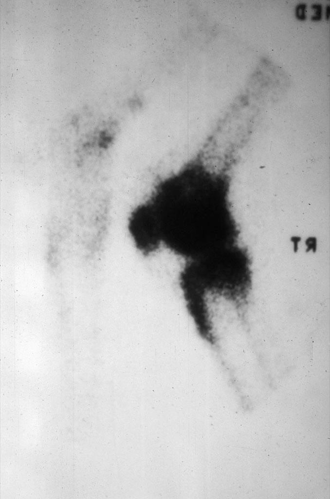
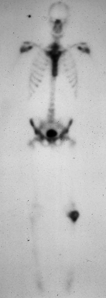
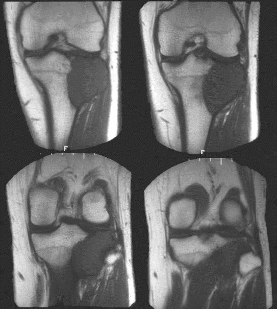
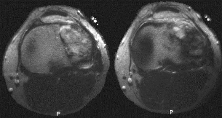
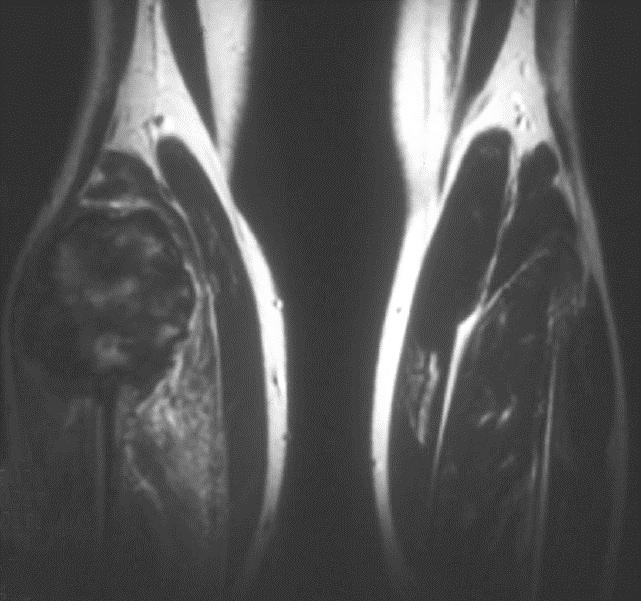
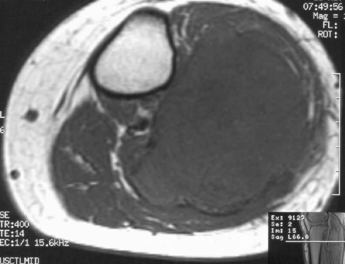
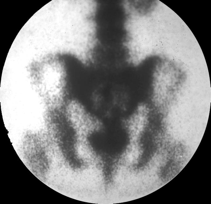
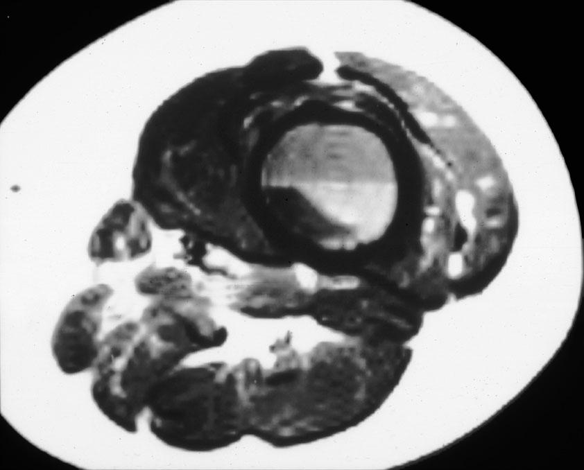
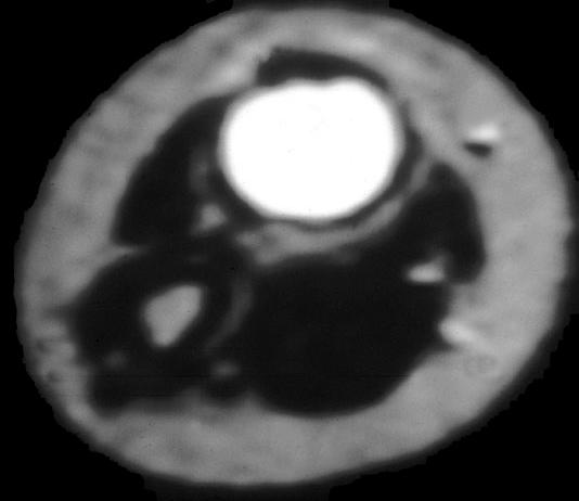
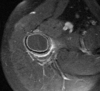
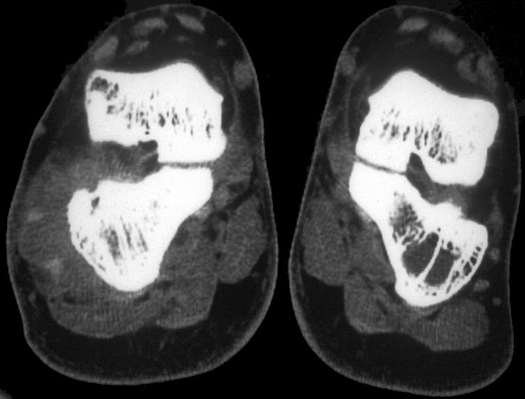
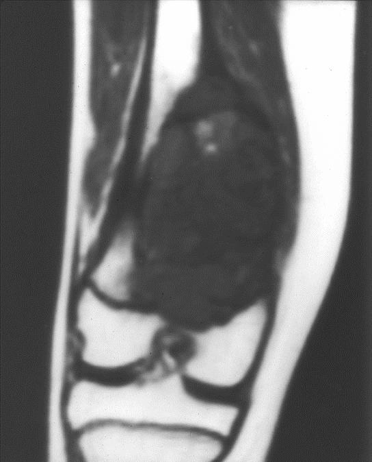
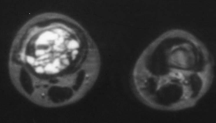
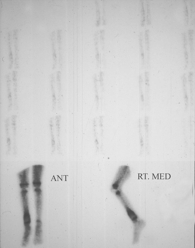
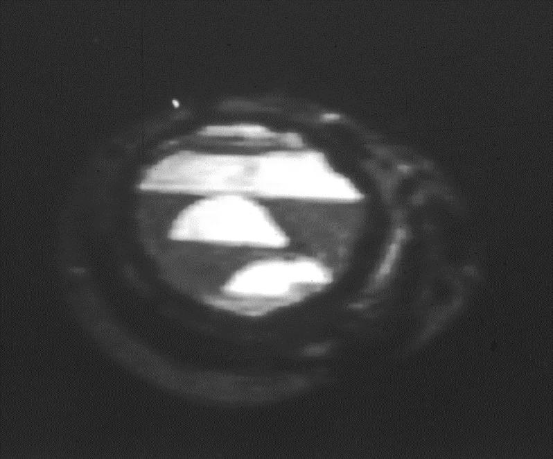
37 GIANT CELL TUMOR RADIOLOGY Bone scan-doughnut sign (57%) Usually a vascular lesion (75%-90%) MRI>CT for evaluation of extent Fluid levels in cystic (ABC) components Low to intermediate intensity usually predominates on T2W images (90%-95%) in solid components
38
39
40
41
42
43
44
45
46 T1
47 T1
48 T1
49 T2
50 T2
51
52
53 T2
54 T2

55 T1 T2
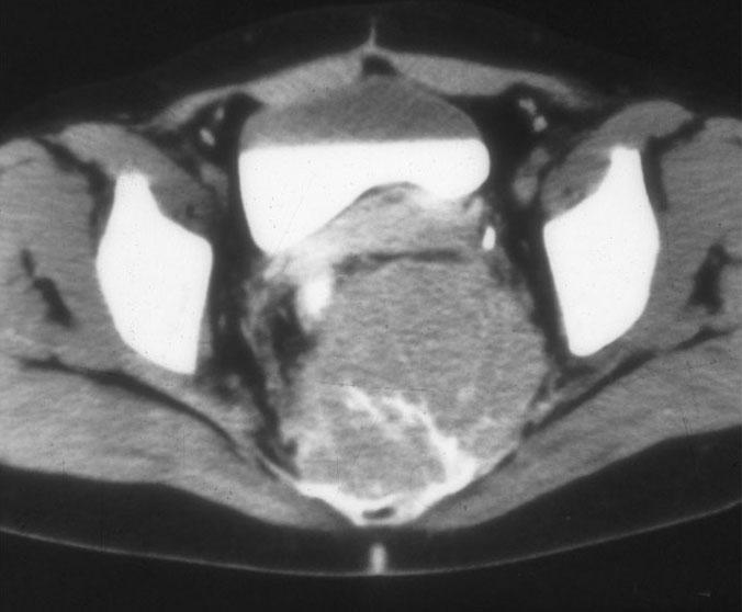
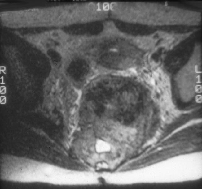
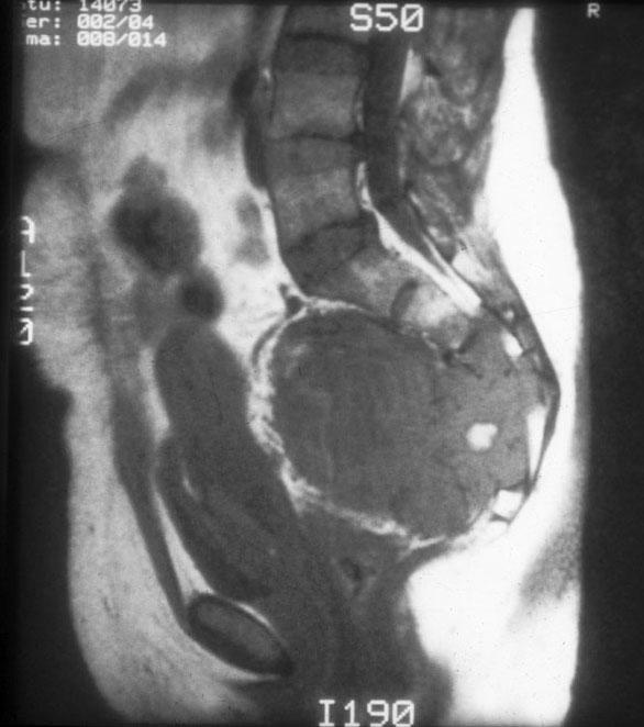
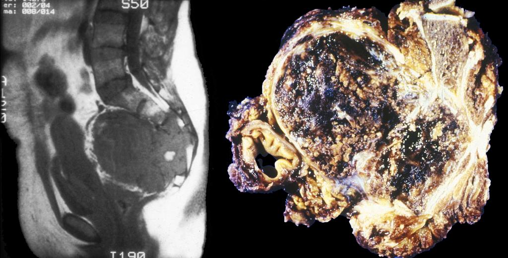
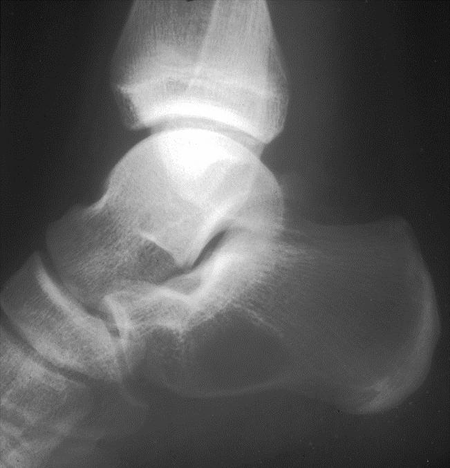
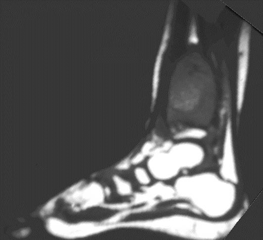
56 T1 T2
57 T1 T2
58
59
60
61
62
63
64 SACRAL LESIONS DIFFERENTIAL DIAGNOSIS GCT/ABC Metastasis Myeloma/plasmacytoma Chordoma Neurogenic tumor
65
66
67
68
69
70 T2
71 T2
72 T1
73 T1
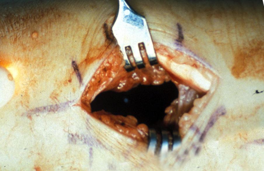
74 T1
75 T1
76 T1
77 T1
78 GIANT CELL TUMOR TREATMENT AND PROGNOSIS Curettage and cryosurgery or en bloc resection and bone graft Local recurrence rate 40% - 60% historically Current recurrence rate 2% - 25% Denosumab medical therapy (86% response) Recurrence does not correspond to radiologic or microscopic appearance
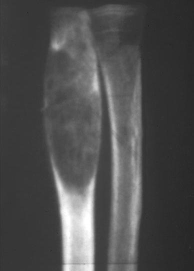
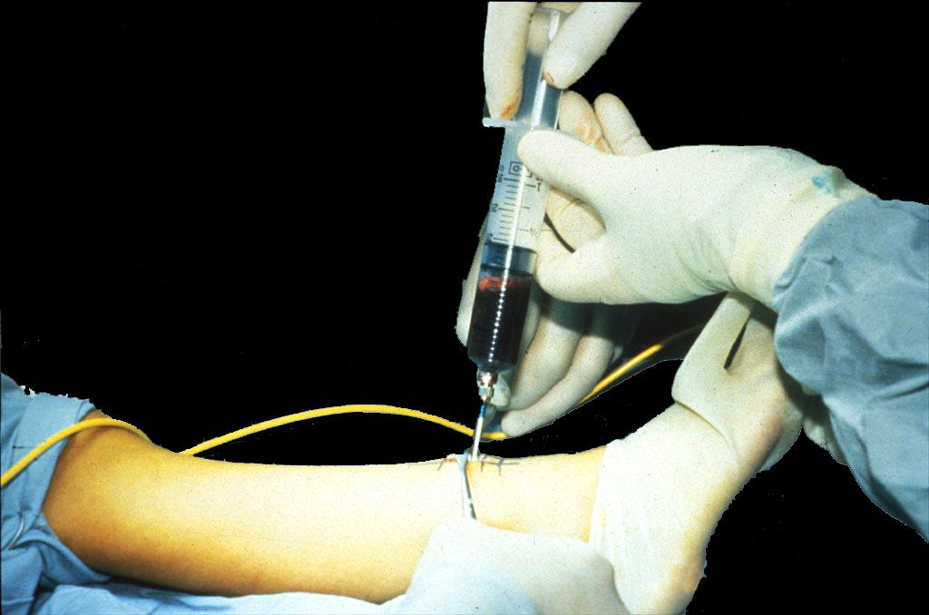
79 UNICAMERAL BONE CYST SIMPLE BONE CYST A fluid-containing lesion lined by mesothelial (epithelial-like) cells usually arising in metaphysis of long bone adjacent to physis
80
81 SIMPLE BONE CYST CLINICAL FEATURES 3% of all biopsied primary osseous neoplasms Young patients 85% <20 years M > F; 2:1 Pathologic fracture 50%
82 SIMPLE BONE CYST PATHOLOGY Clear, straw-colored fluid filled cyst Cyst lining-thin flat epithelial-like cells-mesothelial origin Complicated cysts-hemorrhage, fibro-osseous repair tissue
83
84
85
86 SIMPLE BONE CYST LOCATION/ETIOLOGY Under age 20 - humerus (55% - 65%), femur (25%- 30%), tibia, fibula, radius and ulna rare Over age 20 - iliac bone/ calcaneus Cause - lymphatic or venous obstruction vs. synovial origin
87 SIMPLE BONE CYST RADIOLOGY Geographic IA lesion-originate in central metaphysis (active) Can migrate into the diaphysis (latent) Mild expansile remodeling Not infrequently multilocular
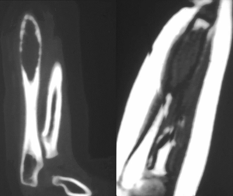
88 SIMPLE BONE CYST RADIOLOGY Pathologic fracture - Fallen fragment sign (5%) CT/MRI-noncomplicated see simple fluid CT/MRI-complicated case Soft tissue Fluid-fluid/gas-fluid level
89
90
91
92
93
94
95
96
97
98 T1
99 T1
100 T1
101
102
103
104
105 T1
106 T2
107 T1 T2 T1 GD
108 T1 T2 T1 GD
109 T1 T2 T1 GD

110 T1 T2 T1 GD
111
112
113
114
115
116 T2
117 T2
118
119
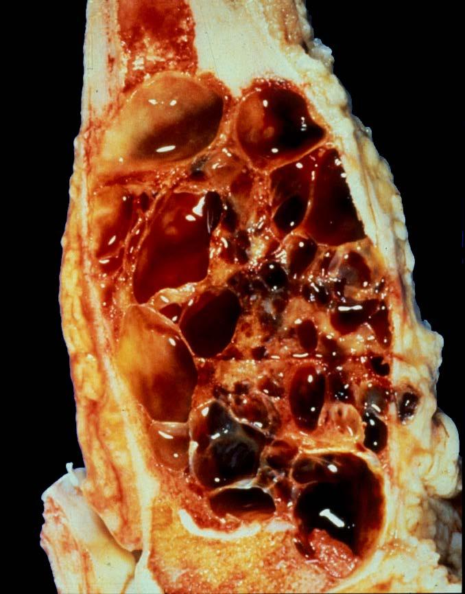
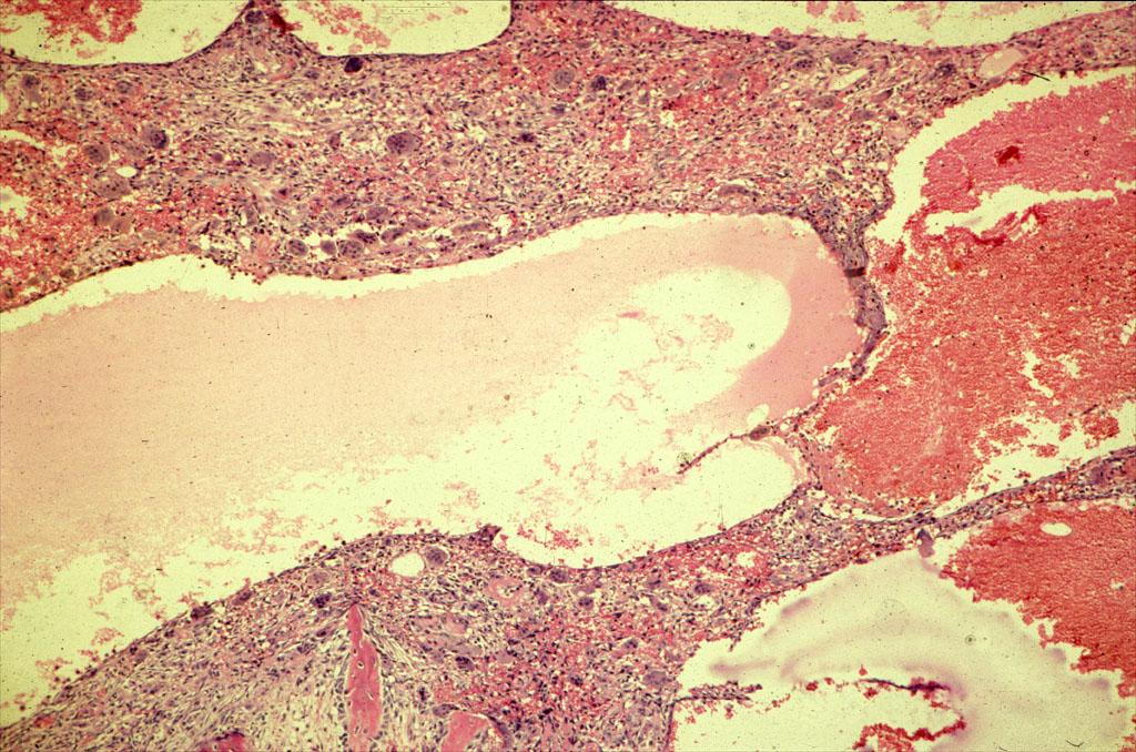
120 T1 T2 T1 GD
121 T1 T2 T1 GD

122 T1 T2 T1 GD
123 SIMPLE BONE CYSTS TREATMENT AND COURSE Spontaneous regression or heal after fracture Curettage and bone grafting Intralesional steroids (70% - 95% effective) Intralesional bone marrow or bone matrix Recurrence 20% - 40% - Increases with younger age and larger size Extremely rare-malignant transformation
124 ANEURYSMAL BONE CYST (ABC) DEFINITION The so called aneurysmal bone cyst is neither a cyst nor a neoplasm; rather it is probably a periosteal to intraosseous arteriovenous malformation not uncommonly seen in association with other well known benign and even malignant lesions Mirra JM. Bone tumors. Lea & Febiger 1989.
125 ANEURYSMAL BONE CYST CLINICAL FEATURES 1%-6% of all biopsied primary osseous neoplasms 80%-85% between ages 5 and 20 years Patients present with pain, swelling, and pathologic fracture (10% - 20%) May be associated with trauma More common in women (1.5-2:1)
126 ANEURYSMAL BONE CYST SECONDARY LESION 1% - 32% of cases Benign lesions-chondroblastoma, CMF, NOF, GCT, fibrous dysplasia, UBC, brown tumor, hemangioma, giant cell reparative granuloma Malignant lesionshemangioendothelioma, telangiectatic osteosarcoma, chondrosarcoma
127 OSSEOUS LESIONS WITH PROMINENT FLUID LEVELS DIFFERENTIAL DIAGNOSIS Aneurysmal bone cyst (only fluid levels) Giant cell tumor (to bone end, metaphyseal center) Chondroblastoma (epiphyseal center) Osteoblastoma (posterior elements spine) Telangiectatic Osteosarcoma (thick walls, osteoid CT) Fibrous dysplasia (diaphysis, ground glass )
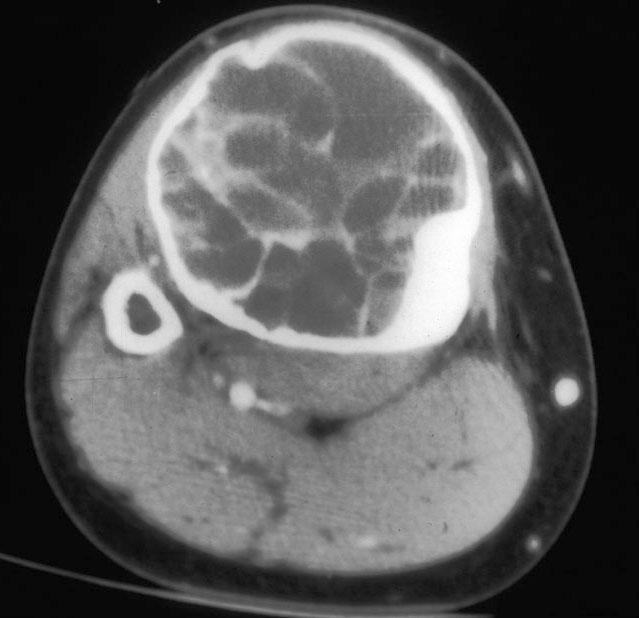
128 ANEURYSMAL BONE CYST PATHOLOGY Gross Blood filled sponge Cavernous blood filled spaces line by fibrous walls May see chondrosseous tissue indicating repair
129
130
131
132
133
134
135
 Pelvis 5% - 10% Hands 10% -")
136 ANEURYSMAL BONE CYST LOCATION Long tubular bone 70% - 80% Spine posterior elements-15% (thoracic, lumbar, cervical, sacral) Pelvis 5% - 10% Hands 10% - 15%
137 ANEURYSMAL BONE CYST RADIOLOGY Only osseous neoplasm named for its radiologic appearance Metaphysis (80% - 90%), eccentric medullary geographic lytic lesion Can appear central with expansion Diaphysis (10% - 20%), often surface lesions
138 ANEURYSMAL BONE CYST RADIOLOGY Expansile remodeling uneven in distribution creating one aggressive margin Spine-expansion can lead to neurologic deficits Periosteal membrane intact on CT/MRI Bone scan-peripheral activity (65%)
139 ANEURYSMAL BONE CYST RADIOLOGY Fluid levels (CT/MRI)-nonspecific representing sedimentation of blood Angiography-hypovascular lesion with localized areas of increased vascularity
140
141
142
143
144
145
146
147
148
149
150
151
152
153
154
155
156
157
158 T1
159 T2
160 T2
161 T2
162
163
164
165
166
167
168 T1
169 T2
170 T2
171 T2
172
173
174
175
176 T1 T1 GD
177 T1 T1 GD
178
179
180
181
182
183 T1 T2
184 T1 T2
185 T1 T2
186
187
188 T1 T2
189 T1 T2
190 T1 T2
191
192 ALPHABET SOUP AND CYSTIC LESIONS OF THE BONE Giant cell tumor (GCT) Unicameral bone cyst (UBC) Aneurysmal bone cyst (ABC) Epidermoid inclusion cyst Subchondral cyst Intraosseous ganglion Post-traumatic cyst
193 ALPHABET SOUP AND CYSTIC LESIONS OF THE BONE MARK D. MURPHEY MD, FACR Physician-in-Chief, AIRP Chief, Musculoskeletal Imaging
The Radiology Assistant : Bone tumor - well-defined osteolytic tumors and tumor-like lesions
 Bone tumor - well-defined osteolytic tumors and tumor-like lesions Henk Jan van der Woude and Robin Smithuis Radiology department of the Onze Lieve Vrouwe Gasthuis, Amsterdam and the Rijnland hospital,
Fluid-fluid levels in bone tumors: A pictorial review
 Fluid-fluid levels in bone tumors: A pictorial review Poster No.: C-578 Congress: ECR 2009 Type: Educational Exhibit Topic: Musculoskeletal Authors: L. Figueroa Nasra, C. Martín Hervás, M. Tapia-Viñé,
Bone Tumors Clues and Cues
 William Herring, M.D. 2002 Bone Tumors Clues and Cues In Slide Show mode, advance the slides by pressing the spacebar All Photos Retain the Copyright of their Authors Clues by Appearance of Lesion Patterns
The Radiology Assistant : Bone tumor - ill defined osteolytic tumors and tumor-like lesions
 Bone tumor - ill defined osteolytic tumors and tumor-like lesions Henk Jan van der Woude and Robin Smithuis Radiology department of the Onze Lieve Vrouwe Gasthuis, Amsterdam and the Rijnland hospital,
Disclosures. Giant Cell Rich Tumors of Bone. Outline. The osteoclast. Giant cell rich tumors 5/21/11
 Disclosures Giant Cell Rich Tumors of Bone Andrew Horvai, MD, PhD Associate Clinical Professor, Pathology This lecture discusses "off label" uses of a number of pharmaceutical agents. The speaker is describing
Introduction to Musculoskeletal Tumors. James C. Wittig, MD Orthopedic Oncologist Sarcoma Surgeon
 Introduction to Musculoskeletal Tumors James C. Wittig, MD Orthopedic Oncologist Sarcoma Surgeon www.tumorsurgery.org Definitions Primary Bone / Soft tissue tumors Mesenchymally derived tumors (Mesodermal)
APMA 2018 Radiology Track Bone Tumors When to say Gulp!
 APMA 2018 Radiology Track Bone Tumors When to say Gulp! DANIEL P. EVANS, DPM, FACFAOM Professor, Department of Podiatric Medicine and Radiology Dr. Wm. Scholl College of Podiatric Medicine Conflict of
Typical skeletal location and differential diagnosis of bone tumors.
 Typical skeletal location and differential diagnosis of bone tumors. Poster No.: C-2418 Congress: ECR 2015 Type: Educational Exhibit Authors: M. Barros, L. A. Ferreira, Y. Costa, P. J. V. Coelho, F. Caseiro
Benign Fibrous Histiocytoma with Cystic Change of the Femur: a Case Report
 pissn 2384-1095 eissn 2384-1109 imri 2016;20:264-268 https://doi.org/10.13104/imri.2016.20.4.264 Benign Fibrous Histiocytoma with Cystic Change of the Femur: a Case Report Jung Ah Park, Sung Gyu Moon,
Primary bone tumors > metastases from other sites Primary bone tumors widely range -from benign to malignant. Classified according to the normal cell
 Primary bone tumors > metastases from other sites Primary bone tumors widely range -from benign to malignant. Classified according to the normal cell counterpart and line of differentiation. Among the
Bubbly Lesions of Bone
 Residents Section Pattern of the Month w79 08.18.09 Eisenberg Residents Section Pattern of the Month Residents inradiology Ronald L. Eisenberg 1 Eisenberg RL Keywords: bubbly lesions, fegnomashic, skeletal
Case Report Giant Cell Tumor of Bone: Documented Progression over 4 Years from Its Origin at the Metaphysis to the Articular Surface
 Volume 2016, Article ID 9786925, 5 pages http://dx.doi.org/10.1155/2016/9786925 Case Report Giant Cell Tumor of Bone: Documented Progression over 4 Years from Its Origin at the Metaphysis to the Articular
Grading of Bone Tumors
 Grading of Bone Tumors Joon Hyuk Choi, M.D. Department of Pathology College of Medicine, Yeungnam University Introduction to grading system of bone tumor used at Mayo Clinic WHO Histologic Classification
Bone and Joint Part 2. Leslie G Dodd, MD
 Bone and Joint Part 2 Leslie G Dodd, MD Relative rates of cancer Sarcomas are relatively uncommon tumors New cancer cases 2007 All sites 1.4 million prostate 218,890 lung 213,380 breast 180,510 Soft tissue
GIANT CELL TUMOR OF LOWER END OF FEMUR IN A SKELETALLY IMMATURE-A RARE CASE
 GIANT CELL TUMOR OF LOWER END OF FEMUR IN A SKELETALLY IMMATURE-A RARE CASE *Surojit Mondal 1, Aniket Chowdhury 2 and Goutam Bandyopadhyay 3 1 Department of Orthopaedics, B.S.Medical College, Bankura,
FEGNOMASHIC: from x-ray to MRI
 FEGNOMASHIC: from x-ray to MRI Poster No.: C-2441 Congress: ECR 2015 Type: Educational Exhibit Authors: S. Fouassier, A. L. C. Duarte, C. Ruivo, J. Velez ; Évora/PT, 1 2 1 2 3 1 3 Coimbra/PT, PT Keywords:
Iliac aneurysmal bone cyst treated by cystoscopic controlled curettage
 Accepted February 13th, 2004 Iliac aneurysmal bone cyst treated by cystoscopic controlled curettage Ludwig Schwering¹, Markus Uhl² and Georg W. Herget( )¹ ¹ Department of Orthopaedics and Traumatology,
Aneurysmal Bone Cyst of the Pelvis: A Challenge in Treatment: Review of the Literature
 ISPUB.COM The Internet Journal of Orthopedic Surgery Volume 8 Number 1 Aneurysmal Bone Cyst of the Pelvis: A Challenge in Treatment: Review of the Literature S Bajracharya, G Khanal, A Sundas, S Pandey,
Primary bone tumors according to the WHO classification: a review of 13 years with illustrative examples
 Primary bone tumors according to the WHO classification: a review of 13 years with illustrative examples Poster No.: C-1741 Congress: ECR 2015 Type: Educational Exhibit Authors: J. Silva, M. A. Ramírez
GIANT CELL TUMOR OF BONE
 GIANT CELL TUMOR OF BONE Definition. First described by Jaffe et al. 1, giant cell tumor of bone is a locally aggressive primary neoplasm of bone that is composed of proliferation of bland looking oval
Endovascular and surgical treatment of giant pelvic tumor
 Endovascular and surgical treatment of giant pelvic tumor Mitrev Z., MD FETCS; Anguseva T., MD; Milev I., MD; Zafiroski G., PhD MD Center for Cardiosurgery, Filip the II, Skopje, Macedonia Background Giant
Cystic Lesions of Bone
 Cystic Lesions of Bone Intraosseous Ganglion Cyst Definition. Intraosseous pseudocyst of unknown etiology that is filled with viscous mucinous material and is not associated with osteoarthritis in the
CASE PRESENTATION. Dr. Faseeh Shahab PGY3 Orthopaedic Resident, Khyber Teaching Hospital, Peshawar, PAKISTAN
 CASE PRESENTATION Dr. Faseeh Shahab PGY3 Orthopaedic Resident, Khyber Teaching Hospital, Peshawar, PAKISTAN CASE PRESENTATION - History Ms. SB, 30yo Afghan National Presented with 3 months history of Swelling
Malignant bone tumors. Incidence Myeloma 45% Osteosarcoma 24% Chondrosarcoma 12% Lyphoma 8% Ewing s Sarcoma 7%
 Malignant bone tumors Incidence Myeloma 45% Osteosarcoma 24% Chondrosarcoma 12% Lyphoma 8% Ewing s Sarcoma 7% Commonest primary bone sarcoma is osteosarcoma X ray Questions to ask 1. Solitary or Multiple
GIANT CELL-RICH OSTEOSARCOMA: A CASE REPORT
 Nagoya J. Med. Sci. 59. 151-157, 1996 CASE REPORTS GIANT CELL-RICH OSTEOSARCOMA: A CASE REPORT KEIJI SATO!, SHIGEKI YAMAMURA!, HISASHI IWATA!, HIDESHI SUGIURA 2, NOBUO NAKASHIMA 3 and TETSURO NAGASAKA
Skeletal Radiology. Solitary (unicameral) bone cyst. The fallen fragment sign revisited
 bone cyst. The fallen fragment sign revisited") Skeletal Radiol (1989) 18:261-265 Skeletal Radiology Solitary (unicameral) bone cyst The fallen fragment sign revisited S. Struhl, M.D., C. Edelson, M.D., H. Pritzker, M.D., L.P. Seimon, M.D., and H.D.
Skeletal Radiol (1989) 18:261-265 Skeletal Radiology Solitary (unicameral) bone cyst The fallen fragment sign revisited S. Struhl, M.D., C. Edelson, M.D., H. Pritzker, M.D., L.P. Seimon, M.D., and H.D.
Radiologic approach to pediatric lytic bone lesions
 Radiologic approach to pediatric lytic bone lesions Poster No.: C-1177 Congress: ECR 2016 Type: Educational Exhibit Authors: J. L. LERMA GALLARDO, I. de la Pedraja, A. Lancharro 1 1 1 2 1 1 Zapata, J.
General Approach to Lytic Bone Lesions D. Lee Bennett, MD, MA, Georges Y. El Khoury, MD Appl Radiol. 2004;33(5)
") General Approach to Lytic Bone Lesions D. Lee Bennett, MD, MA, Georges Y. El Khoury, MD Appl Radiol. 2004;33(5) www.medscape.com Abstract and Introduction Abstract When interpreting musculoskeletal radiographs,
General Approach to Lytic Bone Lesions D. Lee Bennett, MD, MA, Georges Y. El Khoury, MD Appl Radiol. 2004;33(5) www.medscape.com Abstract and Introduction Abstract When interpreting musculoskeletal radiographs,
History. 33 y/o F with hx of palpable anterior tibial mass x 2 years, only painful with palpation
 History 33 y/o F with hx of palpable anterior tibial mass x 2 years, only painful with palpation Imaging Photo Album Patient also had a smaller lesion 1 cm proximal to this lesion, not seen radiographically.
The Radiology Assistant : Bone tumor A-G
 Bone tumor A-G Bone tumors and tumor-like lesions in alphabethic order Henk Jan van de Woude and Robin Smithuis Radiology department of the Onze Lieve Vrouwe Gasthuis, Amsterdam and the Rijnland hospital,
Radiography in the Initial Diagnosis of Primary Bone Tumors
 Residents Section Structured Review Costelloe and Madewell Radiography of Primary Bone Tumors Residents Section Structured Review Colleen M. Costelloe 1 John E. Madewell Costelloe CM, Madewell JE Keywords:
Fluid fluid levels in bone tumors and tumoral lesions - Pictorial essay
 Review Fluid fluid levels in bone tumors and tumoral lesions - Pictorial essay Subbarao Kakarla 1,* 1 KIMS Foundation and Research Centre, Minister Road, Secunderabad - 500003, Telangana, India Abstract
Bone Tumours - a synopsis. Dr Zena Slim SpR in Histopathology QAH 2009
 Bone Tumours - a synopsis Dr Zena Slim SpR in Histopathology QAH 2009 Aims General approach to diagnosis Common entities.and not so common ones. Mini quiz Challenge of bone tumour diagnosis Bone tumours
Bone tumors. RMG: jan
 Bone tumors RMG: jan 217. @Kijohs KIZZA JOHN KIJOHS Diseases arising in bone Lipoma Fibrous cortical defects Non-ossifying fibroma Bone island Benign simple cysts Enchondroma Osteochondroma Osteoid osteoma
CASE STUDY: PRO-DENSE Injectable Regenerative Graft Used to Backfill a Bone Cavity Following Resection of a Giant Cell Tumor
 : PRO-DENSE Used to Backfill a Bone Cavity Following Resection of a Giant Cell Tumor Contributed by: Matthew J. Seidel, MD* Lauren A. Schwartz, NP Scottsdale, AZ *Dr. Seidel is a paid consultant for Wright
Clear Cell Chondrosarcoma of the Sacrum
 Clear Cell Chondrosarcoma of the Sacrum Yasunobu Iwasaki MD 1, Tetsuji Yamamoto MD (!) 2, Mitsuo Tsuji MD 1, Akira Kurihara MD 1, Masaaki Uratsuji MD 1, Norihide Sha MD 1, and Shinichi Yoshiya MD 2 1 Department
MUSCULOSKELETAL RADIOLOGY
 MUSCULOSKELETAL RADOLOGY SECTON www.cambridge.org Achilles tendonopathy/rupture Characteristics Describes pathology of the combined tendon of the gastro-soleus complex, which inserts onto the calcaneum.
Aneurysmal bone cysts (ABCs) are rare, destructive,
 are rare, destructive,") ORIGINAL ARTICLE A Review of 56 Patients Kerem BazarNr, MD,* Ahmet Pizkin,Þ Berk Güc$lü,þ Yusuf YNldNz,* and Yener SağlNk* Background: Aneurysmal bone cysts (ABCs) are benign lesions that are usually treated
ORIGINAL ARTICLE A Review of 56 Patients Kerem BazarNr, MD,* Ahmet Pizkin,Þ Berk Güc$lü,þ Yusuf YNldNz,* and Yener SağlNk* Background: Aneurysmal bone cysts (ABCs) are benign lesions that are usually treated
MRI Findings of Giant Cell Tumors of the Spine
 MRI of Spinal Tumors Musculoskeletal Imaging Clinical Observations Jong Won Kwon 1 Hye Won Chung 1,2 Eun Yoon Cho 3 Sung Hwan Hong 4 Sang-Hee Choi 1 Young Cheol Yoon 1 Sang Kyu Yi 1 Kwon JW, Chung HW,
Osteomyelitis in infancy and childhood: A clinical and diagnostic overview M. Mearadji
 Osteomyelitis in infancy and childhood: A clinical and diagnostic overview M. Mearadji International Foundation for Pediatric Imaging Aid Introduction Osteomyelitis is a relative common disease in infancy
4/28/2010. Fractures. Normal Bone and Normal Ossification Bone Terms. Epiphysis Epiphyseal Plate (physis) Metaphysis
 Metaphysis") Fractures Normal Bone and Normal Ossification Bone Terms Epiphysis Epiphyseal Plate (physis) Metaphysis Diaphysis 1 Fracture Classifications A. Longitudinal B. Transverse C. Oblique D. Spiral E. Incomplete
Fractures Normal Bone and Normal Ossification Bone Terms Epiphysis Epiphyseal Plate (physis) Metaphysis Diaphysis 1 Fracture Classifications A. Longitudinal B. Transverse C. Oblique D. Spiral E. Incomplete
GIANT CELL TUMOR : RARE SITES
 CASE REPORT GIANT CELL TUMOR : RARE SITES Zaheda Khatoon, Rukhsana Tariq, Shahzad Babar Kureshi, Rashid Ahmed Advanced Radiology Clinic (Pvt.) Ltd., Karachi, Pakistan. :13-17 ABSTRACT Giant cell tumor
Differential Diagnosis of Radiolucent Lesions of the Jaws
 Differential Diagnosis of Radiolucent Lesions of the Jaws Multilocular Multilocular Radiolucencies Odontogenic Keratocyst Botryoid Odontogenic Cyst Glandular odontogenic Cyst Invasive Ameloblastoma Central
Malignant Bone Tumors - Part I: a brief revision of diagnostic aspects with conventional radiology
 Malignant Bone Tumors - Part I: a brief revision of diagnostic aspects with conventional radiology Poster No.: C-2473 Congress: ECR 2013 Type: Educational Exhibit Authors: I. Candelaria, L. B. Barbosa,
Primary Bone Tumors: Spine Surgery Live -Video Techniques Mobile Spine
 Primary Bone Tumors: Spine Surgery Live -Video Techniques Mobile Spine Christopher Ames MD Professor of Neurosurgery and Orthopedic Surgery Director of Spine Tumor And Deformity Surgery UCSF Department
Common Primary Tumors of Bone
 Special Report Common Primary Tumors of Bone Primary bone tumors are a relatively rare occurrence, however, they can have serious deleterious consequences. Many possess the ability to degenerate into malignant
Bread and Butter Bone Pathology
 Bread and Butter Bone Pathology NICOLE D. RIDDLE, MD RUFFOLO, HOOPER, AND ASSOC. / UNIVERSITY OF SOUTH FLORIDA Goals: Fundamentals of neoplastic bone pathology Bone Producing Cartilage Producing Miscellaneous
VALORACIÒN RADIOLÓGICA DE LA LESIÒN ÒSEA SOLITARIA IMAGENOLOGIA MEDICA UNIVERSIDAD HISPANOAMERICANA
 VALORACIÒN RADIOLÓGICA DE LA LESIÒN ÒSEA SOLITARIA IMAGENOLOGIA MEDICA UNIVERSIDAD HISPANOAMERICANA TUMORES ÓSEOS SE PRESENTAN POR RANGOS DE EDAD, PRINCIPALMENTE: MENORES DE 20 AÑOS 20 A 40 AÑOS MAYORES
COPYRIGHT 2004 BY THE JOURNAL OF BONE AND JOINT SURGERY, INCORPORATED
 84 COPYRIGHT 2004 BY THE JOURNAL BONE AND JOINT SURGERY, INCORPORATED Radiographic Evaluation of Pathological Bone Lesions: Current Spectrum of Disease and Approach to Diagnosis BY BENJAMIN G. DOMB, MD,
Giant Cell Tumor of the Thoracic Spine Presenting as a Posterior Mediastinal Tumor with Benign Pulmonary Metastases: A Case Report 1
 Giant Cell Tumor of the Thoracic Spine Presenting as a Posterior Mediastinal Tumor with Benign Pulmonary Metastases: A Case Report 1 Tae Hun Kim, M.D., Byung Hak Rho, M.D. 2, Young Eun Bahn, M.D. 2, Won
Solitary Bone Cyst of the Lunate: A Case Report
 Cronicon OPEN ACCESS ORTHOPAEDICS Case Report Solitary Bone Cyst of the Lunate: A Case Report MihirDesai* and Shivanand Bandekar Department of Orthopedics, Goa Medical College, Goa, India *Corresponding
Imaging Findings Of Bone Tumors: A Pictorial Review
 Imaging Findings Of Bone Tumors: A Pictorial Review Poster No.: C-2511 Congress: ECR 2015 Type: Educational Exhibit Authors: M. Limeme, N. Benzina, A. BelKhiria, H. Zaghouani, S. Majdoub, N. Mallat, H.
ORTHOPAEDIC ONCOLOGY OITE REVIEW COURSE
 ORTHOPAEDIC ONCOLOGY OITE REVIEW COURSE Richard D. Lackman, MD FACS Director, Orthopaedic Oncology Center Cancer Institute Introduction In the evaluation of a patient with a bone tumor, there are several
Case Report Lower Limb Reconstruction with Tibia Allograft after Resection of Giant Aneurysmal Bone Cyst
 Case Reports in Orthopedics Volume 2016, Article ID 7980593, 6 pages http://dx.doi.org/10.1155/2016/7980593 Case Report Lower Limb Reconstruction with Tibia Allograft after Resection of Giant Aneurysmal
USCAP 2014 Common problems in bone and soft tissue pathology: Cartilage tumors
 USCAP 2014 Common problems in bone and soft tissue pathology: Cartilage tumors Andrew Horvai MD PhD Clinical Professor, Pathology UCSF, San Francisco, CA Outline Common intramedullary tumors Enchondroma
Imaging Findings of Sacral Tumors 1
 Imaging Findings of Sacral Tumors 1 Seung Ho Kim, M.., Sung Hwan Hong, M.., Ja-Young hoi, M.., Sung Hye Koh, M.., Hye Won hung, M.., Jung-h hoi, M.., Heung Sik Kang, M.. The various pathologic conditions
MRI of Pediatric Ankle and Foot. Mahesh Thapa, MD Associate Professor Seattle Children s University of Washington School of Medicine
 MRI of Pediatric Ankle and Foot Mahesh Thapa, MD Associate Professor Seattle Children s University of Washington School of Medicine Disclosures Under contract with Lippincott Williams and Wilkins (LWW)
A peculiar location of a rare bone tumor: sternal lipoma
 A peculiar location of a rare bone tumor: sternal lipoma Poster No.: P-0033 Congress: ESSR 2016 Type: Authors: Keywords: DOI: Scientific Poster Z. Akkaya, C. Uzun, S. Enon, G. Kocaman, G. Sahin; Ankara/TR
Bone Tumors: In 1 Simple Chart
 Bone Tumors with PowerPoint Interactivity Download this entire slideshow from When running this on your own computer you can jump from slide to slide using these buttons at bottom of each slide: Last slide
Recognizing Cartilaginous Tumors: Spectrum of Imaging Characteristics with Radiologic-Pathologic correlation.
 Recognizing Cartilaginous Tumors: Spectrum of Imaging Characteristics with Radiologic-Pathologic correlation. Poster No.: C-1451 Congress: ECR 2012 Type: Educational Exhibit Authors: E. Barcina García,
Giant cell tumour of the sternum-two cases
 Giant cell tumour of the sternum-two cases Nishaa.P 1, Raghuram.P 2, Navin patil 3, Jaipal B.R 4 Akkamahadevi patel 5 Assistant Professor ESIC medical college and PGIMSR 1 Professor and HOD, 2 Professor
Malignant Bone Tumours. PathoBasic, Daniel Baumhoer
 Malignant Bone Tumours PathoBasic, 20.03.18 Daniel Baumhoer FNCLCC Grading The differentiation score is defined as the extent to which a tumor resembles adult mesenchymal tissue (score 1), the extent to
UCLA UCLA Previously Published Works
 UCLA UCLA Previously Published Works Title Benign bone tumors Permalink https://escholarship.org/uc/item/7h86k14t Journal Radiologic Clinics of North America, 49(6) ISSN 0033-8389 Authors Motamedi, K Seeger,
Effective local and systemic therapy is necessary for the cure of Ewing tumor Most chemotherapy regimens are a combination of cyclophosphamide,
 Ewing Tumor Perez Ewing tumor is the second most common primary tumor of bone in childhood, and also occurs in soft tissues Ewing tumor is uncommon before 8 years of age and after 25 years of age In the
Skeletal metastases are the most common variety of bone tumors and should always be considered in the differential diagnosis, particularly in older
 Dr Brajesh Nandan Skeletal metastases are the most common variety of bone tumors and should always be considered in the differential diagnosis, particularly in older patients. Cancers of the breast, prostate,
Due in Lab. Due next week in lab - Scientific America Article Select one article to read and complete article summary
 Due in Lab 1. Skeletal System 33-34 2. Skeletal System 26 3. PreLab 6 Due next week in lab - Scientific America Article Select one article to read and complete article summary Cell Defenses and the Sunshine
The Skeletal System. Chapter 7a. Skeletal System Introduction Functions of the skeleton Framework of bones The skeleton through life
 The Skeletal System Skeletal System Introduction Functions of the skeleton Framework of bones The skeleton through life Chapter 7a Support Protection Movement Storage areas Minerals Lipids Hemopoiesis
Case report. Giant cell reparative granuloma of the hallux following enchondroma. Open Access
 Case report Open Access Giant cell reparative granuloma of the hallux following enchondroma Khaled Kamoun 1,&, Tarak Sellami 1, Zied Jlailia 1, Layla Abid 2, Mourad Jenzri 1, Mouna Bouaziz 3, Omar Zouar
MRI XR, CT, NM. Principal Modality (2): Case Report # 2. Date accepted: 15 March 2013
: Case Report # 2. Date accepted: 15 March 2013") Radiological Category: Musculoskeletal Principal Modality (1): Principal Modality (2): MRI XR, CT, NM Case Report # 2 Submitted by: Hannah Safia Elamir, D.O. Faculty reviewer: Naga R. Chinapuvvula, M.D.
Radiological Category: Musculoskeletal Principal Modality (1): Principal Modality (2): MRI XR, CT, NM Case Report # 2 Submitted by: Hannah Safia Elamir, D.O. Faculty reviewer: Naga R. Chinapuvvula, M.D.
Small lesions involving scalp and skull in pediatric age.
 Small lesions involving scalp and skull in pediatric age. Poster No.: C-1149 Congress: ECR 2013 Type: Educational Exhibit Authors: M. J. Yi, J. H. Yoo; Seoul/KR Keywords: Education and training, Education,
Paediatric post-traumatic osseous cystic lesion following a distal radial fracture
 Paediatric post-traumatic osseous cystic lesion following a distal radial fracture Joey Chan Yiing Beh 1*, Ehab Shaban Mahmoud Hamouda 1 1. Department of Diagnostic Imaging, KK Women's and Children's Hospital,
Mark D. Murphey, MD, FACR
 Fundamental Concepts of Musculoskeletal Neoplasm: CT and MRI Mark D. Murphey, MD, FACR Important Features in Evaluation of Musculoskeletal Masses Differential diagnosis Preoperative assessment and staging
Small lesions involving scalp and skull in pediatric age.
 Small lesions involving scalp and skull in pediatric age. Poster No.: C-1149 Congress: ECR 2013 Type: Educational Exhibit Authors: M. J. Yi, J. H. Yoo; Seoul/ Keywords: Education and training, Education,
Incidental bone tumors are asymptomatic lesions that are. Incidental Bone Lesions. When to Refer to the Tumor Specialist
 Bulletin of the NYU Hospital for Joint Diseases 2012;70(4):235-40 235 Incidental Bone Lesions When to Refer to the Tumor Specialist LT Suezie Kim, M.D., M.C., U.S.N., Catherine N. Laible, M.D., Leon D.
Aneurysmal bone cyst of the scapula A case report
 Acta Orthop. Belg., 2009, 75, 684-689 CASE REPORT Aneurysmal bone cyst of the scapula A case report Panagiotis MEGAS, Zafiria G. PAPATHANASSIOU, George KASIMATIS, Dionysios J. PAPACHRISTOU From the University
Radiologic Pathologic Correlation of Intraosseous Lipomas. Tim Propeck 1, Mary Anne Bullard 1, John Lin 1, Kei Doi 2, William Martel 1
 Downloaded from www.ajronline.org by 148.251.232.83 on 04/10/18 from IP address 148.251.232.83. opyright RRS. For personal use only; all rights reserved Radiologic Pathologic orrelation of Intraosseous
Section II Musculoskeletal Radiology
 Section II Musculoskeletal Radiology Figure 1 25. You are shown a noncontrast CT (Figure 1) of the thigh. What is the MOST LIKELY diagnosis? A. Synovial sarcoma B. Hemangioma C. Organizing hematoma D.
Comprehensive Case Report Of A Giant Cell Tumor Involving The Proximal Phalanx Of The Left Great Toe Managed With Fibular Grafting
 ISPUB.COM The Internet Journal of Radiology Volume 1 Number 1 Comprehensive Case Report Of A Giant Cell Tumor Involving The Proximal Phalanx Of The Left Great Toe T Dasan, K Vijay, C Satish, B Nagraj,
Bio 103 Skeletal System 45
 45 Lecture Outline: SKELETAL SYSTEM [Chapters 7, 8] Introduction A. Components B. Functions 1. 2. 3. 4. Classification and Parts A. Bone Shapes 1. Long: 2. Short: 3. Flat: 4. Irregular: 5. Sesamoid: B.
Musculoskeletal Sarcomas
 Musculoskeletal Sarcomas Robert C. Orth, M.D., Ph.D. Edward B. Singleton Department of Pediatric Radiology Texas Children s Hospital Page 0 xxx00.#####.ppt 9/23/2012 9:01:18 AM No disclosures Page 1 xxx00.#####.ppt
33 & ABSTRACT
 Surgical Treatment Of Aneurysmal Bone Cyst In AL- Najaf City: Aretrospective study on 33 patients Dr. MOHAMMAD MOHI AL-TALLAL & Dr. HAMDELLA HADI AL-BUSAISY. Department of Orthopedic Surgery, AL-SADDER
Multifocal fibrous Dysplasia with enchondroma-like areas: Fibrocartilaginous Dysplasia
 ISPUB.COM The Internet Journal of Pathology Volume 7 Number 2 Multifocal fibrous Dysplasia with enchondroma-like areas: Fibrocartilaginous Dysplasia V Monappa, R Kudva Citation V Monappa, R Kudva. Multifocal
Human Skeletal System Glossary
 Acromegaly Apatite Acromegaly - is a condition which involves excessive growth of the jaw, hands, and feet. It results from overproduction of somatotropin in adults (after fusion of the ossification centres
This case report describes a rare bony cervical tumor,
 Chondromyxoid Fibroma With Secondary Aneurysmal Bone Cyst in the Cervical Spine* CASE REPORT Brian R. Subach, M.D., F.A.C.S. Anne G. Copay, Ph.D. Marcus M. Martin, Ph.D. Thomas C. Schuler, M.D., F.A.C.S.
Case 8 Soft tissue swelling
 Case 8 Soft tissue swelling 26-year-old female presented with a swelling on the back of the left knee joint since the last 6 months and chronic pain in the calf and foot since the last 2 months. Pain in
University Journal of Surgery and Surgical Specialities
 University Journal of Surgery and Surgical Specialities Volume 1 Issue 1 2015 EXTRA SKELETAL MESENCHYMAL CHONDROSARCOMA :A CASE REPORT Rajaraman R Subbiah S Navin Naushad Kilpaulk Medical College Abstract:
Skeletal System worksheet
 Skeletal System worksheet Name Section A: Intro to Skeletal System The skeletal system performs vital functions that enable us to move through our daily lives. Support - The skeleton provides support and
Bone/Osteoid Producing Lesions
 Chapter 2 Bone/Osteoid Producing Lesions Introduction There are many lesions that are associated with reactive new bone formation; this chapter predominantly covers those in which deposition of osteoid/bone
Malignant Transformation of an Aneurysmal Bone Cyst to Fibroblastic Osteosarcoma
 Case Report & Literature Review Malignant Transformation of an neurysmal one Cyst to Fibroblastic Osteosarcoma kash P. Kansagra, MD, MS, Jennifer J. Wan, MD, Kavi K. Devulapalli, MD, ndrew E. Horvai, MD,
Imaging Gorham's disease (vanishing bone)
") Imaging Gorham's disease (vanishing bone) Poster No.: C-2201 Congress: ECR 2010 Type: Scientific Exhibit Topic: Musculoskeletal Authors: D. Vanel, P. Ruggieri, M. Alberghini; Bologna/IT Keywords: Gorham,
Imaging Gorham's disease (vanishing bone) Poster No.: C-2201 Congress: ECR 2010 Type: Scientific Exhibit Topic: Musculoskeletal Authors: D. Vanel, P. Ruggieri, M. Alberghini; Bologna/IT Keywords: Gorham,
Rama Nada. - Mousa Al-Abbadi. 1 P a g e
 - 1 - Rama Nada - - Mousa Al-Abbadi 1 P a g e Bones, Joints and Soft tissue tumors Before we start: the first 8 minutes was recalling to Dr.Mousa s duties, go over them in the slides. Wherever you see
Vertebral and Paravertebral Diseases
 Department of Radiology University of California San Diego Vertebral and Paravertebral Diseases John R. Hesselink, M.D. Vertebral / Paravertebral Disease (Extradural) Metastatic disease Primary bone tumors
PowerPoint Lecture Slides. Prepared by Patty Bostwick-Taylor, Florence-Darlington Technical College. The Skeletal System Pearson Education, Inc.
 PowerPoint Lecture Slides Prepared by Patty Bostwick-Taylor, Florence-Darlington Technical College CHAPTER 5 The Skeletal System 2012 Pearson Education, Inc. Title Classification of Bones and Gross Anatomy
Radiographic features of Ollier s disease two case reports
 Sadiqi et al. BMC Medical Imaging (2017) 17:58 DOI 10.1186/s12880-017-0230-8 CASE REPORT Radiographic features of Ollier s disease two case reports Jamshid Sadiqi 1,3*, Najibullah Rasouly 1, Hidayatullah
Figure ) The area that causes the lengthwise growth of a long bone is indicated by letter. Diff: 2 Page Ref:
 The area that causes the lengthwise growth of a long bone is indicated by letter. Diff: 2 Page Ref:") Essentials of Anatomy and Physiology, 9e (Marieb) Chapter 5 The Skeletal System Short Answer Figure 5.1 Using Figure 5.1, identify the following: 1) Spongy bone is indicated by letter. Diff: 1 Page Ref:
Essentials of Anatomy and Physiology, 9e (Marieb) Chapter 5 The Skeletal System Short Answer Figure 5.1 Using Figure 5.1, identify the following: 1) Spongy bone is indicated by letter. Diff: 1 Page Ref:
Hip Salvaging Surgery in Complicated Aneurysmal Bone Cyst of Proximal Femur
 JOURNAL OF CASE REPORTS 2013;3(1):71-75 Hip Salvaging Surgery in Complicated Aneurysmal Bone Cyst of Proximal Femur T.B. Dhanasekaraprabu, Sumit Mahajan, Yash Mohan Lal, Himani Sharma 1, Rajesh Chandra
FRACTURE CALLUS ASSOCIATED WITH BENIGN AND MALIGNANT BONE LESIONS AND MIMICKING OSTEOSARCOMA
 THE AMERICAN JOURNAL OF CLINICAL PATHOLOGY Vol. 52, No. 1 Copyright 1969 by The Williams & Wilkins Co. Printed in U.S.A. FRACTURE CALLUS ASSOCIATED WITH BENIGN AND MALIGNANT BONE LESIONS AND MIMICKING