USCAP 2014 Common problems in bone and soft tissue pathology: Cartilage tumors
|
|
|
- Darrell Tate
- 5 years ago
- Views:
Transcription

1 USCAP 2014 Common problems in bone and soft tissue pathology: Cartilage tumors Andrew Horvai MD PhD Clinical Professor, Pathology UCSF, San Francisco, CA
2 Outline Common intramedullary tumors Enchondroma Conventional chondrosarcoma Chondrosarcoma variants Dedifferentiated chondrosarcoma Mesenchymal chondrosarcoma Clear cell chondrosarcoma (Chondroblastoma to be discussed with giant cell rich lesions) Pitfalls and controversies Grading Malignant transformation from benign tumors Cartilage tumors of small bones of hands and feet


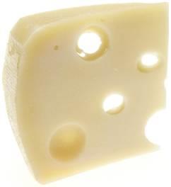
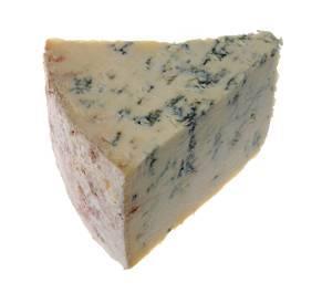
3 Principles: Definitions Unifying characteristics Collagen II Proteoglycan matrix 65% water Aggrecan most common Chondrotoin and Keratan sulfate side chains

4 Principles: Radiology Ring-like calcifications
5 Principles: Treatment Enchondroma Grade 1 chondrosarcoma, chondroblastoma Grade 2-3 chondrosarcoma Observation Curettage and cement Wide local excision +/- chemo Dedifferentiated chondrosarcoma Wide local excision + chemotherapy Mesenchymal chondrosarcoma Wide local excision + chemotherapy Clear cell chondrosarcoma Wide local excision
6 Principles: Syndromes Syndrome MOI* Locus Genes Associated conditions Ollier disease Sporadic 2q33, 15q26 IDH1, IDH2 Maffucci syndrome Sporadic Spindle cell hemangiomas (Glomangiomas Angiosarcomas Hepatobiliary) Malignant transformation 15-20% 20-30% Multiple hereditary exostoses (osteochondromas) Autosomal 8q24, dominant 11p11 EXT1, EXT2 Deformities of forearm, knee, short stature 5-35% *MOI: Mode of inheritance

7 Ollier s / Maffucci s syndrome
8 Malignant transformation of sporadic cartilage tumors Enchondroma: Case reports, <<1% Osteochondroma: 1-8%

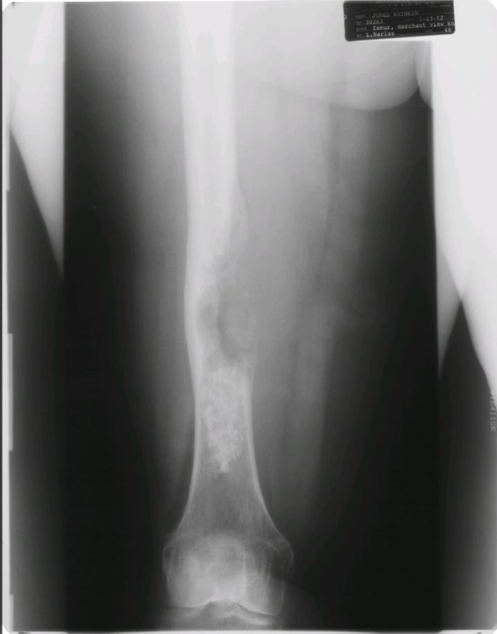
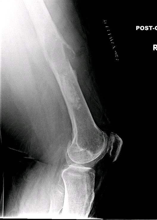
9 Case A 16 year old girl, incidentally discovered proximal tibia lesion No pain, no palpable mass.
10 Case A
11 Case A
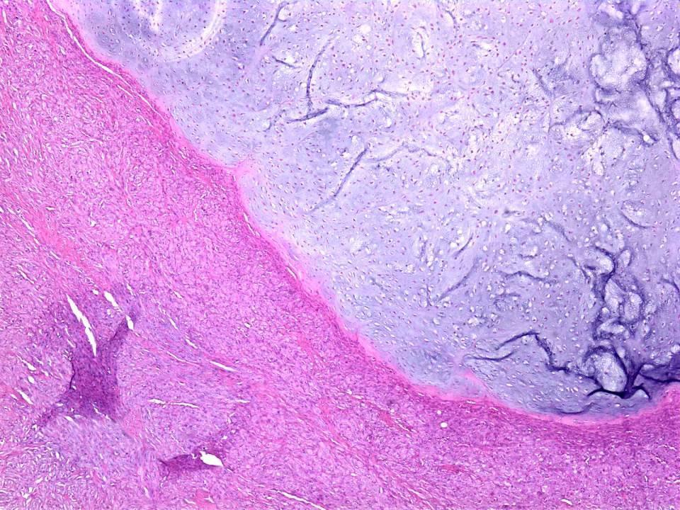
12 Enchondroma Clinical Radiology Histology Ddx Age: All Loc: Hands/feet, long tubular bones ~3% of adults Metaphysis Lytic, lobular Ring calcifications Intact cortex Lobular Peripheral ossification No permeation Low cellularity No atypia/mitoses Grade 1 Chondrosarcoma


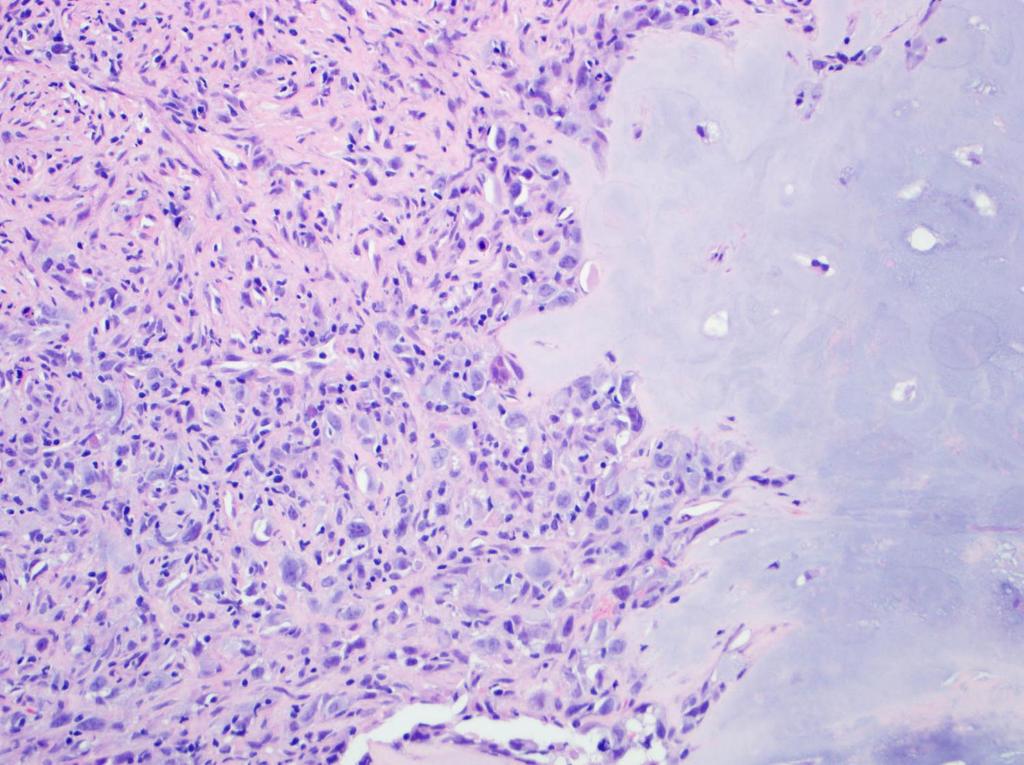
13 Pitfalls: Enchondroma Necrosis Mucin in lacunae
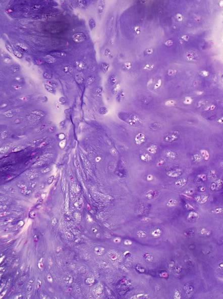
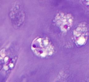
14 Pitfalls: Enchondroma Binucleation enchondroma chondrosarcoma
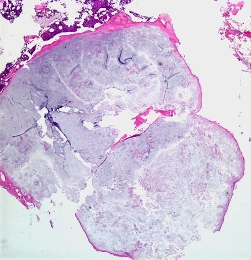
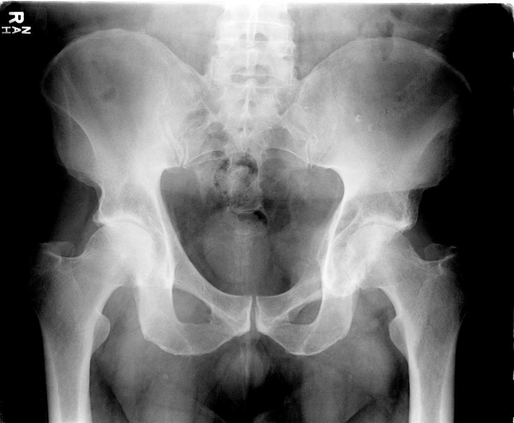
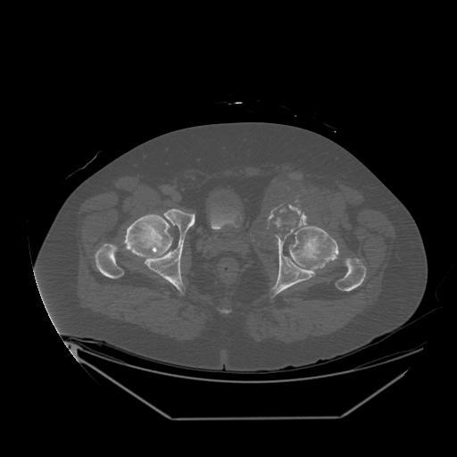
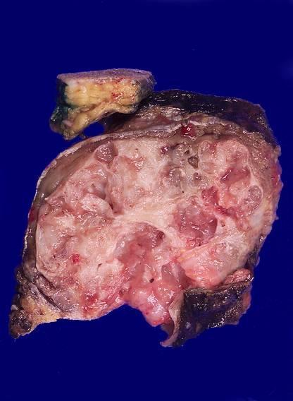
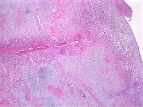
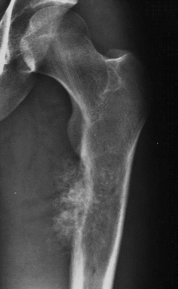
15 Case B 57 year old man with painful hip for 2 months Needle biopsy performed of acetabular mass
16
17
18 Case B
19 Case B
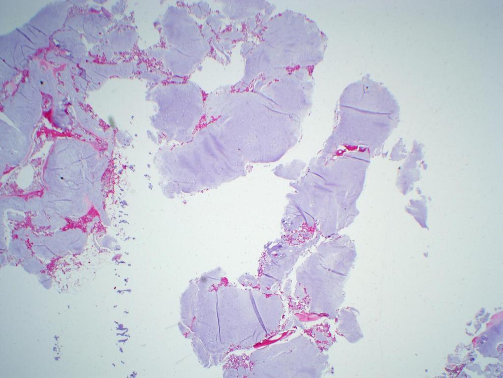
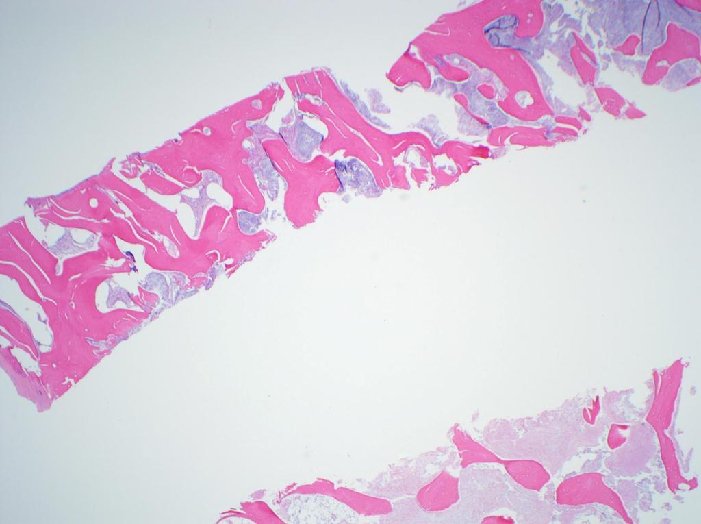
20 Conventional chondrosarcoma Clinical Radiology Histology Ddx Age: 50+ Loc: pelvis, metadiaphysis long bones Large (> 5 cm) Cortical disruption Endosteal scalloping Soft tissue extension Permeates viable, lamellar bone Myxoid change Hypercellular Atypia open chromatin Mitoses Enchondroma Clear cell chondrosarcoma Chondroblastic osteosarcoma


21 Grade 1 chondrosarcoma or enchondroma? Histology Radiology Molecular Pathology
22 Enchondroma or Grade I chondrosarcoma: Histology Kappa ~ 0.5 Two features >95% sensitivity and specificity Permeation = host bone entrapment > 20% myxoid matrix Eefting D et al. Am J Surg Pathol :50-57
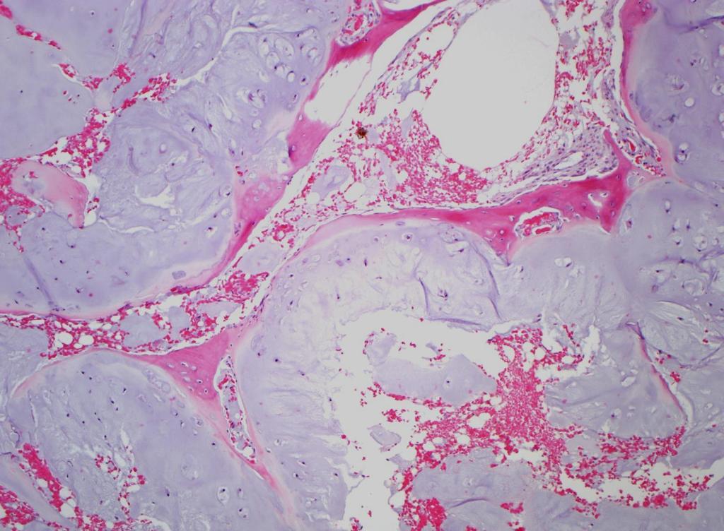
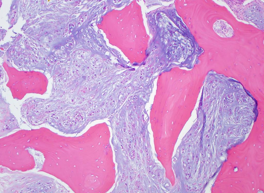
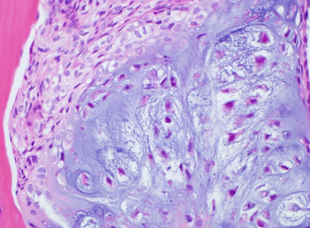
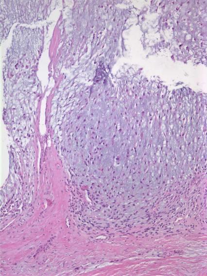
23 Chondrosarcoma: Permeation Enchondroma: Multiple nodules of hyaline cartilage separated by normal marrow (and) plates of lamellar bone that conform to the irregular shapes of cartilage lobules. Chondrosarcoma: Single confluent mass of cartilage trapping host lamellar bone or invasion into Haversian or Volkman systems. - J. Mirra (1985) Pitfalls Small biopsy Curettage Endochondral ossification Mirra JM et al. Clin Ortho Rel Res :

24 Permeation: host bone entrapment


25 Chondrosarcoma: permeation Permeation Enchondral ossification

26 Permeation
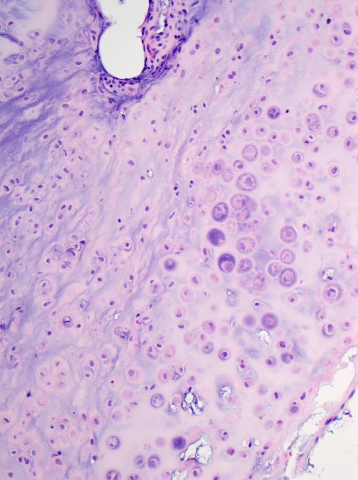
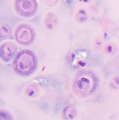
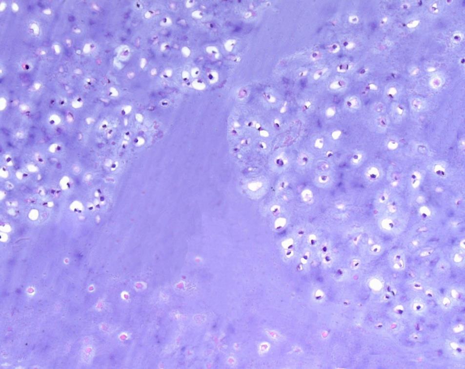
27 Chondrosarcoma: Myxoid change
28 Enchondroma or Grade 1 chondrosarcoma: Radiology Most predictive Least predictive Pathologic fracture Axial skeleton Infiltrating margin Soft tissue mass Size > 5 cm Lobulated contour Periosteal reaction Cortical disruption or thickening Deep endosteal scalloping Murphey MD et al. Radiographics (5): Brien EW et al. Skeletal Rad 1997; 26(6) Giuffrida AY et al. J Bone Joint Surg Am :
29 Enchondroma or Grade I chondrosarcoma: Molecular Genetic and molecular features: Karyotype: (C.H.A.M.P. study) Enchondroma (n=14) and CHUMP (n=3): karyotypically normal Grade I chondrosarcoma: Numerical and structural aberrations (n=3). Comparative genomic hybridization: Enchondroma: 13q21; 19, 22q Chondrosarcoma: 2-11, 14, 15, 18, 21 Similar numbers of aberrations Exome sequencing: COL2A1, IDH1, IDH2, TP53 Tallini G et al. J Pathol, 2002; 196(2):194 Ozaki T et al. Anticancer Res 2004; 1721 Amary MF et al. J Pathol. 2011; 334. Szuhai, et al. Cancer Genet 2012; 193. Tarpey PS et al. Nat Genet 2013; 923.
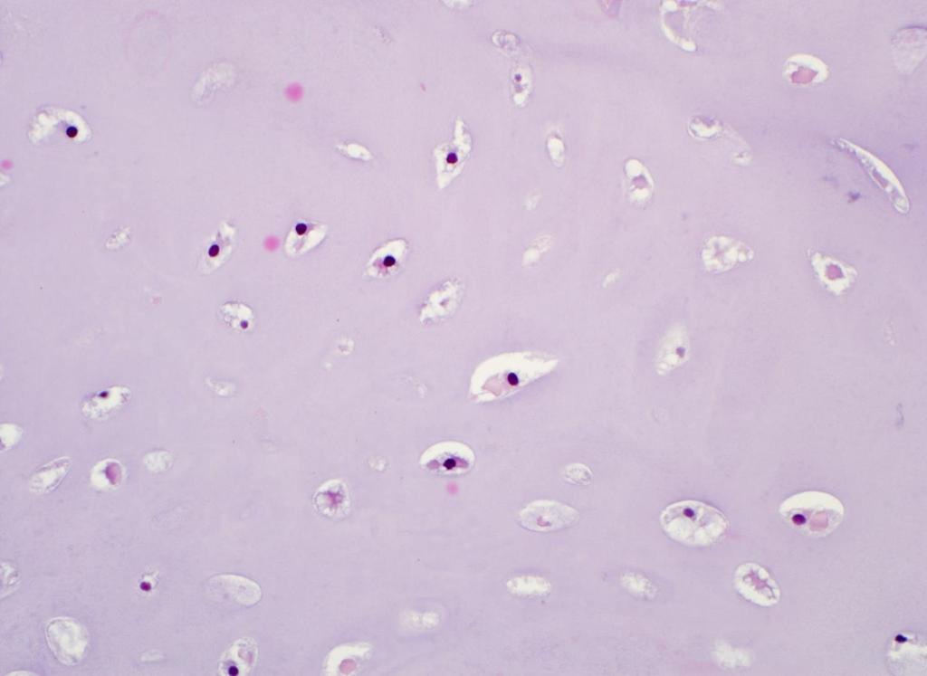
30 Chondrosarcoma: Grading PROGNOSTIC FACTORS IN CHONDROSARCOMA OF BONE A Clinicopathologic Analysis with Emphasis on Histologic Grading HARRY L. EVANS, M.D.* ALBERTO G. AYALA, MD AND MARVIN M. ROMSDAHL, MD, PHD Cancer 40: , Histology 5 year survival 10 year survival Grade 1 Hypercellular, uniform but hyperchromatic nuclei, nuclear detail not visible Grade 2 Diffusely hypercellular, nuclei paler with visible intranuclear detail Grade 3 Sheets of cells, larger nuclei, mitotic activity > 2mf/10 hpf 43 29
31 Grade 1 Chondrosarcoma
32 Grade 2 Chondrosarcoma
33 Grade 3 Chondrosarcoma
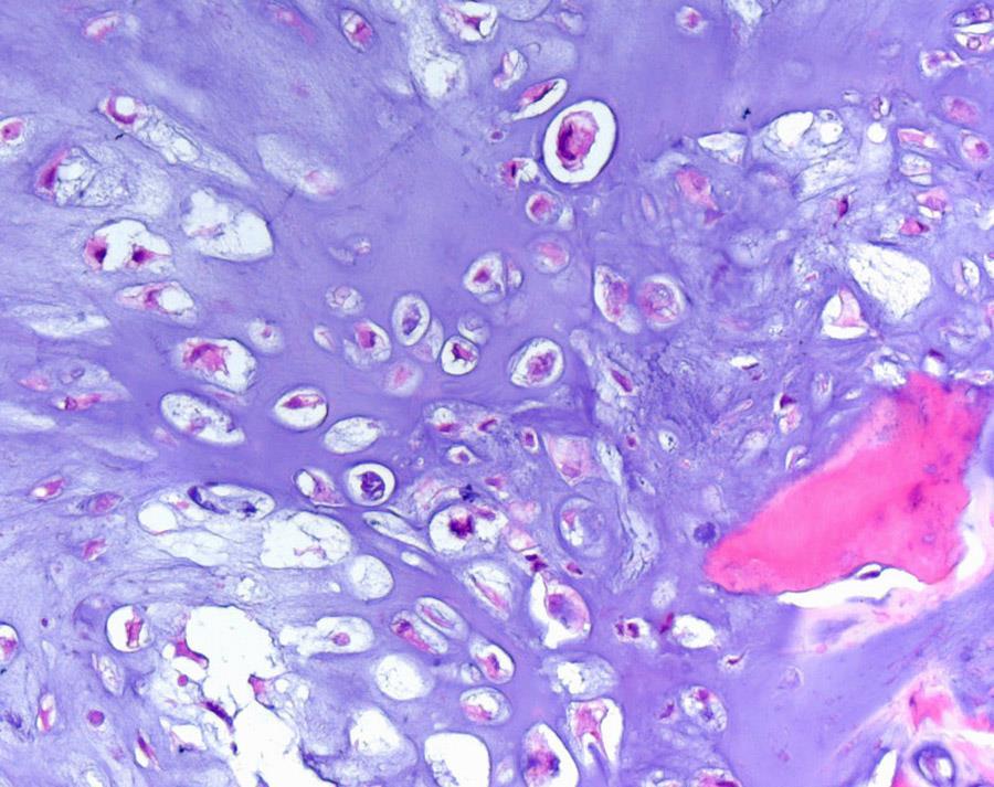
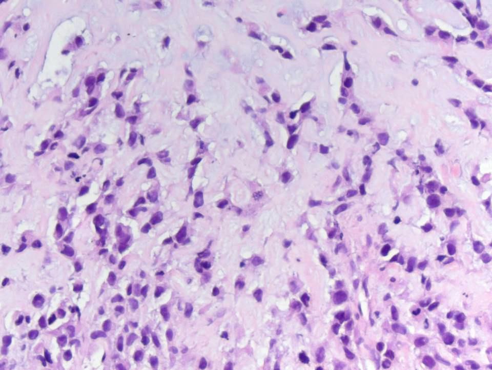
34 Pitfall: cartilage tumors of the hands and feet Enchondroma of hands and feet Clinical: Very common site (~30%), painful, pathologic fracture common Radiographs: Cortical erosion common Histology: Hypercellular, myxoid change, nuclear hyperchromasia, permeation within medulla
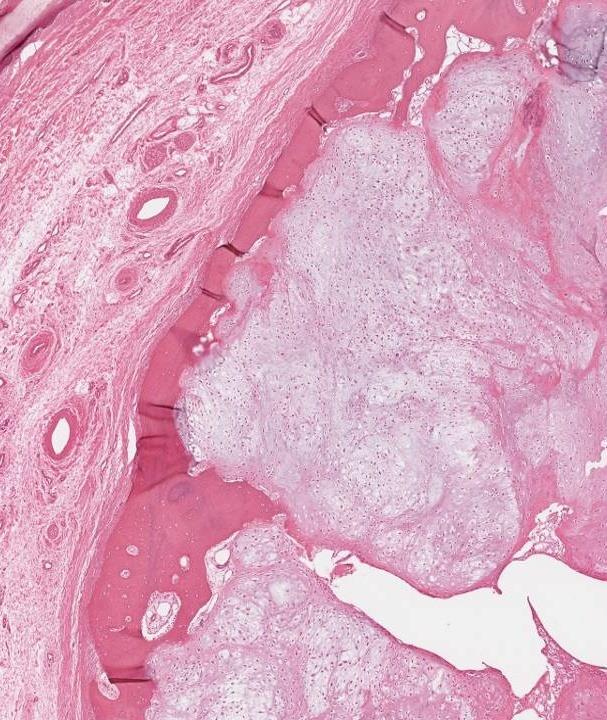
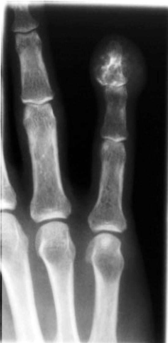
35 Enchondroma of the phalanx

36 Enchondroma of the phalanx
37 Summary: chondrosarcoma of small bones of hands and feet Metacarpals, metatarsals, wrist and ankle: Soft tissue extension, Grade 3 cytology Behavior similar to chondrosarcoma grade 1 at other sites Phalanges: Soft tissue extension Metastatic rate is very low (<2%) Local resection is favored over amputation whenever possible. Ogose A et al. Cancer, (1):50-9 Ostrowski ML et al. Am J Surg Pathol, (6): Bovee JV et al.cancer, (9):
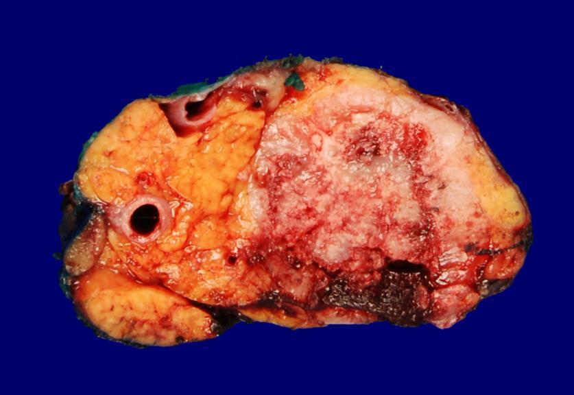
38 Dedifferentiated chondrosarcoma
39 Dedifferentiated chondrosarcoma
40 Dedifferentiated chondrosarcoma
41 Dedifferentiated chondrosarcoma Clinical Radiology Histology Ddx Age: Adult >50 Loc: Pelvis>Femur> Humerus 5 year survival < 10% Metaphysis Bimorphic (especially on MRI) Abrupt transition: 1. Chondrosarcoma grade 1 2. osteosarcoma/ MFH / fibrosarcoma Chondroblastic osteosarcoma -Less abrupt transition -High grade chondroid component Mesenchymal chondrosarcoma -Young patients -Benign cartilage islands -Less abrupt transition
42 Dedifferentiated chondrosarcoma ~100% mortality Chondroblastic osteosarcoma ~30% mortality
43 Mesenchymal chondrosarcoma
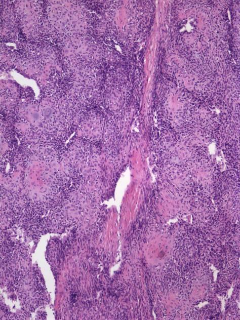
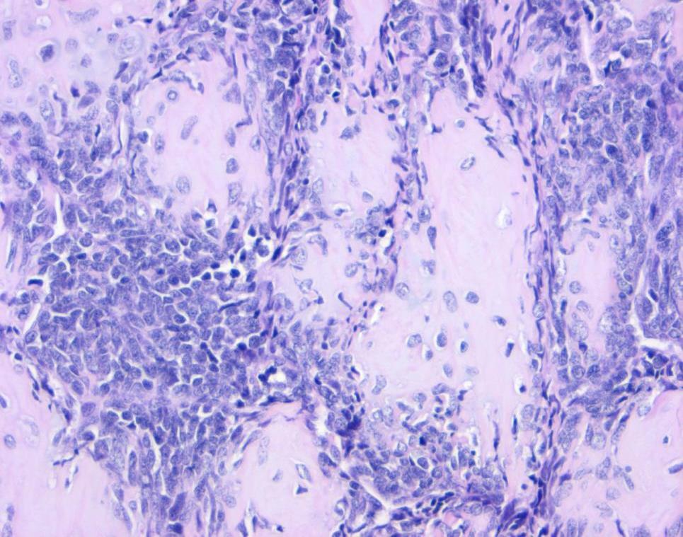
44 Mesenchymal chondrosarcoma

45 Mesenchymal chondrosarcoma
46 Mesenchymal chondrosarcoma Clinical Radiology Histology Ddx Age: Loc: Jaw, rib, pelvis, soft tissue Multifocal 10yr survival 25% Variable course Lytic Ring calcifications Rarely circumscribed 1. Benign cartilage islands/perivascular 2. Small round blue cell tumor HPC-ish vessels common Ewing sarcoma Dediff chondrosarc Small cell osteosarcoma Genetics: Inv(8q21.1;8q13.3) HEY1-NCOA2 Wang L, Genes Chromosomes Cancer 51:127.

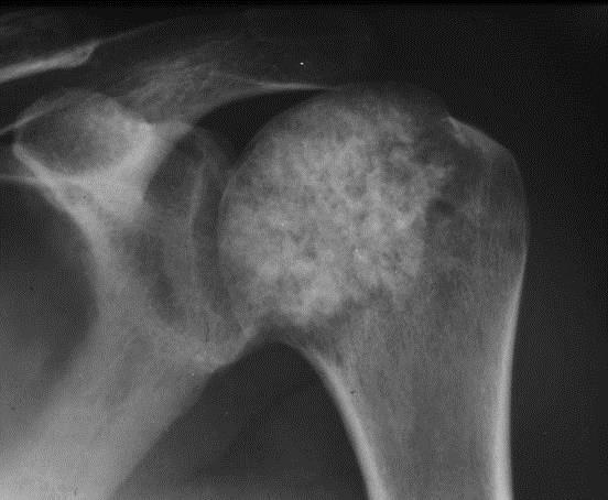
47 Clear cell chondrosarcoma
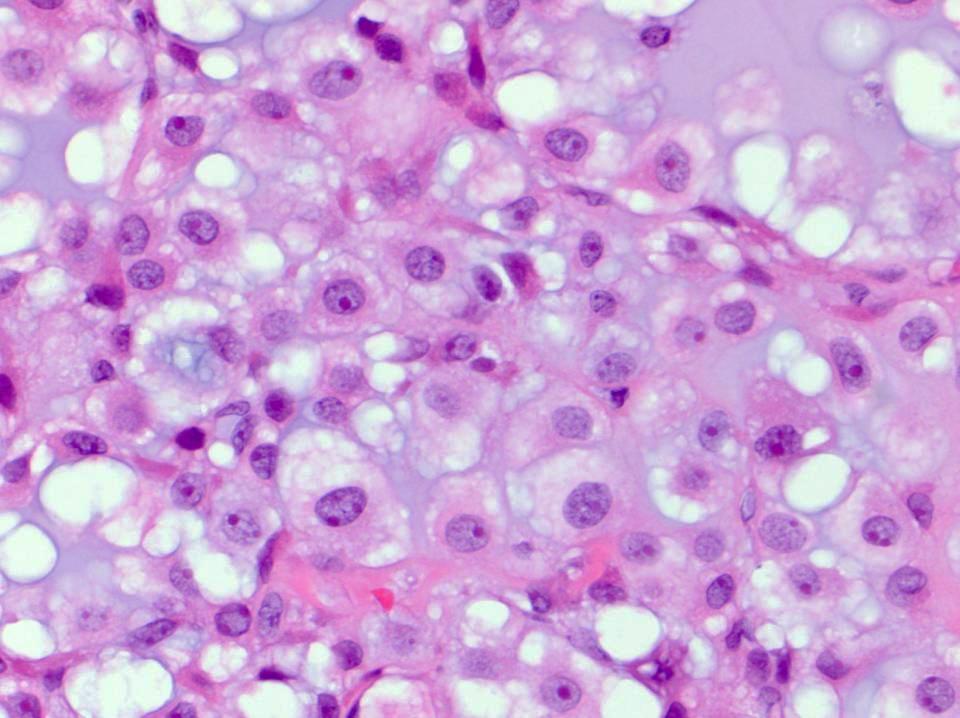
48 Clear cell chondrosarcoma
49 Clear cell chondrosarcoma Clinical Radiology Histology Ddx Age: M>F Loc: Proximal femur Skull, rib, spine Slow growing ~25% Metastasize ~15% mortality Epiphysis Circumscribed Early: lytic, septae Late: sclerotic Permeates Osteogenic No hyaline cartilage Clear cells, well defined membrane, macronucleoli Chondroblastic osteosarcoma Osteoblastoma Chondroblastoma (radiology Ddx)
50 Take-home messages Correlation of radiologic, clinical and histologic features are critical, especially in low-grade cartilage lesions. Permeation is the most sensitive histologic criterion separating enchondroma from chondrosarcoma. Chondrosarcoma of small bones of the hands and feet should be diagnosed with caution. Chondrosarcomas, especially grade 1, are predominantly locally aggressive, but local disease can be fatal even in the absence of metastases
Primary bone tumors > metastases from other sites Primary bone tumors widely range -from benign to malignant. Classified according to the normal cell
 Primary bone tumors > metastases from other sites Primary bone tumors widely range -from benign to malignant. Classified according to the normal cell counterpart and line of differentiation. Among the
Disclosures. Giant Cell Rich Tumors of Bone. Outline. The osteoclast. Giant cell rich tumors 5/21/11
 Disclosures Giant Cell Rich Tumors of Bone Andrew Horvai, MD, PhD Associate Clinical Professor, Pathology This lecture discusses "off label" uses of a number of pharmaceutical agents. The speaker is describing
SC #06. Common Challenging Problems in Bone and Soft Tissue Pathology. Andrew L. Folpe, MD Mayo Clinic Rochester, MN
 SC #06 Common Challenging Problems in Bone and Soft Tissue Pathology Andrew L. Folpe, MD Mayo Clinic Rochester, MN Andrew E. Horvai, MD, PhD University of California, San Francisco San Francisco, CA Short
Bone Tumours - a synopsis. Dr Zena Slim SpR in Histopathology QAH 2009
 Bone Tumours - a synopsis Dr Zena Slim SpR in Histopathology QAH 2009 Aims General approach to diagnosis Common entities.and not so common ones. Mini quiz Challenge of bone tumour diagnosis Bone tumours
The Radiology Assistant : Bone tumor - ill defined osteolytic tumors and tumor-like lesions
 Bone tumor - ill defined osteolytic tumors and tumor-like lesions Henk Jan van der Woude and Robin Smithuis Radiology department of the Onze Lieve Vrouwe Gasthuis, Amsterdam and the Rijnland hospital,
04/27/2017. The Spectrum of Cartilaginous Tumors
 The Spectrum of Cartilaginous Tumors L I S A E R C O L A N O, M D M U S C U L O S K E L E T A L O N C O L O G Y D E P A R T M E N T O F O R T H O P A E D I C S A L L E G H E N Y H E A L T H N E T W O R
Grading of Bone Tumors
 Grading of Bone Tumors Joon Hyuk Choi, M.D. Department of Pathology College of Medicine, Yeungnam University Introduction to grading system of bone tumor used at Mayo Clinic WHO Histologic Classification
The Radiology Assistant : Bone tumor - well-defined osteolytic tumors and tumor-like lesions
 Bone tumor - well-defined osteolytic tumors and tumor-like lesions Henk Jan van der Woude and Robin Smithuis Radiology department of the Onze Lieve Vrouwe Gasthuis, Amsterdam and the Rijnland hospital,
Bone and Joint Part 2. Leslie G Dodd, MD
 Bone and Joint Part 2 Leslie G Dodd, MD Relative rates of cancer Sarcomas are relatively uncommon tumors New cancer cases 2007 All sites 1.4 million prostate 218,890 lung 213,380 breast 180,510 Soft tissue
APMA 2018 Radiology Track Bone Tumors When to say Gulp!
 APMA 2018 Radiology Track Bone Tumors When to say Gulp! DANIEL P. EVANS, DPM, FACFAOM Professor, Department of Podiatric Medicine and Radiology Dr. Wm. Scholl College of Podiatric Medicine Conflict of
What Do You Need to Know About Bone Pathology? Benjamin L. Hoch M.D. Associate Professor Department of Pathology University of Washington
 What Do You Need to Know About Bone Pathology? Benjamin L. Hoch M.D. Associate Professor Department of Pathology University of Washington What s Do You Need To Know About Bone Pathology? Reactive/pseudosarcomatous
Recognizing Cartilaginous Tumors: Spectrum of Imaging Characteristics with Radiologic-Pathologic correlation.
 Recognizing Cartilaginous Tumors: Spectrum of Imaging Characteristics with Radiologic-Pathologic correlation. Poster No.: C-1451 Congress: ECR 2012 Type: Educational Exhibit Authors: E. Barcina García,
Malignant bone tumors. Incidence Myeloma 45% Osteosarcoma 24% Chondrosarcoma 12% Lyphoma 8% Ewing s Sarcoma 7%
 Malignant bone tumors Incidence Myeloma 45% Osteosarcoma 24% Chondrosarcoma 12% Lyphoma 8% Ewing s Sarcoma 7% Commonest primary bone sarcoma is osteosarcoma X ray Questions to ask 1. Solitary or Multiple
Typical skeletal location and differential diagnosis of bone tumors.
 Typical skeletal location and differential diagnosis of bone tumors. Poster No.: C-2418 Congress: ECR 2015 Type: Educational Exhibit Authors: M. Barros, L. A. Ferreira, Y. Costa, P. J. V. Coelho, F. Caseiro
MARK D. MURPHEY MD, FACR. Physician-in-Chief, AIRP. Chief, Musculoskeletal Imaging
 ALPHABET SOUP AND CYSTIC LESIONS OF THE BONE MARK D. MURPHEY MD, FACR Physician-in-Chief, AIRP Chief, Musculoskeletal Imaging ALPHABET SOUP AND CYSTIC LESIONS OF THE BONE Giant cell tumor (GCT) Unicameral
Multifocal fibrous Dysplasia with enchondroma-like areas: Fibrocartilaginous Dysplasia
 ISPUB.COM The Internet Journal of Pathology Volume 7 Number 2 Multifocal fibrous Dysplasia with enchondroma-like areas: Fibrocartilaginous Dysplasia V Monappa, R Kudva Citation V Monappa, R Kudva. Multifocal
Introduction to Musculoskeletal Tumors. James C. Wittig, MD Orthopedic Oncologist Sarcoma Surgeon
 Introduction to Musculoskeletal Tumors James C. Wittig, MD Orthopedic Oncologist Sarcoma Surgeon www.tumorsurgery.org Definitions Primary Bone / Soft tissue tumors Mesenchymally derived tumors (Mesodermal)
Chondrosarcoma of 5 th metatarsal Right Foot: An Unusual Presentation and Review of Literature
 IOSR Journal of Dental and Medical Sciences (IOSR-JDMS) e-issn: 2279-0853, p-issn: 2279-0861.Volume 17, Issue 8 Ver. 13 (August. 2018), PP 27-33 www.iosrjournals.org Chondrosarcoma of 5 th metatarsal Right
What is chondrosarcoma?
 Return to This Guide to chondrosarcoma was authored by DAVIDE DONATI M.D. LUCA SANGIORGI M.D., PH.D., Rizzoli Orthopaedic Institute, Bologna Italy & The MHE Research Foundation. What is chondrosarcoma?
Bread and Butter Bone Pathology
 Bread and Butter Bone Pathology NICOLE D. RIDDLE, MD RUFFOLO, HOOPER, AND ASSOC. / UNIVERSITY OF SOUTH FLORIDA Goals: Fundamentals of neoplastic bone pathology Bone Producing Cartilage Producing Miscellaneous
warwick.ac.uk/lib-publications
 A Thesis Submitted for the Degree of PhD at the University of Warwick Permanent WRAP URL: http://wrap.warwick.ac.uk/102063/ Copyright and reuse: This thesis is made available online and is protected by
Case Report Intramedullary Chondrosarcoma of Proximal Humerus
 Hindawi Publishing Corporation Case Reports in Radiology Volume 2012, Article ID 642062, 7 pages doi:10.1155/2012/642062 Case Report Intramedullary Chondrosarcoma of Proximal Humerus Pratiksha Yadav, Dolly
Primary bone tumors according to the WHO classification: a review of 13 years with illustrative examples
 Primary bone tumors according to the WHO classification: a review of 13 years with illustrative examples Poster No.: C-1741 Congress: ECR 2015 Type: Educational Exhibit Authors: J. Silva, M. A. Ramírez
Non-monomelic synchronous primary multicentric chondrosarcoma : A case report
 Acta Orthop. Belg., 2005, 71, 242-248 CASE REPORT Non-monomelic synchronous primary multicentric chondrosarcoma : A case report Yolanda HERNANZ GONZÁLEZ, Javier SALAMANCA, Carlos RESINES ERASUN, Francisco
Fun with Fat. General Rules. Case
 Fun with Fat General Rules Imaging: location (deep vs. superficial) Superficial lesions are seldom liposarcomas Deep lesions may be benign or malignant Myxoid stroma is common in benign and malignant lesions
Bone Tumors Clues and Cues
 William Herring, M.D. 2002 Bone Tumors Clues and Cues In Slide Show mode, advance the slides by pressing the spacebar All Photos Retain the Copyright of their Authors Clues by Appearance of Lesion Patterns
Bone/Osteoid Producing Lesions
 Chapter 2 Bone/Osteoid Producing Lesions Introduction There are many lesions that are associated with reactive new bone formation; this chapter predominantly covers those in which deposition of osteoid/bone
COPYRIGHT 2004 BY THE JOURNAL OF BONE AND JOINT SURGERY, INCORPORATED
 84 COPYRIGHT 2004 BY THE JOURNAL BONE AND JOINT SURGERY, INCORPORATED Radiographic Evaluation of Pathological Bone Lesions: Current Spectrum of Disease and Approach to Diagnosis BY BENJAMIN G. DOMB, MD,
Musculoskeletal Sarcomas
 Musculoskeletal Sarcomas Robert C. Orth, M.D., Ph.D. Edward B. Singleton Department of Pediatric Radiology Texas Children s Hospital Page 0 xxx00.#####.ppt 9/23/2012 9:01:18 AM No disclosures Page 1 xxx00.#####.ppt
University Journal of Surgery and Surgical Specialities
 University Journal of Surgery and Surgical Specialities Volume 1 Issue 1 2015 EXTRA SKELETAL MESENCHYMAL CHONDROSARCOMA :A CASE REPORT Rajaraman R Subbiah S Navin Naushad Kilpaulk Medical College Abstract:
Clinical Study Enchondroma versus Low-Grade Chondrosarcoma in Appendicular Skeleton: Clinical and Radiological Criteria
 Oncology Volume 2012, Article ID 437958, 6 pages doi:10.1155/2012/437958 Clinical Study Enchondroma versus Low-Grade Chondrosarcoma in Appendicular Skeleton: Clinical and Radiological Criteria Eugenio
Bizarre parosteal osteochondromatous proliferation
 * * Bizarre Parosteal Osteochondromatous Proliferation A Case Report with Literature Review Chi-Fu Kao Yang-Chih Lin Yu-Hung Wu Be-Fong Chen* We report the case of a 12-year-old female with a slowly erythematous
Malignant Bone Tumours. PathoBasic, Daniel Baumhoer
 Malignant Bone Tumours PathoBasic, 20.03.18 Daniel Baumhoer FNCLCC Grading The differentiation score is defined as the extent to which a tumor resembles adult mesenchymal tissue (score 1), the extent to
JMSCR Vol 3 Issue 11 Page November 2015
 www.jmscr.igmpublication.org Impact Factor 3.79 Index Copernicus Value: 5.88 ISSN (e)-2347-176x ISSN (p) 2455-0450 DOI: http://dx.doi.org/10.18535/jmscr/v3i11.36 Diagnostic Dilemmas in Cytodiagnosis of
3/27/2017. Disclosure of Relevant Financial Relationships
 Ophthalmic Pathology Evening Specialty Conference USCAP 2017 5 th March, 2017 Mukul K. Divatia, MD Assistant Professor Department of Pathology & Genomic Medicine Weill Cornell Medical College Houston Methodist
GIANT CELL-RICH OSTEOSARCOMA: A CASE REPORT
 Nagoya J. Med. Sci. 59. 151-157, 1996 CASE REPORTS GIANT CELL-RICH OSTEOSARCOMA: A CASE REPORT KEIJI SATO!, SHIGEKI YAMAMURA!, HISASHI IWATA!, HIDESHI SUGIURA 2, NOBUO NAKASHIMA 3 and TETSURO NAGASAKA
Bone tumors. RMG: jan
 Bone tumors RMG: jan 217. @Kijohs KIZZA JOHN KIJOHS Diseases arising in bone Lipoma Fibrous cortical defects Non-ossifying fibroma Bone island Benign simple cysts Enchondroma Osteochondroma Osteoid osteoma
Chondrosarcoma in Childhood: The Radiologic and Clinical Conundrum
 Chondrosarcoma in Childhood: The Radiologic and Clinical Conundrum Susan M. Mosier 1, Tanvi Patel 1, Karen Strenge 2, Andrew D. Mosier 3* 1. Department of Pediatrics, Ireland Army Community Hospital, Fort
RESEARCH INFORMATION AWARENESS SUPPORT PRIMARY BONE CANCER CHONDROSARCOMA. Visit bcrt.org.uk for more information
 RESEARCH INFORMATION AWARENESS SUPPORT PRIMARY BONE CANCER CHONDROSARCOMA Visit bcrt.org.uk for more information CONTENTS What is it? Who does it affect? Symptoms Types of Chondrosarcoma Cause and Risk
Fluid-fluid levels in bone tumors: A pictorial review
 Fluid-fluid levels in bone tumors: A pictorial review Poster No.: C-578 Congress: ECR 2009 Type: Educational Exhibit Topic: Musculoskeletal Authors: L. Figueroa Nasra, C. Martín Hervás, M. Tapia-Viñé,
ORTHOPAEDIC ONCOLOGY OITE REVIEW COURSE
 ORTHOPAEDIC ONCOLOGY OITE REVIEW COURSE Richard D. Lackman, MD FACS Director, Orthopaedic Oncology Center Cancer Institute Introduction In the evaluation of a patient with a bone tumor, there are several
Department of Radiology, University of Szeged. Imaging of the skeleton
 Imaging of the skeleton Methods of examination: plain x-ray (radiography, densitometry) x-ray with contrast material (fistulography, angiography) ultrasound (b-mode, Doppler, color, duplex) computed tomography
Essential Dermatopathology. Jinah Kim, MD, PhD Department of Pathology and Dermatology Stanford University Medical Center
 Essential Dermatopathology Jinah Kim, MD, PhD Department of Pathology and Dermatology Stanford University Medical Center OBJECTIVES Review clinical, pathologic and molecular aspects of bone and fat tumors
The Relevance of Cytologic Atypia in Cutaneous Neural Tumors
 The Relevance of Cytologic Atypia in Cutaneous Neural Tumors Recent Findings - New Developments New Problems Zsolt B. Argenyi, M.D. Professor of Pathology & Dermatology Director of Dermatopathology Department
Imaging Findings Of Bone Tumors: A Pictorial Review
 Imaging Findings Of Bone Tumors: A Pictorial Review Poster No.: C-2511 Congress: ECR 2015 Type: Educational Exhibit Authors: M. Limeme, N. Benzina, A. BelKhiria, H. Zaghouani, S. Majdoub, N. Mallat, H.
Notochordal cell tumors
 Notochordal cell tumors G. Petur Nielsen Department of Pathology Massachusetts General Hospital Boston, MA gnielsen@partners.org Introduction Chordoma is a rare primary malignant neoplasm of bone that
Bubbly Lesions of Bone
 Residents Section Pattern of the Month w79 08.18.09 Eisenberg Residents Section Pattern of the Month Residents inradiology Ronald L. Eisenberg 1 Eisenberg RL Keywords: bubbly lesions, fegnomashic, skeletal
A. Nahal MD,* A. Ajlan MD, T. Alcindor MD, and R. Turcotte MD P A T H O L O G Y ABSTRACT 2. CASE DESCRIPTION KEY WORDS 1.
 P A T H O L O G Y Dedifferentiated giant cell tumour of bone in the form of low-grade fibroblastic osteogenic sarcoma: case report of a unique presentation with follow-up A. Nahal MD,* A. Ajlan MD, T.
SMALL ROUND BLUE CELL LESION OF BONE
 DISCLOSURE SMALL ROUND BLUE CELL LESION OF BONE Dr. Alistair Jordan University of South Alabama No financial support or endorsement OBJECTIVES Describe the more common small round cell lesions of bone
Radiology-Pathology Conference
 July 31, 2009 Radiology-Pathology Conference Daniel T Ginat, M.D., M.S. Sharlin Johnykutty,, M.D. Presentation material is for education purposes only. All rights reserved. 2009 URMC Radiology Page 1 of
The Radiology Assistant : Bone tumor A-G
 Bone tumor A-G Bone tumors and tumor-like lesions in alphabethic order Henk Jan van de Woude and Robin Smithuis Radiology department of the Onze Lieve Vrouwe Gasthuis, Amsterdam and the Rijnland hospital,
ISSN: DISTRIBUTION OF BONE AND CARTILAGINOUS TUMORS IN PEDIATRIC AGE GROUP IN WESTERN UTTAR-PRADESH: AN EVALUATIVE STUDY
 : 289-295 ISSN: 2277 4998 DISTRIBUTION OF BONE AND CARTILAGINOUS TUMORS IN PEDIATRIC AGE GROUP IN WESTERN UTTAR-PRADESH: AN EVALUATIVE STUDY QADRI S, HASAN M, AKHTAR K * AND SHERWANI RK The Departments
Update On Lipomatous Tumors: Old Standbys and New Concepts
 Update On Lipomatous Tumors: Old Standbys and New Concepts John R. Goldblum, M.D. Chairman, Department of Anatomic Pathology Cleveland Clinic Professor of Pathology Cleveland Clinic Lerner College of Medicine
Radiologic approach to pediatric lytic bone lesions
 Radiologic approach to pediatric lytic bone lesions Poster No.: C-1177 Congress: ECR 2016 Type: Educational Exhibit Authors: J. L. LERMA GALLARDO, I. de la Pedraja, A. Lancharro 1 1 1 2 1 1 Zapata, J.
The Scandinavian Sarcoma Group annual report on extremity and trunk wall soft tissue and bone sarcomas
 The Scandinavian Sarcoma Group annual report on extremity and trunk wall soft tissue and bone sarcomas 2012-2016 1 The SSG annual report on extremity and trunk wall soft tissue and bone sarcomas. The Scandinavian
2. Assessment of interobserver variability and histological parameters to improve. central cartilaginous tumours.
 . Assessment of interobserver variability and histological parameters to improve central cartilaginous tumours. Daniel Eefting, Yvonne M. Schrage, Maartje J. Geirnaerdt, Saskia Le Cessie, Antonie H.M.
Diagnostic problems in uterine smooth muscle tumors
 Diagnostic problems in uterine smooth muscle tumors Marina Kos Ljudevit Jurak Clinical Department of Pathology, Clinical Hospital Center Sestre milosrdnice, Zagreb Institute of Pathology, University of
The Skeletal System. Chapter 7a. Skeletal System Introduction Functions of the skeleton Framework of bones The skeleton through life
 The Skeletal System Skeletal System Introduction Functions of the skeleton Framework of bones The skeleton through life Chapter 7a Support Protection Movement Storage areas Minerals Lipids Hemopoiesis
Common Primary Tumors of Bone
 Special Report Common Primary Tumors of Bone Primary bone tumors are a relatively rare occurrence, however, they can have serious deleterious consequences. Many possess the ability to degenerate into malignant
Lytic Lesion in the Distal Phalanx of the Hand
 Shafa Ortho J. 2015 February; 2(1):e441. Published online 2015 February 15. DOI: 10.5812/soj.441 Research Article Lytic Lesion in the Distal Phalanx of the Hand Khodamorad Jamshidi 1 ; Farid Najd Mazhar
Evening Specialty Conference Bone and Soft Tissue Pathology. Diagnostic pitfalls in bone and soft tissue pathology
 Evening Specialty Conference Bone and Soft Tissue Pathology. Case 1 Elizabeth G Demicco, MD, PhD Mount Sinai Hospital, New York Disclosure of Relevant Financial Relationships USCAP requires that all planners
Effective local and systemic therapy is necessary for the cure of Ewing tumor Most chemotherapy regimens are a combination of cyclophosphamide,
 Ewing Tumor Perez Ewing tumor is the second most common primary tumor of bone in childhood, and also occurs in soft tissues Ewing tumor is uncommon before 8 years of age and after 25 years of age In the
Immunohistochemistry in Bone and Soft Tissue Tumors. Sahar Rassi Zankoul, MD
 Immunohistochemistry in Bone and Soft Tissue Tumors Sahar Rassi Zankoul, MD Introduction Bone tumors represent a wide variety of tumors of various origins and malignant potentials. These different tumor
Bone Tumors: Epidemiology, Classification, Pathology
 Bone Tumors: Epidemiology, Classification, Pathology 1 Lars Gunnar Kindblom C O N T E N T S 1.1 Introduction 2 1.2 Epidemiology 2 1.3 Morphologic Diagnosis of Bone Tumors 5 1.4 Types of Bone Tumor Specimens
Skeletal System worksheet
 Skeletal System worksheet Name Section A: Intro to Skeletal System The skeletal system performs vital functions that enable us to move through our daily lives. Support - The skeleton provides support and
Therapy Related Changes in Osteosarcoma and Ewing Sarcoma of Bone
 The Open Pathology Journal, 2009, 3, 99-105 99 Open Access Therapy Related Changes in Osteosarcoma and Ewing Sarcoma of Bone Hye Sook Min 1, Hyun Guy Kang 2 and Jae Y. Ro *,3 1 Department of Pathology
The Skeletal System. Mosby items and derived items 2010, 2006, 2002, 1997, 1992 by Mosby, Inc., an affiliate of Elsevier Inc.
 The Skeletal System Functions of Skeletal System Provides internal framework that supports the body Protects internal organs Helps fight disease by producing white blood cells 2 Functions of Skeletal System
A review of Tumoral lesions of the shoulder
 A review of Tumoral lesions of the shoulder Poster No.: P-0109 Congress: ESSR 2013 Type: Scientific Exhibit Authors: M. M. Milán Rodríguez, Á. E. Moreno Puertas, J. M. Giménez, 1 1 1 1 2 1 A. Rubio Fernández,
Myxo-inflammatory Fibroblastic sarcoma
 AKA Myxo-inflammatory Fibroblastic sarcoma Acral Myxoinflammatory fibroblastic sarcomaam.j.surg.path1998; 22; 911-924 Inflammatory myxoid tumour of soft parts with bizarre giant cells [Pathol.Res.Pract.
Fibrocartilaginous Dysplasia of the Bone: A Rare Variant of Fibrous Dysplasia
 Open Access Case Report DOI: 10.7759/cureus.448 Fibrocartilaginous Dysplasia of the Bone: A Rare Variant of Fibrous Dysplasia Raju Vaishya 1, Amit Kumar Agarwal 1, Nishint Gupta 2, Vipul Vijay 1 1. Department
Calcifying Aponeurotic Fibroma of the Knee: a Case Report with Radiographic and MRI Finding
 pissn 2384-1095 eissn 2384-1109 imri 2017;21:259-263 Calcifying Aponeurotic Fibroma of the Knee: a Case Report with Radiographic and MRI Finding Seung Hyun Lee 1,2, In Sook Lee 1,2, You Seon Song 1,2,
Fluid fluid levels in bone tumors and tumoral lesions - Pictorial essay
 Review Fluid fluid levels in bone tumors and tumoral lesions - Pictorial essay Subbarao Kakarla 1,* 1 KIMS Foundation and Research Centre, Minister Road, Secunderabad - 500003, Telangana, India Abstract
ACCME/Disclosures ALK FUSION-POSITIVE MESENCHYMAL TUMORS. Tumor types with ALK rearrangements. Anaplastic Lymphoma Kinase. Jason L.
 Companion Meeting of the International Society of Bone and Soft Tissue Pathology The Evolving Concept of Mesenchymal Tumors ALK FUSION-POSITIVE MESENCHYMAL TUMORS Jason L. Hornick, MD, PhD March 13, 2016
Current Thinking of the Osteochondroses. Diego Jaramillo, M.D., M.P.H. Department of Radiology Stanford Children s Hospital
 Current Thinking of the Osteochondroses Diego Jaramillo, M.D., M.P.H. Department of Radiology Stanford Children s Hospital What is an osteochondrosis? Abnormal endochondral ossification and epiphyseal
WHAT IS MDM2? (MDMTWOMICS) MDM2 IN SARCOMAS? (MDMTWOMAS) MDM2MICS? NO CONFLICT OF INTERESTS 5/07/2018 MDM2 IN SOFT TISSUE AND BONE SARCOMAS
 MDM2 IN SARCOMAS? (MDMTWOMAS) MDM2MICS? NO CONFLICT OF INTERESTS 5/07/2018 MDM2 IN SOFT TISSUE AND BONE SARCOMAS") IN SOFT TISSUE AND BONE SARCOMAS WHAT IS? (MDMTWOMICS) Raf Sciot, M.D., PhD. Department of Pathology, University Hospitals Katholieke Universiteit Leuven, LEUVEN, Belgium IN SARCOMAS? (MDMTWOMAS) MICS?
IN SOFT TISSUE AND BONE SARCOMAS WHAT IS? (MDMTWOMICS) Raf Sciot, M.D., PhD. Department of Pathology, University Hospitals Katholieke Universiteit Leuven, LEUVEN, Belgium IN SARCOMAS? (MDMTWOMAS) MICS?
Giant cell tumour of the sternum-two cases
 Giant cell tumour of the sternum-two cases Nishaa.P 1, Raghuram.P 2, Navin patil 3, Jaipal B.R 4 Akkamahadevi patel 5 Assistant Professor ESIC medical college and PGIMSR 1 Professor and HOD, 2 Professor
Case 8 Soft tissue swelling
 Case 8 Soft tissue swelling 26-year-old female presented with a swelling on the back of the left knee joint since the last 6 months and chronic pain in the calf and foot since the last 2 months. Pain in
Due in Lab. Due next week in lab - Scientific America Article Select one article to read and complete article summary
 Due in Lab 1. Skeletal System 33-34 2. Skeletal System 26 3. PreLab 6 Due next week in lab - Scientific America Article Select one article to read and complete article summary Cell Defenses and the Sunshine
Cytology of Neoplasms that Occur on the Limbs Rick Alleman, DVM, PhD, DABVP, DACVP
 Cytology of Neoplasms that Occur on the Limbs Rick Alleman, DVM, PhD, DABVP, DACVP I. Introduction The purpose of this material is to provide information that may be useful in the identification of tumors
Clear Cell Chondrosarcoma of the Sacrum
 Clear Cell Chondrosarcoma of the Sacrum Yasunobu Iwasaki MD 1, Tetsuji Yamamoto MD (!) 2, Mitsuo Tsuji MD 1, Akira Kurihara MD 1, Masaaki Uratsuji MD 1, Norihide Sha MD 1, and Shinichi Yoshiya MD 2 1 Department
Mody. AIS vs. Invasive Adenocarcinoma of the Cervix
 Common Problems in Gynecologic Pathology Michael T. Deavers, M.D. Houston Methodist Hospital, Houston, Texas Common Problems in Gynecologic Pathology Adenocarcinoma in-situ (AIS) of the Cervix vs. Invasive
AIRP Best Cases in Radiologic- Pathologic Correlation
 Note: This copy is for your personal non-commercial use only. To order presentation-ready copies for distribution to your colleagues or clients, contact us at www.rsna.org/rsnarights. MUSCULOSKELETAL IMAGING
Advertisement. Osteochondroma
 Advertisement Osteochondroma An osteochondroma is a benign (noncancerous) tumor that develops during childhood or adolescence. It is an abnormal growth that forms on the surface of a bone near the growth
2003 PIP-A A Cases. Paul K. Shitabata, M.D. APMG May 21, 2003
 2003 PIP-A A Cases Paul K. Shitabata, M.D. APMG May 21, 2003 Case 1 47F pelvic mass involving the right fallopian tube 5.5 cm intraluminal mass Fallopian Tube Adenocarcinoma Risk factors Breast, endometrial,
Disseminated Primary Non-Hodgkin s Lymphoma of Bone : A Case Re p o r t 1
 Disseminated Primary Non-Hodgkin s Lymphoma of Bone : A Case Re p o r t 1 Hee-Jin Park, M.D., Sung-Moon Lee, M.D., Hee-Jung Lee, M.D., Jung-Sik Kim, M.D., Hong Kim, M.D. Primary lymphoma of bone is uncommon
Incidental bone tumors are asymptomatic lesions that are. Incidental Bone Lesions. When to Refer to the Tumor Specialist
 Bulletin of the NYU Hospital for Joint Diseases 2012;70(4):235-40 235 Incidental Bone Lesions When to Refer to the Tumor Specialist LT Suezie Kim, M.D., M.C., U.S.N., Catherine N. Laible, M.D., Leon D.
CASE year old male with a PET avid nodule in the left adrenal gland
 CASE 1 55 year old male with a PET avid nodule in the left adrenal gland Case 1 Adrenal gland parenchyma partly replaced by a spindle cell tumour with mild nuclear pleomorphism Atypical mitoses present
LAC + USC.
 Jeff McDavit,, M.D. LAC + USC mcdavit@usc.edu Clinical History 55 year old male with large, deep, non- tender left thigh mass. Seen at LAC+USC Med Ctr FNA clinic No h/o trauma or radiation Vimentin
Review Course «Musculoskeletal Oncology» October 6, 2011 UNIKLINIK BALGRIST. Imaging of Bone and Soft Tissue. Tumors
 Imaging of Bone and Soft Tissue Tumors Approach from a radiologist s point of view Florian Buck Radiology Radio- Radio- Oncologist Oncologist Orthopedist Orthopedist Patient Management Oncologist Oncologist
Radiography in the Initial Diagnosis of Primary Bone Tumors
 Residents Section Structured Review Costelloe and Madewell Radiography of Primary Bone Tumors Residents Section Structured Review Colleen M. Costelloe 1 John E. Madewell Costelloe CM, Madewell JE Keywords:
Clinical History. TumorBoard: Recurrent Chondrosarcoma of the Ethmoid Sinus. chondrosarcoma. Clinical History (cont ) Present Illness
 Present Illness") TumorBoard: Recurrent Chondrosarcoma of the Ethmoid Sinus Part 1 of 2 Najat Daw, MD Sandeep Samant, MD Jeffrey Buchsbaum, MD, PhD Christine Fuller, MD Kathleen Helton, MD May 9, 2003 Clinical History 7
TumorBoard: Recurrent Chondrosarcoma of the Ethmoid Sinus Part 1 of 2 Najat Daw, MD Sandeep Samant, MD Jeffrey Buchsbaum, MD, PhD Christine Fuller, MD Kathleen Helton, MD May 9, 2003 Clinical History 7
Chondroblastoma of bone
 Chronology Chondroblastoma of bone LONG-TERM RESULTS AND FUNCTIONAL OUTCOME AFTER INTRALESIONAL CURETTAGE R. Suneja, R. J. Grimer, M. Belthur, L. Jeys, S. R. Carter, R. M. Tillman, A. M. Davies From The
SKELETAL METASTASES FROM SOFT-TISSUE SARCOMAS
 INCIDENCE, PATTERNS, AND RADIOLOGICAL FEATURES HIDEKI YOSHIKAWA, TAKAFUMI UEDA, SHIGEKI MORI, NOBUHITO ARAKI, SHIGEYUKI KURATSU, ATSUMASA UCHIDA, TAKAHIRO OCHI From Osaka Medical Centre for Cancer and
MRI XR, CT, NM. Principal Modality (2): Case Report # 2. Date accepted: 15 March 2013
: Case Report # 2. Date accepted: 15 March 2013") Radiological Category: Musculoskeletal Principal Modality (1): Principal Modality (2): MRI XR, CT, NM Case Report # 2 Submitted by: Hannah Safia Elamir, D.O. Faculty reviewer: Naga R. Chinapuvvula, M.D.
Radiological Category: Musculoskeletal Principal Modality (1): Principal Modality (2): MRI XR, CT, NM Case Report # 2 Submitted by: Hannah Safia Elamir, D.O. Faculty reviewer: Naga R. Chinapuvvula, M.D.
GIANT CELL TUMOR OF BONE
 GIANT CELL TUMOR OF BONE Definition. First described by Jaffe et al. 1, giant cell tumor of bone is a locally aggressive primary neoplasm of bone that is composed of proliferation of bland looking oval
Endometrial Stromal Tumors
 Endometrial Stromal Tumors WHO Categories: Endometrial Stromal Nodule (ESN) Endometrial Stromal Sarcoma, low grade (LGESS) Endometrial Stromal Sarcoma, high grade (HGESS) Undifferentiated Uterine Sarcoma
What is bone? Specialized form of connective tissue: mineralized collagen matrix, therefore very rigid and strong while still retaining some degree of
 Bone What is bone? Specialized form of connective tissue: mineralized collagen matrix, therefore very rigid and strong while still retaining some degree of flexibility Other types of connective tissue:
General Approach to Lytic Bone Lesions D. Lee Bennett, MD, MA, Georges Y. El Khoury, MD Appl Radiol. 2004;33(5)
") General Approach to Lytic Bone Lesions D. Lee Bennett, MD, MA, Georges Y. El Khoury, MD Appl Radiol. 2004;33(5) www.medscape.com Abstract and Introduction Abstract When interpreting musculoskeletal radiographs,
General Approach to Lytic Bone Lesions D. Lee Bennett, MD, MA, Georges Y. El Khoury, MD Appl Radiol. 2004;33(5) www.medscape.com Abstract and Introduction Abstract When interpreting musculoskeletal radiographs,
Bone, soft tissue and skin tumors. By: Shefaa qa qa
 Bone, soft tissue and skin tumors By: Shefaa qa qa Bone tumors Most bone neoplasms develop during the first several decades of life and have a propensity for the long bones of the extremities. The occurrence
Disclosure of Relevant Financial Relationships
 Surgical Pathology Companion Meeting Case 5: Locally Recurrent Chest wall Mass Cristina Antonescu, MD Department of Pathology, Memorial Sloan Kettering Cancer Center Disclosure of Relevant Financial Relationships