26/9/2016. Anatomy. 1 Nour Erekat Wejdan Amer
|
|
|
- Jocelin Oliver
- 5 years ago
- Views:
Transcription

1 26/9/2016 Anatomy st 1 Nour Erekat Wejdan Amer
2 Notes before we start the lecture. Bring any colored Atlas with you to the lab. The main reference is clinical anatomy by regions by Richard snell the 9 th edition and the handout. There will be a quiz everyday on the material of the previous lecture. I included the handout and the slides. Between the two lectures there is no difference except in the sequence of the lectures. Objectives Describe the principle distinguishing features of scapula, clavicle, and humerus Discuss the intermuscular spaces related to the scapula and their contents List the rotator cuff muscles The axilla: boundaries and contents Describe the components of the shoulder joint Describe the muscles acting on the shoulder joint according to the type and movement they perform Describe the bursae in relation to the shoulder joint List the blood supply and nerve supply of the shoulder joint P a g e 1

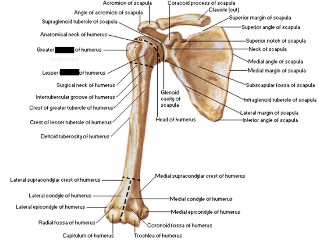
3 Regarding the last point in the objectives.. synovial joints are avascular they do not have direct blood supply, however they are supplied indirectly by neighboring structures and that s why you can see anastomosis of arteries surrounding synovial joints but there is still gas exchange nutrients and waste exchange via diffusion. *Shoulder girdle consists of clavicle and scapula -which articulate with one another at the acromioclavicular joint *Clavicle : Long slender subcutaneous bone, that lies horizontally across the root of the neck, it articulates with : -sternum and 1 st the clavicle) costal cartilage medially.(the sternal or medial end of - Acromion process of scapula laterally.(acromial or lateral end of the clavicle) Clavicle medial two thirds is convex forward and the lateral third is concave forward. P a g e 2
4 Scapula: A flat triangular bone, it has a superior angle, inferior angle, medial boarder (vertebral boarder), lateral boarder (axillary boarder). It lies between on the posterior chest wall between the 2 nd and 7 th ribs. -It has a spine on its posterior surface, that projects backward and its lateral end is free and forms the Acromian which articulates with the clavicle. -glenoid cavity or fossa The superolateral angle forms what is called the glenoid cavity or fossa. This is the part that articulates with the head of the humerus to form the shoulder joint. -coracoid process medial to glenoid cavity. That projects upward and forward above the glenoid cavity and it provides attachment for muscles and ligaments. -Suprascapular notch Medial to the base of the coracoid process -The anterior surface of the scapula is concave and forms >> the shallow subscapular fossa. -The posterior surface is devided by the spine into supraspinous fossa above the spine and infraspinous fossa below the spine. -The inferior angle can be palpated easily in the living subject (the only part of scapula that is subcutaneous) and marks the level of the 7 th rib and the spine of the 7 th thoracic vertebra. Note from NH lecture The scapula is kept in position by muscles particularly serratus anterior muscle, that is innervated by long thoracic nerve. If paralysis happen to that muscle a condition results that is called winged scapula, the scapula will be protruding in a way that results in wing like resemblance. Winged scapula is normal posture in children but abnormal in adults. P a g e 3
5 Humerus >> is not part of the shoulder girldle -Articulates with the scapula at the shoulder joint and with the radius and ulna at the elbow joint. -its upper end has a head which articulate with the glenoid cavity of the scapula. The head is immediately above the anatomical neck -greater and lesser tuberosities. Are below the neck and separated from each other by the bicipital groove (bicipital groove is called intertubercular groove in other books because they refer to greater and lesser tuberosities as greater and lesser tubercles) -narrow surgical neck. Where the upper end of the humerus joins the shaft. -deltoid tuberosity. Which is roughened elevation about halfway down the lateral aspect of the shaft. -spiral groove. Behind and below tuberosity. which accommodates the radial nerve. -lower end has medial and lateral epicondyles for the attachment of muscles and ligaments. -rounded capitulum, for articulation with the head of the radius at the elbow joint. -pulley-shaped trochlea, for articulation with the trochlear notch of the ulna. -radial fossa. Above the capitulum. And receives the head of the radius when the elbow is flexed. -coronoid fossa. Above the trochlea anteriorly. And receives the coronoid process of the ulna. -Olecranon fossa. Above the trochlea posteriorly. And receives the olecranon process of the ulna when the elbow joint is extended. P a g e 4
6 P a g e 5

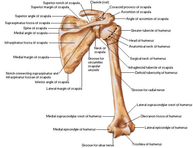
7 Quadrangular space Intramuscular spaces related to scapula. -Is an intramuscular space located immediately below the shoulder joint -It is bounded *above by the teres minor *below by the teres major *medially by the long head of the triceps brachii *laterally by the surgical neck of the humerus. Structures in this space>> axillary nerve, posterior circumflex humeral vessels. This space is clinically significant because those structures can P a g e 6
8 get injured or can be compressed due to trauma resulting in different degrees of weakness of the deltoid muscle. Triangular space the most medial space -it is bounded *inferiorly by the superior border of the teres major. *laterally by the long head of triceps. *superiorly by teres minor. Structures in the space>> it contains scapular circumflex vessels only. The triangular interval It is bounded to *superiorly by teres major. *medially by long head of triceps brachii. *laterally by lateral head of the triceps brachii. The structures in the space > radial nerve and profunda(deep) brachii artery. Axilla -between is a pyramid-shaped space between the upper part of the arm and the side of the chest. -it forms an important passage for nerves, blood and lymph vessels, as they travel from the root of the neck to the upper limb. -its apex is the upper end that is directed into the root of the neck and it is bounded *in front by the clavicle *behind by the upper border of the scapula *medially by the outer border of the first rib P a g e 7
 * behind by the posterior axillary fold (formed by the tendon of latissimus dorsi and the teres major muscle) *medially by the chest wall.")
9 -the lower end of the axilla is called Base, is bounded *in front by the anterior axillary fold, called also the anterior axillary line (formed by the lower boarder of the pectoralis major) * behind by the posterior axillary fold (formed by the tendon of latissimus dorsi and the teres major muscle) *medially by the chest wall. Apex of axilla P a g e 8

10 Walls of the axilla. Anterior walls is formed by -pectoralis major, subclavius and pectoralis minor muscles. Posterior wall is formed by -subcapsularis, latissimus dorsi and teres major muscles from above down. Medial wall is formed by -the upper four or five ribs and the intercostal spaces covered by the serratus anterior muscle. P a g e 9
11 lateral wall is formed by -coracobrachial and biceps muscles, in the bicipital groove of the humerus. The base is formed by -skin stretching between the anterior and posterior walls Axilla contains.. Contents of the Axilla 1) axillary artery and its branches which supply blood to the upper limb. 2) axillary vein and its tributaries which drain blood from the upper limb. 3) lymph vessels and lymph nodes which drain lymph from the upper limb and the breast, and from the skin of the trunk, down as far as the level of the umbilicus. 4) brachial plexus(formed by the anterior primary rime of spinal nerves C4 through T1), which innervates the upper limb. Note >> structures in the axilla are embedded in fat. P a g e 10

12 In the picture above pectoralis minor muscle and axillary artery. **Axillary artery is divided into three parts by the pectoralis minor muscle, so from the outer boarder of the first rib to the upper boarder of pectoralis minor muscle you can see the first part of axillary artery and then you can see the second part of axillary artery behind the pectoralis minor muscle, and finally the third part is from the lower boarder of pectoralis minor to the boarder of teres major where the axillary artery continues as the brachial artery. Rotator cuff >> tendons of these muscles are fused with the underlying capsule of the shoulder joint and strengthen the capsule. Those muscles are 4 >> *supraspinatus ( origin from supraspinous fossa),*infraspinatus ( origin from infraspinous fossa), *teres minor and *subscapularis(you can t find it in a posterior view only in anterior view of shoulder joint.) -it plays a very important role in stabilizing the shoulder joint. P a g e 11

13 -it lies on the anterior, superior, and posterior aspects of the joint. -it is deficient inferiorly which is a site of potential weakness. Rotator cuff muscles, it s a posterior view because you can see the spine of the scapula. Shoulder joint. Is formed by articulation between head of humerus and glenoid cavity of scapula. Its articular surfaces are covered by hyaline articular cartilage. Glenoid cavity is deepened by the presence of glenoid labrum, which is fibrocartilaginous rim. -Type: synovial (ball-and socket) joint -It is surrounded by capsule, which is attached medially to the margin of the glenoid cavity outside the labrum, and laterally to the anatomical neck of the humerus. P a g e 12

14 The capsule is also thin and lax, allowing a wide range of movment>> flexion, extention, adduction, abduction, medial rotation, lateral rotation and circumduction. The capsule is strengthened by fibrous slips from the tendons of the rotator cuff muscles. The capsule is lined by synovial membrane, - that is attached to the margins of the cartilage covering the articular surfaces. -It forms a tubular sheath around the tendon of the long head of biceps brachii -and it extends through the anterior wall of the capsule to form the subscapularis bursa(beneath the subscapularis muscle) The shoulder joint is innervated by axillary and suprascapular nerves. This is a sagittal view of the shoulder joint. Sagittal section is a vertical plane that devides an organ or structure into right and left parts. -the glenoid fossa is deepened by the presence of fibrocartilaginous Rim known as glenoid labrum In this view you can see 2 bursae 1)subacromian bursa 2)subscapularis bursa. Bursa are clinically important because they get inflamed bursitis A very painful condition that happens because of trauma or infection. -the capsule is attached to the margins of glenoid cavity medially and to the neck of the humerus laterally, it s outside the glenoid labrum. -bursa is a small sack filled with lubricating fluid to prevent frection between the two surfaces of bone. P a g e 13

15 Shoulder joint has ligaments Glenohumeral ligaments (superior, inferior and middle glenohumeral ligaments) Transverse humeral ligament Corachohumeral ligament Which are three weak bands of fibrous tissue that strengthen the front of the capsule. These muscles are hidden beneath the capsule of the joint, to find them you need to open the capsule and remove the head of the humerus. Which strengthens the capsule and bridges the gap between the two tuberosities of the humerus. Which strengthens the capsule above and stretches from the root of the coracoid process to the greater tuberosity of the humerus. Accessory ligaments are the coracoacromial ligament (extends from coracoid process to acromian process) It extendes between the coracoid process and the acromion, its function is to protect the superior aspect of the joint. P a g e 14
.")
16 Arterial Anastomosis Around shoulder joint, also the joint gets its needs by diffusion indirectly by neighboring tissues. Is formed by *anterior and posterior circumflex humeral arteries(which comes from the axillary artery which is a continuation of the subclavian artery). *circumflex scapular artery(branch of the subscapular artery which is the largest branch of the Axillary artery) *suprascapular artery (branch of the thyrocervical trunk that comes of the subclavian artery.) P a g e 15


17 Radio graphically you can see the two boney parts that articulate forming the shoulder joint you can see the head of the humerus, the glenoid cavity, coracoid process, acromian process, the clavical, the two boarders of the scapula, axillary(lateral boarder of scapula), vertebra and you can also see the first rib, second, third and forth. Because radio graphs are just two dimensional so there is superimposition between anterior and posterior parts. P a g e 16
18 Movements of the shoulder joint Flexion (forward movment of the upper limb) Preformed by the anterior fibers of deltoid, pectoralis major, biceps and coracobrachialis muscles Extension (backward movement of the upper limb) Which is performed by the posterior fibers of the deltoid, latissimus dorsi and teres major muscles. Abduction (moving the upper limb away from body or midline) -Involves middle fibers of the deltoid assisted by the supraspinatus. -it is initiated by supraspinatus muscle > that hold the head of the humerus against the glenoid fossa of the scapula, which allows the deltoid muscle to contract and abduct the humerus at the shoulder joint. Adduction (moving the upper limb toward body or midline) Performed by the pectoralis major, latissimus dorsi, teres major, and teres minor muscles. Lateral rotation Performed by the infraspinatus, teres minor, posterior fibers of the deltoid muscle. Medial rotation (results in the anterior surface of the arm facing medially) Which is performed by the subscapularis, the latissimus dorsi, teres major, and the anterior fibers of the deltoid muscle Circumduction which is a combination of the above movements P a g e 17


19 Now each muscle is attached to two sites there is site of origin and site of insertion, the picture below is an example.. this is the clavicle, The deltoid muscle takes its origin from the lateral part of the clavicle. Pectoralis major muscle takes its origin from more medial part of clavicle. another example, muscles attachments to scapula. P a g e 18
20 Subscapularis muscle origin is subscapular fossa and inserts on the lesser tuberosity of humerus. On the inferior angle and medial boarder of the scapula the serratus anterior muscle inserts. The deltoid muscle inserst on the acromian of scapula and the spine of the scapula. Teres minor and teres major muscles originate from the lateral boarder of scapula. * the site of origin of teres minor is above that of teres major. Supraspinatus originate from supraspinous fossa and inserts on greater tuberosity of humerus. Infraspinatous originate from infraspinous fossa. Teres major and latissimus dorsi insert on the medial lip of bicipital groove of humerus. Pectoralis major inserts on the lateral lip of bicipital groove of humerus. Posterior view you can see the three muscles of the rotator cuff Supraspinatus, infraspinatus and teres minor inserting on greater tuberosity in addition to the capsule of the shoulder joint, whereas subscapularis inserts on lesser tuberosity of humerus, in addition to the capsule of the joint. So all rotator cuff muscles insert on the capsule. *teres minor is one of the rotator cuff muscles not teres major. Dr nour said that you need to know all these muscles however the most important ones are the circled ones. Remmember P a g e 19


21 This is the first tafreegh in our 3 rd year of medicine : ) Good luck. P a g e 20
FUNCTIONAL ANATOMY OF SHOULDER JOINT
 FUNCTIONAL ANATOMY OF SHOULDER JOINT ARTICULATION Articulation is between: The rounded head of the Glenoid cavity humerus and The shallow, pear-shaped glenoid cavity of the scapula. 2 The articular surfaces
3 Mohammad Al-Mohtasib Areej Mosleh
 3 Mohammad Al-Mohtasib Areej Mosleh ***Muscles Connecting the Upper Limb to the Vertebral Column 1.Trapezius Muscle ***The first muscle on the back is trapezius muscle, it s called so according
Anatomy of the Shoulder Girdle. Prof Oluwadiya Kehinde FMCS (Orthop)
") Anatomy of the Shoulder Girdle Prof Oluwadiya Kehinde FMCS (Orthop) www.oluwadiya.com Bony Anatomy Shoulder Complex: Sternum(manubrium) Clavicle Scapula Proximal humerus Manubrium Sterni Upper part of
Anatomy of the Shoulder Girdle Prof Oluwadiya Kehinde FMCS (Orthop) www.oluwadiya.com Bony Anatomy Shoulder Complex: Sternum(manubrium) Clavicle Scapula Proximal humerus Manubrium Sterni Upper part of
Upper Limb Muscles Muscles of Axilla & Arm
 Done By : Saleh Salahat Upper Limb Muscles Muscles of Axilla & Arm 1) Muscles around the axilla A- Muscles connecting the upper to thoracic wall (4) 1- pectoralis major Origin:- from the medial half of
THE SHOULDER JOINT T H E G L E N O H U M E R A L ( G H ) J O I N T
 J O I N T") THE SHOULDER JOINT T H E G L E N O H U M E R A L ( G H ) J O I N T CLARIFICATION OF TERMS Shoulder girdle = scapula and clavicle Shoulder joint (glenohumeral joint) = scapula and humerus Lippert, p115
THE SHOULDER JOINT T H E G L E N O H U M E R A L ( G H ) J O I N T CLARIFICATION OF TERMS Shoulder girdle = scapula and clavicle Shoulder joint (glenohumeral joint) = scapula and humerus Lippert, p115
7/31/2012 THE SHOULDER JOINT CLARIFICATION OF TERMS OSTEOLOGY OF THE GH JOINT(BONES)
") THE SHOULDER JOINT T H E G L E N O H U M E R AL ( G H ) J O I N T CLARIFICATION OF TERMS Shoulder girdle = scapula and clavicle Shoulder joint (glenohumerual joint) = scapula and Lippert, p115 OSTEOLOGY
THE SHOULDER JOINT T H E G L E N O H U M E R AL ( G H ) J O I N T CLARIFICATION OF TERMS Shoulder girdle = scapula and clavicle Shoulder joint (glenohumerual joint) = scapula and Lippert, p115 OSTEOLOGY
MUSCLES OF SHOULDER REGION
 Dr Jamila EL Medany OBJECTIVES At the end of the lecture, students should: List the name of muscles of the shoulder region. Describe the anatomy of muscles of shoulder region regarding: attachments of
SHOULDER JOINT ANATOMY AND KINESIOLOGY
 SHOULDER JOINT ANATOMY AND KINESIOLOGY SHOULDER JOINT ANATOMY AND KINESIOLOGY The shoulder joint, also called the glenohumeral joint, consists of the scapula and humerus. The motions of the shoulder joint
Scapular and Deltoid Regions
 M1 Gross and Developmental Anatomy Scapular and Deltoid Regions Dr. Peters 1 Outline I. Skeleton of the Shoulder and Attachment of the Upper Extremity to Trunk II. Positions and Movements of the Scapula
Region of upper limb attachment to the trunk Proximal segment of limb overlaps parts of the trunk (thorax and back) and lower lateral neck.
 and lower lateral neck.") Region of upper limb attachment to the trunk Proximal segment of limb overlaps parts of the trunk (thorax and back) and lower lateral neck. includes Pectoral Scapular Deltoid regions of the upper limb
Region of upper limb attachment to the trunk Proximal segment of limb overlaps parts of the trunk (thorax and back) and lower lateral neck. includes Pectoral Scapular Deltoid regions of the upper limb
The Shoulder. Anatomy and Injuries PSK 4U Unit 3, Day 4
 The Shoulder Anatomy and Injuries PSK 4U Unit 3, Day 4 Shoulder Girdle Shoulder Complex is the most mobile joint in the body. Scapula Clavicle Sternum Humerus Rib cage/thorax Shoulder Girdle It also includes
MUSCLES. Anconeus Muscle
 LAB 7 UPPER LIMBS MUSCLES Anconeus Muscle anconeus origin: distal end of dorsal surface of humerus insertion: lateral surface of ulna from distal margin of the semilunar notch to proximal end of the olecranon
Connects arm to thorax 3 joints. Glenohumeral joint Acromioclavicular joint Sternoclavicular joint
 Connects arm to thorax 3 joints Glenohumeral joint Acromioclavicular joint Sternoclavicular joint Scapula Elevation Depression Protraction (abduction) Retraction (adduction) Downward Rotation Upward Rotation
Upper limb Arm & Cubital region 黃敏銓
 Upper limb Arm & Cubital region 黃敏銓 1 Arm Lateral intermuscular septum Anterior (flexor) compartment: stronger Medial intermuscular septum Posterior (extensor) compartment 2 Coracobrachialis Origin: coracoid
Pectoral region. Lecture 2
 Pectoral region Lecture 2 Muscle Action Each muscle has: Origin Beginning. Insertion End. Body (belly). Law: When a muscle performs its action, its insertion, moves towards its origin. Spring 2016 Dr.
Upper limb Pectoral region & Axilla
 Upper limb Pectoral region & Axilla 黃敏銓 mchuang@ntu.edu.tw 1 Pectoral region Intercostal nerve Anterior branch of lateral cutaneous branch Lateral cutaneous branch Anterior cutaneous branch Anterior cutaneous
This figure (of humerus) is from Dr. Maher's newest slides. -Its added here just for consideration-
 is from Dr. Maher's newest slides. -Its added here just for consideration-") This figure (of humerus) is from Dr. Maher's newest slides. -Its added here just for consideration- Slides of Anatomy Please note : These slides are Dr. Maher Hadidi s slides of spring 2016 and were edited
This figure (of humerus) is from Dr. Maher's newest slides. -Its added here just for consideration- Slides of Anatomy Please note : These slides are Dr. Maher Hadidi s slides of spring 2016 and were edited
Pectoral region. Lecture 2
 Pectoral region Lecture 2 Muscle Action Each muscle has: Origin Beginning. Insertion End. Body (belly). Law: When a muscle performs its action, its insertion, moves towards its origin. Spring 2016 Dr.
Muscle Action Origin Insertion Nerve Innervation Chapter Page. Deltoid. Trapezius. Latissimus Dorsi
 Muscle Action Origin Insertion Nerve Innervation Chapter Page All Fibers Abduct the shoulder (glenohumeral joint) Deltoid Anterior Fibers Flex the shoulder (G/H joint) Horizontally adduct the shoulder
Chapter 8. The Pectoral Girdle & Upper Limb
 Chapter 8 The Pectoral Girdle & Upper Limb Pectoral Girdle pectoral girdle (shoulder girdle) supports the arm consists of two on each side of the body // clavicle (collarbone) and scapula (shoulder blade)
Axilla and Brachial Region
 L 4 A B O R A T O R Y Axilla and Brachial Region BRACHIAL PLEXUS 5 Roots/Rami (ventral rami C5 T1) 3 Trunks Superior (C5, C6) Middle (C7) Inferior (C8, T1) 3 Cords Lateral Cord (Anterior Superior and Anterior
Practical 2 Worksheet
 Practical 2 Worksheet Upper Extremity BONES 1. Which end of the clavicle is on the lateral side (acromial or sternal)? 2. Describe the difference in the appearance of the acromial and sternal ends of the
Gateway to the upper limb. An area of transition between the neck and the arm.
 Gateway to the upper limb An area of transition between the neck and the arm. Pyramidal space inferior to shoulder @ junction of arm & thorax Distribution center for the neurovascular structures that serve
Pectoral girdle, SUPERIEUR ARM AND HAND. Danil Hammoudi.MD
 Pectoral girdle, SUPERIEUR ARM AND HAND Danil Hammoudi.MD The pectoral girdle is the set of bones which connect the upper limb to the axial skeleton on each side. It consists of the clavicle scapula in
The Arm and Cubital Fossa
 The Arm and Cubital Fossa Dr. Andrew Gallagher School of Anatomical Sciences University of the Witwatersrand Introduction The ARM (BRACHIUM) is the most proximal segment of the upper limb musculoskeletal
Anatomy and Physiology II. Review Shoulder Girdle New Material Upper Extremities - Bones
 Anatomy and Physiology II Review Shoulder Girdle New Material Upper Extremities - Bones Anatomy and Physiology II Shoulder Girdle Review Questions From Last Lecture Can you identify the following muscles?
Muscles of the Upper Limb
 Muscles of the Upper Limb anterior surface of ribs 3 5 coracoid process Pectoralis minor pectoral nerves protracts / depresses scapula Serratus anterior Subclavius ribs 1-8 long thoracic nerve rib 1 ----------------
Muscles in the Shoulder, Chest, Arm, Stomach, and Back
 Muscles in the Shoulder, Chest, Arm, Stomach, and Back Shoulder Muscles Deltoid Supraspinatus Infraspinatus Teres Major Teres Minor Subscapularis Deltoid (Delts) Function: Raises the upper arm Origin:
SUPERIEUR ARM AND HAND
 Pectoral girdle, SUPERIEUR ARM AND HAND Danil Hammoudi.MD The pectoral girdle is the set of bones which connect the upper limb to the axial skeleton on each side. It consists of the clavicle scapula in
Figure 1: Bones of the upper limb
 BONES OF THE APPENDICULAR SKELETON The appendicular skeleton is composed of the 126 bones of the appendages and the pectoral and pelvic girdles, which attach the limbs to the axial skeleton. Although the
The Upper Limb II. Anatomy RHS 241 Lecture 11 Dr. Einas Al-Eisa
 The Upper Limb II Anatomy RHS 241 Lecture 11 Dr. Einas Al-Eisa Sternoclavicular joint Double joint.? Each side separated by intercalating articular disc Grasp the mid-portion of your clavicle on one side
Netter's Anatomy Flash Cards Section 6 List 4 th Edition
 Netter's Anatomy Flash Cards Section 6 List 4 th Edition https://www.memrise.com/course/1577581/ Section 6 Upper Limb (66 cards) Plate 6-1 Humerus and Scapula: Anterior View 1.1 Acromion 1.2 Greater tubercle
region of the upper limb between the shoulder and the elbow Superiorly communicates with the axilla.
 1 region of the upper limb between the shoulder and the elbow Superiorly communicates with the axilla. Inferiorly, a number of important structures pass between arm & forearm through cubital fossa. 2 medial
The arm: *For images refer back to the slides
 The arm: *For images refer back to the slides Muscles of the arm: deltoid, triceps (which is located at the back of the arm), biceps and brachialis (it lies under the biceps), brachioradialis (it lies
Joint G*H. Joint S*C. Joint A*C. Labrum. Humerus. Sternum. Scapula. Clavicle. Thorax. Articulation. Scapulo- Thoracic
 A*C Joint Scapulo- Thoracic Articulation Thorax Sternum Clavicle Scapula Humerus S*C Joint G*H Joint Labrum AC Ligaments SC Ligaments SC JOINT AC Coracoacromial GH GH Ligament Complex Coracoclavicular
Returning the Shoulder Back to Optimal Function. Scapula. Clavicle. Humerus. Bones of the Shoulder (Osteology) Joints of the Shoulder (Arthrology)
 Joints of the Shoulder (Arthrology)") Returning the Shoulder Back to Optimal Function Sternum Clavicle Ribs Scapula Humerus Bones of the Shoulder (Osteology) By Rick Kaselj Clavicle Scapula Medial Left Anterior Clavicle Inferior View 20 degree
Returning the Shoulder Back to Optimal Function Sternum Clavicle Ribs Scapula Humerus Bones of the Shoulder (Osteology) By Rick Kaselj Clavicle Scapula Medial Left Anterior Clavicle Inferior View 20 degree
Glenohumeral Joint. Glenohumeral Joint. Glenohumeral Joint. Glenohumeral Joint. Glenohumeral Joint. Glenohumeral Joint
 The Shoulder Joint Chapter 5 The Shoulder Joint Manual of Structural Kinesiology R.T. Floyd, EdD, ATC, CSCS McGraw-Hill Higher Education. All rights reserved. 5-1 Shoulder joint is attached to axial skeleton
Kinesiology of the Upper Extremity
 P A R T II Kinesiology of the Upper Extremity Pectoralis Latissimus dorsi UNIT 1: SHOULDER UNIT: THE SHOULDER COMPLEX Chapter 8: Structure and Function of the Bones and Joints of the Shoulder Girdle Chapter
Sports Medicine Part II : ANATOMY OF THE SPINE, ABDOMEN AND SHOULDER COMPLEX
 Sports Medicine 25 1.1 Part II : ANATOMY OF THE SPINE, ABDOMEN AND SHOULDER COMPLEX c.w.p. Wagner High School, Sports Medicine, A. Morgan, T. Morgan & A. Eastlake, 2008 Muscles of the Upper Limbs In this
Gross Anatomy Questions That Should be Answerable After October 27, 2017
 Gross Anatomy Questions That Should be Answerable After October 27, 2017 1. The inferior angle of the scapula of a woman who was recently in an automobile accident seems to protrude making a ridge beneath
Fascial Compartments of the Upper Arm
 Fascial Compartments of the Upper Arm The upper arm is enclosed in a sheath of deep fascia and has two fascial septa: 1- Medial fascial septum (medial intermuscular septum): attached to the medial supracondylar
Joints of the upper limb II
 Joints of the upper limb II Prof. Abdulameer Al-Nuaimi E-mail: a.al-nuaimi@sheffield.ac.uk E. mail: abdulameerh@yahoo.com Elbow joint The elbow joint is connecting the upper arm to the forearm. It is classed
THE SKELETAL SYSTEM. Focus on the Pectoral Girdle
 THE SKELETAL SYSTEM Focus on the Pectoral Girdle Appendicular Skeleton 126 bones Includes bones of the limbs (arms and legs) Pectoral girdle (shoulder) Pelvic girdle (hip) Pectoral Girdle (the shoulder)
The Elbow and the cubital fossa. Prof Oluwadiya Kehinde
 The Elbow and the cubital fossa Prof Oluwadiya Kehinde www.oluwadiya.com Elbow and Forearm Anatomy The elbow joint is formed by the humerus, radius, and the ulna Bony anatomy of the elbow Distal Humerus
THE SHORT DESCRIPTION OF THE JOINTS 1. THE UPPER LIMB (Dr. Dóra Reglődi*, version )
") THE SHORT DESCRIPTION OF THE JOINTS 1. THE UPPER LIMB (Dr. Dóra Reglődi*, version 02-2007) Shoulder girdle The shoulder girdle consists of the clavicle and scapula on both sides. The two sides are connected
THE SHORT DESCRIPTION OF THE JOINTS 1. THE UPPER LIMB (Dr. Dóra Reglődi*, version 02-2007) Shoulder girdle The shoulder girdle consists of the clavicle and scapula on both sides. The two sides are connected
REFERENCE DIAGRAMS OF UPPER LIMB MUSCLES: NAMES, LOCATIONS, ATTACHMENTS, FUNCTIONS MUSCLES CONNECTING THE UPPER LIMB TO THE AXIAL SKELETON
 REFERENCE DIAGRAMS OF UPPER LIMB MUSCLES: NAMES, LOCATIONS, ATTACHMENTS, FUNCTIONS MUSCLES CONNECTING THE UPPER LIMB TO THE AXIAL SKELETON A25LAB EXERCISES: UPPER LIMB MUSCLES Page 1 MUSCLES CONNECTING
Introduction to anatomy
 Introduction to anatomy Dr. Maher Hadidi Fareed Halteh 3 7/2/2013 Subscapularis: It is located on the anterior side of the scapula. It has a triangular shape. It is like Pectoralis major and Teres major
Functional Anatomy. CHAPTER 5 Functional Anatomy of the Upper Extremity. CHAPTER 6 Functional Anatomy of the Lower Extremity
 Hamill_ch05_137-186.qxd 11/2/07 3:55 PM Page 137 S E C T I O N II Functional Anatomy CHAPTER 5 Functional Anatomy of the Upper Extremity CHAPTER 6 Functional Anatomy of the Lower Extremity CHAPTER 7 Functional
The skeleton consists of: Bones: special connective tissue, hard. Cartilage: special connective tissue, less hard than bones. Joints: joint is the
 The skeleton consists of: Bones: special connective tissue, hard. Cartilage: special connective tissue, less hard than bones. Joints: joint is the location at witch two bones make contact, whereas ligaments
Anatomy of the Musculoskeletal System
 Anatomy of the Musculoskeletal System Kyle E. Rarey, Ph.D. Department of Anatomy & Cell Biology and Otolaryngology University of Florida College of Medicine Outline of Presentation Vertebral Column Upper
David G. Simpson, Ph.D.
 David G. Simpson, Ph.D. ARM & CUBITAL FOSSA Revised 7/08 Text References Moores 3 rd ed., p402 408, 436 439, 439 443, 478, 481 LEARNING OBJECTIVES: 1. Describe the humerus, indicating the sites of muscle
Motion of Left Upper Extremity During A Right- Handed Golf Swing
 Motion of Left Upper Extremity During A Right- Handed Golf Swing Description of Movement While the movement required for a golf swing requires many muscles, joints, & ligaments throughout the body, the
The shoulder girdle consists of the glenohumeral, acromioclavicular, sternoclavicular and scapulothoracic joints
 Anatomy of Shoulder Girdle The shoulder girdle consists of the glenohumeral, acromioclavicular, sternoclavicular and scapulothoracic joints Glenohumeral Joint A ball and socket synoval joint with a large
Glenohumeral. Laura Leonetti Genna Moak Taylor Hansen
 Glenohumeral Laura Leonetti Genna Moak Taylor Hansen Surface anatomy Genna Anterior axillary fold Clavicle Clavicular head of Pectoralis major Clavipectoral triangle Acromial part of Deltoid Manubrium
#12. Joint نبيل خوري
 #12 30 Anatomy Joint هيام الر جال 9/10/2015 نبيل خوري Salam Awn Some notes before starting : ** Not all slides are included, so I recommend having a look at the slides beside this sheet ** If you find
Human Anatomy Biology 351
 1 Human Anatomy Biology 351 Upper Limb Exam Please place your name on the back of the last page of this exam. You must answer all questions on this exam. Because statistics demonstrate that, on average,
BRACHIAL PLEXUS. DORSAL SCAPULAR NERVE (C5) supraclavicular branch innervates rhomboids (major and minor) and levator scapulae
 supraclavicular branch innervates rhomboids (major and minor) and levator scapulae") THE BRACHIAL PLEXUS DORSAL SCAPULAR NERVE (C5) supraclavicular branch innervates rhomboids (major and minor) and levator scapulae SCHEMA OF THE BRACHIAL PLEXUS THE BRACHIAL PLEXUS PHRENIC NERVE supraclavicular
THE BRACHIAL PLEXUS DORSAL SCAPULAR NERVE (C5) supraclavicular branch innervates rhomboids (major and minor) and levator scapulae SCHEMA OF THE BRACHIAL PLEXUS THE BRACHIAL PLEXUS PHRENIC NERVE supraclavicular
WEEKEND THREE HOMEWORK
 WEEKEND THREE HOMEWORK READING ASSIGNMENTS Salvo Massage Therapy Principles and Practice 4 th Edition Muscolino The Muscular System Manual Muscolino The Muscle and Bone Palpation Manual Ch. 19 Skeletal
Muscular Nomenclature and Kinesiology - One
 Chapter 16 Muscular Nomenclature and Kinesiology - One Lessons 1-3 (with lesson 4) 1 Introduction 122 major muscles covered in this chapter Chapter divided into nine lessons Kinesiology study of human
Copyright 2010 Pearson Education, Inc.
 E. VERTEBRAL COLUMN 1. The vertebral column extends from the skull to the pelvis and forms the vertical axis of the skeleton. 2. The vertebral column is composed of vertebrae that are separated by intervertebral
The pectoral region. University of Babylon College of Medicine Dr.HaythemAli Alsayigh M.B.CH.B.-F.I.M.B.S. Surgical Clinical Anatomy
 The pectoral region University of Babylon College of Medicine Dr.HaythemAli Alsayigh M.B.CH.B.-F.I.M.B.S. Surgical Clinical Anatomy Objective Study the Bones and Joints A. Clavicle (collarbone) B. Scapula
Anatomy Workshop Upper Extremity David Ebaugh, PT, PhD Workshop Leader. Lab Leaders: STATION I BRACHIAL PLEXUS
 Anatomy Workshop Upper Extremity David Ebaugh, PT, PhD Workshop Leader Lab Leaders: STATION I BRACHIAL PLEXUS A. Posterior cervical triangle and axilla B. Formation of plexus 1. Ventral rami C5-T1 2. Trunks
BLUE SKY SCHOOL OF PROFESSIONAL MASSAGE AND THERAPEUTIC BODYWORK. Musculoskeletal Anatomy & Kinesiology II REVIEW
 BLUE SKY SCHOOL OF PROFESSIONAL MASSAGE AND THERAPEUTIC BODYWORK Musculoskeletal Anatomy & Kinesiology II REVIEW MSAK101-II Session 4 LEARNING OBJECTIVES: By the end of this session, the student will be
Musculoskeletal Ultrasound. Technical Guidelines SHOULDER
 Musculoskeletal Ultrasound Technical Guidelines SHOULDER 1 Although patient s positioning for shoulder US varies widely across different Countries and Institutions reflecting multifaceted opinions and
LIST OF STRUCTURES TO BE IDENTIFIED IN LAB: UPPER EXTREMITY REVIEW 2016
 LIST OF STRUCTURES TO BE IDENTIFIED IN LAB: UPPER EXTREMITY REVIEW 2016 BONES Ribs, sternum, clavicle Humerus: Head, greater tubercle, lesser tubercle, intertubercular sulcus, surgical neck, anatomical
Shoulder joint Assessment and General View
 Shoulder joint Assessment and General View Done by; Mshari S. Alghadier BSc Physical Therapy RHPT 366 m.alghadier@sau.edu.sa http://faculty.sau.edu.sa/m.alghadier/ Functional anatomy The shoulder contains
Shoulder: Clinical Anatomy, Kinematics & Biomechanics
 Shoulder: Clinical Anatomy, Kinematics & Biomechanics Dr. Alex K C Poon Department of Orthopaedics & Traumatology Pamela Youde Nethersole Eastern Hospital Clinical Anatomy the application of anatomy to
Key Relationships in the Upper Limb
 Key Relationships in the Upper Limb This list contains some of the key relationships that will help you identify structures in the lab. They are organized by dissection assignment as defined in the syllabus.
medial half of clavicle; Sternum; upper six costal cartilages External surfaces of ribs 3-5
 MUSCLE ORIGIN INSERTION ACTION NERVE Pectoralis Major medial half of clavicle; Sternum; upper six costal cartilages Lateral lip of intertubercular groove of horizontal adduction Medial and lateral pectoral
Chapter 7: Skeletal System: Gross Anatomy
 Chapter 7: Skeletal System: Gross Anatomy I. General Considerations A. How many bones in an average adult skeleton? B. Anatomic features of bones are based on II. Axial Skeleton A. Skull 1. Functionally
G24: Shoulder and Axilla
 G24: Shoulder and Axilla Syllabus - Pg. 2 ANAT 6010- Medical Gross Anatomy David A. Morton, Ph.D. Objectives Upper limb Systemically: Bones (joints) Muscles Nerves Vessels (arteries/veins) Fascial compartments
Continuing Education: Shoulder Stability
 Continuing Education: Shoulder Stability Anatomy & Kinesiology: The GHJ consists of the articulation of three bones: the scapula, clavicle and humerus. The scapula has three protrusions: the coracoid,
213: HUMAN FUNCTIONAL ANATOMY: PRACTICAL CLASS 1: Proximal bones, plexuses and patterns
 213: HUMAN FUNCTIONAL ANATOMY: PRACTICAL CLASS 1: Proximal bones, plexuses and patterns CLAVICLE Examine an isolated clavicle and compare it with a clavicle on an articulated skeleton. Viewed from above,
Introduction to anatomy Lecture # 2
 Introduction to anatomy Dr. Maher Hadidi Tareq AlTal 2 6/2/2013 Quick revision: The body is built on the skeleton The skeleton is divided into two parts >>>> axial skeleton (80 bones) >>>> appendicular
An Introduction to the Appendicular Skeleton
 An Introduction to the Appendicular Skeleton The Appendicular Skeleton is composed of the 126 bones of the appendages (limbs) and the pectoral and pelvic girdles, which attach to the axial skeleton. Each
Lina Al Qerem Zaid R Al Najdawi Ahmad Gharaibeh Farah Khreisat Satani Sharkas Ahmad Ar ar
 Lina Al Qerem Zaid R Al Najdawi Ahmad Gharaibeh Farah Khreisat Satani Sharkas Ahmad Ar ar Osama Al Zoubi Mamoon Qatameen Lecture 1 Done by Lina Al Qerem Cytology: the science of the cell. Lysosome-> Lyso:
*the Arm* -the arm extends from the shoulder joint (proximal), to the elbow joint (distal) - it has one bone ; the humerus which is a long bone
, to the elbow joint (distal) - it has one bone ; the humerus which is a long bone") *the Arm* -the arm extends from the shoulder joint (proximal), to the elbow joint (distal) - it has one bone ; the humerus which is a long bone - muscles in the arm : *brachialis muscle *Biceps brachii
*the Arm* -the arm extends from the shoulder joint (proximal), to the elbow joint (distal) - it has one bone ; the humerus which is a long bone - muscles in the arm : *brachialis muscle *Biceps brachii
Lab Activity 11: Group II
 Lab Activity 11: Group II Muscles Martini Chapter 11 Portland Community College BI 231 Origin and Insertion Origin: The place where the fixed end attaches to a bone, cartilage, or connective tissue. Insertion:
Supplied in part by the musculocutaneous nerve. Forms the axis of rotation in movements of pronation and supination
 Anatomy: Upper limb (15 questions) 1. Latissimus Dorsi: Is innervated by the dorsal scapular nerve Lies above feres major muscle Medially rotates the humerus All of the above 2. Supinator muscle is: Deep
Introduction. Rarely does a single muscle act in isolation at the shoulder complex.
 Shoulder complex 1 Introduction Our study of the upper limb begins with the shoulder complex, a set of four articulations involving the sternum, clavicle, ribs, scapula, and humerus. Rarely does a single
CLINICAL SUMMARY AND RECOMMENDATIONS 378
 Shoulder 9 CLINICAL SUMMARY AND RECOMMENDATIONS 378 Anatomy 379 Osteology 379 Arthrology 380 Scapulohumeral Rhythm 381 Ligaments 382 Muscles 384 Nerves 387 Patient History 389 Initial Hypotheses Based
STRUCTURAL BASIS OF MEDICAL PRACTICE EXAMINATION 5 October 6, 2006
 STRUCTURAL BASIS OF MEDICAL PRACTICE EXAMINATION 5 October 6, 2006 PART l. Answer in the space provided. (8 pts) 1. Identify the structures. (2 pts) B C A. _pisiform B. _ulnar artery A C. _flexor carpi
Acknowledgement. Here are some flash cards all set up in a "pdf" format for you! Thanks to Laura H. (spring 08)
") Acknowledgement Here are some flash cards all set up in a "pdf" format for you! Thanks to Laura H. (spring 08) for her donation to all my anatomy students! t Here is her suggestion for making flashcards
Acknowledgement Here are some flash cards all set up in a "pdf" format for you! Thanks to Laura H. (spring 08) for her donation to all my anatomy students! t Here is her suggestion for making flashcards
Dr.Israa H. Mohsen. Lecture 5. The vertebral column
 Anatomy Lecture 5 Dr.Israa H. Mohsen The vertebral column The vertebral column a flexible structure consisting of 33 vertebrae holds the head and torso upright, serves as an attachment point for the legs,
Acland's DVD Atlas of Human Anatomy. Transcript for Volume Robert D Acland
 Acland's DVD Atlas of Human Anatomy Transcript for Volume 1 2007 Robert D Acland This free downloadable pdf file is to be used for individual study only. It is not to be reproduced in any form without
Anatomical Considerations/ Pathophysiology The shoulder is the most mobile joint in the body. : Three bones:
 Introduction Musculoskeletal training is generally underrepresented in medical training and residency curriculums. There is a general deficit in musculoskeletal knowledge amongst current medical students,
STEP #1: Anatomy STEP #2: Awareness STEP #3: Action
 Stabilize the Scapula in Three Easy Steps by NFPT Idea World 2016: Session 611 Saturday July 16th 7:30-9:20am Beverly Hosford, MA STEP #1: Anatomy STEP #2: Awareness STEP #3: Action 1. Anatomy *Memorize
Traditional Thai Acupressure Points. The anterior aspect of the body THE ANATOMICAL ATLAS
 Traditional Thai Acupressure Points The anterior aspect of the body THE ANATOMICAL ATLAS lines of the SHOULDER BLADES AND POSTERIOR ARM Scapula Line This line runs through landmarks: 1. Above the midpoint
The Clavicle Right clavicle Deltoid tubercle: Conoid tubercle, conoid ligamen Impression for the
 The Clavicle Muscle Attachment Sites in the Upper Limb Pectoralis major Right clavicle Smooth superior surface of the shaft, under the platysma muscle tubercle: attachment of the deltoid Acromial facet
SCHOOL OF ANATOMICAL SCIENCES. ANATOMY FOR BSc PT II and BSc OT II (ANAT 2033) TEST 1: 28 th March 2012
 TEST 1: 28 th March 2012") SCHOOL OF ANATOMICAL SCIENCES ANATOMY FOR BSc PT II and BSc OT II (ANAT 2033) TEST 1: 28 th March 2012 TOTAL TIME: 2 Hours TOTAL MARKS: 100 This test consists of two papers; Paper I (60 marks) will consist
SCHOOL OF ANATOMICAL SCIENCES ANATOMY FOR BSc PT II and BSc OT II (ANAT 2033) TEST 1: 28 th March 2012 TOTAL TIME: 2 Hours TOTAL MARKS: 100 This test consists of two papers; Paper I (60 marks) will consist
Shoulder Biomechanics
 Shoulder Biomechanics Lecture originally developed by Bryan Morrison, Ph.D. candidate Arizona State University Fall 2000 1 Outline Anatomy Biomechanics Problems 2 Shoulder Complex Greatest Greatest Predisposition
The Thoracic Cage ANATOMY 2: THORACIC CAGE AND VERTEBRAL COLUMN
 ANATOMY 2: THORACIC CAGE AND VERTEBRAL COLUMN PSK 4U Mr. S. Kelly North Grenville DHS The Thoracic Cage 7 true ribs 3 false ribs 2 floating ribs Clavicle = collarbone Manubrium Sternum Xiphoid Process
The online version of this article, along with updated information and services, can
 Functional Anatomy of the Shoulder Complex Malcolm Peat PHYS THER. 1986; 66:1855-1865. The online version of this article, along with updated information and services, can be found online at: http://ptjournal.apta.org/content/66/12/1855
Dr. Mahir Alhadidi Anatomy Lecture #9 Feb,28 th 2012
 Quick Revision: Upper arm is divided into two compartments: 1. Anterior Compartment: Contains three muscles (Biceps brachii, Coracobrachialis, Brachialis). Innervated by Musculocutaneous nerve. 2. Posterior
Medical Language. Dr. Maher Hadidi, University of Jordan 1. Spring 2016
 Medical Language Most derived from Latin and/ Greek language. Important for clear communication in health sciences. To describe the body clearly and indicate the position of its parts in relative to each
Chapter 8 The Skeletal System: The Appendicular Skeleton. Copyright 2009 John Wiley & Sons, Inc.
 Chapter 8 The Skeletal System: The Appendicular Skeleton Appendicular Skeleton The primary function is movement It includes bones of the upper and lower limbs Girdles attach the limbs to the axial skeleton
PowerPoint Lecture Slides prepared by Janice Meeking, Mount Royal College C H A P T E R. Joints: Part A. Copyright 2010 Pearson Education, Inc.
 PowerPoint Lecture Slides prepared by Janice Meeking, Mount Royal College C H A P T E R 8 Joints: Part A Warm Up 11/28/16 Happy Thanksgiving welcome back! J (be ready to share something fun you did over
The Free Upper Limb. Bone of the Arm. aus: Platzer, Locomotor System (ISBN ), 2009 Georg Thieme Verlag KG
, 2009 Georg Thieme Verlag KG") : ones, Ligaments, Joints The Free The bones of the free upper limb are The humerus The radius and ulna The carpal bones The metacarpal bones The phalanges one of the Arm Humerus (A H) The humerus articulates
: ones, Ligaments, Joints The Free The bones of the free upper limb are The humerus The radius and ulna The carpal bones The metacarpal bones The phalanges one of the Arm Humerus (A H) The humerus articulates
A&P 1 Skeletal Lab Guide Week 2 - Appendicular Skeleton and Joints Lab Exercises: Pectoral Girdle
 A&P 1 Skeletal Lab Guide Week 2 - Appendicular Skeleton and Joints Lab Exercises: Pectoral Girdle PLEASE NOTE: Your group will need an articulated skeleton, a disarticulated skeleton, and the joint models
Important Parts of Bones
 Important Parts of Bones For 2015 Know: Humerus (posterior) Clavical Femur (Anterior) Foot Hand Mandible Os Coxa Scapula Skull (Anterior, Inferior, Lateral) Sternum Humerus (posterior) A. olecranon fossa