Human Anatomy. Spinal Cord and Spinal Nerves
|
|
|
- Brenda Clarke
- 5 years ago
- Views:
Transcription
1 Human Anatomy Spinal Cord and Spinal Nerves 1
2 The Spinal Cord Link between the brain and the body. Exhibits some functional independence from the brain. The spinal cord and spinal nerves serve two functions: 1. pathway for sensory and motor impulses 2. responsible for reflexes 16-2
 median sulcus the anterior")
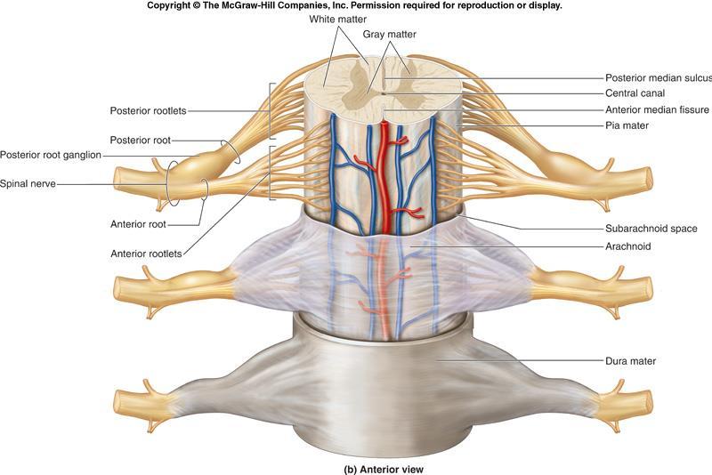
3 Structure of the Spinal Cord Typical adult spinal cord ranges between 42 and 45 centimeters (cm) (16 to 18 inches) in length. In cross section roughly cylindrical External surface has two longitudinal depressions: the posterior (dorsal) median sulcus the anterior (ventral) median fissure 16-3
4 16-4
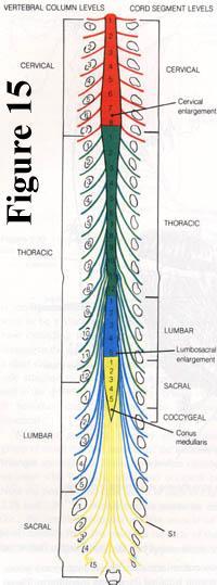
5 31 Regions of the Spinal Cord The cervical region continuous with the medulla oblongata contains neurons whose axons form the cervical spinal nerves (8) The thoracic region attached to this region are the thoracic spinal nerves (12) The lumbar region contains the neurons for the lumbar spinal nerves (5) The sacral region contains the neurons for the sacral spinal nerves (5) The coccygeal region one pair of coccygeal spinal nerves arises from this region 16-5
6 31Regions of the Spinal Cord each is identified by a number and letter designation. For example C3 is the third cervical segment. 6

7 Structure of the Spinal Cord The spinal cord is shorter than the vertebral canal that houses it. Conus medullaris: tapered inferior end of the spinal cord marks the official end of the spinal cord proper. Cauda equina Inferior to conus medularis nerve roots (groups of axons) that project inferiorly from the spinal cord. Filum terminale Within the cauda equina thin strand of pia mater helps anchor the conus medullaris to the coccyx. 16-7


8 Inferior End of Spinal Cord
9 Structure of the Spinal Cord The spinal cord is associated with 31 pairs of spinal nerves Each side of the spinal cord contains: 8 cervical nerves (called C1 C8) 12 thoracic nerves (T1 T12) 5 lumbar nerves (L1 L5) 5 sacral nerves (S1 S5) 1 coccygeal nerve (Co1) 16-9
10 10

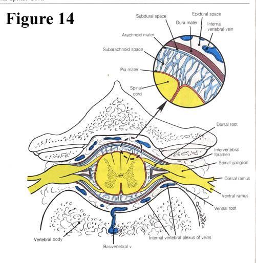
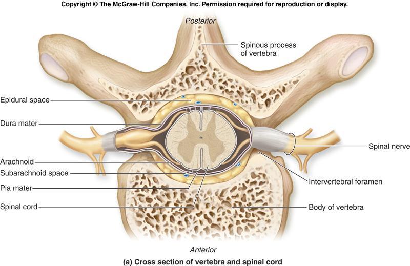
11 Arrangement and Functions of the Spinal Meninges Are continuous with the cranial meninges. Structures that encircle the spinal cord, listed from superficial to deep are: vertebra epidural space dura mater subdural space arachnoid subarachnoid space pia mater 16-11
12 12
13 13
14 Nucleus: A group of neuron cell bodies in the central nervous system Ganglia: A group of neuron cell bodies in the Peripheral nervous system Tract: A bundle of axons within the central nervous system that share a common function, origin and destination Column: a group of tracts found within a specific region of spinal tracts. Nerve: A bundle of axons within the Peripheral nervous system. 14
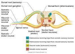
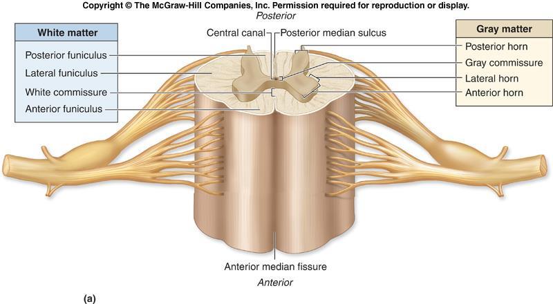
15 Location and Distribution of Gray Matter In the spinal cord, it is centrally located. Its shape resembles a letter H or a butterfly. Gray Matter is dominated by cell bodies of neurons and glial cells. The Gray Matter surrounds a central canal. The projections of the Gray Matter are called horns. The gray matter may be subdivided into the following components: 1. anterior horns 2. lateral horns 3. posterior horns 4. the gray commissure 16-15
16 The Gray Matter is the area where many types of neurons synapse. In dorsal horns (posterior horns) many types of incoming sensory neurons synapse with interneurons. Some interneurons use the gray commissure to cross to the other side of the spinal cord. In ventral horns (anterior horns) the descending upper level motor neuron (and other types of neurons ) synapse with outgoing motor neuron. In lateral horn descending autonomic pathway neurons synapse with outgoing sympathetic motor neuron
17 16-17
18 Location and Distribution of White Matter The white matter of the spinal cord is external to the gray matter. Three regions (referred to as funiculi or columns) : Composed of Ascending and Descending tracts 1- Posterior funiculus/column: 2- Lateral funiculus/column: 3- Anterior f funiculus/column: 1- A posterior funiculus: lies between the posterior gray horns and the posterior median sulcus. 2- Lateral funiculus. lies between the posterior funiculi and the anterior funiculi 3- Anterior funiculus between the anterior gray horns and the anterior median fissure
19 The white commissure is located anterior to the gray commissure. A specific tract convey either sensory or motor information Axons in sensory tracts carry information up to spinal cord and brain Axons in motor tracts carry information from the brain down to spinal cord

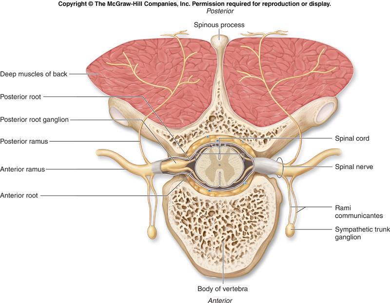
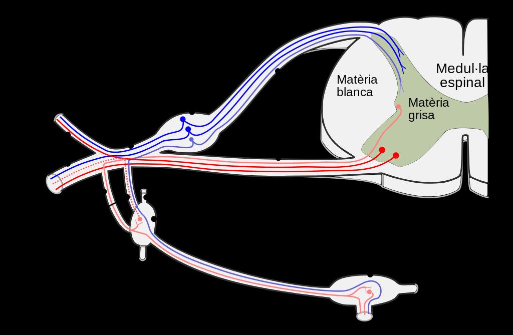
20 Two pairs of nerve roots extend from each segment of spinal cord: The ventral root allows motor neurons to exit the spinal cord. The dorsal root allows sensory neurons to enter the spinal cord. Along each root is a small bulge called dorsal root ganglia that contains the cell body of sensory neuron The ventral root and dorsal root fuse to form the spinal nerve, thus called Mixed spinal nerve 16-20


21 Opening between the vertebrae called intervertebral foramine allow the spinal nerve to enter and exit the spinal cord
22 22
23 Spinal Nerves 31 pairs connect the CNS to: receptors effectors (muscle and glands) Each spinal nerve is mixed: thousands of motor and sensory axons. Sensory axons originate from receptors Motor axons originate from the spinal cord. Spinal nerve is associated with the vertebra of the same number
24 Rami of Spinal Nerves Each spinal nerve forms though the fusion of Anterior root and posterior root as these roots pass through an intervertebral foramen. Then the spinal nerve divides into two branches a dorsal ramus and a ventral rami ramus. Posterior (or Dorsal) ramus Innervates muscles and skin of the neck and the back Anterior (or ventral rami ) Ramus Largest branch Forms plexuses Innervates anterior and lateral trunk, upper and lower limbs. For spinal nerves T1 to L2 there are 4 branches: a white ramus and gray ramus collectively known as Rami communicantes, Posterior and ramus Anterior ramus. Rami communicantes carry visceral motor fibers to and from a near by Autonomic ganglion associated with sympathetic division of Autonomic nervous system (sympathetic) 16-24
25 16-25
26 26

27 16-27
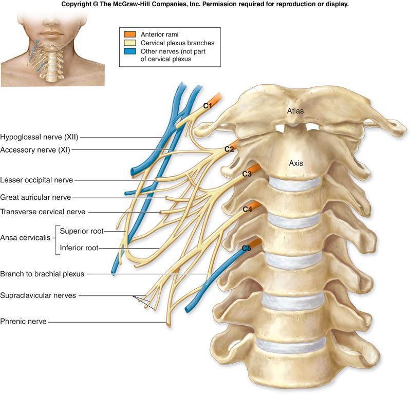
28 Nerve Plexuses A network of interweaving anterior rami of spinal nerves. nerve plexuses on both the right and left sides of the body. Nerve plexuses then split into multiple named nerves that innervate various body structures. Principal plexuses 1. cervical plexuses 2. brachial plexuses 3. lumbar plexuses 4. sacral plexuses


29 4 Principal Plexuses 1-Cervical Plexus Cervical Plexus Consists of cutaneous and muscular branches in the ventral rami of spinal nerves C1-C4 and some of C5 Phrenic nerve - innervates diaphragm Lesions of one or more nerve roots result in neurologic defects (muscle weakness, abnormal sensation, changes in reflexes)
30 30

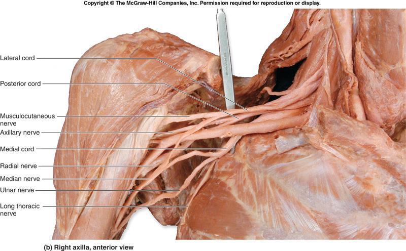
31 Brachial Plexus Musculocutaneous nerve - innervates biceps and brachialis muscles Median nerve - Ulnar nerve - Radial nerve Axillary nerve study in lab!!


32 Brachial Plexus is formed from ventral rami of spinal nerves 32 C5 to T1
33 33
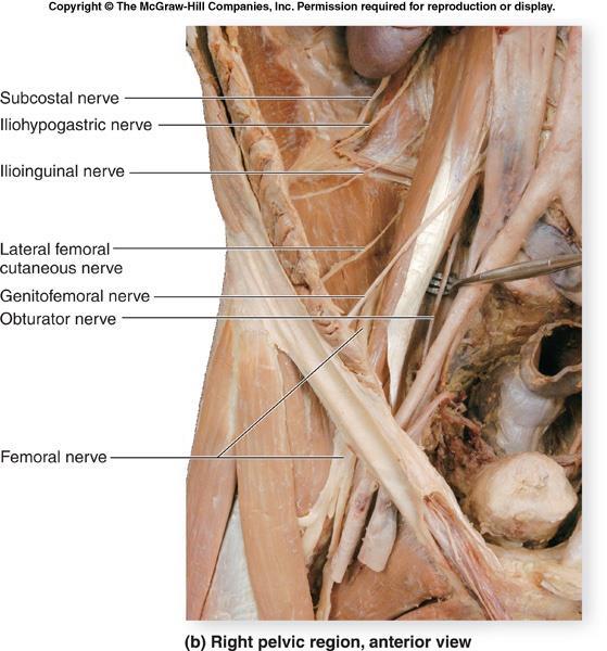
34 1. lumbar plexuses 2. sacral plexuses Collectively called lumbosacral plexuses The ventral rami supply the pelvis and lower limbs. lumbar plexuses is formed from ventral rami of spinal nerves T12 tol4 The major nerves of lumbar plexuses are Genifemoral nerve Lateral femoral cutaneous nerve Femoral nerve. 34


35 :lumbar plexuses is formed from ventral rami of spinal nerves T12 tol4 lumbar plexuses 35

36 36
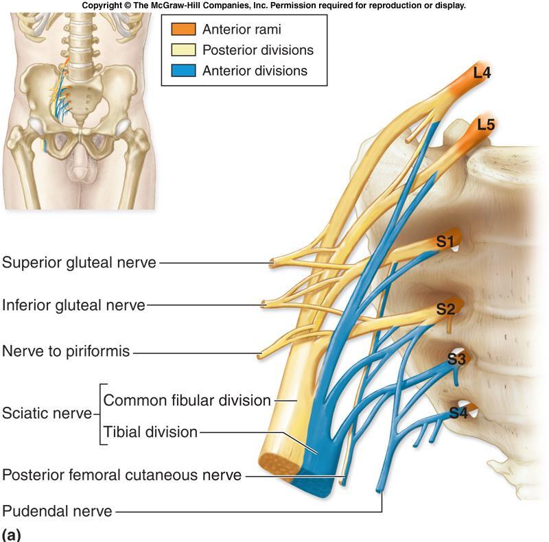
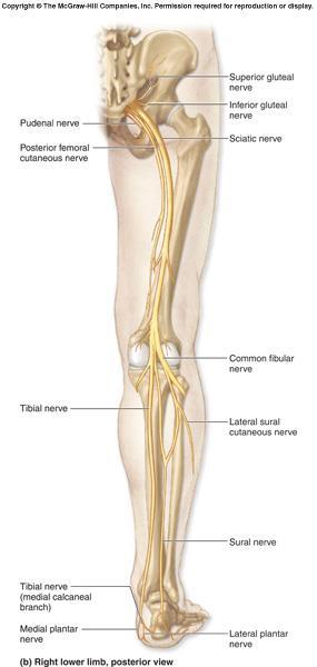
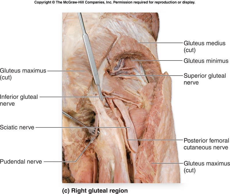
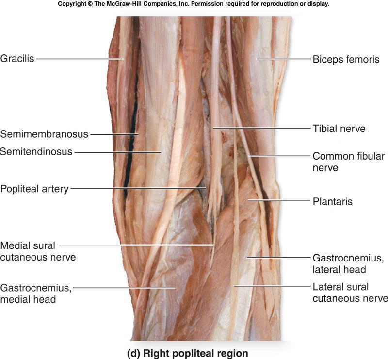
37 Sacral plexuses: is formed from ventral rami of spinal nerves L4 to S4 Sciatic nerve pass to biceps femoris muscle Pudendal nerve 37
38 38
39 39
40 40
41 41
42 42
43 Reflexes A reflex is a response: Rapid, automatic involuntary reactions of effectors to a stimulus. Properties. a stimulus required to initiate a response to sensory input a rapid response requires that few neurons be involved synaptic delay be minimal an automatic response occurs the same way every time An involuntary response requires no intent or pre-awareness of the reflex activity. Reflexes usually can not be suppressed. Awareness of the stimulus occurs after the reflex action in time to correct or avoid a potentially dangerous situation
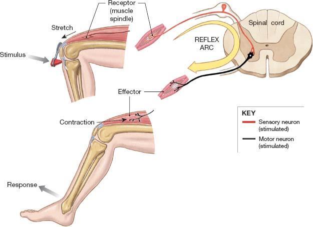
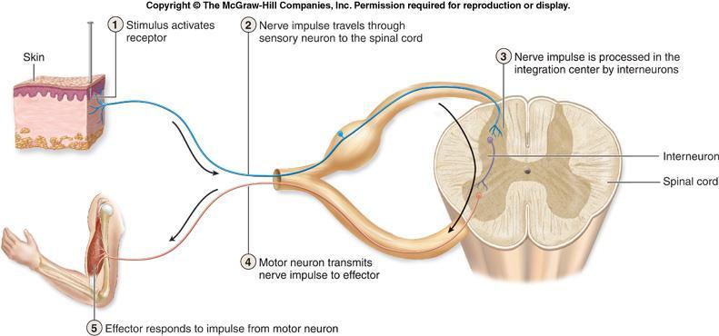
44 Components of a Reflex Arc The neural wiring of a single reflex. Always begins at a receptor in the PNS Sensory afferent Communicates with the CNS. May involve interneurons Ends at a peripheral effector (muscle or gland) Motor efferent 16-44

45 Reflex arc is a pathway involving: receptor, sensory neuron, motor neuron, one or more association neurons, and an effecter (muscle or gland).
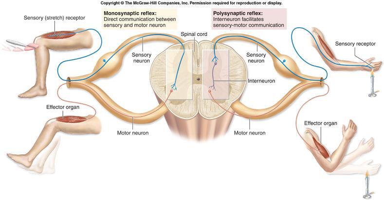
46 Ipsilateral and Contralateral Reflex Arcs Ipsilateral: both the receptor and effector organs of the reflex are on the same side of the spinal cord. Contralateral the sensory impulses from a receptor organ cross over through the spinal cord to activate effector organs in the opposite side 16-46
47 Monosynaptic Reflexes The simplest of all reflexes. No interneurons. The patellar (knee-jerk) reflex is a monosynaptic reflex physicians use to assess the functioning of the spinal cord. tap the patellar ligament with a reflex hammer muscle spindles in the quadriceps muscles are stretched. Produces a noticeable kick of the leg
48
49 Polysynaptic Reflexes Have more complex neural pathways exhibit a number of synapses involve interneurons within the reflex arc. Has more components more prolonged delay between stimulus and response
50 50
51 51
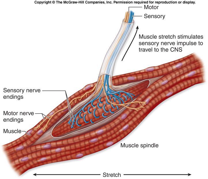
52 Stretch Reflexes Monosynaptic reflex that monitors and regulates skeletal muscle length. When a stimulus results in the stretching of a muscle, that muscle reflexively contracts. The patellar (knee-jerk) reflex is an example of a stretch reflex. The stimulus (the tap on the patellar tendon) initiates contraction of the quadriceps femoris muscle and extension of the knee joint
53 53
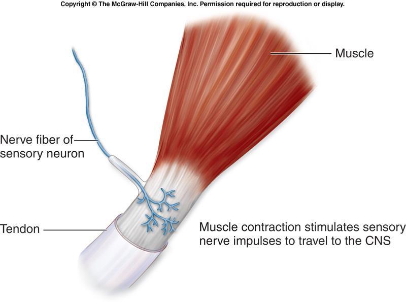
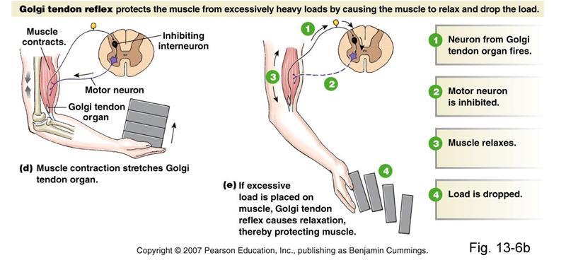
54 Golgi Tendon Reflex Prevents skeletal muscles from tensing excessively. Golgi tendon organs are nerve endings located within tendons near a muscle tendon junction. activation of the Golgi tendon organ signal interneurons in the spinal cord, which in turn inhibit the actions of the motor neurons The associated muscle is allowed to relax, thus protecting the muscle and tendon from excessive tension damage
55 55
56 16-56
57 Reflex Testing in a Clinical Setting Reflexes can be used to test specific muscle groups and specific spinal nerves or segments of the spinal cord. Consistently abnormal reflex response may indicate damage to the nervous system or muscles. A reflex response may be normal, hypoactive, or hyperactive
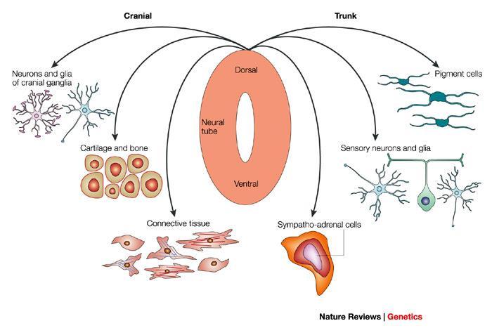
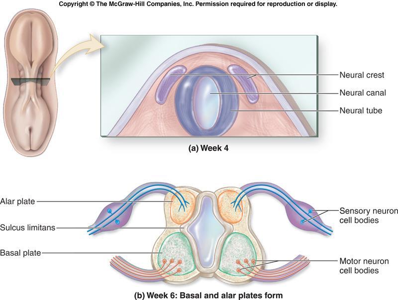
58 Spinal Cord Development The central nervous system forms from the embryonic neural tube. Cranial and spinal nerves form from neural crest cells that have split off from the developing neural tube. The cranial (superior) part of the neural tube expands and develops into the brain. The caudal (inferior) part of the neural tube forms the spinal cord
59 16-59
60 60
Chapter 13: The Spinal Cord and Spinal Nerves
 Chapter 13: The Spinal Cord and Spinal Nerves Spinal Cord Anatomy Protective structures: Vertebral column and the meninges protect the spinal cord and provide physical stability. a. Dura mater, b. Arachnoid,
Note: Please refer to handout Spinal Plexuses and Representative Spinal Nerves for
 Chapter 13 Outline Note: Please refer to handout Spinal Plexuses and Representative Spinal Nerves for what you need to know from Exhibits 13.1 13.4 I. INTRODUCTION A. The spinal cord and spinal nerves
Chapter 14. The Nervous System. The Spinal Cord and Spinal Nerves. Lecture Presentation by Steven Bassett Southeast Community College
 Chapter 14 The Nervous System The Spinal Cord and Spinal Nerves Lecture Presentation by Steven Bassett Southeast Community College Introduction The Central Nervous System (CNS) consists of: The spinal
Lecture 14: The Spinal Cord
 Lecture 14: The Spinal Cord M/O Chapters 16 69. Describe the relationship(s) between the following structures: root, nerve, ramus, plexus, tract, nucleus, and ganglion. 70. Trace the path of information
ANATOMY OF SPINAL CORD. Khaleel Alyahya, PhD, MEd King Saud University School of
 ANATOMY OF SPINAL CORD Khaleel Alyahya, PhD, MEd King Saud University School of Medicine @khaleelya OBJECTIVES At the end of the lecture, students should be able to: Describe the external anatomy of the
Human Anatomy - Problem Drill 11: The Spinal Cord and Spinal Nerves
 Human Anatomy - Problem Drill 11: The Spinal Cord and Spinal Nerves Question No. 1 of 10 Instructions: (1) Read the problem statement and answer choices carefully, (2) Work the problems on paper as needed,
Chapter 13! Chapter 13 Spinal Cord and Spinal Nerves! The Spinal Cord and Spinal Nerves!
 Chapter 13! The Spinal Cord and Spinal Nerves! SECTION 13-1! The brain and spinal cord make up the central nervous system, and the cranial nerves and spinal nerves constitute the peripheral nervous system!
CHAPTER 13 LECTURE OUTLINE
 CHAPTER 13 LECTURE OUTLINE I. INTRODUCTION A. The spinal cord and spinal nerves mediate reactions to environmental changes. B. The spinal cord has several functions. 1. It processes reflexes. 2. It is
Chapter 13. The Spinal Cord & Spinal Nerves. Spinal Cord. Spinal Cord Protection. Meninges. Together with brain forms the CNS Functions
 Spinal Cord Chapter 13 The Spinal Cord & Spinal Nerves Together with brain forms the CNS Functions spinal cord reflexes integration (summation of inhibitory and excitatory) nerve impulses highway for upward
Gross Anatomy of Lower Spinal Cord
 Chapter 13 Spinal Cord, Spinal Nerves and Somatic Reflexes Spinal cord Spinal nerves Somatic reflexes Gross Anatomy of Lower Spinal Cord Meninges of Vertebra & Spinal Cord Spina Bifida Congenital defect
The Spinal Cord, Spinal Nerves, and Spinal Reflexes
 13 The Spinal Cord, Spinal Nerves, and Spinal Reflexes PowerPoint Lecture Presentations prepared by Jason LaPres Lone Star College North Harris An Introduction to the Spinal Cord, Spinal Nerves, and Spinal
Fig Cervical spinal nerves. Cervical enlargement C7. Dural sheath. Subarachnoid space. Thoracic. Spinal cord Vertebra (cut) spinal nerves
 spinal nerves") Fig. 13.1 C1 Cervical enlargement C7 Cervical spinal nerves Dural sheath Subarachnoid space Thoracic spinal nerves Spinal cord Vertebra (cut) Lumbar enlargement Medullary cone T12 Spinal nerve Spinal nerve
Fig. 13.1 C1 Cervical enlargement C7 Cervical spinal nerves Dural sheath Subarachnoid space Thoracic spinal nerves Spinal cord Vertebra (cut) Lumbar enlargement Medullary cone T12 Spinal nerve Spinal nerve
Lab Activity 13. Spinal Cord. Portland Community College BI 232
 Lab Activity 13 Spinal Cord Portland Community College BI 232 Definitions Tracts: collections of axons in CNS Nerves:collections of axons in PNS Ganglia: collections of neuron cell bodies in PNS Nucleus
The Spinal Cord and Spinal Nerves!
 Chapter 13! The Spinal Cord and Spinal Nerves! SECTION 13-1! The brain and spinal cord make up the central nervous system, and the cranial nerves and spinal nerves constitute the peripheral nervous system!
TEST BANK FOR FUNDAMENTALS OF ANATOMY & PHYSIOLOGY 9TH EDITION BY MARTINI
 Link download :https://testbankservice.com/download/test-bank-forfundamentals-of-anatomy-and-physiology-9th-edition-by-martini TEST BANK FOR FUNDAMENTALS OF ANATOMY & PHYSIOLOGY 9TH EDITION BY MARTINI
Spinal Cord H. Ruth Clemo, Ph.D.
 Spinal Cord H. Ruth Clemo, Ph.D. OBJECTIVES After studying the material of this lecture, the student should be familiar with: 1. Surface anatomy of the spinal cord. 2. Internal structure and organization
Spinal Cord Protection. Chapter 13 The Spinal Cord & Spinal Nerves. External Anatomy of Spinal Cord. Structures Covering the Spinal Cord
 Spinal Cord Protection Chapter 13 The Spinal Cord & Spinal Nerves We are only going to cover Pages 420-434 and 447 Together with brain forms the CNS Functions spinal cord reflexes integration (summation
Spinal Cord and Spinal Nerves. Spinal Cord. Chapter 12
 Chapter 12 Spinal Cord and Spinal Nerves 1 Spinal Cord Extends from foramen magnum to second lumbar vertebra Segmented: Cervical, Thoracic, Lumbar & Sacral Gives rise to 31 pairs of spinal nerves Not uniform
Anatomy of the Nervous System. Brain Components
 Anatomy of the Nervous System Brain Components NERVOUS SYSTEM INTRODUCTION Is the master system of human body, controlling the functions of rest of the body systems Nervous System CLASSIFICATION A. Anatomical
Department of Neurology/Division of Anatomical Sciences
 Spinal Cord I Lecture Outline and Objectives CNS/Head and Neck Sequence TOPIC: FACULTY: THE SPINAL CORD AND SPINAL NERVES, Part I Department of Neurology/Division of Anatomical Sciences LECTURE: Monday,
BIOH111. o Cell Module o Tissue Module o Integumentary system o Skeletal system o Muscle system o Nervous system o Endocrine system
 BIOH111 o Cell Module o Tissue Module o Integumentary system o Skeletal system o Muscle system o Nervous system o Endocrine system Endeavour College of Natural Health endeavour.edu.au 1 Textbook and required/recommended
The Spinal Cord. The Nervous System. The Spinal Cord. The Spinal Cord 1/2/2016. Continuation of CNS inferior to foramen magnum.
 The Nervous System Spinal Cord Continuation of CNS inferior to foramen magnum Simpler than the brain Conducts impulses to and from brain Two way conduction pathway Reflex actions Passes through vertebral
With other members of your lab group, discuss the following questions: - The spinal cord connects directly to which part of the brain?
 BIOLOGY 211: HUMAN ANATOMY & PHYSIOLOGY ************************************************************************************************************************* SPINAL CORD, SPINAL NERVES, AND REFLEXES
Synapse Homework. Back page last question not counted. 4 pts total, each question worth 0.18pts. 26/34 students answered correctly!
 Synapse Homework Back page last question not counted 26/34 students answered correctly! 4 pts total, each question worth 0.18pts Business TASS hours extended! MWF 1-2pm, Willamette 204 T and Th 9:30-10:30am,
ANATOMY OF THE SPINAL CORD. Structure of the spinal cord Tracts of the spinal cord Spinal cord syndromes
 SPINAL CORD ANATOMY OF THE SPINAL CORD Structure of the spinal cord Tracts of the spinal cord Spinal cord syndromes The Nervous System Coordinates the activity of muscles, organs, senses, and actions Made
Central Nervous System: Part 2
 Central Nervous System: Part 2 1. Meninges 2. CSF 3. Spinal Cord and Spinal Nerves Explain spinal cord anatomy, including gray and white matter and meninges (give the general functions of this organ).
The functional Anatomy of the Nervous System. DR. OKSANA PETRICHKO Department of Human Anatomy
 The functional Anatomy of the Nervous System DR. OKSANA PETRICHKO Department of Human Anatomy Coordination and Regulation of Body Systems Nervous system. Conducts nerve impulses maintaining homeostasis
Chapter 12b. Overview
 Chapter 12b Spinal Cord Overview Spinal cord gross anatomy Spinal meninges Sectional anatomy Sensory pathways Motor pathways Spinal cord pathologies 1 The Adult Spinal Cord About 18 inches (45 cm) long
The Spinal Cord & Spinal Nerves
 The Spinal Cord & Spinal Nerves Together with brain forms the CNS Functions spinal cord reflexes integration (summation of inhibitory and excitatory) nerve impulses highway for upward and downward travel
THE BACK THE SPINAL CORD
 THE BACK THE SPINAL CORD The structures in the vertebral canal: the spinal cord spinal nerve roots spinal meninges the neurovascular structures THE SPINAL CORD The spinal cord occupies the superior 2/3
Faculty of Dental Medicine and Surgery. Sem 4 Peripheral nervous system and nerve plexus Dr. Abbas Garib Alla
 Faculty of Dental Medicine and Surgery Sem 4 Peripheral nervous system and nerve plexus Dr. Abbas Garib Alla PNS Terminology Ganglia neuron cell bodies Peripheral nerves neuronal axons PNS neuroglia Satellite
Spinal nerves. Aygul Shafigullina. Department of Morphology and General Pathology
 Spinal nerves Aygul Shafigullina Department of Morphology and General Pathology Spinal nerve a mixed nerve, formed in the vicinity of an intervertebral foramen, where fuse a dorsal root and a ventral root,
The Nervous System: The
 C h a p t e r 14 The Nervous System: The Spinal Cord and Spinal Nerves PowerPoint Lecture Slides prepared by Jason LaPres North Harris College Houston, Texas Copyright 2009 Pearson Education, Inc., publishing
Spinal Cord- Medulla Spinalis. Cuneyt Mirzanli Istanbul Gelisim University
 Spinal Cord- Medulla Spinalis Cuneyt Mirzanli Istanbul Gelisim University Spinal Column Supports the skull, pectoral girdle, upper limbs and thoracic cage by way of the pelvic girdle. Transmits body weight
Gross Morphology of Spinal Cord
 Gross Morphology of Spinal Cord Done By : Rahmeh Alsukkar ** I did my best and sorry for any mistake ** the sheet does not contain pictures, tables and some slides so please be careful and go back to slides
Laboratory 08 Peripheral Nervous System
 Laboratory 08 Peripheral Nervous System Goals: Structure and function of the cranial nerves. 1. List the cranial nerves by name and number. 2. Describe the specific functions of each of the cranial nerves
Nervous System: Spinal Cord and Spinal Nerves (Chapter 13)
") Nervous System: Spinal Cord and Spinal Nerves (Chapter 13) Lecture Materials for Amy Warenda Czura, Ph.D. Suffolk County Community College Eastern Campus Primary Sources for figures and content: Marieb,
Nervous System: Spinal Cord and Spinal Nerves (Chapter 13) Lecture Materials for Amy Warenda Czura, Ph.D. Suffolk County Community College Eastern Campus Primary Sources for figures and content: Marieb,
Chapter 14 Lecture Outline
 Chapter 14 Lecture Outline See separate PowerPoint slides for all figures and tables preinserted into PowerPoint without notes. Copyright McGraw-Hill Education. Permission required for reproduction or
Gross Morphology of Spinal Cord
 Gross Morphology of Spinal Cord Lecture Objectives Describe the gross anatomical features of the spinal cord. Describe the level of the different spinal segments compared to the level of their respective
Copyright McGraw-Hill Education. Permission required for reproduction or display. C1. Cervical spinal ner ves. Thor acic. T12 Spinal nerve rootlets
 Fig. 13.1 C1 Cervical enlar gem ent C7 Cervical spinal ner ves Dural sheath Subarachnoi d space Thor acic spinal ner ves Vertebra (cut) Lum bar enlar gem ent Medullar y T12 rootlets cone Posterior median
BIOH111. o Cell Module o Tissue Module o Skeletal system o Muscle system o Nervous system o Endocrine system o Integumentary system
 BIOH111 o Cell Module o Tissue Module o Skeletal system o Muscle system o Nervous system o Endocrine system o Integumentary system Endeavour College of Natural Health endeavour.edu.au 1 Textbook and required/recommended
Introduction and Basic structural organization of the nervous system
 Introduction and Basic structural organization of the nervous system **the slides are in bold and the book is in red Done by : razan krishan & marah marahleh INTRODUCTION The nervous system, along with
Organization of The Nervous System PROF. SAEED ABUEL MAKAREM
 Organization of The Nervous System PROF. SAEED ABUEL MAKAREM Objectives By the end of the lecture, you should be able to: List the parts of the nervous system. List the function of the nervous system.
The Spinal Cord, Spinal Nerves, and Spinal Reflexes
 12 The Spinal Cord, Spinal Nerves, and Spinal Reflexes Lecture Presentation by Lori Garrett Section 1: Functional Organization of the Spinal Cord Learning Outcomes 12.1 Describe how the spinal cord can
NERVOUS SYSTEM ANATOMY
 INTRODUCTION to NERVOUS SYSTEM ANATOMY M1 - Gross and Developmental Anatomy Dr. Milton M. Sholley Professor of Anatomy and Neurobiology and Dr. Michael H. Peters Professor of Chemical and Life Science
The Nervous System S P I N A L R E F L E X E S
 The Nervous System S P I N A L R E F L E X E S Reflexes Rapid, involuntary, predictable motor response to a stimulus Spinal Reflexes Spinal somatic reflexes Integration center is in the spinal cord Effectors
NERVOUS SYSTEM ANATOMY
 NTRODUCTON to NERVOUS SYSTEM ANATOMY M1 - Gross and Developmental Anatomy Dr. Milton M. Sholley Professor of Anatomy and Neurobiology and Dr. Michael H. Peters Professor of Chemical and Life Science Engineering
Chapter 13 Lecture Outline
 Chapter 13 Lecture Outline See separate PowerPoint slides for all figures and tables preinserted into PowerPoint without notes. Copyright McGraw-Hill Education. Permission required for reproduction or
Spinal cord. We have extension of the pia mater below L1-L2 called filum terminale
 Spinal cord Part of the CNS extend from foramen magnum to the level of L1-L2 (it is shorter than the vertebral column) it is covered by spinal meninges. It is cylindrical in shape. It s lower end become
Anatomy and Physiology 1 Chapters 12 and 13 self quiz Pro, Dima Darwish,MD.
 Anatomy and Physiology 1 Chapters 12 and 13 self quiz Pro, Dima Darwish,MD. 1) Which of the following is a function of the nervous system? A) sense the internal and external environments B) integrate sensory
Nervous System. The Peripheral Nervous System Agenda Review of CNS v. PNS PNS Basics Cranial Nerves Spinal Nerves Reflexes Pathways
 Nervous System Agenda Review of CNS v. PNS PNS Basics Cranial Nerves Spinal Nerves Sensory Motor Review of CNS v. PNS Central nervous system (CNS) Brain Spinal cord Peripheral nervous system (PNS) All
Human Anatomy Biology 351
 nnnnn 1 Human Anatomy Biology 351 Exam #2 Please place your name on the back of the last page of this exam. You must answer all questions on this exam. Because statistics demonstrate that, on average,
Chapter 17 Nervous System
 Chapter 17 Nervous System 1 The Nervous System Two Anatomical Divisions Central Nervous System (CNS) Brain and Spinal Cord Peripheral Nervous System (PNS) Two Types of Cells Neurons Transmit nerve impulses
CHAPTER 11: NERVOUS SYSTEM II: DIVISIONS OF THE NERVOUS SYSTEM. 1. Outline the major divisions of the nervous system.
 CHAPTER 11: NERVOUS II: DIVISIONS OF THE NERVOUS OBJECTIVES: 1. Outline the major divisions of the nervous system. NERVOUS CENTRAL NERVOUS (BRAIN & SPINAL CORD) (INTERNEURONS) PERIPHERAL NERVOUS (CRANIAL
Chapter 13 Lecture Outline
 Chapter 13 Lecture Outline See separate PowerPoint slides for all figures and tables preinserted into PowerPoint without notes. Copyright McGraw-Hill Education. Permission required for reproduction or
Chapter 9. Nervous System
 Chapter 9 Nervous System Central Nervous System (CNS) vs. Peripheral Nervous System(PNS) CNS Brain Spinal cord PNS Peripheral nerves connecting CNS to the body Cranial nerves Spinal nerves Neurons transmit
Human Anatomy and Physiology I Laboratory Spinal and Peripheral Nerves and Reflexes
 Human Anatomy and Physiology I Laboratory Spinal and Peripheral Nerves and Reflexes 1 This lab involves the second section of the exercise Spinal Cord, Spinal Nerves, and the Autonomic Nervous System,
I. Autonomic Nervous System (ANS) A. Dual Innervation B. Autonomic Motor Pathway 1. Preganglionic Neuron a. Preganglionic Fibers (Axons) (1)
 A. Dual Innervation B. Autonomic Motor Pathway 1. Preganglionic Neuron a. Preganglionic Fibers (Axons) (1)") I. Autonomic Nervous System (ANS) A. Dual Innervation B. Autonomic Motor Pathway 1. Preganglionic Neuron a. Preganglionic Fibers (Axons) (1) Acetylcholine - ACh 2. Ganglion (Ganglia) 3. Ganglionic Neuron
I. Autonomic Nervous System (ANS) A. Dual Innervation B. Autonomic Motor Pathway 1. Preganglionic Neuron a. Preganglionic Fibers (Axons) (1) Acetylcholine - ACh 2. Ganglion (Ganglia) 3. Ganglionic Neuron
Sympathetic Nervous System
 Sympathetic Nervous System Lecture Objectives Review the subdivisions of the nervous system. Review the general arrangement and compare the sympathetic and parasympathetic parts. Describe the following
THE BACK. Dr. Ali Mohsin. Spinal Cord
 Spinal Cord THE BACK Dr. Ali Mohsin The spinal cord is the elongated caudal part of the CNS. It starts as the inferior continuation of the medulla oblongata at the level of foramen magnum, & ends as an
Chapter 13 *Lecture PowerPoint
 Chapter 13 *Lecture PowerPoint The Spinal Cord, Spinal Nerves, and Somatic Reflexes *See separate FlexArt PowerPoint slides for all figures and tables preinserted into PowerPoint without notes. Copyright
Lesson 33. Objectives: References: Chapter 16: Reading for Next Lesson: Chapter 16:
 Lesson 33 Lesson Outline: Nervous System Structure and Function Neuronal Tissue Supporting Cells Neurons Nerves Functional Classification of Neuronal Tissue Organization of the Nervous System Peripheral
Organisation of the nervous system
 Chapter1 Organisation of the nervous system 1. Subdivisions of the nervous system The nervous system is divided: i) Structurally The central nervous system (CNS) composed of the brain and spinal cord.
Unit Three. The brain includes: cerebrum, diencephalon, brain stem, & cerebellum. The brain lies within the cranial cavity of the skull.
 Human Anatomy & Physiology 11 Divisions of the Nervous System Karen W. Smith, Instructor Unit Three BRAIN & SPINAL CORD Refer to the following URLs. Be sure to study these along with your book. http://www.sirinet.net/~jgjohnso/nervous.html
Cranial Nerves and Spinal Cord Flashcards
 1. Name the cranial nerves and their Roman numeral. 2. What is Cranial Nerve I called, and what does it 3. Scientists who are trying to find a way to make neurons divide to heal nerve injuries often study
Spinal Cord Anatomy. Key Points. What is the spine? Areas of the spine: Spinal Cord Anatomy
 Spinal Cord Anatomy Authors: SCIRE Community Team Reviewed by: Riley Louie, PT Last updated: Sept 21, 2017 This page provides an overview of the structures of the spinal cord and how the spinal cord works.
Peripheral Nervous System
 Peripheral Nervous System Sensory Receptors Motor Endings Cranial Nerves The Four Plexuses Extremities Review of Reflexes Fast, preprogrammed, inborn, automatic responses Occur in the CNS at the spinal
Spinal nerves and cervical plexus Prof. Abdulameer Al Nuaimi. E mail: a.al E. mail:
 Spinal nerves and cervical plexus Prof. Abdulameer Al Nuaimi E mail: a.al nuaimi@sheffield.ac.uk E. mail: abdulameerh@yahoo.com Branches of ophthalmic artery Muscles of face A spinal nerve Spinal
Chapter 9 The Nervous System: The Spinal Cord and Spinal Nerves
 Chapter 9 The Nervous System: The Spinal Cord and Spinal Nerves Copyright 2015 Wolters Kluwer Health Lippincott Williams & Wilkins Overview Key Terms acetylcholine motor presynaptic action potential nerve
Organization of The Nervous System PROF. MOUSAED ALFAYEZ & DR. SANAA ALSHAARAWY
 Organization of The Nervous System PROF. MOUSAED ALFAYEZ & DR. SANAA ALSHAARAWY Objectives At the end of the lecture, the students should be able to: List the parts of the nervous system. List the function
The neurvous system senses, interprets, and responds to changes in the environment. Two types of cells makes this possible:
 NERVOUS SYSTEM The neurvous system senses, interprets, and responds to changes in the environment. Two types of cells makes this possible: the neuron and the supporting cells ("glial cells"). Neuron Neurons
Chapter 13 PNS and reflex activity
 Chapter 13 PNS and reflex activity I. Peripheral nervous system A. PNS links CNS to the body B. Sensory: the afferent division C. Motor: the efferent division D. Ganglia: collections of cell bodies in
Part 1: Communication between CNS & PNS
 Ch. 6: Peripheral Nervous System Objectives: 1. Communication between CNS & PNS: afferent (sensory) pathway versus efferent (motor) pathway of information. 2. Regulation of somatic (voluntary) motor system
Chapter 13. The Nature of Muscle Spindles, Somatic Reflexes, and Posture
 Chapter 13 The Nature of Muscle Spindles, Somatic Reflexes, and Posture Nature of Reflexes A reflex is an involuntary responses initiated by a sensory input resulting in a change in the effecter tissue
Brain Stem. Nervous System (Part A-3) Module 8 -Chapter 14
 Module 8 -Chapter 14") Nervous System (Part A-3) Module 8 -Chapter 14 Overview Susie Turner, M.D. 1/9/13 Cellular structure of the nervous system Neurons Neuroglia Nervous System Divisions Central nervous system Peripheral nervous
Nervous System (Part A-3) Module 8 -Chapter 14 Overview Susie Turner, M.D. 1/9/13 Cellular structure of the nervous system Neurons Neuroglia Nervous System Divisions Central nervous system Peripheral nervous
Dendrites Receive impulse from the axon of other neurons through synaptic connection. Conduct impulse towards the cell body Axon
 Dendrites Receive impulse from the axon of other neurons through synaptic connection. Conduct impulse towards the cell body Axon Page 22 of 237 Conduct impulses away from cell body Impulses arise from
Classification of the nervous system. Prof. Dr. Nikolai Lazarov 2
 1 1. Formation and general organization 2. Spinal ganglia 3. Zonal and segmental innervation 4. Dorsal rami of the spinal nerves 5. Ventral rami of the spinal nerves 6. Cervical plexus Classification of
SHORT ANSWER. Write the word or phrase that best completes each statement or answers the question.
 Exam Name 1) A change in the conditions in the synaptic terminal can influence the soma as a result of axoplasmic transport. 2) The nervous system is composed of the brain and spinal cord. A) efferent
Cerebral hemisphere. Parietal Frontal Occipital Temporal
 Cerebral hemisphere Sulcus / Fissure Central Precental gyrus Postcentral gyrus Lateral (cerebral) Parieto-occipital Cerebral cortex Frontal lobe Parietal lobe Temporal lobe Insula Amygdala Hippocampus
Lab # 2: Spinal Cord & Nerves, Reflexes and General Senses. A & P II Spring, 2014
 Lab # 2: Spinal Cord & Nerves, Reflexes and General Senses A & P II Spring, 2014 Objectives Be able to identify specified spinal cord structures and spinal nerves on models Be familiar with spinal nerve
50a A&P: Nervous System -! Peripheral Nervous System
 50a A&P: Nervous System -! Peripheral Nervous System 50a A&P: Nervous System -! Peripheral Nervous System! Class Outline" 5 minutes" "Attendance, Breath of Arrival, and Reminders " 10 minutes "Lecture:"
Spinal Cord Organization. January 12, 2011
 Spinal Cord Organization January 12, 2011 Spinal Cord 31 segments terminates at L1-L2 special components - conus medullaris - cauda equina no input from the face Spinal Cord, Roots & Nerves Dorsal root
Tymaa Al-zaben & Amin Al-ajalouni
 Done by: Tymaa Al-zaben & Amin Al-ajalouni ** Hello SERTONIN! SLIDE 3 note:: the slide included within the sheet but make sure back to slide for pictures The Autonomic Nervous System Function : Regulate
Nervous System. Overview, Tissues and Spinal Nerves
 Nervous System Overview, Tissues and Spinal Nerves neurology - study of the nervous system - nervous system and endocrine system work together to maintain homeostasis - More than 80 major nerves and billions
4/5/2015. Peripheral nervous system (PNS)
") Peripheral Nervous System Learn and Understand: Peripheral nerves connect the edges of the body and outside world to the CNS. Most nerves carry specific types of information to/from specific locations.
Peripheral Nervous System Learn and Understand: Peripheral nerves connect the edges of the body and outside world to the CNS. Most nerves carry specific types of information to/from specific locations.
Brain and spinal nerve. By: shirin Kashfi
 Brain and spinal nerve By: shirin Kashfi Nervous system: central nervous system (CNS) peripheral nervous system (PNS) Brain (cranial) nerves Spinal nerves Ganglions (dorsal root ganglions, sympathetic
Group of students. - Rawan almujabili د. محمد المحتسب - 1 P a g e
 - 14 - Group of students - Rawan almujabili د. محمد المحتسب - 1 P a g e Nerves of the posterior abdominal wall The spinal cord gives off spinal nerves between the vertebrae. In the abdomen, through the
Nervous Systems: Diversity & Functional Organization
 Nervous Systems: Diversity & Functional Organization Diversity of Neural Signaling The diversity of neuron structure and function allows neurons to play many roles. 3 basic function of all neurons: Receive
Chapter 12. APR Enhanced Lecture Slides
 Chapter 12 APR Enhanced Lecture Slides See separate PowerPoint slides for all figures and tables pre-inserted into PowerPoint without notes and animations. Copyright The McGraw-Hill Companies, Inc. Permission
Chapter 8 Nervous System
 Chapter 8 Nervous System Two message centers: Functions of these systems: 1. * 2. * Overview of the Nervous System Parts: General Functions: Functions Sensory input: Sensation via nerves Integration: interpretation
Lecture - Chapter 13: Central Nervous System
 Lecture - Chapter 13: Central Nervous System 1. Describe the following structures of the brain, what is the general function of each: a. Cerebrum b. Diencephalon c. Brain Stem d. Cerebellum 2. What structures
The Nervous System 7PART B. PowerPoint Lecture Slide Presentation by Patty Bostwick-Taylor, Florence-Darlington Technical College
 PowerPoint Lecture Slide Presentation by Patty Bostwick-Taylor, Florence-Darlington Technical College The Nervous System 7PART B What is a reflex? What is a reflex? What is meant by the statement that
Peripheral Nervous System Dr. Gary Mumaugh
 Peripheral Nervous System Dr. Gary Mumaugh Spinal Nerves Overview Thirty-one pairs of spinal nerves are connected to the spinal cord No special names; numbered by level of vertebral column at which they
Biological Bases of Behavior. 3: Structure of the Nervous System
 Biological Bases of Behavior 3: Structure of the Nervous System Neuroanatomy Terms The neuraxis is an imaginary line drawn through the spinal cord up to the front of the brain Anatomical directions are
The Nervous System PART A
 7 The Nervous System PART A PowerPoint Lecture Slide Presentation by Jerry L. Cook, Sam Houston University ESSENTIALS OF HUMAN ANATOMY & PHYSIOLOGY EIGHTH EDITION ELAINE N. MARIEB Structural Classification
Brachial plexuses and axillary lymph nodes
 Brachial plexuses and axillary lymph nodes Introduction about nervous system nervous system central nervous system periphral nervous system brain spinal cord 31 pairs of spinal nerves 12 paris of cranial
Pathways of proprioception
 The Autonomic Nervous Assess Prof. Fawzia Al-Rouq Department of Physiology College of Medicine King Saud University Pathways of proprioception System posterior column& Spinocerebellar Pathways https://www.youtube.com/watch?v=pmeropok6v8
Nervous system. The main regulation mechanism of organism's functions
 Nervous system The main regulation mechanism of organism's functions Questions Neuron The reflex arc The nervous centers Properties of the nervous centers The general principles of coordination Inhibition
cardiac plexus is continuous with the coronary and no named branches pain from the heart and lungs
 Nerves of the Thoracic Region Nerve Source Branches Motor Sensory Notes cardiac plexus cardiac brs. of the vagus n. and cervical ; thoracic l nn. the heart and lungs cardiac, cervical cardiac, vagal vagus