Objective. Reducing a displaced Syndesmosis 2/11/2016. Ankle Fractures Common Misconceptions. Common Myths in ankle fracture management
|
|
|
- Dayna May
- 5 years ago
- Views:
Transcription
1 Ankle Fractures Common Misconceptions Jackson Lee, MD Associate Professor Clinical Orthopedics Keck School of Medicine of the University of Southern California Objective Common Myths in ankle fracture management Reducing a displaced Syndesmosis Find the largest clamp you can find and use it to squeeze the fibula up against the tibia 1
2 High Fibula Fracture No need to reduce and stabilize a fibula fracture if it is more than halfway up the leg Order of Reduction Always reduce and stabilize the fibula first before doing anything else Posterior Malleous No need to reduce and stabilize unless is is at least 25% of the articular surface on a lateral x-ray 2
3 Why do we operate on Ankle fractures? To restore stability to a joint Contrary to popular belief, not to make crooked bones straight To Restore Joint Stability Address the Boney Injury Address the Ligament Injury All ankle fractures involve both Important Ligament Anatomy Often neglected 3
 Syndesmosis-Boney Attachments Posterior inferior tibfib ligament is attached to the posterior malleolus Anterior inferior tib-fib ligament is attached to the Chaput 4")
4 Important Ligaments for Joint Stability Deep Deltoid ligament Syndesmosis AITF PITF Syndesmosis ligament(tib-fib) Interosseous Membrane Syndesmosis Anterior tibio-fibular ligament (ATFL) Posterior tibio-fibular ligament (PTFL) Syndesmosis-Boney Attachments Posterior inferior tibfib ligament is attached to the posterior malleolus Anterior inferior tib-fib ligament is attached to the Chaput 4

5 Deltoid Superficial Deltoid attached to anterior colliculis of the Medial Malleolus Deep Deltoid is attached to the posterior colliculis of the Medial Malleolus Understanding the ligaments helps in understanding how to restore stability of the ankle joint Syndesmosis and the Posterior Malleolus Fracture 5


6 Posterior Malleolus Fracture Has the Posterior Inferior Tib Fib ligament attached In the presence of a Posterior Malleolus Fracture, the ligament is intact 100% of the time Opportunity If we reduce and stabilize the posterior malleous, we have in effect repaired the PITFL(syndesmosis) So which ones should we fix? All of them! Late fractures Indirect reduction of the articular surface but direct reduction of the fragment Absolute stability with anti-glide +/- lag 6


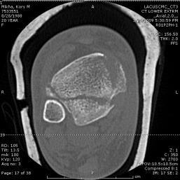
7 What about size? Traditionally fix if 25% on a lateral x-ray Problem - These fractures are oblique in nature and not in the pure coronal plane 25% on a lateral x-ray does not provide any information on the actual size of the fragment 15 patients with posterior malleous fx studied with MRI-posterior tib-fib ligament intact Cadaveric PLR IV fractures either fixed with ORIF of Posterior Malleolus or Syndesomotic Screws ---Fixing posterior malleous restored stiffness to 70% compared to only 40% with syndesmotic screws Size does not matter Stability Does Reducing and stabilizing a posterior malleous will in effect restore the PITFL Avoid Syndesmotic stabilization 7


8 Syndesmotic Injury All rotational ankle fractures have some degree of syndesmotic injury(ser, PER) Reduction and stabilization of a posterior malleous will repair the syndesmotic component of the injury How to reduce Indirect Reduction Posterolateral approach(harmon)- between Peroneals and FHL Posteromedial Reduce looking at posterior cortex Effective if no articular impaction Supine with bump under ipsilateral hip Prone - not good if there is a concomitant medial mal fracture Use implant in an anti-glide manner to reduce Case Example 8

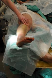
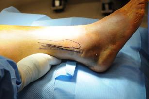
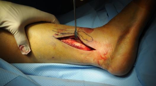
9 Case Example Patient Positioning Approach 9
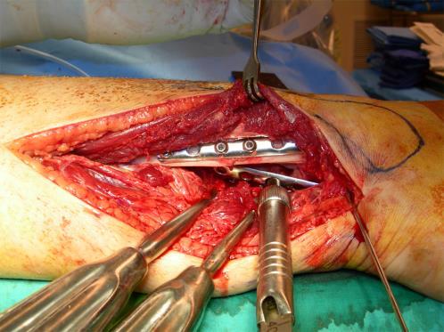
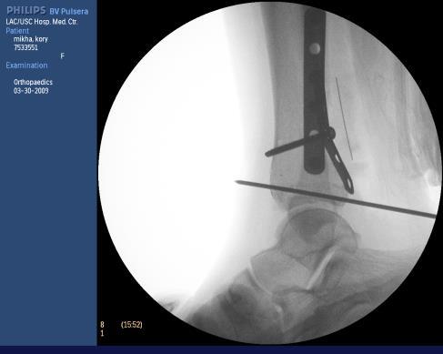
10 Anti-glide Reduction 10


11 Final Construct Stable to ER stress - no syndesmotic stabilization indicated! Medial Malleolus Medial Malleolus Deep Deltoid attaches to Posterior Colliculis Reducing and stabilizing a MM with a posterior Colliculis will in effect repair the deltoid injury 11


12 Medial Malleolus Most Rotational injures the MM is pulled off Fracture at or below level of the plafond Tension Failure Lag to stabilize Cancellous Four cortical Has only superficial Deltoid attached Deep Deltoid most likely torn Reducing and stabilizing this anterior Colliculis will contribute very little to ankle stability Medial Malleolus Anterior Colliculis Medial Malleolus With some mechanisms,(pab, SAD), mm is pushed off Sheer injury So need to push these back on or Antiglide 12
13 Syndesmosis What is the Problem? High rate of mal-reduced syndesmosis Mis-understood anatomy Often an after-thought during ORIF ankle fx To much noise in regards to implants Screws vs. Tight-rope Number and size of screws Not enough emphasis on reduction Too much talk about fixation methods 13

14 Reduction is the Problem Definition of Reduction Syndesmosis Fibula needs to be out to length Fibula needs to be correctly aligned in three planes Fibula needs to be corrected Rotated Fibula needs to be in the correct place within it s sulcus-incisura Old wive s tale If fibula fracture more than half way up from ankle joint, no need to fix 14


15 Restoring Length, Alignment Rotation Directly Reduce Fibula regardless fracture s location Only reliable way to achieve reduction as defined Reducing Fibula Fibula now out to length Fibula now at correct rotation Fibula correctly aligned Issues to reducing Fibula Very Proximal fracture-fear of nerve Approach and reduce +/- stabilize Once reduced with respect to length and rotation Address syndemosis reduction and stabilize syndemosis in a reduced state 15
, reduce and stabilize MM first")

16 Issues to reducing Fibula Very Comminuted Fibula Rely on Talo-cural angle from uninjured side? Other tactics If there is a medial Malleolus fracture that is not comminuted(likelytension failure), reduce and stabilize MM first Posterior Malleolus Comminuted Fibula Can you rely on Talo-Crural angle from other side? Maybe, but individual variation can vary from 75 to 86 (Foot Ankle, 1989, Rolfe et. Al) Reduce and stabilize a non-comminuted Medial Malleolus FIRST! Ligametotaxis will help restore fibula length Comminuted Fibula Other Tactics 16


17 Comminuted Fibula Other Tactics Reduce and Stabilize posterior Malleolus FIRST! PM has posteriorinferior tib-fib ligament Bonus-Reducing and stabilizing PM will often restore syndesmosis stability Reduction in the Sulcus Open reduce Allows debridement of the tib-fib joint Allows direct reduction Put fibula into sulcus Pay attention to the rotation within the sulcus No guess Clamp position Reduce Syndesmosis Start with a fibula that is reduced with respect to length alignment and rotation Fibula resides in a sulcus If you reduce a posterior Malleolus, it will place the fibula in the correct location, then just need to correct rotation within sulcus If no posterior malleous, then consider open reduce 17


18 Articular Impaction Injuries Often missed, not recognized, or not addressed Articular Impaction Injuries Look out in SAD and PAB injuries Evaluate with CT scan Articular Impaction SAD and PAB Need to reduce 18


19 What about Late fractures? 3 weeks Plus Late Fractures Must take down partially healed Posterior Malleous in order to restore length of fibula Take down high fibula s Open debride syndesmosis J 19


20 Soft Tissues Straight incisions Avoid Curving, J or any other letter incision Traumatized tissue best with straight incisions done sharply(knife, not spread(tear)) Just a few words about the fibula fracture component The Fibula in Rotational Injures SER, PER 20
 Simple")
 amenable to IM lag screw, tension banding, tension band")
21 Most rotational injuries(short oblique/spiral) very amenable to open reduction under direct vision, lag and neutralization plate or posterio-lateral anti glide plate absolute stability Rotational (SLR, PER) Simple fracture patterns Lag Screws and Neutralization Plates Posterior Anti-glide Operative Lateral Malleolus Fibula s that fail under tension(transverse- PAB) amenable to IM lag screw, tension banding, tension band plate 21


22 Lateral Malleolus Lag Screw for transverse Lateral Malleolus Low transverse High Fibula(diaphyseal) OK to stabilize with a stouter Plate(like LCP or similar) 22
23 Conclusions Reduce fibula with respect to length, alignment and rotation Reduce Syndesmosis Reduce and stabilize posterior malleous Reduce articular impaction injuries Straight Incisions 23
FIBULAR & SYNDESMOSIS MALUNIONS
 FIBULAR & SYNDESMOSIS MALUNIONS MICHAEL P. CLARE, MD FLORIDA ORTHOPAEDIC INSTITUTE TAMPA, FL USA MORTISE INHERENTLY UNSTABLE LATERAL MALLEOLUS ACTS AS BUTTRESS / POST RESIST LATERAL TRANSLATION OF TALUS
High Ankle Sprains: Diagnosis & Treatment
 High Ankle Sprains: Diagnosis & Treatment Mark J. Mendeszoon, DPM, FACFAS, FACFAOM Precision Orthopaedic Specialties University Regional Hospitals Advanced Foot & Ankle Fellowship- Director It Is Only
Ankle Fractures in the Elderly: How to Deal with Poor Bone Quality
 : How to Deal with Poor Bone Quality Richard T. Laughlin, MD Professor of Orthopaedic Surgery University of Cincinnati College of Medicine No disclosures relative to this presentation acknowledgement Some
Syndesmotic Ankle Injuries: Diagnosis and Treatment
 Syndesmotic Ankle Injuries: Diagnosis and Treatment John A. Scolaro, M.D., M.A. Assistant Professor of Orthopaedic Surgery University of California, Irvine California Orthopaedic Association - 2016 Disclosures
CURRENT TREATMENT OPTIONS
 CURRENT TREATMENT OPTIONS Fix single column or both: Always fix both. A study by Svend-Hansen corroborated the poor results associated with isolated medial malleolar fixation in bimalleolar ankle fractures.
Radiographic assessment. Functional. Paul Tornetta III Professor 11/21/2016. Fracture not in coronal plane May need CT to evaluate
 The Posterior Malleolus Paul Tornetta III Professor Boston Medical Center Publications: Disclosures! Rockwood and Green, Tornetta and Einhorn; Subspecialty series, Court-Brown, Tornetta; Trauma, AAOS;
PRONATION-ABDUCTION FRACTURES
 C H A P T E R 1 2 PRONATION-ABDUCTION FRACTURES George S. Gumann, DPM (The opinions of the author should not be considered as reflecting official policy of the US Army Medical Department.) Pronation-abduction
Ankle Fracture: Tips and Tricks
 Ankle Fracture: Tips and Tricks Christiaan N. Mamczak, DO LCDR, MC, USN Naval Medical Center Portsmouth Department of Orthopaedic Surgery Assistant Professor Uniformed Services University of the Health
5/3/2016 DISCLOSURES. Outline. Hassan R. Mir, MD, MBA, FACS. Ankle Fractures Lateral Malleolus Medial Malleolus Posterior Malleolus Chaput Syndesmosis
 DISCLOSURES Hassan R. Mir, MD, MBA, FACS Medical/Orthopaedic Publications Editorial/Governing Board OTA Newsletter Editor OsteoSynthesis, The JOT Online Discussion Forum Editor JOT Associate Editor JAAOS
BIOMECHANICS OF ANKLE FRACTURES
 BIOMECHANICS OF ANKLE FRACTURES William R Reinus, MD MBA FACR Significance of Ankle Fractures Most common weight-bearing Fx 70% of all Fxs Incidence is increasing Bimodal distribution Men 15-24 Women over
Increasing surgical freedom Restoring patient function
 Increasing surgical freedom Restoring patient function Fracture specific plating solutions for the most common tibia and fibula fractures Frequency of fracture occurrences* 66% 61% 36% 36% 28% 14% 20%
X-Ray Rounds: (Plain) Radiographic Evaluation of the Ankle.
 Radiographic Evaluation of the Ankle.") X-Ray Rounds: (Plain) Radiographic Evaluation of the Ankle www.fisiokinesiterapia.biz Anatomy Complex hinge joint Articulations among: Fibula Tibia Talus Tibial plafond Distal tibial articular surface
X-Ray Rounds: (Plain) Radiographic Evaluation of the Ankle www.fisiokinesiterapia.biz Anatomy Complex hinge joint Articulations among: Fibula Tibia Talus Tibial plafond Distal tibial articular surface
The Syndesmosis. Syndesmosis: How to Reduce and How Perfect? Boston Medical Center. Indications. Technique 11/19/2018.
 Syndesmosis: How to Reduce and How Perfect? Paul Tornetta III Professor Boston Medical Center Boston Medical Center The Syndesmosis Indications Subluxation Instability Technique Fluoroscopic Open 1 Weber
Ankle Ligament Injury: Don t Worry- It s Only a Sprain Wes Jackson MD Orthopaedic Foot & Ankle
 Ankle Ligament Injury: Don t Worry- It s Only a Sprain Wes Jackson MD Orthopaedic Foot & Ankle Outline I. Epidemiology II. Classification and Types of Sprains III. Anatomy IV. Clinical Assessment and Imaging
1/27/2016. Background. Background. Seth R. Yarboro University of Virginia January 29, Distal tibio fibular joint
 Seth R. Yarboro January 29, 2015 Background Distal tibio fibular joint maintains ankle stability while allowing motion Dorsiflexion/external rotation mechanism Poor alignment ankle arthritis Background
Disclosures! The Syndesmosis. Syndesmosis: How and When to Reduce. Boston Medical Center. Indications. Technique.
 Syndesmosis: How and When to Reduce Paul Tornetta III Professor Boston Medical Center Boston Medical Center Publications: Disclosures! Rockwood and Green, Tornetta and Einhorn; Subspecialty series, Court-Brown,
Outline. Ankle/Foot Anatomy Ankle Sprains Ottawa Ankle Rules DDx: The Sprain That Wasn t
 Ankle Injuries Outline Ankle/Foot Anatomy Ankle Sprains Ottawa Ankle Rules DDx: The Sprain That Wasn t Anatomy: Ankle Mortise Bony Anatomy Lateral Ligament Complex Medial Ligament Complex Ankle Sprains
Case Report The Utility and Limitations of the Transfibular Approach in Ankle Trauma Surgery
 Case Reports in Orthopedics, Article ID 234369, 4 pages http://dx.doi.org/10.1155/2014/234369 Case Report The Utility and Limitations of the Transfibular Approach in Ankle Trauma Surgery Mustafa Yassin,
Burwood Road, Concord Dora Street, Hurstville Lethbridge Street, Penrith 160 Belmore Road, Randwick
 www.orthosports.com.au 47 49 Burwood Road, Concord 29 31 Dora Street, Hurstville 119 121 Lethbridge Street, Penrith 160 Belmore Road, Randwick Update on Syndesmosis Ankle Sprains By Todd Gothelf Foot,
Competence of the Deltoid Ligament in Bimalleolar Ankle Fractures After Medial Malleolar Fixation *
 Competence of the Deltoid Ligament in Bimalleolar Ankle Fractures After Medial Malleolar Fixation * BY PAUL TORNETTA, III, M.D. Investigation performed at Kings County Hospital, New York, N.Y. Abstract
Donald Stewart, MD. Lateral ligament injuries Chronic lateral ligament instability Syndesmosis Injuries
 Donald Stewart, MD Arlington Orthopedic Associates Lateral ligament injuries Chronic lateral ligament instability Syndesmosis Injuries Anatomy Mechanism of Injury Classification Diagnostic Tests Management
Foot and Ankle Update
 Foot and Ankle Update 2019 Instructional Course Hiro Tanaka It s your on-call weekend Objectives We are going to apply evidence based treatment for 2 patients who are admitted under your care 1. Dislocated
AOFAS Resident Review Course September 28, Justin Greisberg, MD Associate Professor of Orthopaedic Surgery Columbia University
 Course September 28, 2013 Justin Greisberg, MD Associate Professor of Orthopaedic Surgery Columbia University Disclosures Consultant to Extremity Medical Receive royalties from Saunders/Mosby-Elsevier
Orthopedics in Motion Tristan Hartzell, MD January 27, 2016
 Orthopedics in Motion 2016 Tristan Hartzell, MD January 27, 2016 Humerus fractures Proximal Shaft Distal Objectives 1) Understand the anatomy 2) Epidemiology and mechanisms of injury 3) Types of fractures
Disclosures. Syndesmosis Injury. Syndesmosis Ligaments. Objectives. Mark M. Casillas, M.D.
 Disclosures Syndesmosis Injury No relevant disclosures Mark M. Casillas, M.D. 1 Objectives Syndesmosis Ligaments Understand the syndesmosis anatomy and function Classify syndesmosis injuries Describe treatment
EVOS MINI with IM Nailing
 Case Series Dr. John A. Scolaro EVOS MINI with IM Nailing A series of studies Introduction Intramedullary nailing has become the standard for many long bone fractures. Fracture reduction prior to nail
A Patient s Guide to Ankle Anatomy
 A Patient s Guide to Ankle Anatomy Pond View Professional Park 301 Professional View Drive Freehold, NJ 07728 Phone: 732-720-2555 DISCLAIMER: The information in this booklet is compiled from a variety
Fractures of the Ankle Region in the Skeletally Immature Patient. The Salter Classification is Worthless!!
 Fractures of the Ankle Region in the Skeletally Immature Patient. The Salter Classification is Worthless!! Kaye E Wilkins D.V.M,M.D. President's Council/Dielmann Chair in Pediatric Orthopedics Professor
SURGICAL AND APPLIED ANATOMY
 Página 1 de 9 Copyright 2001 Lippincott Williams & Wilkins Bucholz, Robert W., Heckman, James D. Rockwood & Green's Fractures in Adults, 5th Edition SURGICAL AND APPLIED ANATOMY Part of "47 - ANKLE FRACTURES"
Intramedullary Rodding of Distal Tibial Shaft Fractures with Intra Articular Extension
 Intramedullary Rodding of Distal Tibial Shaft Fractures with Intra Articular Extension My Name is Claude Sagi CSOT Tampa, FL 2018 Disclosures: None, I am just a simple man. This talk is about treating
LCP Anterior Ankle Arthrodesis Plates. Part of the Synthes Locking Compression Plate (LCP) System.
 System.") LCP Anterior Ankle Arthrodesis Plates. Part of the Synthes Locking Compression Plate (LCP) System. Technique Guide Instruments and implants approved by the AO Foundation Table of Contents Introduction
LCP Anterior Ankle Arthrodesis Plates. Part of the Synthes Locking Compression Plate (LCP) System. Technique Guide Instruments and implants approved by the AO Foundation Table of Contents Introduction
Craig S. Radnay, M.D. 1/27/2016. Access to the Talus for Treatment of Osteochondral Lesions. Epidemiology of OLT. Treatment of OLT
 Access to the Talus for Treatment of Osteochondral Lesions Craig S. Radnay, MD, MPH ISK Institute for Orthopaedics and Sports Medicine NYU/Hospital for Joint Diseases Tampa, FL January 23, 2016 Epidemiology
Pilon fractures. Pat Yoon, MD Minneapolis Veterans Affairs Medical Center Associate Professor, University of Minnesota
 Pilon fractures Pat Yoon, MD Minneapolis Veterans Affairs Medical Center Associate Professor, University of Minnesota Disclosures Reviewer Foot and Ankle International Journal of the American Academy of
Practical Reduction Techniques: Diaphyseal Reduction. Philip Wolinsky University of California at Davis Medical Center
 OTA Specialty Day 2016 Practical Reduction Techniques: Diaphyseal Reduction Philip Wolinsky University of California at Davis Medical Center 8:55 am 9:55 am Tips and Tricks: Practical Reduction Techniques
A Patient s Guide to Ankle Anatomy
 A Patient s Guide to Ankle Anatomy 1436 Exchange Street Middlebury, VT 05753 Phone: 802-388-3194 Fax: 802-388-4881 cvo@champlainvalleyortho.com DISCLAIMER: The information in this booklet is compiled from
A Patient s Guide to Ankle Anatomy
 A Patient s Guide to Ankle Anatomy 245 North College Lafayette, LA 70506 Phone: 337.232.5301 Fax: 337.237.6504 DISCLAIMER: The information in this booklet is compiled from a variety of sources. It may
Talus Fractures: When and Why on Screws and Plates
 Talus Fractures: When and Why on Screws and Plates Frank A. Liporace, MD Associate Professor Director of Orthopaedic Research New York University / Hospital for Joint Diseases, NY, NY Director Orthopaedic
Arthroscopy Of the Ankle.
 Arthroscopy Of the Ankle www.fisiokinesiterapia.biz Ankle Arthroscopy Anatomy Patient setup Portal placement Procedures Complications Anatomy Portals Anterior Anteromedial Anterolateral Anterocentral Posterior
Surgery-Ortho. Fractures of the tibia and fibula. Management. Treatment of low energy fractures. Fifth stage. Lec-6 د.
 Fifth stage Lec-6 د. مثنى Surgery-Ortho 28/4/2016 Indirect force: (low energy) Fractures of the tibia and fibula Twisting: spiral fractures of both bones Angulatory: oblique fractures with butterfly segment.
UvA-DARE (Digital Academic Repository) Treatment of osteochondral defects of the talus van Bergen, C.J.A. Link to publication
 Treatment of osteochondral defects of the talus van Bergen, C.J.A. Link to publication") UvA-DARE (Digital Academic Repository) Treatment of osteochondral defects of the talus van Bergen, C.J.A. Link to publication Citation for published version (APA): van Bergen, C. J. A. (2014). Treatment
UvA-DARE (Digital Academic Repository) Treatment of osteochondral defects of the talus van Bergen, C.J.A. Link to publication Citation for published version (APA): van Bergen, C. J. A. (2014). Treatment
.org. Ankle Fractures (Broken Ankle) Anatomy
 Anatomy") Ankle Fractures (Broken Ankle) Page ( 1 ) A broken ankle is also known as an ankle fracture. This means that one or more of the bones that make up the ankle joint are broken. A fractured ankle can range
Ankle Fractures (Broken Ankle) Page ( 1 ) A broken ankle is also known as an ankle fracture. This means that one or more of the bones that make up the ankle joint are broken. A fractured ankle can range
ROTATIONAL PILON FRACTURES
 CHAPTER 31 ROTATIONAL PILON FRACTURES George S. Gumann, DPM The opinions and commentary of the author should not be construed as refl ecting offi cial U.S. Army Medical Department policy. Pilon injuries
Locking Ankle Plating System. Surgical Technique
 Locking Ankle Plating System Surgical Technique Acumed is a global leader of innovative orthopaedic and medical solutions. We are dedicated to developing products, service methods, and approaches that
Open Reduction Internal Fixation of Posterior Malleolus Fractures and Iatrogenic Injuries: A Cadaveric Study
 Open Reduction Internal Fixation of Posterior Malleolus Fractures and Iatrogenic Injuries: A Cadaveric Study JOHN KARBASSI, MD, MPH ANDREW BRAZIEL, MD MICHAEL HEFFERNAN, MD ABHAY PATEL, MD UNIVERSITY OF
.org. Tibia (Shinbone) Shaft Fractures. Anatomy. Types of Tibial Shaft Fractures
 Shaft Fractures. Anatomy. Types of Tibial Shaft Fractures") Tibia (Shinbone) Shaft Fractures Page ( 1 ) The tibia, or shinbone, is the most common fractured long bone in your body. The long bones include the femur, humerus, tibia, and fibula. A tibial shaft fracture
Tibia (Shinbone) Shaft Fractures Page ( 1 ) The tibia, or shinbone, is the most common fractured long bone in your body. The long bones include the femur, humerus, tibia, and fibula. A tibial shaft fracture
Radiographic Evaluation of Calcaneal Fractures. Kali Luker, PGY-1
 Radiographic Evaluation of Calcaneal Fractures Kali Luker, PGY-1 Anatomy Extraarticular Fractures Involve body, anterior process or tuberosity Treated with immobilization and NWB x 6 wks UNLESS Displaced
11/2/17. Lateral Collateral Complex Medial Collateral Complex Distal Tibiofibular Syndesmosis Spring Ligament
 Andrew J Grainger Leeds, UK Lateral Collateral Complex ial Collateral Complex Distal Tibiofibular Syndesmosis Spring Ligament Brief anatomy review Scan tips and tricks Pathological appearances andrewgrainger@nhs.net
The Flower Straight Fibula Plate
 The Flower Straight Fibula Plate PROCEDURE GUIDE www.flowerortho.com The Flower Foot & Ankle Application STRAIGHT LOCKING FIBULA PLATE ANTERIOR LATERAL DISTAL TIBIAL PLATE MEDIAL DISTAL TIBIAL PLATE ANATOMIC
Ankle Sprains and Their Imitators
 Ankle Sprains and Their Imitators Mark Halstead, MD Dr. Mark Halstead is the Associate Professor of the Departments of Orthopedics and Pediatrics at Washington University School of Medicine; Director of
Technique Guide. 2.7 mm/3.5 mm LCP Distal Fibula Plates. Part of the Synthes locking compression plate (LCP) system.
 system.") Technique Guide 2.7 mm/3.5 mm LCP Distal Fibula Plates. Part of the Synthes locking compression plate (LCP) system. Table of Contents Introduction 2.7 mm/3.5 mm LCP Distal Fibula Plates 2 AO Principles
Technique Guide 2.7 mm/3.5 mm LCP Distal Fibula Plates. Part of the Synthes locking compression plate (LCP) system. Table of Contents Introduction 2.7 mm/3.5 mm LCP Distal Fibula Plates 2 AO Principles
7/23/2018 DESCRIBING THE FRACTURE. Pattern Open vs closed Location BASIC PRINCIPLES OF FRACTURE MANAGEMENT. Anjan R. Shah MD July 21, 2018.
 BASIC PRINCIPLES OF FRACTURE MANAGEMENT Anjan R. Shah MD July 21, 2018 DESCRIBING THE FRACTURE Pattern Open vs closed Location POLL OPEN HOW WOULD YOU DESCRIBE THIS FRACTURE PATTERN? 1 Spiral 2 Transverse
Surgical Technique. Foot and Ankle Technique Guide Ankle Syndesmosis Repair, Operative Technique
 Surgical Technique Foot and Ankle Technique Guide Ankle Syndesmosis Repair, Operative Technique INVISIKNOT Ankle Syndesmosis Repair Surgical Technique The following technique guide was prepared under close
Surgical Technique. Fibula Rod System
 Surgical Technique Fibula Rod System Acumed is a global leader of innovative orthopaedic and medical solutions. We are dedicated to developing products, service methods, and approaches that improve patient
Copyright 2004, Yoshiyuki Shiratori. All right reserved.
 Ankle and Leg Evaluation 1. History Chief Complaint: A. What happened? B. Is it a sharp or dull pain? C. How long have you had the pain? D. Can you pinpoint the pain? E. Do you have any numbness or tingling?
Ankle Fracture in the Athlete: Should I scope? What about the Deltoid? Do I have to repair?
 Ankle Fracture in the Athlete: Should I scope? What about the Deltoid? Do I have to repair? DAVID A PORTER, MDPHD METHODIST SPORTS MEDICINE/THE ORTHOPEDIC SPECIALISTS 201 PENNSYLVANIA PKWY INDIANAPOLIS,
Isolated Syndesmotic Instability The High Ankle Sprain Robert B. Anderson, MD
 Isolated Syndesmotic Instability The High Ankle Sprain Robert B. Anderson, MD Chief, Foot & Ankle Service Carolinas Medical Center OrthoCarolina Team Orthopaedist, Carolina Panthers Charlotte, North Carolina
Paul Alley MD,DPM,MS,FACS,FAAOS,BFD Eby Orthopaedics,Jasper,Indiana
 Paul Alley MD,DPM,MS,FACS,FAAOS,BFD Eby Orthopaedics,Jasper,Indiana Very common Bone=fractures Description (cracked,broke,busted,or smashed) A=anatomic area of bone eg: head,neck,shaft B=bone involved
Ankle Fixation System. System Brochure
 Ankle Fixation System System Brochure Anatomy Fracture Implant Transverse Ankle Hook Plate Semi-Tubular Plate Oblique Sidewinder Plate Semi-Tubular Plate Fibula Comminuted Semi-Tubular Plate Transverse
2.7 mm/3.5 mm LCP Distal Fibula Plate
 Part of the DePuy Synthes Locking Compression Plate (LCP ) System 2.7 mm/3.5 mm LCP Distal Fibula Plate Surgical Technique Table of Contents Introduction 2.7 mm/3.5 mm LCP Distal Fibula Plates 2 AO Principles
Saudi Journal of Medicine (SJM)
") Saudi Journal of Medicine (SJM) Scholars Middle East Publishers Dubai, United Arab Emirates Website: http://scholarsmepub.com/ ISSN 2518-3389 (Print) ISSN 2518-3397 (Online) Surgical Management of Bimalleolar
Saudi Journal of Medicine (SJM) Scholars Middle East Publishers Dubai, United Arab Emirates Website: http://scholarsmepub.com/ ISSN 2518-3389 (Print) ISSN 2518-3397 (Online) Surgical Management of Bimalleolar
Proximal fibular fracture icd-10
 Proximal fibular fracture icd-10 Search Free, official information about 2014 (and also 2015) ICD-9-CM diagnosis code 824.8, including coding notes, detailed descriptions, index cross-references and ICD-10.
Knee Injury Assessment
 Knee Injury Assessment Clinical Anatomy p. 186 Femur Medial condyle Lateral condyle Femoral trochlea Tibia Intercondylar notch Tibial tuberosity Tibial plateau Fibula Fibular head Patella Clinical Anatomy
Technique Guide. LCP Distal Fibula Plates. Part of the Synthes locking compression plate (LCP) system.
 system.") Technique Guide LCP Distal Fibula Plates. Part of the Synthes locking compression plate (LCP) system. Table of Contents Introduction LCP Distal Fibula Plates 2 AO Principles 4 Indications 5 Surgical Technique
Technique Guide LCP Distal Fibula Plates. Part of the Synthes locking compression plate (LCP) system. Table of Contents Introduction LCP Distal Fibula Plates 2 AO Principles 4 Indications 5 Surgical Technique
A STUDY OF SURGICAL MANAGEMENT OF MALLEOLAR FRACTURES IN ADULTS Srinivas Nagendra G 1, Prabhakar Venkataramana 2, Siddarth Mahesh 3
 A STUDY OF SURGICAL MANAGEMENT OF MALLEOLAR FRACTURES IN ADULTS Srinivas Nagendra G 1, Prabhakar Venkataramana 2, Siddarth Mahesh 3 HOW TO CITE THIS ARTICLE: Srinivas Nagendra G, Prabhakar Venkataramana,
Disclosures. OTA Resident Advanced Trauma Techniques Course: Ankle Fractures. No relevant disclosures. William H. Harvin, MD Dallas, TX
 OTA Resident Advanced Trauma Techniques Course: Ankle Fractures William H. Harvin, MD Dallas, TX January 31, 2017 Disclosures No relevant disclosures 1 Ankle Anatomy: Lateral ankle ligaments Ankle Anatomy:
Commonly Missed Foot and Ankle Conditions. David Miller, DPM AMG Podiatry
 Commonly Missed Foot and Ankle Conditions David Miller, DPM AMG Podiatry Lisfranc Injuries Wide spectrum of injuries High energy Subtle subluxation which could be easily missed injuries Men are 2-4x s
Fractures and dislocations around elbow in adult
 Lec: 3 Fractures and dislocations around elbow in adult These include fractures of distal humerus, fracture of the capitulum, fracture of the radial head, fracture of the olecranon & dislocation of the
Total Ankle Arthroplasty. Joseph P. McCormick, M.D. Affinity Orthopedics & Sports Medicine the original 2014
 Total Ankle Arthroplasty Joseph P. McCormick, M.D. Affinity Orthopedics & Sports Medicine the original 2014 Ankle Anatomy The ankle is a hinge or ginglymus joint Made up of the tibia, fibula, & talus
Ankle fracture: The operative outcome of 30 patients
 2018; 4(1): 947-951 ISSN: 2395-1958 IJOS 2018; 4(1): 947-951 2018 IJOS www.orthopaper.com Received: 27-11-2017 Accepted: 28-12-2017 Purushotham K Professor and HOD, Department of Swet Ranjan Shoaib Mohammed
Fibula Rod System. Lateral Malleolus Fracture Indications:
 Fibula Rod System Fibula Rod System Since 1988, Acumed has been designing solutions for the demanding situations facing orthopaedic surgeons, hospitals and their patients. Our strategy has been to know
LCP Anterolateral Distal Tibia Plate 3.5. The low profile anatomic fixation system with optimal plate placement and angular stability.
 LCP Anterolateral Distal Tibia Plate 3.5. The low profile anatomic fixation system with optimal plate placement and angular stability. Technique Guide LCP Small Fragment System Table of Contents Introduction
Clinical evaluation where no obvious fracture a. Squeeze test
 7:43 am The Syndesmotic Injury: From Subtle to Severe Robert B. Anderson, MD Chief, Foot and Ankle Carolinas Medical Center OrthoCarolina (Charlotte, North Carolina) 7:30-8:25 am Symposium 1: Management
Sports Injuries of the Foot and Ankle Dominic Nielsen. Parkside Hospital Ashtead Hospital St George s
 Sports Injuries of the Foot and Ankle Dominic Nielsen Parkside Hospital Ashtead Hospital St George s Themes Ankle instability Ankle impingement Stress fractures 5 th MT fractures Peroneal subluxation Ankle
LCP Anterolateral Distal Tibia Plate 3.5. The low profile anatomic fixation system with optimal plate placement and angular stability.
 LCP Anterolateral Distal Tibia Plate 3.5. The low profile anatomic fixation system with optimal plate placement and angular stability. Technique Guide LCP Small Fragment System Table of Contents Introduction
Midfoot - Reduction & Fixation - ORIF - screw fixation - AO Surgery Reference. ORIF - screw fixation
 Midfoot - TMT (Lisfranc) injury 1. Diagnosis ORIF - screw fixation Authors Mechanism of the injury Tarso-metatarsal (Lisfranc) injuries may be caused by direct or indirect forces. Direct forces include
Imaging the musculoskeletal system. An Introduction
 Imaging the musculoskeletal system An Introduction Objectives Discuss: commonly used imaging modalities in the musculoskeletal system normal imaging anatomy in the extremities fracture description Imaging
Case Presentation: Comminuted Fractures of the Proximal Ulna 11/28/2017. Disclosures. Surgical Strategy. Implant Choice. Melvin P.
 Current Solutions in Orthopaedic Trauma Case Presentation: Comminuted Fracture of the Proximal Ulna Melvin P. Rosenwasser, MD Robert E. Carroll Professor of Surgery of the Hand Chief, Orthopaedic Hand
OTA Resident Core Curriculum Lecture Series Updated November 2010 Matt Graves, M.D. University of Mississippi Medical Center
 Ankle Fracture Update OTA Resident Core Curriculum Lecture Series Updated November 2010 Matt Graves, M.D. University of Mississippi Medical Center Objectives Following this session, you should be able
Nearly all of these fractures are displaced, given the paucity of soft tissue attachments.
 CAPITELLAR FRACTURE Vasu Pai Nearly all of these fractures are displaced, given the paucity of soft tissue attachments. Nonsurgical management is fraught with complications including chronic pain, mechanical
A Patient s Guide to Ankle Syndesmosis Injuries
 A Patient s Guide to Ankle Syndesmosis Injuries Introduction An ankle injury common to athletes is the ankle syndesmosis injury. This type of injury is sometimes called a high ankle sprain because it involves
PediLoc Extension Osteotomy Plate (PLEO)
") PediLoc Extension Osteotomy Plate (PLEO) Left PLEO Plates Sizes: 6, 8 and 10 hole plates Right PLEO Plates Sizes: 6, 8 and 10 hole plates PediLoc Extension Osteotomy Plate The technique description herein
PediLoc Extension Osteotomy Plate (PLEO) Left PLEO Plates Sizes: 6, 8 and 10 hole plates Right PLEO Plates Sizes: 6, 8 and 10 hole plates PediLoc Extension Osteotomy Plate The technique description herein
Section Three: The Leg, Ankle, and Foot Lecture: Review of Clinical Anatomy, Patterns of Dysfunction and Injury, and
 Section Three: The Leg, Ankle, and Foot Lecture: Review of Clinical Anatomy, Patterns of Dysfunction and Injury, and Treatment Implications for the Leg, Ankle, and Foot Levels I and II Demonstration and
LCP Distal Fibula Plates. Part of the Synthes locking compression plate (LCP) system.
 system.") LCP Distal Fibula Plates. Part of the Synthes locking compression plate (LCP) system. Surgical Technique This publication is not intended for distribution in the USA. Instruments and implants approved
LCP Distal Fibula Plates. Part of the Synthes locking compression plate (LCP) system. Surgical Technique This publication is not intended for distribution in the USA. Instruments and implants approved
musculoskeletal system anatomy muscles of foot sheet done by: dina sawadha & mohammad abukabeer
 musculoskeletal system anatomy muscles of foot sheet done by: dina sawadha & mohammad abukabeer Extensor retinaculum : A- superior extensor retinaculum (SER) : originates from the distal ends of the tibia
A Patient s Guide to Adult Finger Fractures
 A Patient s Guide to Adult Finger Fractures 2350 Royal Boulevard Suite 200 Elgin, IL 60123 Phone: 847.931.5300 Fax: 847.931.9072 1 DISCLAIMER: The information in this booklet is compiled from a variety
2.7 mm/3.5 mm Variable Angle LCP. Ankle Trauma System
 Part of the DePuy Synthes Variable Angle Locking Compression Plate (VA LCP ) System 2.7 mm/3.5 mm Variable Angle LCP Ankle Trauma System Surgical Technique Table of Contents Introduction 2.7 mm/3.5 mm
VA-LCP Ankle Trauma System 2.7/3.5. Our most comprehensive ankle plating system.
 VA-LCP Ankle Trauma System 2.7/3.5. Our most comprehensive ankle plating system. Surgical Technique This publication is not intended for distribution in the USA. Instruments and implants approved by the
Surgical Technique International Version
 Surgical Technique International Version PERI-LOC VLP Variable-Angle Locked Plating System Surgical Technique Table of Contents Product Overview...2 Introduction...2 Indications and Contraindications...3
LCP Distal Fibula Plates. Part of the Synthes locking compression plate (LCP) system.
 system.") LCP Distal Fibula Plates. Part of the Synthes locking compression plate (LCP) system. Surgical Technique This publication is not intended for distribution in the USA. Instruments and implants approved
LCP Distal Fibula Plates. Part of the Synthes locking compression plate (LCP) system. Surgical Technique This publication is not intended for distribution in the USA. Instruments and implants approved
Impingement Syndromes of the Ankle. Noaman W Siddiqi MD 5/4/2006
 Impingement Syndromes of the Ankle Noaman W Siddiqi MD 5/4/2006 Ankle Impingement Overview Clinical DX Increasingly recognized cause of chronic ankle pain Etiology can be soft tissue or osseous Professional
Minimally Invasive Plating of Fractures:
 Minimally Invasive Plating of Fractures: Advantages, Techniques and Trade-offs Matthew Garner, MD Created January 2016 OUTLINE Principles of fracture management The importance of vascular supply Equipment
Ankle and Foot Orthopaedic Tests Orthopedics and Neurology DX 612
 Ankle and Foot Orthopaedic Tests Orthopedics and Neurology DX 612 James J. Lehman, DC, MBA, DABCO University of Bridgeport College of Chiropractic Ankle & Foot Anatomy Stability of the ankle is dependent
11/16/2015. No disclosures or conflicts of interest relevant to the presentation
 Travis C. Burns, MD SAMMC, Ft Sam Houston, Tx Chief, Sports Medicine Advanced Concepts in Sports Medicine Nov 6 8, 2015 Las Vegascourse.com No disclosures or conflicts of interest relevant to the presentation
Cost and Time Considerations: Are Minifragment Plates Worth It? Disclosure. More Disclosures. Are minifragment plates worth it? it depends!
 Cost and Time Considerations: Are Minifragment Plates Worth It? Andrew Choo, MD Vumedi Webinar November 15, 2016 Disclosure Paid speaker: Depuy Synthes More Disclosures Price quotes are estimates only!
PILON FRACTURES Mechanism of injury
 PILON FRACTURES The term pilon is from the French language and refers to a pestle and Plafond, meaning ceiling in French. Ruedi's obtained best results were obtained by open reduction and internal fixation
3.5 mm LCP Distal Tibia T-Plates
 Part of the DePuy Synthes Locking Compression Plate (LCP ) System 3.5 mm LCP Distal Tibia T-Plates Surgical Technique Table of Contents Introduction 3.5 mm LCP Distal Tibia T-Plates 2 AO Principles 4 Indications
Osteosynthesis involving a joint Thomas P Rüedi
 Osteosynthesis involving a joint Thomas P Rüedi How to use this handout? The left column contains the information given during the lecture. The column at the right gives you space to make personal notes.
AO / Synthes Proximal Posterior Medial Tibia Plate. Tibial Plateau Fx. Osteosynthesis
 Tibial Plateau Fx. Osteosynthesis Articular Fractures Osteosynthesis Characteristics of the LCP system LCP 3.5 system LCP 4.5 system Many different traditional plates (small, large) Lag screws Rafting
Deltoid and Syndesmosis Ligament Injury of the Ankle Without Fracture
 Deltoid and Syndesmosis Ligament Injury of the Ankle Without Fracture Chris D. Miller, MD, Walter R. Shelton,* MD, Gene R. Barrett, MD, F. H. Savoie, MD, and Andrea D. Dukes, MS From the Mississippi Sports
4/28/2010. Fractures. Normal Bone and Normal Ossification Bone Terms. Epiphysis Epiphyseal Plate (physis) Metaphysis
 Metaphysis") Fractures Normal Bone and Normal Ossification Bone Terms Epiphysis Epiphyseal Plate (physis) Metaphysis Diaphysis 1 Fracture Classifications A. Longitudinal B. Transverse C. Oblique D. Spiral E. Incomplete
Fractures Normal Bone and Normal Ossification Bone Terms Epiphysis Epiphyseal Plate (physis) Metaphysis Diaphysis 1 Fracture Classifications A. Longitudinal B. Transverse C. Oblique D. Spiral E. Incomplete