Ultrasound of the Hip: Anatomy, Pathology, and Procedures
|
|
|
- Doris Richard
- 5 years ago
- Views:
Transcription


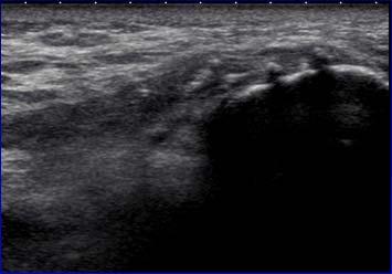
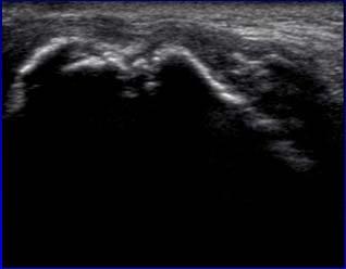
1 Ultrasound of the Hip: Anatomy, Pathology, and Procedures Jon A. Jacobson, M.D. Professor of Radiology Director, Division of Musculoskeletal Radiology University of Michigan Outline Hip Joint Native hip Arthroplasty Greater Trochanter Tendon abnormalities Bursal pathology Snapping Hip Hip: anterior recess Anterior and posterior layers Fibrous tissue + minute layer of synovium Hyperechoic Each 2-4 mm thick Radiology 1999; 210:499 Hip: anterior recess Anterior Posterior Femur Hip Joint Hip Effusion: A H Separation of anterior and posterior layers 1 Capsule distention at femoral neck > 7 mm or difference of 1 mm from opposite side 2 Extension & abduction improves visualization 3 Do not internally rotate hip: capsule thickens Sagittal-oblique 1 Radiology 1999; 210:449 2 Scand J Rheumatology 1989; 18:113 3 Acta Radiologica 1997; 38:867 1


2 Hip Joint: septic effusion FH * * Long Axis * * Hip Effusion: misconception It is incorrect to assume that joint fluid may not be seen anterior due to gravity Native hip: joint fluid distributes around femoral neck In no cases was fluid only seen posterior Exception: after hip surgery Moss et al. Radiology 1998; 208:43 Hip Effusion: Cannot predict infection by ultrasound Negative power color Doppler does not exclude infection* Guided aspiration * AJR 1998; 206:731 * * Joint injection Anterior recess In plane Transducer: Parallel to femoral neck Consider curvilinear Needle: distal to proximal 97% accuracy 1 1 Smith J. J Ultrasound Med 2009; 28:329 F Joint Injection Femoral neck target Preferred over aiming for femoral head Allows higher injection volumes Less extra-articular contrast From Kantarci F et al. Skeletal Radiol 2013; 42:37. Joint injection Transducer: in plane Lateral to medial Horizontal and parallel to sound beam Courtesy of Mark Cresswell, Vancouver F H N N 2


3 Pigmented Villonodular Synovitis Juvenile Rheumatoid Arthritis Erosion Hip Labrum Normal: Hyperechoic, triangular Degeneration: hypoechoic Tear: anterior Anechoic cleft Sensitivity 82%, specificity 60%, accuracy 80%* Chondrocalcinosis Acetab Labral Tear Femoral Femoroacetabular Impingement Pincer-type: deep acetabulum Cam-type Broad irregular femoral neck Possible cortical irregularity at US Associated with anterior labrum tear Consider dynamic evaluation Detachment *Jin W et al. J Ultrasound Med 2012; 31:439 Radiology 2005; 236:588 Labral Tear and Paralabral Cyst Associated with labral tear Full-thickness or detachment Anechoic to hypoechoic Multilocular Hip Arthroplasty: Prosthesis identifiable May use sonography to guide hip aspiration Most useful: non-communicating abscess, bursitis, incision infection Courtesy of D. Fessell, Ann Arbor, MI 3


4 Total Hip Arthroplasty: Metal components demonstrate posterior reverberation Artifact occurs deep to prosthesis away from fluid collection (unlike MRI, CT) Acet H Femur Hip Arthroplasty: Ultrasound cannot differentiate small effusion from post-op change 1 Suspect infection: Pseudocapsule > 3.2 mm: suspect infection 2 Extra-articular fluid collection Not visualized with arthrography if noncommunication A > 3.2 mm 1 Weybright PN et al. AJR 2003; 181:215 2 AJR 1994; 163:381 Hip Arthroplasty: infection Hip Arthroplasty: infection Superior Inferior Femur Sagittal Native Femur Coronal Radiograph Teaching Point: Always screen soft tissues about an arthroplasty prior to fluoroscopic joint aspiration Metal-on-Metal Arthroplasty: pseudotumor Troch Cup Cup Iliopsoas Bursa Hip joint communication in 10% Increased with hip joint pathology After joint replacement May extend cephalad into abdomen May be mistaken for psoas abscess Look for hip joint communication Anterior Lateral Radiology 1995; 197:853 4


 Gluteus minimus (blue)")


5 Iliopsoas Bursal Fluid Iliopsoas Bursa IP Oblique-axial plane: Superior to femoral head Lateral to medial Inject between tendon and ilium 1 Pain relief = successful iliopsoas surgical release 2 Femoral Axial T1w post-gadolinium 1 Dauffenbach J et al. J Ultrasound Med 2014; 33:405 2 Blankenbaker DG et al. Skeletal Radiol 2006; 35: 565 Ilium I Outline Greater Trochanter: gluteal tendons Anterior Lateral Posterior Hip Joint Native hip Arthroplasty Greater Trochanter Tendon abnormalities Bursal pathology Snapping Hip Gluteus medius (red) Gluteus minimus (blue) Greater Trochanter Greater Trochanter Subgluteus Medius Bursa Trochanteric Bursa TFL Gluteus Medius Gluteus Minimus Subgluteus Minimus Bursa Glut Max PF : anterior facet : lateral facet PF: posterior facet FACETS: = anterior; = lateral; SPF = superoposterior; PF = posterior Pfirrmann et al. Radiology 2001; 221:469 5


 Associated with tendon tear")
6 Gluteus Minimus and Medius: Long Axis Gluteus Minimus: Long Axis Gluteus Medius Gluteus Minimus Gmed Gmed PF Anterior Facet Gluteus Medius: Long Axis Iliotibial Tract Lateral Facet Trochanteric Pain Syndrome: Most commonly caused by gluteus minimus and medius tendon abnormalities 1 Trochanteric bursitis: uncommon 20% of symptomatic patients 2 Not actually inflamed 3 Not associated with pain 4 1 Kong A et al. Eur Rad 2007; 17: Long SS et al. AJR 2013; 201: Sylva F et al. Clin Rheumatol 2008; 14:82 4 Blankenbaker DG et al. Skeletal Radiol 2008; 37:903 Tendinosis: Gluteus Minimus Gluteal Tendon Pathology: Tendinosis: hypoechoic, no defects Partial tear: anechoic clefts Complete tear: discontinuous tendon >2 mm cortical irregularity (depth) Associated with tendon tear Positive predictive value = 90% (xray)* *Steinert et al. Radiology 2010; 257:754 6


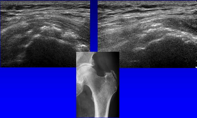
7 Tear: Gluteus Minimus Tendinosis: Gluteus Medius SPF Tear: Gluteus Medius Tear: Gluteus Medius after THA SPF >2 mm cortical irregularity depth (x-ray) = 90% positive predictive value for gluteus tendon tear Steinert et al. Radiology 2010; 257:754 Post-operative: Gluteus Medius Calcific Tendinosis: Gluteus Medius SPF Long Axis Short Axis 7
, hamstring (8),")


8 Fenestration: pelvis 22 tendons in 21 patients Gluteus medius (11), hamstring (8), gluteus minimus (2), tensor fascia lata (1) Marked or some improvement: 82% Greater Trochanter Gluteus Medius Needle Jacobson JA et al. J Ultrasound Med 2015; 34:2029 Normal PRP and Tendon Injection Trochanteric Bursal Fluid + Glut Min Tear Gluteal Tendons: greater trochanter Randomized controlled: 30 patients PRP versus fenestration alone Significant improvement at weeks 1 and 2 Approximately 80% had long term improvement: up to 1 year follow-up No difference between treatment groups Glut Max PF Jacobson JA et al. J Ultrasound Med 2016; 35:2413 Axial Trochanteric Bursitis Trochanteric Bursa: infection + gas T1w Greater Trochanter 8


9 Trochanteric Region Bursae Outline Trochanteric: deep to gluteus maximus Subgluteus medius Subgluteus minimus Axial or coronal plane PF Hip Joint Native hip Arthroplasty Greater Trochanter Tendon abnormalities Bursal pathology Snapping Hip Iliopsoas Complex A Snapping Hip Syndrome AIIS Painful snap with hip motion Intraarticular Extraarticular: Anterior: iliopsoas tendon Lateral: iliotibial tract or gluteus maximus A B Ilium Red: psoas major Orange: medial iliacus fibers Purple: lateral iliacus fibers Femoral Pubis Short Axis From: Guillin R. et al. Eur Rad 2009; 19:995 Snapping Hip Syndrome: iliopsoas Snapping Hip Syndrome: iliopsoas Image long axis to inguinal ligament superior to femoral head Extension of flexed abducted and externally rotated hip Abrupt movement of iliopsoas as iliacus muscle interposed between tendon and bone moves Deslandes et al. AJR 2008; 190: Deslandes et al. AJR 2008; 190:576 9


10 Snapping Hip Syndrome: iliopsoas Snapping Hip: lateral Transverse over greater trochanter Hip external rotation / flexion Abrupt motion of iliotibial tract or gluteus maximus over greater trochanter Gluteus Maximus Snapping Gluteus Maximus / Iliotibial Band Gluteus Medius TFL Gluteus Maximus Iliotibial Band Gmin Snapping Hip Syndrome: iliotibial tract Iliotibial Band Gmax Gmed Gmin Take-home points: Hip: Native: focus on anterior recess Arthroplasty: pseudotumor, iliopsoas bursa Greater trochanter: Bursitis uncommon Tendinosis and tendon tear Snapping Hip: Iliopsoas and iliotibial tract/gluteus maximus See for syllabus and other educational material 10
Greater Trochanter: Anatomy and Pathology
 Greater Trochanter: Anatomy and Pathology Jon A. Jacobson, M.D. Professor of Radiology Director, Division of Musculoskeletal Radiology University of Michigan Disclosures: Consultant: Bioclinica Book Royalties:
Snapping Hip and Impingement
 Snapping Hip and Impingement Jon A. Jacobson, M.D. Professor of Radiology Director, Division of Musculoskeletal Radiology University of Michigan Disclosures: Consultant: Bioclinica Advisory Board: GE,
MRI of the Hip. Jon A. Jacobson, M.D. Professor of Radiology Director, Division of Musculoskeletal Radiology University of Michigan
 MRI of the Hip Jon A. Jacobson, M.D. Professor of Radiology Director, Division of Musculoskeletal Radiology University of Michigan Take Home Points Joint effusion: does not collect dependently Imaging
Complex Fractures and Hip Dislocations
 IMAGING OF HIP PAIN Patients may present with acute (< 2 weeks) or chronic hip pain. Acute pain may be related or not related to an acute traumatic event such as fall or trauma from a motor vehicle accident.
MR Imaging in Athlete s Hip/Pelvis
 MR Imaging in Athlete s Hip/Pelvis Tara Lawrimore, MD FRCPC Department of Radiology Musculoskeletal Division Massachusetts General Hospital Harvard Medical School No disclosures MR and Hip Pain in the
Common Applications for Sonography and Guided Intervention: Shoulder
 Common Applications for Sonography and Guided Intervention: Shoulder Jon A. Jacobson, M.D. Professor of Radiology Director, Division of Musculoskeletal Radiology University of Michigan Disclosures: Consultant:
Rotator Cuff and Biceps Pathology
 Rotator Cuff and Biceps Pathology Jon A. Jacobson, M.D. Professor of Radiology Director, Division of Musculoskeletal Radiology University of Michigan Disclosures: Consultant: Bioclinica Advisory Board:
The Hip (Iliofemoral) Joint. Presented by: Rob, Rachel, Alina and Lisa
 Joint. Presented by: Rob, Rachel, Alina and Lisa") The Hip (Iliofemoral) Joint Presented by: Rob, Rachel, Alina and Lisa Surface Anatomy: Posterior Surface Anatomy: Anterior Bones: Os Coxae Consists of 3 Portions: Ilium Ischium Pubis Bones: Pubis Portion
The Hip (Iliofemoral) Joint Presented by: Rob, Rachel, Alina and Lisa Surface Anatomy: Posterior Surface Anatomy: Anterior Bones: Os Coxae Consists of 3 Portions: Ilium Ischium Pubis Bones: Pubis Portion
Ultrasound Evaluation of Masses
 Ultrasound Evaluation of Masses Jon A. Jacobson, M.D. Professor of Radiology Director, Division of Musculoskeletal Radiology University of Michigan Disclosures: Consultant: Bioclinica Advisory Panel: GE,
Viviane Khoury, MD. Assistant Professor Department of Radiology University of Pennsylvania
 U Penn Diagnostic Imaging: On the Cape Chatham, MA July 11-15, 2016 Viviane Khoury, MD Assistant Professor Department of Radiology University of Pennsylvania Hip imaging has changed in recent years: new
Ultrasound of the Knee
 Ultrasound of the Knee Jon A. Jacobson, M.D. Professor of Radiology Director, Division of Musculoskeletal Radiology University of Michigan Disclosures: Consultant: Bioclinica Book Royalties: Elsevier Advisory
CAN SOFT TISSUES STRUCTURES DIFFERENTIATE BETWEEN DYSPLASIA AND CAM-FAI OF THE HIP?
 CAN SOFT TISSUES STRUCTURES DIFFERENTIATE BETWEEN DYSPLASIA AND CAM-FAI OF THE HIP? A Le Bouthillier, KS Rakhra 1, PE Beaulé 2, RCB Foster 1 1 Department of Medical Imaging 2 Division of Orthopaedic Surgery
Knee, Ankle, and Foot: Normal and Abnormal Features with MRI and Ultrasound Correlation. Disclosures. Outline. Joint Effusion. Suprapatellar recess
 Knee, Ankle, and Foot: Normal and Abnormal Features with MRI and Ultrasound Correlation Jon A. Jacobson, M.D. Professor of Radiology Director, Division of Musculoskeletal Radiology University of Michigan
Tendon Fenestration. Disclosures. Outline: questions. Introduction: Peritendon Steroid Injections. Jon A. Jacobson, MD. Patellar Tendon: tendinosis
 Tendon Fenestration Jon A. Jacobson, MD Professor of Radiology Director, Division of Musculoskeletal Radiology University of Michigan Disclosures Consultant: Bioclinica Advisory Board: GE, Philips Book
CLINICS IN SPORTS MEDICINE
 Clin Sports Med 25 (2006) 365 369 CLINICS IN SPORTS MEDICINE A Acetabular labrum, tears of, hip arthroscopy in, 264 Acetabular rim, trimming of, and labral repair, new method for, 293 297 Acetabulum, femoral
Musculoskeletal Ultrasound Fundamentals
 Fundamentals Benjamin D. Levine, M.D. Associate Professor of Radiology Musculoskeletal Imaging Dept. of Radiological Sciences UCLA Health System I. Image Optimization II. Image Interpretation Artifacts
Main Menu. Joint and Pelvic Girdle click here. The Power is in Your Hands
 1 Hip Joint and Pelvic Girdle click here Main Menu K.6 http://www.handsonlineeducation.com/classes//k6entry.htm[3/23/18, 2:01:12 PM] Hip Joint (acetabular femoral) Relatively stable due to : Bony architecture
Lateral Elbow Pathology
 Lateral Elbow Pathology Jon A. Jacobson, M.D. Professor of adiology Director, Division of Musculoskeletal adiology University of Michigan Disclosures: Consultant: Bioclinica Advisory Board: GE, Philips
Hip joint and pelvic girdle. Lower Extremity. Pelvic Girdle 6/5/2017
 Hip joint and pelvic girdle Lower Extremity The relationship between the pelvic girdle and hip is similar to that between the shoulder girdle and shoulder joint. The lower limbs are attached to the axial
The Young Adult Hip: FAI. Jason Snibbe, M.D. Snibbe Orthopedics Team Physician, University of Southern California
 The Young Adult Hip: FAI Jason Snibbe, M.D. Snibbe Orthopedics Team Physician, University of Southern California Introduction Femoroacetabular Impingment(FAI) Presentation and Exam Imaging Surgical Management
Bony Anatomy. Femur. Femoral Head Femoral Neck Greater Trochanter Lesser Trochanter Intertrochanteric Crest Intertrochanteric Line Gluteal Tuberosity
 Hip Anatomy Bony Anatomy Femur Femoral Head Femoral Neck Greater Trochanter Lesser Trochanter Intertrochanteric Crest Intertrochanteric Line Gluteal Tuberosity Bony Anatomy Pelvic Girdle Acetabulum 3 bones
Lectures of Human Anatomy
 Lectures of Human Anatomy Lower Limb Gluteal Region and Hip Joint By DR. ABDEL-MONEM AWAD HEGAZY M.B. with honor 1983, Dipl."Gynecology and Obstetrics "1989, Master "Anatomy and Embryology" 1994, M.D.
Joints of the lower limb
 Joints of the lower limb 1-Type: Hip joint Synovial ball-and-socket joint 2-Articular surfaces: a- head of femur b- lunate surface of acetabulum Which is deepened by the fibrocartilaginous labrum acetabulare
Non-arthritic anterior hip pain in the younger patient: examination and intervention strategies
 Non-arthritic anterior hip pain in the younger patient: examination and intervention strategies Melodie Kondratek, PT, DScPT, OMPT Bryan Kuhlman, PT, DPT, OMPT Oakland University Orthopedic Spine and Sports
Mr Simon Jennings BSc, MB BS, FRCS, Dip Sports Med FRCS (Trauma & Orthopaedics)
") Mr Simon Jennings BSc, MB BS, FRCS, Dip Sports Med FRCS (Trauma & Orthopaedics) Consultant Orthopaedic Surgeon Northwick Park Hospital 107 Harley Street RSM 16 th September 2010 Orthopaedic Surgeon Knee
Mr Simon Jennings BSc, MB BS, FRCS, Dip Sports Med FRCS (Trauma & Orthopaedics) Consultant Orthopaedic Surgeon Northwick Park Hospital 107 Harley Street RSM 16 th September 2010 Orthopaedic Surgeon Knee
FUNCTIONAL ANATOMY AND EXAM OF THE HIP, GROIN AND THIGH
 FUNCTIONAL ANATOMY AND EXAM OF THE HIP, GROIN AND THIGH Peter G Gerbino, MD, FACSM Orthopedic Surgeon Monterey Joint Replacement and Sports Medicine Monterey, CA TPC, San Diego, 2017 The lecturer has no
The psoas minor is medial to the psoas major. The iliacus is a fan-shaped muscle that when contracted helps bring the swinging leg forward in walking
 1 p.177 2 3 The psoas minor is medial to the psoas major. The iliacus is a fan-shaped muscle that when contracted helps bring the swinging leg forward in walking and running. The iliopsoas and adductor
Hip Injuries & Arthroscopy in Athletes
 Hip Injuries & Arthroscopy in Athletes John P Salvo, MD Sports Medicine Rothman Institute Philadelphia, PA EATA Annual Meeting January, 2011 Hip Injuries & Arthroscopy in Anatomy History Physical Exam
ANTERIOR TOTAL HIP ARTHOPLASTY
 ANTERIOR TOTAL HIP ARTHOPLASTY And Other Approaches Bill Rhodes PTA 236 Total Hip Arthoplasty (THA) Background THA, also know as Total Hip Replacement Regarded as the most valued development in orthopedics
STAIRS. What s Hip: Top 5 Hip Problems in Primary Care. I have no relevant disclosures. Top 5 (or 6) Pathologies. Big 3- Questions to Ask
 Pathologies. Big 3- Questions to Ask") I have no relevant disclosures. What s Hip: Top 5 Hip Problems in Primary Care Alan Zhang MD Assistant Professor Sports Medicine and Hip Arthroscopy UCSF Department of Orthopaedic Surgery December, 2015
I have no relevant disclosures. What s Hip: Top 5 Hip Problems in Primary Care Alan Zhang MD Assistant Professor Sports Medicine and Hip Arthroscopy UCSF Department of Orthopaedic Surgery December, 2015
Greater Trochanteric Pain Syndrome
 43 Andrea S. Klauser, MD 1 Carlo Martinoli, MD 2 Alberto Tagliafico, MD 3 Rosa Bellmann-Weiler, MD 4 Gudrun M. Feuchtner, MD, PhD 1 Marius Wick, MD 1 Werner R. Jaschke, MD, PhD 1 1 Department of Diagnostic
Lesson 24. A & P Hip
 Lesson 24 A & P Hip 1 Aims of the Session This session will allow candidates to have an understanding of the bony prominences and soft tissues of the hip 2 Learning Outcomes By the end of the lesson the
What s Hip: Common Hip Problems and Kids and Adults
 What s Hip: Common Hip Problems and Kids and Adults Alan Zhang MD Assistant Professor Sports Medicine and Hip Arthroscopy UCSF Department of Orthopaedic Surgery I have no relevant disclosures. 2 1 Most
Ultrasound of the Shoulder
 Ultrasound of the Shoulder Patrick Battaglia, DC, DACBR Logan University, Department of Radiology Outline Review ultrasound appearance of NMSK tissues Present indications for ultrasound of the shoulder.
The Hip Joint. Shenequia Howard David Rivera
 The Hip Joint Shenequia Howard David Rivera Topics Of Discussion Movement Bony Anatomy Ligamentous Anatomy Muscular Anatomy Origin/Insertion/Action/Innervation Common Injuries MOVEMENT Flexion Extension
Ultrasound of Shoulder Pathology and Intervention 서울대학교병원재활의학과 김기원
 Ultrasound of Shoulder Pathology and Intervention 서울대학교병원재활의학과 김기원 Ultrasound for Shoulder Disorder Advantage Dynamic evaluation Immediate clinical correlation + Intervention Weakness Diagnostic accuracy?
SURGICAL AND APPLIED ANATOMY
 Página 1 de 6 Copyright 2001 Lippincott Williams & Wilkins Bucholz, Robert W., Heckman, James D. Rockwood & Green's Fractures in Adults, 5th Edition SURGICAL AND APPLIED ANATOMY Part of "37 - HIP DISLOCATIONS
First practical session. Bones of the gluteal region
 First practical session 2017 Bones of the gluteal region The Hip bone The hip bone is made of: 1 The ilium: superior in position 2 The ischium:postero-inferior in position 3 The pubis: antero-inferior
Human Anatomy Biology 255
 Human Anatomy Biology 255 Exam #4 Please place your name and I.D. number on the back of the last page of this exam. You must answer all questions on this exam. Because statistics demonstrate that, on average,
MRI of the Hips and Pelvis
 MRI of the Hips and Pelvis Hips and Pelvis Protocols Vascular abnormalities Fractures Soft tissues Labrum and FAI Hips and Pelvis Protocols Vascular abnormalities Fractures Soft tissues Labrum and FAI
Ultrasonographic Examination of the Adult Hip
 Journal of Medical Ultrasound (2012) 20, 201e209 Available online at www.sciencedirect.com journal homepage: www.jmu-online.com REVIEW ARTICLE Ultrasonographic Examination of the Adult Hip Yun-Tai Lin
Specialists in Joint Replacement, Spinal Surgery, Orthopaedics and Sport Injuries. The Hip.
 Specialists in Joint Replacement, Spinal Surgery, Orthopaedics and Sport Injuries The Hip INTRODUCTION THE HIP The hip is a ball-and-socket joint. The socket is formed by the acetabulum, which is part
THE HIP. Cooler than cool, the pinnacle of what is "it". Beyond all trends and conventional coolness.
 THE HIP Cooler than cool, the pinnacle of what is "it". Beyond all trends and conventional coolness. Objectives Hip anatomy Causes of hip pain Hip exam Anatomy Bones Ilium Anterior Superior Iliac Spine
Evaluation of the Hip
 Evaluation of the Hip Adam Lewno, DO PCSM Fellow, University of Michigan Primary Care Sports Update 2017 Disclosures Financial: None Images: I would like to acknowledge the work of the original owners
Greater Trochanteric Pain Syndrome
 ORIGINAL RESEARCH Greater Trochanteric Pain Syndrome Percutaneous Tendon Fenestration Versus Platelet-Rich Plasma Injection for Treatment of Gluteal Tendinosis Jon A. Jacobson, MD, Corrie M. Yablon, MD,
FAI syndrome with or without labral tear.
 Case This 16-year-old female, soccer athlete was treated for pain in the right groin previously. Now has acute onset of pain in the left hip. The pain was in the groin that was worse with activities. Diagnosis
CONSERVATIVE MANAGEMENT OF FEMOROACETABULAR IMPINGEMENT
 SPORTS REHABILITATION CONSERVATIVE MANAGEMENT OF FEMOROACETABULAR IMPINGEMENT A case study and rationale for treatment Written by Joanne Kemp and Kay Crossley, Australia BACKGROUND The hip joint and FAI
Hip Cases from Clinic: Refining your history and physical
 Hip Cases from Clinic: Refining your history and physical Alan Zhang MD Assistant Professor Sports Medicine and Hip Arthroscopy UCSF Department of Orthopaedic Surgery 11/20/2017 Case #1 Healthy 21 M College
Review causes of hip and groin pain in athlete Discuss indications for hip arthroscopy Review, if any, history & physical findings of a patient who
 Review causes of hip and groin pain in athlete Discuss indications for hip arthroscopy Review, if any, history & physical findings of a patient who may benefit from hip arthroscopy Review portal placement
Figure 1 - Hip and Pelvis
 Hip Figure 1 - Hip and Pelvis The terms hip and pelvis are frequently used interchangeably, but strictly speaking, the pelvis is a girdle of bones and the hip is a joint. The pelvis consists of The sacrum
Applied anatomy of the hip and buttock
 CHAPTER CONTENTS The hip joint e9 Capsule and ligaments e9 s e0 Flexor muscles................... e0 Extensor muscles.................. e Abductor muscles.................. e Adductor muscles..................
Young Adult Hip problems. Aresh Hashemi-Nejad FRCS(Orth)
") Young Adult Hip problems Aresh Hashemi-Nejad FRCS(Orth) RNOH founded 1837 by William Little 14 year old presenting with limp Knee pain on and off 4 months Limps Aresh Hashemi-Nejad FRCS(Orth) The Royal
Young Adult Hip problems Aresh Hashemi-Nejad FRCS(Orth) RNOH founded 1837 by William Little 14 year old presenting with limp Knee pain on and off 4 months Limps Aresh Hashemi-Nejad FRCS(Orth) The Royal
HIP_CASE 2_OA. Hip Forces. Function of the Hip. Property of VOMPTI, LLC. For Use of Participants Only. No Use or Reproduction Without Consent 1
 HIP_CASE 2_OA Orthopaedic Manual Physical Therapy Series Charlottesville 2017-2018 Eric Magrum DPT, OCS, FAAOMPT 62 yo female AM stiffness Hip pain diffuse, variable ant>lateral>post Gradual onset Tennis
Overview. Overview. Introduction. Introduction Anatomy History Examination Common Disorders. Introduction Anatomy History Examination Common Disorders
 Common Hip Disorders in Figure Skaters 14 th Annual Meeting of Sports Medicine and Science in Figure Skating January 25, 2009 8:15-8:45am Robert J. Dimeff, MD Medical Director of Sports Medicine Overview
Current Concepts in Magnetic Resonance Imaging of the Hip. Ray Hong
 Current Concepts in Magnetic Resonance Imaging of the Hip Ray Hong Overview Technique Basic Anatomy/Normal Variants Osseous Soft Tissue Pathology FAI RC/Hamstring Tears Ligamentum Teres Adhesive Capsulitis
Human Anatomy Biology 351
 Human Anatomy Biology 351 Lower Limb Please place your name on the back of the last page of this exam. You must answer all questions on this exam. Because statistics demonstrate that, on average, between
Gluteal region DR. GITANJALI KHORWAL
 Gluteal region DR. GITANJALI KHORWAL Gluteal region The transitional area between the trunk and the lower extremity. The gluteal region includes the rounded, posterior buttocks and the laterally placed
The Evaluation of Hip pain in the Athlete
 The Evaluation of Hip pain in the Athlete DREW ROGERS,MD The Evaluation of Hip pain in the Athlete Andrew Rogers, MD (Drew) Orthopedic Care Physician Network Chief of Orthopedics Morton Hospital Team Physician
Human Anatomy Biology 351
 Human Anatomy Biology 351 Lower Limb Please place your name on the back of the last page of this exam. You must answer all questions on this exam. Because statistics demonstrate that, on average, between
Beyond the Bump: The Spectrum of Extra-articular Pathology in Hip MRI for Clinical Femoroacetabular Impingement
 Beyond the Bump: The Spectrum of Extra-articular Pathology in Hip MRI for Clinical Femoroacetabular Impingement Poster No.: C-2239 Congress: ECR 2012 Type: Authors: Keywords: DOI: Educational Exhibit K.
CHAPTER 8: THE BIOMECHANICS OF THE HUMAN LOWER EXTREMITY
 CHAPTER 8: THE BIOMECHANICS OF THE HUMAN LOWER EXTREMITY _ 1. The hip joint is the articulation between the and the. A. femur, acetabulum B. femur, spine C. femur, tibia _ 2. Which of the following is
Muscles of Lesson Five. Muscular Nomenclature and Kinesiology - Two. Muscles of Lesson Five, cont. Chapter 16
 Chapter 16 Muscular Nomenclature and Kinesiology - Two Lessons 5-6 Muscles of Lesson Five Iliopsoas (psoas major, iliacus) Hip outward rotators (piriformis, gemellus superior, gemellus inferior, obturator
Index. Note: Page numbers of article titles are in boldface type.
 Magn Reson Imaging Clin N Am 12 (2004) 185 189 Index Note: Page numbers of article titles are in boldface type. A Acromioclavicular joint, MR imaging findings concerning, 161 Acromion, types of, 77 79
Baraa Ayed حسام أبو عوض. Ahmad Salman. 1 P a g e
 4 Baraa Ayed حسام أبو عوض Ahmad Salman 1 P a g e Today we are going to cover these concepts: Iliotibial tract Anterior compartment of the thigh and the hip Medial compartment of the thigh Femoral triangle
The University Of Jordan Faculty Of Medicine THE LOWER LIMB. Dr.Ahmed Salman Assistant Prof. of Anatomy. The University Of Jordan
 The University Of Jordan Faculty Of Medicine THE LOWER LIMB Dr.Ahmed Salman Assistant Prof. of Anatomy. The University Of Jordan Gluteal Region Cutaneous nerve supply of (Gluteal region) 1. Lateral cutaneous
Musculoskeletal Ultrasound. Technical Guidelines SHOULDER
 Musculoskeletal Ultrasound Technical Guidelines SHOULDER 1 Although patient s positioning for shoulder US varies widely across different Countries and Institutions reflecting multifaceted opinions and
SURGICAL EXPOSURES SURGERY OF THE HIP
 1 of 24 11/19/03 1:11 PM SURGICAL EXPOSURES SURGERY OF THE HIP by R. CALANDRUCCIO In: Atlas of Orthopaedic Surgery Volume 3 Lower Extremity; Editors: Laurin, CA, Riley Jr. LH, Roy-Camille R Reprinted with
A Guide for Patients with Hip and Groin Pain. By - Rob Lawton & Ajay Malviya. Overview
 A Guide for Patients with Hip and Groin Pain By - Rob Lawton & Ajay Malviya Overview - Introduction - Hip Anatomy - Is the pain coming from the hip joint? - Intra-articular causes of hip pain o Impingement
lesser trochanter of femur lesser trochanter of femur iliotibial tract (connective tissue) medial surface of proximal tibia
 medial surface of proximal tibia") LOWER LIMB MUSCLES OF THE APPENDICULAR SKELETON The muscles that act on the lower limb fall into three groups: those that move the thigh, those that move the lower leg, and those that move the ankle, foot,
LOWER LIMB MUSCLES OF THE APPENDICULAR SKELETON The muscles that act on the lower limb fall into three groups: those that move the thigh, those that move the lower leg, and those that move the ankle, foot,
MRI of the Shoulder What to look for and how to find it? Dr. Eric Handley Musculoskeletal Radiologist Cherry Creek Imaging
 MRI of the Shoulder What to look for and how to find it? Dr. Eric Handley Musculoskeletal Radiologist Cherry Creek Imaging MRI of the Shoulder Benefits of Ultrasound: * Dynamic * Interactive real time
When the patient presents at the GP surgery with Hip pain. Investigations, Input & Referral
 When the patient presents at the GP surgery with Hip pain Investigations, Input & Referral GP with an interest in Pain Management How common is hip pain? Around 450 patients per 100,000 population will
ELENI ANDIPA General Hospital of Athens G. Gennimatas
 ELENI ANDIPA General Hospital of Athens G. Gennimatas Technological advances over the last years have caused a dramatic improvement in ultrasound quality and resolution An established imaging modality
APPROPRIATE USE GUIDELINES
 APPROPRIATE USE GUIDELINES Appropriateness of Advanced Imaging Procedures (MRI, CT, Bone Scan/PET) in Patients with Shoulder Pain CDI QUALITY INSTITUTE: PROVIDER LED ENTITY (PLE) Compiled by Rob Liddell,
Ultrasound Guided Therapeutic Injections in the Treatment of Shoulder Pain: A Multimedia Review
 Ultrasound Guided Therapeutic Injections in the Treatment of Shoulder Pain: A Multimedia Review Poster No.: P-0127 Congress: ESSR 2015 Type: Educational Poster Authors: A. Karsandas, J. Tuckett, R. Sinha,
Evaluation of Hip Pain in Adults. Jerry Ahluwalia, M.D. November 13, 2015
 Evaluation of Hip Pain in Adults Jerry Ahluwalia, M.D. November 13, 2015 Objectives Develop a better understanding of the differential diagnosis of hip pain in active young adults Appreciate key points
To classify the joints relative to structure & shape
 To classify the joints relative to structure & shape To describe the anatomy of the hip joint To describe the ankle joint To memorize their blood & nerve supply JOINTS: Joints are sites where skeletal
Labral Tears/FAI. Andrew Parker, MD
 Labral Tears/FAI Andrew Parker, MD Athletic Hip Injuries Incidence of hip injuries has increased dramatically over the last decade In part due to better recognition with improved imaging and arthroscopy,
Femoroacetabular Impingement in the Throwing Athlete. Michael Banffy, MD Sports Medicine, Hip Preservation Kerlan Jobe Institute
 Femoroacetabular Impingement in the Throwing Athlete Michael Banffy, MD Sports Medicine, Hip Preservation Kerlan Jobe Institute Disclosures None Baseball Hip Injuries - Background Abdominal/groin injuries
GET HIP! CAPA 2015 Annual Conference WHAT IS HIP? HIP JOINT. Bradford H. Stiles, M.D., FAAFP
 GET HIP! Bradford H. Stiles, M.D., FAAFP WHAT IS HIP? HIP JOINT Synovial ball-and-socket joint Articulation between femoral head and acetabulum Acetabulum formed by the confluence of pelvis bones (ilium,
MRI SHOULDER WHAT TO SEE
 MRI SHOULDER WHAT TO SEE DR SHEKHAR SRIVASTAV Sr. Consultant- Knee & Shoulder Arthroscopy Sant Parmanand Hospital Normal Anatomy Normal Shoulder MRI Coronal Oblique Sagital Oblique Axial Cuts Normal Coronal
Ultrasound assessment of most frequent shoulder disorders
 Ultrasound assessment of most frequent shoulder disorders Poster No.: C-2026 Congress: ECR 2014 Type: Educational Exhibit Authors: S. P. Ivanoski; Ohrid/MK Keywords: Trauma, Athletic injuries, Arthritides,
Hip pain rating after preforming MRI with gadolinium arthrography and intra-articular lidocaine
 Hip pain rating after preforming MRI with gadolinium arthrography and intra-articular lidocaine Poster No.: C-1352 Congress: ECR 2014 Type: Scientific Exhibit Authors: J. García Yavar, J. Cabezudo, S.
The Shoulder. Systematically scanning the shoulder provides extremely useful diagnostic information. The Shoulder
 1 ! The most ACCESSIBLE to sonographic exam! The most MOBILE and VULNERABLE extremity AND Systematically scanning the shoulder provides extremely useful diagnostic information! The Goal for this section
Muscles of the lower extremities. Dr. Nabil khouri MD, MSc, Ph.D
 Muscles of the lower extremities Dr. Nabil khouri MD, MSc, Ph.D Posterior leg Popliteal fossa Boundaries Biceps femoris (superior-lateral) Semitendinosis and semimembranosis (superior-medial) Gastrocnemius
This presentation is the intellectual property of the author. Contact them for permission to reprint and/or distribute.
 MRI of the Knee Jennifer Swart, M.D. Musculoskeletal Radiology South Texas Radiology Group Outline Coils, Patient Positioning Acquisition Parameters, Planes and Pulse Sequences Knee Arthrography Normal
Hip Impingement and Arthritis: Preservation vs. Total Hip Arthroplasty. Faculty Disclosures. Objectives 11/17/2017
 Hip Impingement and Arthritis: Preservation vs. Total Hip Arthroplasty Jonathan R. Schiller, MD Assistant Professor of Orthopedics Warren Alpert Medical School of Brown University Director, Adolescent
Surgical Anatomy of the Hip. Joseph H. Dimon
 Surgical Anatomy of the Hip Joseph H. Dimon The hip joint is a deep joint surrounded by large and powerful muscles necessary for its proper function. Essential neurovascular structures lie in front and
The thigh. Prof. Oluwadiya KS
 The thigh Prof. Oluwadiya KS www.oluwadiya.com The Thigh: Boundaries The thigh is the region of the lower limb that is approximately between the hip and knee joints Anteriorly, it is separated from the
Pigmented Villonodular Synovitis PVNS
 February 2002 Pigmented Villonodular Synovitis PVNS Amy Gillis, Harvard Medical School Year III 47 year old female Our Patient Right hip pain since age 20 No history of trauma Diagnosed with DJD of R hip
This presentation is the intellectual property of the author. Contact them at for permission to reprint and/or distribute.
 MRI of the Knee Jennifer Swart, M.D. Musculoskeletal Radiology South Texas Radiology Group Financial Disclosure Dr. Jennifer Swart has no relevant financial relationships with commercial interests to disclose.
Imaging in Groin Pain What the Team Physician Needs to Know
 Imaging in Groin Pain What the Team Physician Needs to Know Üstün Aydıngöz, MD Professor of Radiology Hacettepe University School of Medicine Ankara, Turkey ustunaydingoz@yahoo.com No conflicts of interest
Urgent Cases and Foreign Bodies
 Urgent Cases and Foreign Bodies Catherine J. Brandon, MD, MS University of Michigan Ann Arbor, MI, USA Introduction: Patients added on to the schedule from the emergency department or as urgent add-on
MSK Radiology Interesting Case Presentation
 MSK Radiology Interesting Case Presentation Sitt Ching Man Jacqueline 27 th Feb 2015 Patient 1: YPY F/27 Professional badminton player C/o sudden onset of right shoulder pain after a very vigorous match
External Snapping Hip Syndrome:
 J Korean Soc Radiol 2010;62:185-190 External Snapping Hip Syndrome: Emphasis on the MR Imaging 1 Jung Eun Choi, M.D., Mi Sook Sung, M.D. 2, Ki Haeng Lee, M.D. 3, ae Young Lee, M.D., Jeong Mi Park, M.D.
Imaging features of complications following hip replacement: A pictorial assay
 Imaging features of complications following hip replacement: A pictorial assay Poster No.: C-2041 Congress: ECR 2014 Type: Educational Exhibit Authors: K. Pilania, B. Jankharia ; Mumbai, maharashtra/in,
rotation of the hip Flexion of the knee Iliac fossa of iliac Lesser trochanter Femoral nerve Flexion of the thigh at the hip shaft of tibia
 Anatomy of the lower limb Anterior & medial compartments of the thigh Dr. Hayder The fascia lata encloses the entire thigh like a sleeve/stocking. Three intramuscular fascial septa (lateral, medial, and
4/1/2016. Total Hip Arthroplasty. DAHR Procedure. Direct Anterior Hip Replacement. DAHR Procedure. DAHR Procedure
 Mercy Orthopedist Types of Approaches Total Hip Arthroplasty Mercy Has a total of 16 Orthopedist that perform all three different approaches Posterior Anterior Lateral Direct Anterior Direct Anterior Hip
Hip Anatomy. Bony. The Athletic Hip: Anatomy and Common Injuries. Kyle Wilkens MSPAS, PA-C, ATC/L October 8, 2013
 The Athletic Hip: Anatomy and Common Injuries Kyle Wilkens MSPAS, PA-C, ATC/L October 8, 2013 Hip Anatomy Bony Femur Ischium Ilium Pubis Sacrum Coccyx Soft Tissue Joints Muscles -Pubic Symphisis Labrum
The Muscular System. Chapter 10 Part D. PowerPoint Lecture Slides prepared by Karen Dunbar Kareiva Ivy Tech Community College
 Chapter 10 Part D The Muscular System Annie Leibovitz/Contact Press Images PowerPoint Lecture Slides prepared by Karen Dunbar Kareiva Ivy Tech Community College Table 10.14: Muscles Crossing the Hip and