Osteology of the Elbow and Forearm Complex
|
|
|
- Carol Holland
- 5 years ago
- Views:
Transcription
 d epends upon the")
1 Osteology of the Elbow and Forearm Complex The ability to perform m any activities of daily living (ADL) d epends upon the elbow.


2 Activities of Daily Living (ADL) Can you think of anything that you do to take care of yourself that would not require you to flex your elbow?
3 Bone Markings Epicondyle Prominence above or on a condyle Condyle(rounded knucklelike projection) Head Rounded projection beyond a narrow, necklike portion of bone Fossa Hollow or depression Spine Long, thin projection Tubercle Small, rounded projection Tuberosity Large, rounded projection Process a projecting part

4 Elbow/Forearm Joints Elbow Humeroulnar Humeroradial Forearm Proximal radioulnar Distal radioulnar

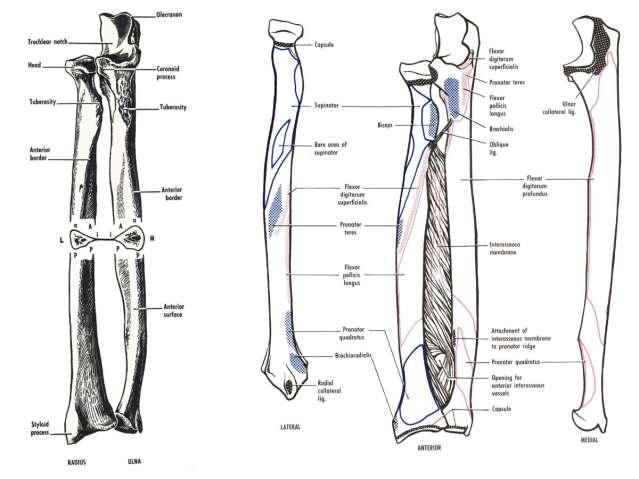
5 Osteology of the Structures
6

7
8
9
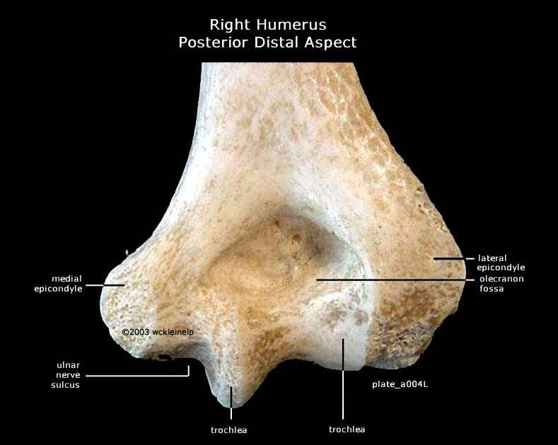
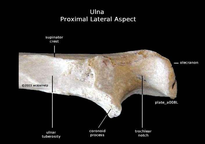
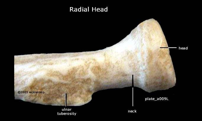
10 Osteology of the Proximal Structures of the Elbow & Forearm Ulna Radius
11 Osteology of the Distal Structures of the Forearm Ulna Radius
 and extension (0 ) Hyperextension is common (5-10")
12 The Elbow Joint 2 articulations The humeroulnar /humeroradial joints Structural stability for the joint: trochlear notch Hinge joint Allows flexion (145 ) and extension (0 ) Hyperextension is common (5-10 degrees)
13 So how much elbow ROM do you need? Usually about 100 o for most ADLs, but it occurs between 30 and 130 degrees of flexion
 With the forearm supinated")

14 Carrying Angle? (normal cubitus valgus) With the forearm supinated and elbow fully extended, the forearm projects laterally about o relative to the humerus. This is normal, but tends to be greater in females.
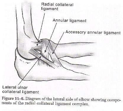
15 Supporting Structures of the Elbow Bony interlocking of humerus, ulna, and radius Coronoid process /fossa in flexion Olecranon process/fossa in extension Joint Capsule: Fairly lax, resists valgus and distraction forces Medial Collateral Ligament: Help provide stability in resisting valgus producing forces Most important soft tissue stabilizing structure Lateral Collateral Ligament: From the lateral epicondyle to the proximal forearm, resists varus Not an important stabilizing structure because varus forces are resisted mostly by bony constraints Interosseus Membrane

16 Medial Lateral
17 Interosseous Membrane

18 Arthrology of the Forearm Composed of proximal and distal radioulnar joints Acts as one uniaxial joint Supination and Pronation occur at humeroradial and in radioulnar joints

19 Osteology Note: The proximal radius rotates around the ulna which is stationary The distal radius is larger and broader than the distal ulna


20 Supination & Pronation Shoulder rotation can often be functionally substituted for each motion But not if the humerus is held tight against the thorax and the elbow is in 90 o of flexion

21 Colle s Fracture Fracture of the distal radius The weight of the body is transmitted through the hand and wrist, exceeding the strength of the radius The interosseous membrane dissipates some of the force
22 Redundancy is a fact of life/function Innervation The musculocutaneous n. Supplies most of the elbow flexors.. EXCEPT the brachioradialis and pronator teres The radial n. Supplies the elbow extensors The median n. Supplies all the pronators of the forearm
23 Myology of the Elbow Biceps Brachii Origin Insertion Long Head: Supraglenoid tubercle of the scapula Short Head: Coracoid process of the scapula Bicipital tuberosity of the radius Innervation Musculocutaneous n. Action tidbit Elbow flexion, sh flexion and forearm supination corkscrew muscle Biceps Brachii Muscle Bellies Biceps Brachii Biceps Brachii Tendons

24 Myology of the Elbow Brachialis Origin Insertion Anterior aspect of the distal humerus Coronoid process of the ulna Innervation Musculocutaneous n. Action tidbit Elbow flexion workhorse for elbow flexion


25 Myology of the Elbow Brachioradialis Origin Insertion Innervation Radial n. Action Lateral supracondylar ridge of the humerus Near the styloid process of the distal radius Elbow flexion, Pronation or supination of the forearm to the neutral position

26 Myology of the Elbow Triceps Brachii Origin Insertion Innervation Radial n. Action Long Head: infraglenoid tubercle of the scapula Lateral Head: posterior aspect of the superior humerus, lateral to the radial groove Medial Head: posterior aspect of the superior humerus, medial to the radial groove Olecranon process of the ulna Elbow extension Sh extension: Long head only

27 Myology of the Elbow Anconeus Origin Insertion Innervation Radial n. Action tidbit Posterior aspect of the laterals epicondyle of the humerus Olecranon process of the ulna Elbow extension? Believed to clear the joint space of soft tissue to permit full elbow extension. Too small to create torque for elbow extension.
28 Myology of the Elbow Supinator Origin Insertion Innervatio n Action Lateral epicondyle of the humerus and supinator crest of the ulna Lateral surface of the proximal radius Radial n. Forearm supination,
29 Myology of the Forearm Pronator Teres Origin Insertion Innervation Median n. Action Humeral head; medial epicondyle of the humerus Lateral surface of the midshaft of the radius Forearm pronation, secondary elbow flexion
30 Myology of the Forearm Pronator Quadratus Origin Insertion Innervatio n Action Anterior surface of the distal ulna Anterios surface of the distal radius Median n. Forearm pronation

31 Superficial/Deep Biceps brachii Brachialis Coracobrachialis Pronator Teres

32 Superficial/Deep Biceps Brachii Brachialis Brachioradialis Pronator Teres Pronator Quadratus
Osteology of the Elbow and Forearm Complex. The ability to perform many activities of daily living (ADL) depends upon the elbow.
 depends upon the elbow.") Osteology of the Elbow and Forearm Complex The ability to perform many activities of daily living (ADL) depends upon the elbow. Activities of Daily Living (ADL) Can you think of anything that you do to
Osteology of the Elbow and Forearm Complex The ability to perform many activities of daily living (ADL) depends upon the elbow. Activities of Daily Living (ADL) Can you think of anything that you do to
Elbow & Forearm H O W V I T A L I S T H E E L B O W T O O U R D A I L Y L I V E S?
 Elbow & Forearm H O W V I T A L I S T H E E L B O W T O O U R D A I L Y L I V E S? Clarification of Terms The elbow includes: 3 bones (humerus, radius, and ulna) 2 joints (humeroulnar and humeroradial)
The Elbow and the cubital fossa. Prof Oluwadiya Kehinde
 The Elbow and the cubital fossa Prof Oluwadiya Kehinde www.oluwadiya.com Elbow and Forearm Anatomy The elbow joint is formed by the humerus, radius, and the ulna Bony anatomy of the elbow Distal Humerus
The Elbow and Radioulnar Joints Kinesiology. Dr Cüneyt Mirzanli Istanbul Gelisim University
 The Elbow and Radioulnar Joints Kinesiology Dr Cüneyt Mirzanli Istanbul Gelisim University 1 The Elbow & Radioulnar Joints Most upper extremity movements involve the elbow & radioulnar joints. Usually
Main Menu. Elbow and Radioulnar Joints click here. The Power is in Your Hands
 1 The Elbow and Radioulnar Joints click here Main Menu K.4 http://www.handsonlineeducation.com/classes//k4entry.htm[3/23/18, 1:29:53 PM] Bones Ulna is much larger proximally than radius Radius is much
Elbow Elbow Anatomy. Flexion extension. Pronation Supination. Anatomy. Anatomy. Romina Astifidis, MS., PT., CHT
 Elbow Elbow Anatomy Romina Astifidis, MS., PT., CHT Curtis National Hand Center Baltimore, MD October 6-8, 2017 Link between the arm and forearm to position the hand in space Not just a hinge Elbow = 70%
The Biomechanics of the Human Upper Extremity-The Elbow Joint C. Mirzanli Istanbul Gelisim University
 The Biomechanics of the Human Upper Extremity-The Elbow Joint C. Mirzanli Istanbul Gelisim University Structure of The Elbow Joint A simple hinge joint, actually categorized as a trochoginglymus joint
Lab Activity 11: Group II
 Lab Activity 11: Group II Muscles Martini Chapter 11 Portland Community College BI 231 Origin and Insertion Origin: The place where the fixed end attaches to a bone, cartilage, or connective tissue. Insertion:
MUSCLES OF THE ELBOW REGION
 MUSCLES OF THE ELBOW REGION Dr Bronwen Ackermann COMMONWEALTH OF AUSTRALIA Copyright Regulation WARNING This material has been reproduced and communicated to you by or on behalf of the University of Sydney
Functional Anatomy of the Elbow
 Functional Anatomy of the Elbow Orthopedic Institute Daryl C. Osbahr, M.D. Chief of Sports Medicine, Orlando Health Chief Medical Officer, Orlando City Soccer Club Orthopedic Consultant, Washington Nationals
David G. Simpson, Ph.D.
 David G. Simpson, Ph.D. ARM & CUBITAL FOSSA Revised 7/08 Text References Moores 3 rd ed., p402 408, 436 439, 439 443, 478, 481 LEARNING OBJECTIVES: 1. Describe the humerus, indicating the sites of muscle
Muscular Nomenclature and Kinesiology - One
 Chapter 16 Muscular Nomenclature and Kinesiology - One Lessons 1-3 (with lesson 4) 1 Introduction 122 major muscles covered in this chapter Chapter divided into nine lessons Kinesiology study of human
ARM Brachium Musculature
 ARM Brachium Musculature Coracobrachialis coracoid process of the scapula medial shaft of the humerus at about its middle 1. flexes the humerus 2. assists to adduct the humerus Blood: muscular branches
Joints of the upper limb II
 Joints of the upper limb II Prof. Abdulameer Al-Nuaimi E-mail: a.al-nuaimi@sheffield.ac.uk E. mail: abdulameerh@yahoo.com Elbow joint The elbow joint is connecting the upper arm to the forearm. It is classed
Connects arm to thorax 3 joints. Glenohumeral joint Acromioclavicular joint Sternoclavicular joint
 Connects arm to thorax 3 joints Glenohumeral joint Acromioclavicular joint Sternoclavicular joint Scapula Elevation Depression Protraction (abduction) Retraction (adduction) Downward Rotation Upward Rotation
region of the upper limb between the shoulder and the elbow Superiorly communicates with the axilla.
 1 region of the upper limb between the shoulder and the elbow Superiorly communicates with the axilla. Inferiorly, a number of important structures pass between arm & forearm through cubital fossa. 2 medial
Fascial Compartments of the Upper Arm
 Fascial Compartments of the Upper Arm The upper arm is enclosed in a sheath of deep fascia and has two fascial septa: 1- Medial fascial septum (medial intermuscular septum): attached to the medial supracondylar
Chapter 6 The Elbow and Radioulnar Joints
 The Elbow & Radioulnar Chapter 6 The Elbow and Radioulnar Manual of Structural Kinesiology R.T. Floyd, EdD, ATC, CSCS Most upper extremity movements involve the elbow & radioulnar joints Usually grouped
The arm: *For images refer back to the slides
 The arm: *For images refer back to the slides Muscles of the arm: deltoid, triceps (which is located at the back of the arm), biceps and brachialis (it lies under the biceps), brachioradialis (it lies
The Arm and Cubital Fossa
 The Arm and Cubital Fossa Dr. Andrew Gallagher School of Anatomical Sciences University of the Witwatersrand Introduction The ARM (BRACHIUM) is the most proximal segment of the upper limb musculoskeletal
Practical 2 Worksheet
 Practical 2 Worksheet Upper Extremity BONES 1. Which end of the clavicle is on the lateral side (acromial or sternal)? 2. Describe the difference in the appearance of the acromial and sternal ends of the
Sports Medicine Unit 16 Elbow
 Sports Medicine Unit 16 Elbow I. Bones a. b. c. II. What movements does the elbow perform? a. Flexion b. c. Pronation d. III. Muscles in motion a. FLEXION (supinated) i Brachialis (pronated) ii (neutral)
Upper limb Arm & Cubital region 黃敏銓
 Upper limb Arm & Cubital region 黃敏銓 1 Arm Lateral intermuscular septum Anterior (flexor) compartment: stronger Medial intermuscular septum Posterior (extensor) compartment 2 Coracobrachialis Origin: coracoid
Ligaments of Elbow hinge: sagittal plane so need lateral and medial ligaments
 Ligaments of Elbow hinge: sagittal plane so need lateral and medial ligaments Ulnar Collateral ligament on medial side; arising from medial epicondyle and stops excess valgus movement (lateral movement)
Biceps Brachii. Muscles of the Arm and Hand 4/4/2017 MR. S. KELLY
 Muscles of the Arm and Hand PSK 4U MR. S. KELLY NORTH GRENVILLE DHS Biceps Brachii Origin: scapula Insertion: radius, fascia of forearm (bicipital aponeurosis) Action: supination and elbow flexion Innervation:
Axilla and Brachial Region
 L 4 A B O R A T O R Y Axilla and Brachial Region BRACHIAL PLEXUS 5 Roots/Rami (ventral rami C5 T1) 3 Trunks Superior (C5, C6) Middle (C7) Inferior (C8, T1) 3 Cords Lateral Cord (Anterior Superior and Anterior
Human Anatomy Biology 351
 1 Human Anatomy Biology 351 Upper Limb Exam Please place your name on the back of the last page of this exam. You must answer all questions on this exam. Because statistics demonstrate that, on average,
Motion of Left Upper Extremity During A Right- Handed Golf Swing
 Motion of Left Upper Extremity During A Right- Handed Golf Swing Description of Movement While the movement required for a golf swing requires many muscles, joints, & ligaments throughout the body, the
THE SHOULDER JOINT T H E G L E N O H U M E R A L ( G H ) J O I N T
 J O I N T") THE SHOULDER JOINT T H E G L E N O H U M E R A L ( G H ) J O I N T CLARIFICATION OF TERMS Shoulder girdle = scapula and clavicle Shoulder joint (glenohumeral joint) = scapula and humerus Lippert, p115
THE SHOULDER JOINT T H E G L E N O H U M E R A L ( G H ) J O I N T CLARIFICATION OF TERMS Shoulder girdle = scapula and clavicle Shoulder joint (glenohumeral joint) = scapula and humerus Lippert, p115
REFERENCE DIAGRAMS OF UPPER LIMB MUSCLES: NAMES, LOCATIONS, ATTACHMENTS, FUNCTIONS MUSCLES CONNECTING THE UPPER LIMB TO THE AXIAL SKELETON
 REFERENCE DIAGRAMS OF UPPER LIMB MUSCLES: NAMES, LOCATIONS, ATTACHMENTS, FUNCTIONS MUSCLES CONNECTING THE UPPER LIMB TO THE AXIAL SKELETON A25LAB EXERCISES: UPPER LIMB MUSCLES Page 1 MUSCLES CONNECTING
MUSCLES. Anconeus Muscle
 LAB 7 UPPER LIMBS MUSCLES Anconeus Muscle anconeus origin: distal end of dorsal surface of humerus insertion: lateral surface of ulna from distal margin of the semilunar notch to proximal end of the olecranon
Muscles of the Upper Limb
 Muscles of the Upper Limb anterior surface of ribs 3 5 coracoid process Pectoralis minor pectoral nerves protracts / depresses scapula Serratus anterior Subclavius ribs 1-8 long thoracic nerve rib 1 ----------------
7/31/2012 THE SHOULDER JOINT CLARIFICATION OF TERMS OSTEOLOGY OF THE GH JOINT(BONES)
") THE SHOULDER JOINT T H E G L E N O H U M E R AL ( G H ) J O I N T CLARIFICATION OF TERMS Shoulder girdle = scapula and clavicle Shoulder joint (glenohumerual joint) = scapula and Lippert, p115 OSTEOLOGY
THE SHOULDER JOINT T H E G L E N O H U M E R AL ( G H ) J O I N T CLARIFICATION OF TERMS Shoulder girdle = scapula and clavicle Shoulder joint (glenohumerual joint) = scapula and Lippert, p115 OSTEOLOGY
The Elbow Scanning Protocol
 The Elbow Scanning Protocol Diagnostic Imaging of the Elbow: Introduction The elbow maybe considered as consisting of four quadrants, anterior, medial, lateral and posterior. Ultrasound would normally
Netter's Anatomy Flash Cards Section 6 List 4 th Edition
 Netter's Anatomy Flash Cards Section 6 List 4 th Edition https://www.memrise.com/course/1577581/ Section 6 Upper Limb (66 cards) Plate 6-1 Humerus and Scapula: Anterior View 1.1 Acromion 1.2 Greater tubercle
medial half of clavicle; Sternum; upper six costal cartilages External surfaces of ribs 3-5
 MUSCLE ORIGIN INSERTION ACTION NERVE Pectoralis Major medial half of clavicle; Sternum; upper six costal cartilages Lateral lip of intertubercular groove of horizontal adduction Medial and lateral pectoral
*the Arm* -the arm extends from the shoulder joint (proximal), to the elbow joint (distal) - it has one bone ; the humerus which is a long bone
, to the elbow joint (distal) - it has one bone ; the humerus which is a long bone") *the Arm* -the arm extends from the shoulder joint (proximal), to the elbow joint (distal) - it has one bone ; the humerus which is a long bone - muscles in the arm : *brachialis muscle *Biceps brachii
*the Arm* -the arm extends from the shoulder joint (proximal), to the elbow joint (distal) - it has one bone ; the humerus which is a long bone - muscles in the arm : *brachialis muscle *Biceps brachii
Upper Limb Muscles Muscles of Axilla & Arm
 Done By : Saleh Salahat Upper Limb Muscles Muscles of Axilla & Arm 1) Muscles around the axilla A- Muscles connecting the upper to thoracic wall (4) 1- pectoralis major Origin:- from the medial half of
Acknowledgement. Here are some flash cards all set up in a "pdf" format for you! Thanks to Laura H. (spring 08)
") Acknowledgement Here are some flash cards all set up in a "pdf" format for you! Thanks to Laura H. (spring 08) for her donation to all my anatomy students! t Here is her suggestion for making flashcards
Acknowledgement Here are some flash cards all set up in a "pdf" format for you! Thanks to Laura H. (spring 08) for her donation to all my anatomy students! t Here is her suggestion for making flashcards
BLUE SKY SCHOOL OF PROFESSIONAL MASSAGE AND THERAPEUTIC BODYWORK. Musculoskeletal Anatomy & Kinesiology II REVIEW
 BLUE SKY SCHOOL OF PROFESSIONAL MASSAGE AND THERAPEUTIC BODYWORK Musculoskeletal Anatomy & Kinesiology II REVIEW MSAK101-II Session 4 LEARNING OBJECTIVES: By the end of this session, the student will be
Gross Anatomy Questions That Should be Answerable After October 27, 2017
 Gross Anatomy Questions That Should be Answerable After October 27, 2017 1. The inferior angle of the scapula of a woman who was recently in an automobile accident seems to protrude making a ridge beneath
Slides of Anatomy. Spring Dr. Maher Hadidi, University of Jordan
 Slides of Anatomy Please note : These slides are Dr. Maher Hadidi s slides of spring 2016 and were edited by the Premed Academic Team to fit the slides of spring 2019. Spring 2019 Dr. Maher Hadidi, University
Elbow. Chapter 2 LISTEN. Mechanism of Injury (If Applicable) Pain
 Pain") Chapter 2 Elbow LISTEN Mechanism of Injury (If Applicable) Patient usually remembers their position at the time of injury Certain mechanisms of injury result in characteristic patterns Fall on outstretched
Chapter 2 Elbow LISTEN Mechanism of Injury (If Applicable) Patient usually remembers their position at the time of injury Certain mechanisms of injury result in characteristic patterns Fall on outstretched
Lecture 9: Forearm bones and muscles
 Lecture 9: Forearm bones and muscles Remember, the region between the shoulder and the elbow = brachium/arm, between elbow and wrist = antebrachium/forearm. Forearm bones : Humerus (distal ends) Radius
Elbow Joint Anatomy ELBOW ANATOMY, BIOMECHANICS. Bone Anatomy. Bone Anatomy. Property of VOMPTI, LLC
 ELBOW ANATOMY, BIOMECHANICS AND PATHOLOGY Kristin Kelley, DPT, OCS, FAAOMPT Elbow Joint Anatomy Joint articulations Humeroulnar Radiohumeral Radioulnar (proximal and distal) Orthopaedic Manual Physical
Chapter 8. The Pectoral Girdle & Upper Limb
 Chapter 8 The Pectoral Girdle & Upper Limb Pectoral Girdle pectoral girdle (shoulder girdle) supports the arm consists of two on each side of the body // clavicle (collarbone) and scapula (shoulder blade)
The Muscular System. Chapter 10 Part C. PowerPoint Lecture Slides prepared by Karen Dunbar Kareiva Ivy Tech Community College
 Chapter 10 Part C The Muscular System Annie Leibovitz/Contact Press Images PowerPoint Lecture Slides prepared by Karen Dunbar Kareiva Ivy Tech Community College Table 10.9: Muscles Crossing the Shoulder
SUPERIEUR ARM AND HAND
 Pectoral girdle, SUPERIEUR ARM AND HAND Danil Hammoudi.MD The pectoral girdle is the set of bones which connect the upper limb to the axial skeleton on each side. It consists of the clavicle scapula in
Pectoral girdle, SUPERIEUR ARM AND HAND. Danil Hammoudi.MD
 Pectoral girdle, SUPERIEUR ARM AND HAND Danil Hammoudi.MD The pectoral girdle is the set of bones which connect the upper limb to the axial skeleton on each side. It consists of the clavicle scapula in
Anatomy and Physiology II. Review Shoulder Girdle New Material Upper Extremities - Bones
 Anatomy and Physiology II Review Shoulder Girdle New Material Upper Extremities - Bones Anatomy and Physiology II Shoulder Girdle Review Questions From Last Lecture Can you identify the following muscles?
Anatomy of the Shoulder Girdle. Prof Oluwadiya Kehinde FMCS (Orthop)
") Anatomy of the Shoulder Girdle Prof Oluwadiya Kehinde FMCS (Orthop) www.oluwadiya.com Bony Anatomy Shoulder Complex: Sternum(manubrium) Clavicle Scapula Proximal humerus Manubrium Sterni Upper part of
Anatomy of the Shoulder Girdle Prof Oluwadiya Kehinde FMCS (Orthop) www.oluwadiya.com Bony Anatomy Shoulder Complex: Sternum(manubrium) Clavicle Scapula Proximal humerus Manubrium Sterni Upper part of
#12. Joint نبيل خوري
 #12 30 Anatomy Joint هيام الر جال 9/10/2015 نبيل خوري Salam Awn Some notes before starting : ** Not all slides are included, so I recommend having a look at the slides beside this sheet ** If you find
Dr. Mahir Alhadidi Anatomy Lecture #9 Feb,28 th 2012
 Quick Revision: Upper arm is divided into two compartments: 1. Anterior Compartment: Contains three muscles (Biceps brachii, Coracobrachialis, Brachialis). Innervated by Musculocutaneous nerve. 2. Posterior
Anatomy Workshop Upper Extremity David Ebaugh, PT, PhD Workshop Leader. Lab Leaders: STATION I BRACHIAL PLEXUS
 Anatomy Workshop Upper Extremity David Ebaugh, PT, PhD Workshop Leader Lab Leaders: STATION I BRACHIAL PLEXUS A. Posterior cervical triangle and axilla B. Formation of plexus 1. Ventral rami C5-T1 2. Trunks
CHAPTER 6: THE UPPER EXTREMITY: THE ELBOW, FOREARM, WRIST, AND HAND
 CHAPTER 6: THE UPPER EXTREMITY: THE ELBOW, FOREARM, WRIST, AND HAND KINESIOLOGY Scientific Basis of Human Motion, 12 th edition Hamilton, Weimar & Luttgens Presentation Created by TK Koesterer, Ph.D.,
Forearm and Wrist Regions Neumann Chapter 7
 Forearm and Wrist Regions Neumann Chapter 7 REVIEW AND HIGHLIGHTS OF OSTEOLOGY & ARTHROLOGY Radius dorsal radial tubercle radial styloid process Ulna ulnar styloid process ulnar head Carpals Proximal Row
Ultrasound of the elbow joint - anatomical review of normal structures
 Ultrasound of the elbow joint - anatomical review of normal structures Poster No.: C-2089 Congress: ECR 2015 Type: Educational Exhibit Authors: D. Castelo, E. Matos, F. C. Pires ; Vila Nova de Gaia/PT,
Arm and elbow. Musculoskeletal block- Anatomy-lecture 7. Editing file
 Arm and elbow Musculoskeletal block- Anatomy-lecture 7 Editing file Objectives Describe the attachments, actions and innervations of: a. Biceps brachii b. Coracobrachialis c. Brachialis d. Triceps brachii
Supplied in part by the musculocutaneous nerve. Forms the axis of rotation in movements of pronation and supination
 Anatomy: Upper limb (15 questions) 1. Latissimus Dorsi: Is innervated by the dorsal scapular nerve Lies above feres major muscle Medially rotates the humerus All of the above 2. Supinator muscle is: Deep
Learning Objectives. 07 Aug 12. Article E-1. At the end of this section the learner will be able to:
 Module 1: Comparative Functional Anatomy and Biomechanics Article E-1 Learning Objectives At the end of this section the learner will be able to: Describe the bones of the equine thoracic Describe the
Muscles in the Shoulder, Chest, Arm, Stomach, and Back
 Muscles in the Shoulder, Chest, Arm, Stomach, and Back Shoulder Muscles Deltoid Supraspinatus Infraspinatus Teres Major Teres Minor Subscapularis Deltoid (Delts) Function: Raises the upper arm Origin:
Anatomage Table Instructors Guide- Upper Limb
 The Upper Limb Anatomage Table Instructors Guide- Upper Limb Table of Contents Upper Limb 1- The Skeletal System...3 1: Clavicle...3 2: Scapula...5 3: Shoulder (Glenohumeral) and Proximal Humerus...7 4:
Systematic Anatomy (For international students)
") Systematic Anatomy (For international students) Department of Anatomy,Fudan University Teaching contents Muscles of abdomen & upper limbs Dr.Hongqi Zhang ( 张红旗 ) Email: zhanghq58@126.com 1 Muscles of abdomen
Systematic Anatomy (For international students) Department of Anatomy,Fudan University Teaching contents Muscles of abdomen & upper limbs Dr.Hongqi Zhang ( 张红旗 ) Email: zhanghq58@126.com 1 Muscles of abdomen
THE SKELETAL SYSTEM. Focus on the Pectoral Girdle
 THE SKELETAL SYSTEM Focus on the Pectoral Girdle Appendicular Skeleton 126 bones Includes bones of the limbs (arms and legs) Pectoral girdle (shoulder) Pelvic girdle (hip) Pectoral Girdle (the shoulder)
STRUCTURAL BASIS OF MEDICAL PRACTICE EXAMINATION 5 October 6, 2006
 STRUCTURAL BASIS OF MEDICAL PRACTICE EXAMINATION 5 October 6, 2006 PART l. Answer in the space provided. (8 pts) 1. Identify the structures. (2 pts) B C A. _pisiform B. _ulnar artery A C. _flexor carpi
The Clavicle Right clavicle Deltoid tubercle: Conoid tubercle, conoid ligamen Impression for the
 The Clavicle Muscle Attachment Sites in the Upper Limb Pectoralis major Right clavicle Smooth superior surface of the shaft, under the platysma muscle tubercle: attachment of the deltoid Acromial facet
The Elbow 3/5/2015. The Elbow Scanning Sequence. * Anterior Joint (The anterior Pyramid ) * Lateral Epicondyle * Medial Epicondyle * Posterior Joint
 * Lateral Epicondyle * Medial Epicondyle * Posterior Joint") Scanning Sequence * Anterior Joint (The anterior Pyramid ) * Lateral Epicondyle * Medial Epicondyle * Posterior Joint Anterior Elbow Pyramid Courtesy of Jay Smith, MD. Vice chair PMR Mayo Clinic Rochester,
Scanning Sequence * Anterior Joint (The anterior Pyramid ) * Lateral Epicondyle * Medial Epicondyle * Posterior Joint Anterior Elbow Pyramid Courtesy of Jay Smith, MD. Vice chair PMR Mayo Clinic Rochester,
Elbow. Chapter 2 LISTEN. Mechanism of Injury (If Applicable) Pain
 Pain") Preface The first decade of the twenty-first century has witnessed the continuation of an explosion in our knowledge and understanding of all aspects of disease. Accompanying this has been the increasing
Preface The first decade of the twenty-first century has witnessed the continuation of an explosion in our knowledge and understanding of all aspects of disease. Accompanying this has been the increasing
LIST OF STRUCTURES TO BE IDENTIFIED IN LAB: UPPER EXTREMITY REVIEW 2016
 LIST OF STRUCTURES TO BE IDENTIFIED IN LAB: UPPER EXTREMITY REVIEW 2016 BONES Ribs, sternum, clavicle Humerus: Head, greater tubercle, lesser tubercle, intertubercular sulcus, surgical neck, anatomical
Key Relationships in the Upper Limb
 Key Relationships in the Upper Limb This list contains some of the key relationships that will help you identify structures in the lab. They are organized by dissection assignment as defined in the syllabus.
Lab Activity 9. Appendicular Skeleton Martini Chapter 8. Portland Community College BI 231
 Lab Activity 9 Appendicular Skeleton Martini Chapter 8 Portland Community College BI 231 Appendicular Skeleton Upper & Lower extremities Shoulder Girdle Pelvic Girdle 2 Humerus 3 Humerus: Proximal End
An Introduction to the Appendicular Skeleton
 An Introduction to the Appendicular Skeleton The Appendicular Skeleton is composed of the 126 bones of the appendages (limbs) and the pectoral and pelvic girdles, which attach to the axial skeleton. Each
Bio 113 Anatomy and Physiology The Muscles. Muscles of the Head and Neck. Masseter. Orbicularis occuli. Orbicularis oris. Sternocleidomastoid
 Bio 113 Anatomy and Physiology The Muscles Muscles of the Head and Neck Masseter Orbicularis occuli Orbicularis oris Sternocleidomastoid Temporalis BIO 113 Fall 2011 Muscles Page 1 of 5 Muscles of the
Elbow, Wrist & Hand Evaluation.
 Elbow, Wrist & Hand Evaluation www.fisiokinesiterapia.biz Common Injuries to the Elbow, Wrist, Hand & Fingers Lateral epicondylitis tennis elbow Medial epicondylitis golfer s s elbow, little league elbow
Disclaimer. Evaluation & Treatment of Shoulder and Elbow Pain in the Adult Patient. Objectives. Anatomy
 Evaluation & Treatment of Shoulder and Elbow Pain in the Adult Patient William T. Crowe, RN-C, FNP, MSN, MBA Disclaimer! I, William T Crowe, have relevant financial relationships to be discussed, directly
Anatomy Upper Limb Muscles
 Anatomy Upper Limb Muscles Rotator cuff/scapulohumeral muscles 4 muscles (SITS) form musculotendinous rotator cuff around glenohumeral joint, provide stability of joint Supraspinatus Course: med 2/3 supraspinatous
Copyright 2003 Pearson Education, Inc. publishing as Benjamin Cummings. Dr. Nabil khouri
 Dr. Nabil khouri Appendicular Skeleton The appendicular skeleton is made up of the bones of the upper and lower limbs and their girdles Two girdles: Pectoral girdles attach the upper limbs to the body
THE ANATOMY of the canine elbow has been fully
 Veterinary Surgery 38:135 143, 2009 INVITED REVIEW A Clinically Oriented Comprehensive Pictorial Review of Canine Elbow Anatomy GHEORGHE M. CONSTANTINESCU, DVM, PhD, mult Dr h c and ILEANA A. CONSTANTINESCU,
Prime movers provide the major force for producing a specific movement Antagonists oppose or reverse a particular movement Synergists
 Dr. Gary Mumaugh Prime movers provide the major force for producing a specific movement Antagonists oppose or reverse a particular movement Synergists Add force to a movement Reduce undesirable or unnecessary
Pilates for Brachialis Tendonitis (Tennis Elbow)
") Pilates for Brachialis Tendonitis (Tennis Elbow) Sally Dunford September 2017 Wimbledon, UK Abstract Tennis Elbow is a term used to describe a painful condition in which the tendons of the elbow are overloaded
Pilates for Brachialis Tendonitis (Tennis Elbow) Sally Dunford September 2017 Wimbledon, UK Abstract Tennis Elbow is a term used to describe a painful condition in which the tendons of the elbow are overloaded
Due in Lab weeks because of Thanksgiving Prelab #10. Homework #8. Both sides! Both sides!
 Lab 8 MUSCLES Due in Lab 10 2 weeks because of Thanksgiving Prelab #10 Both sides! Homework #8 Both sides! Refer to Muscles 22-23 Naming of muscles Origin Site of muscle attachment that doesn t move during
Figure 1: Bones of the upper limb
 BONES OF THE APPENDICULAR SKELETON The appendicular skeleton is composed of the 126 bones of the appendages and the pectoral and pelvic girdles, which attach the limbs to the axial skeleton. Although the
divided by the bones ( redius and ulna ) and interosseous membrane into :
 and interosseous membrane into :") fossa Cubital Has: * floor. * roof : - Skin - superficial fasica - deep fascia ( include bicipital aponeurosis ) Structures within the roof : -cephalic and basilic veins -and between them median cubital
fossa Cubital Has: * floor. * roof : - Skin - superficial fasica - deep fascia ( include bicipital aponeurosis ) Structures within the roof : -cephalic and basilic veins -and between them median cubital
MCQWeek2. All arise from the common flexor origin. The posterior aspect of the medial epicondyle is the common flexor origin.
 MCQWeek2. 1. Regarding superficial muscles of anterior compartment of the forearm: All arise from the common flexor origin. The posterior aspect of the medial epicondyle is the common flexor origin. Flexor
Figure 27: The synovial membrane of the shoulder joint (anterior view)
") The coracoacromial ligament; is an accessory ligament that protects the superior aspect of the joint extending from the coracoid process to the acromion over the tendon of supraspinatus. The synovial membrane
The coracoacromial ligament; is an accessory ligament that protects the superior aspect of the joint extending from the coracoid process to the acromion over the tendon of supraspinatus. The synovial membrane
RADIOGRAPHY OF THE ELBOW & HUMERUS
 RADIOGRAPHY OF THE ELBOW & HUMERUS Patient Position: ELBOW AP Projection in same plane Part Position: Hand in ; patient Centered to Humeral epicondyles Central Ray: Structures Shown: AP Elbow Criteria
Amy Warenda Czura, Ph.D. 1 SCCC BIO130 Lab 7 Appendicular Skeleton & Articulations
 The Skeletal System II: Appendicular Skeleton and Articulations Exercises 11, 13 (begins: page 145 in 9 th and 10 th editions) Exercises 10, 11 (begins: page 147 in 11 th edition, page 149 in 12 th edition)
Chapter 8 The Skeletal System: The Appendicular Skeleton. Copyright 2009 John Wiley & Sons, Inc.
 Chapter 8 The Skeletal System: The Appendicular Skeleton Appendicular Skeleton It includes bones of the upper and lower limbs Girdles attach the limbs to the axial skeleton The pectoral girdle consists
M C P T. "Excellence in Education" SHOULDER AND UPPER LIMB. Part 1. Advanced Anatomy 2 Part 2. Assessment
 M C P T Melbourne College of Professional Therapists "Excellence in Education" SHOULDER AND UPPER LIMB Part 1. Advanced Anatomy 2 Part 2. Assessment 2006 MCPT Advanced Anatomy and Assessment 1 Diploma
JOINT MOBILITY Joint Mobility of Upper Extremity
 Kinesiology 2017#5: JOINT MOBILITY Joint Mobility of Upper Extremity Huei-Ming Chai, Ph.D., PT School of Physical Therapy National Taiwan University Functions of Synovial Joints Joint Mobility Osteokinematic
THE SHORT DESCRIPTION OF THE JOINTS 1. THE UPPER LIMB (Dr. Dóra Reglődi*, version )
") THE SHORT DESCRIPTION OF THE JOINTS 1. THE UPPER LIMB (Dr. Dóra Reglődi*, version 02-2007) Shoulder girdle The shoulder girdle consists of the clavicle and scapula on both sides. The two sides are connected
THE SHORT DESCRIPTION OF THE JOINTS 1. THE UPPER LIMB (Dr. Dóra Reglődi*, version 02-2007) Shoulder girdle The shoulder girdle consists of the clavicle and scapula on both sides. The two sides are connected
GENERAL SCOPE AND USES OF PHYSICAL/BIOLOGICAL ANTHROPOLOGY. Paper No. & Title: B.A./B.Sc. (Honours) 2 dn semester. (Practical)
 2 dn semester. (Practical)") GENERAL SCOPE AND USES OF PHYSICAL/BIOLOGICAL ANTHROPOLOGY Course name: Physical Anthropology Paper No. & Title: B.A./B.Sc. (Honours) 2 dn semester (Practical) Topic No. & Title: 5/12 (Part-I) Drawing
GENERAL SCOPE AND USES OF PHYSICAL/BIOLOGICAL ANTHROPOLOGY Course name: Physical Anthropology Paper No. & Title: B.A./B.Sc. (Honours) 2 dn semester (Practical) Topic No. & Title: 5/12 (Part-I) Drawing
Elbow Anatomy, Growth and Physical Exam. Donna M. Pacicca, MD Section of Sports Medicine Division of Orthopaedic Surgery Children s Mercy Hospital
 Elbow Anatomy, Growth and Physical Exam Donna M. Pacicca, MD Section of Sports Medicine Division of Orthopaedic Surgery Children s Mercy Hospital Contributing Factors to Elbow Injury The elbow is affected
11/15/2018. Temporalis Elevates & retracts mandible. Masseter = Prime mover of jaw closure. Levator scapulae Supraspinatus Clavicle.
 Due in Lab 10 Lab 8 MUSCLES 2 weeks because of Thanksgiving Prelab #10 Both sides! Homework #8 Both sides! Refer to Muscles 22-23 Examples of Origin & Insertion Naming of muscles Origin Site of muscle
Thank You for Your Support! Hosford Muscle Tables
 Thank You for Your Support! This PDF document has been placed online for your enjoyment and I hope you find it useful. These tables are both a teaching tool, and a study / review tool. I created these
Traditional Thai Acupressure Points. The anterior aspect of the body THE ANATOMICAL ATLAS
 Traditional Thai Acupressure Points The anterior aspect of the body THE ANATOMICAL ATLAS lines of the SHOULDER BLADES AND POSTERIOR ARM Scapula Line This line runs through landmarks: 1. Above the midpoint
Appendix. Useful Anatomical Data of Clinical Significance
 Appendix Useful Anatomical Data of Clinical Significance Appendix Outline Respiratory System 426 Table I. Important Airway Distances (Adult) 426 Table II. Important Data Concerning the Trachea 426 Musculoskeletal
The skeleton consists of: Bones: special connective tissue, hard. Cartilage: special connective tissue, less hard than bones. Joints: joint is the
 The skeleton consists of: Bones: special connective tissue, hard. Cartilage: special connective tissue, less hard than bones. Joints: joint is the location at witch two bones make contact, whereas ligaments
Acland's DVD Atlas of Human Anatomy. Transcript for Volume Robert D Acland
 Acland's DVD Atlas of Human Anatomy Transcript for Volume 1 2007 Robert D Acland This free downloadable pdf file is to be used for individual study only. It is not to be reproduced in any form without