SUPERIEUR ARM AND HAND
|
|
|
- Ruth Ryan
- 5 years ago
- Views:
Transcription


1 Pectoral girdle, SUPERIEUR ARM AND HAND Danil Hammoudi.MD The pectoral girdle is the set of bones which connect the upper limb to the axial skeleton on each side. It consists of the clavicle scapula in humans in those species with three bones in the pectoral girdle, the coracoid.. No joint exists between each clavicle and the thorax, instead the muscular connection between the two permits relatively great mobility of the shoulder girdle in relation to the pelvic girdle. In humans, the only joints between shoulder girdle and axial skeleton are the sternoclavicular joints on each side. 1


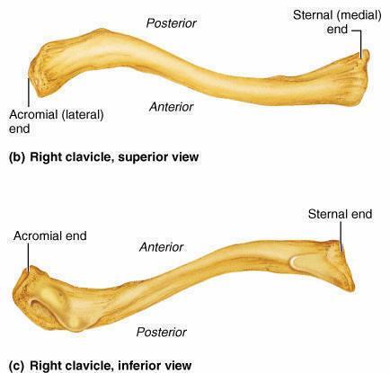
2 scapula Pectoral Girdle clavicle humerus acromial end Clavicle sternal end 2


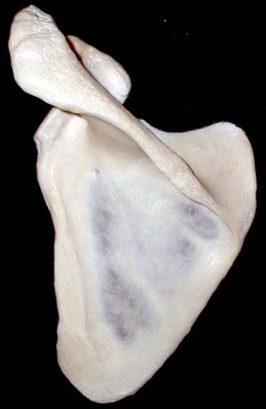


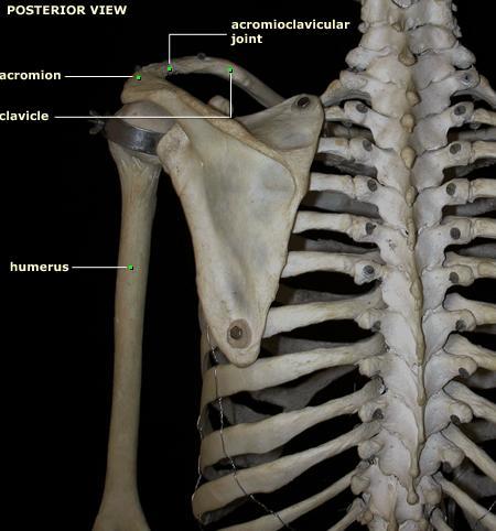
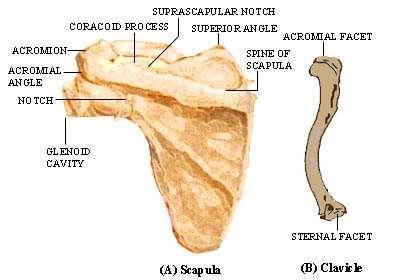
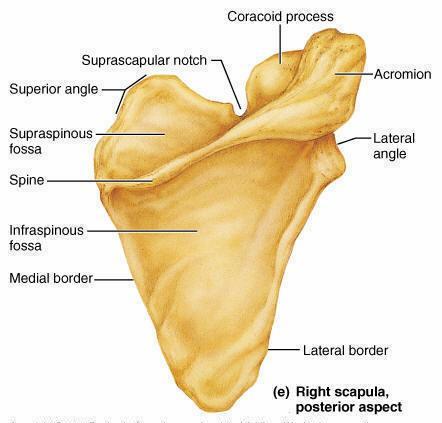
3 Anterior Scapula coracoid process superior angle acromion process glenoid cavity subscapular fossa inferior angle Posterior Scapula acromion process supraspinous fossa infraspinous fossa spine lateral border medial border 3


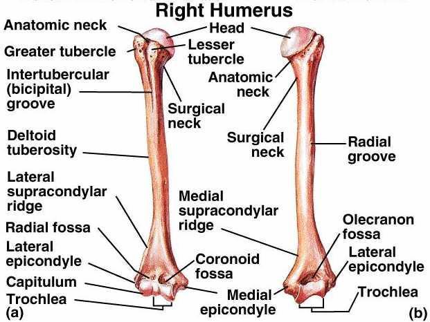
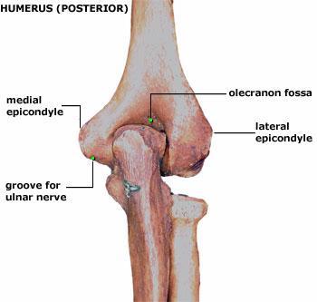
4 head neck Humerus olecranon fossa medial epicondyle trochlea Anterior Humerus deltoid tuberosity lesser tubercle coronoid fossa capitulum lateral epicondyle intertubercular groove greater tubercle 4


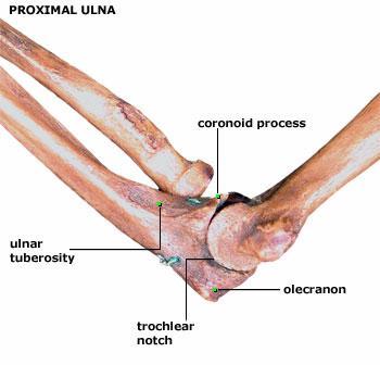
5 trochlear notch Ulna coronoid process head olecranon process radial notch styloid process Radius radial tuberosity head styloid process 5
 scapula: shoulder blade; flat bone; coracoid process/spine: sites for muscle attachments to arm/thorax; extension of quadr limb (scapula glide); glenoid fossa forms synovial jt")
6 Pectoral girdle pectoral girdle = 2 clavicles + 2 scapulae clavicle: collar bone; keeps shoulders apart; vestigial or absent in quadrupeds; synovial jts with acromion process of scapula, and manubrium (sternum) scapula: shoulder blade; flat bone; coracoid process/spine: sites for muscle attachments to arm/thorax; extension of quadr limb (scapula glide); glenoid fossa forms synovial jt (shoulder) with humerus 1. Clavicle 2. Scapula 3. Humerus 4. Sternum 5. Cervical vertebrae 6. Thoracic vertebra 6


7 1. First rib 2. Scapula 3. Humerus 4. Cervical vertebrae 5. Thoracic vertebrae 7
8 8


9 clavicle, coracoid process and acromion. Immediately under the skin, the pectoralis major, deltoid, and trapezius muscles can be palpated. The clavipectoral triangle (or deltopectoral triangle), which contains the cephalic vein, is bordered by the clavicle, pectoralis major muscle and deltoid muscle. Finally, remember that the neurovascular bundle containing the axillary artery, axillary vein and brachial plexus courses under the clavicle and deep to the pectoralis major and minor before coursing into the arm. 9


10 Bony landmarks include the spine of the scapula, medial border of the scapula, and the acromion. Superficial muscles include the three parts of the trapezius, the deltoid, the teres major and the latissimus dorsi 10
11 11
12 12
13 13

.")
14 A). clavicle acromial end is flat and has a small facet for articulation with the acromion; sternal end has a large facet for articulation with the manubrium, and first costal cartilage; conoid tubercle conoid ligament of the coracoclavicular ligament attaches here; trapezoid line trapezoid portion of the coracoclavicular ligament attaches here. B). scapula 1). posterior surface spine acromion coracoid process suprascapular notch supraspinous fossae infraspinous fossae 2). borders superior border medial border lateral border 3). anterior surface scapular fossae 4). lateral end glencoid cavity 14

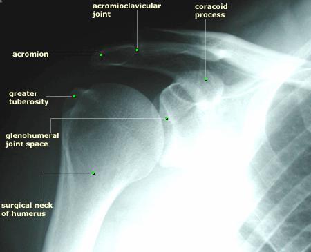
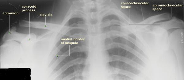
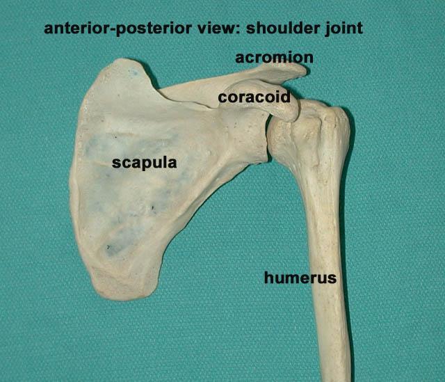
15 Joints Glenohumeral humerus articulating with glenoid fossa of scapula Sternoclavicular (SC) proximal clavicle articulating with manubrium and cartilage of rib 1 Acromioclavicular (AC) acromian process of scapula articulating with distal clavicle Coracoclavicular coracoid process of scapula articulating with inferior clavicle Scapulothoracic anterior scapula articulating with thoracic wall 15
, pectoralis minor, subclavius Elevation trapezius (upper), levatro scapulae, rhomboid")
16 Movements of Scapula and Muscles Causing Movement: Protraction (scapular abduction) serratus anterior, pectoralis minor Retraction (scapular adduction) trapezius, rhomboid, levator scapulae Downward Rotation rhomboids, pectoralis minor Upward rotation trapezius, serratus anterior Depression trapezius (lower), pectoralis minor, subclavius Elevation trapezius (upper), levatro scapulae, rhomboid 16
; infraspinous fossa the infraspinatous muscles")

17 acromion lateral extension of spine of scapula; spine of scapula the trapezius and deltoid attach here; greater scapular notch point at which the spine of the scapula ends, but the acromion continues; coracoid process partially seen as it projects anteriorly; supraspinous fossa the supraspinatus muscle originates here (part of rotator cuff); infraspinous fossa the infraspinatous muscles originates here (part of rotator cuff); lateral border teres minor muscle attaches here (part of rotator cuff), as does the teres major and the long head of the triceps brachii. supraglenoid tubercle the long head of the biceps brachii attaches here; infraglenoid tubercle the long head of the triceps brachii attaches here; spinous process divides the supraspinous and infraspinous fossae, and serves as attachment for the deltoid and trapezius muscles; acromion articulates with the clavicle and is an attachment for the trapezius and deltoid musscles; superior and inferior angles; coracoid process serves as an attachment point for the short head of the biceps brachii, corachobrachialis, and pectoralis minor. 17
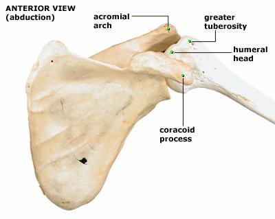
18 Glenohumeral joint 18
 Thirty seven bones form the skeletal framework of each upper limb 19")
19 The Upper Limb The upper limb consists of the arm (brachium), forearm (antebrachium), and hand (manus) Thirty seven bones form the skeletal framework of each upper limb 19
20 Arm The humerus is the sole bone of the arm It articulates with the scapula at the shoulder, and the radius and ulna at the elbow Major markings Arm Proximal humerus includes the head, anatomical and surgical necks, greater and lesser tubercles, and the intertubercular groove Distal humerus includes the capitulum, trochlea, medial and lateral epicondyles, and the coronoid and olecranon fossae Medial portion includes the radial groove and the deltoid process 20

21 Humerus of the Arm Figure 7.23 Forearm The bones of the forearm are the radius and ulna They articulate proximally with the humerus and distally with the wrist bones They also articulate with each other proximally and distally at small radioulnar joints Interosseous membrane connects the two bones along their entire length 21
22 Bones of the Forearm Figure 7.24 Ulna The ulna lies medially in the forearm and is slightly longer than the radius Forms the major portion of the elbow joint with the humerus Its major markings include the olecranon, coronoid process, trochlear notch, radial notch, and the styloid process 22
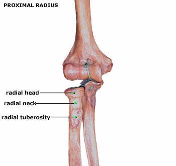
23 Radius The radius lies opposite (lateral to) the ulna and is thin at its proximal end, widened distally The superior surface of the head articulates with the capitulum of the humerus Medially, the head articulates with the radial notch of the ulna Major markings include the radial tuberosity, ulnar notch, and styloid process Radius and Ulna Figure
, and bones of the fingers (phalanges) Hand Figure 7.")


24 Skeleton of the hand contains wrist bones (carpals), bones of the palm (metacarpals), and bones of the fingers (phalanges) Hand Figure 7.26a Upper Arm: humerus A variety of muscles attach to the humerus. These enable movement at the elbow and at the shoulder. The rotator cuff muscles attach at the proximal humerus, and can rotate and abduct the arm at the shoulder. Some of the forearm muscles, (such as pronator teres, and the flexors and extensors of the wrist) also attach to the distal humerus 24
.")
.")
25 1). proximal end head of the humerus greater & lesser tubercle 2). distal end condyle capitulum trochlea epicondyle 3). fossa coronoid fossa olecranon 25
26 26


27 1. Head Anatomical Neck Lesser Tubercle Intertubercular Groove Greater Tubercle Surgical Neck Deltoid Tuberosity 1. Radial Fossa Lateral Epicondyle Capitulum Trochlea Medial Epicondyle Coronoid Fossa Olecranon Fossa 27
, posteriorly (subspinous) or downward (subglenoid) are three most common common when humerus is abducted and externally rotated Rotator")
28 Epiphysial lines of humerus in a young adult. Anterior aspect. The lines of attachment of the articular capsules are in blue. Common Shoulder Injuries Dislocation anteriorly (subcoracoid), posteriorly (subspinous) or downward (subglenoid) are three most common common when humerus is abducted and externally rotated Rotator Cuff Damage (impingement syndrome, tears, especially "throwers" [javelin, tennis, pitchers, swimmers]) Subscapular Neuropathy denervation of infraspinatus with accompanying loss of strength during external rotation of humerus that is common in volleyball 28
. distal head of the ulna styloid process B). radius 1).")
29 Forearm line radius up with thumb line ulna up with little finger A). ulna 1). proximal olecranon coronoid process troclear notch radial notch 2). distal head of the ulna styloid process B). radius 1). proximal head of the radius radial tuberosity 2). distal ulnar notch styloid process 29
 Humeroradial Joint gliding joint (between")

30 Joints Humeroulnar Joint hinge joint (between trochlea and trochlear notch of ulna = "elbow joint") Humeroradial Joint gliding joint (between capitulum and proximal head of radius) Proximal Radioulnar Joint pivot joint (annular ligament binds radial head of radius to radial notch of ulna) 30
31 31
32 Ulna Proximal & Distal End (Anterior Aspect) 1. Olecranon Process Semilunar Notch Coronoid Process Tuberosity Radial Notch Ulna (Shaft) Head of Ulna Styloid Process RADIUS 1. Head of Radius Neck of Radius Radial Tuberosity Radius (Shaft) Styloid Process Ulnar Notch 32
 trapezium, trapezoid, capitate, hamate metacarpals phalanges A).")
33 Wrist and Hand Bones (29 including radius and ulna) radius/ulna carpals proximal row (medial to lateral) scaphoid, lunate, triquetrum, pisiform distal row (medial to lateral) trapezium, trapezoid, capitate, hamate metacarpals phalanges A). carpals 2 rows of 4 each lateral to medial HAND 33
. pisiform 2). distal (articulate with metacarpals) a). trapezium b).")
.")
34 A). carpals 1). proximal (articulate radius and ulna) a). scaphoid b). luna c). triquetral d). pisiform 2). distal (articulate with metacarpals) a). trapezium b). trapezoid c). capitate d). hamate B). metacarpals numbered 1 to 5 starting with the thumb side C). phalanges numbered 1 to 5 starting with the thumb side proximal phalanx medial phalanx distal phalanx 34


35 Bones of the Right Hand (Dorsal Surface) 1. Styloid Process of Radius Navicular (Scaphoid) Lunate Triquetral Pisiform Trapezium Trapezoid Capitate Hamate Metacarpal Proximal Phalange Middle Phalange Distal Phalange Styloid Process of Ulna Bones of the Right Hand (Palmar Surface) 1. Navicular (Scaphoid) Lunate Triquetral Pisiform Trapezium Trapezoid Capitate Hamate Metacarpal Proximal Phalange Middle Phalange Distal Phalange 35
![Some upper extremity bones and landmarks to be familiar with Clavicle: Acromial extremity Acromioclavicular [AC] joint (acromian process and distal clavicle) Conoid tubercle Subclavian groove Costal](/docs-images/89/98420776/images/36-0.jpg "tuberosity Sternal extremity Sternoclavicular [SC] joint (proximal clavicle and manubrium + 1st rib) Scapula: Acromian process Coracoid process Coracovicular joint (coracoid process and inferior")
36 Some upper extremity bones and landmarks to be familiar with Clavicle: Acromial extremity Acromioclavicular [AC] joint (acromian process and distal clavicle) Conoid tubercle Subclavian groove Costal tuberosity Sternal extremity Sternoclavicular [SC] joint (proximal clavicle and manubrium + 1st rib) Scapula: Acromian process Coracoid process Coracovicular joint (coracoid process and inferior clavicle) Scapular notch Superior border Medial border Scaupolothoracic joint (anterior scapula and thoracic wall) Lateral border Superior angle Inferior angle Subscapular fossa Infraspinous fossa Supraspinous fossa Glenoid fossa Glenohumeral joint (humeral head and glenoid fossa) Spine 36
37 Humerus: Head Neck Greater tubercle Lesser tubercle Intertubercular (bicipital) groove Deltoid tuberosity Shaft (diaphysis) Lateral supracondylar ridge Lateral epicondyle Capitulum Humeroradial joint (gliding joint between capitulum and radial head) Radial fossa Medial supracondylar ridge Medial epicondyle Trochlea Humeroulnar joint (humeral trochlea and trochlear notch of ulna) "elbow joint" Coronoid fossa Olecranon fossa Radius: Head Radioulnar joints Neck Radial tuberosity Shaft (diaphysis) Styloid process Ulna: Olecranon process Semilunar (trochlear) notch Coronoid process Ulnar tuberosity Shaft (diaphysis) Head Styloid process Wrist/Hand: Wrist joint (condyloid between distal radius and proximal carpals) Carpals: (radial to ulnar) Proximal Row Intercarpals (gliding or plane joints between carpal bone) Scaphoid Lunate Triquetrum Pisiform Disal Row Trapezium Trapezoid Capitate Hamate 37
38 5 Metacarpals (1 = thumb) Carpometacarpal (CMC) (saddle for 1, plane for 2 5) Phalanges/digits (14 per hand) 5 proximal phalanges Proximal Interphalangeal (PIP) (hinge joint between 1st and 2nd phalanges of 2 5) Interphalangeal (IP) (hinge joint on thumb only between distal and proximal phalanx) 4 middle phalanges (thumb doesn't have) 5 distal phalanges Distal Interphalangeal (DIP) (hinge joint between middle and distal phalanges of 2 5) Want an easy way to remember the carpal bones?!? Remember this... Naughty (Navicular) Lovers (Lunate) Try (Triquetral) Positions (Pisiform) That (Trapezium) They (Trapezoid) Can't (Capitate) Handle (Hamate) 38


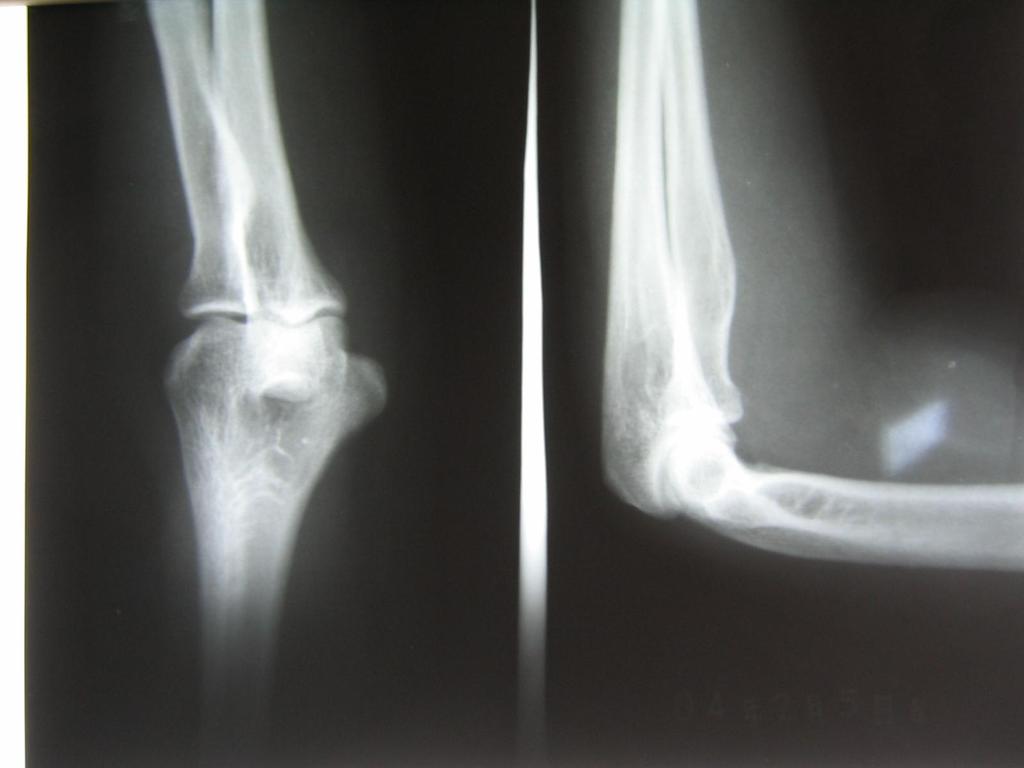
39 WHAT S WRONG Comminuted clavicle fracture; Note 5 th rib fracture in addition 39


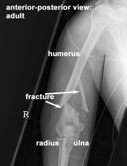
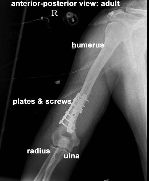
40 Fracture Dislocations of the Proximal Humerus: 40
41 41
Pectoral girdle, SUPERIEUR ARM AND HAND. Danil Hammoudi.MD
 Pectoral girdle, SUPERIEUR ARM AND HAND Danil Hammoudi.MD The pectoral girdle is the set of bones which connect the upper limb to the axial skeleton on each side. It consists of the clavicle scapula in
Chapter 8. The Pectoral Girdle & Upper Limb
 Chapter 8 The Pectoral Girdle & Upper Limb Pectoral Girdle pectoral girdle (shoulder girdle) supports the arm consists of two on each side of the body // clavicle (collarbone) and scapula (shoulder blade)
Anatomy and Physiology II. Review Shoulder Girdle New Material Upper Extremities - Bones
 Anatomy and Physiology II Review Shoulder Girdle New Material Upper Extremities - Bones Anatomy and Physiology II Shoulder Girdle Review Questions From Last Lecture Can you identify the following muscles?
Axilla and Brachial Region
 L 4 A B O R A T O R Y Axilla and Brachial Region BRACHIAL PLEXUS 5 Roots/Rami (ventral rami C5 T1) 3 Trunks Superior (C5, C6) Middle (C7) Inferior (C8, T1) 3 Cords Lateral Cord (Anterior Superior and Anterior
Connects arm to thorax 3 joints. Glenohumeral joint Acromioclavicular joint Sternoclavicular joint
 Connects arm to thorax 3 joints Glenohumeral joint Acromioclavicular joint Sternoclavicular joint Scapula Elevation Depression Protraction (abduction) Retraction (adduction) Downward Rotation Upward Rotation
THE SKELETAL SYSTEM. Focus on the Pectoral Girdle
 THE SKELETAL SYSTEM Focus on the Pectoral Girdle Appendicular Skeleton 126 bones Includes bones of the limbs (arms and legs) Pectoral girdle (shoulder) Pelvic girdle (hip) Pectoral Girdle (the shoulder)
Muscles of the Upper Limb
 Muscles of the Upper Limb anterior surface of ribs 3 5 coracoid process Pectoralis minor pectoral nerves protracts / depresses scapula Serratus anterior Subclavius ribs 1-8 long thoracic nerve rib 1 ----------------
Figure 1: Bones of the upper limb
 BONES OF THE APPENDICULAR SKELETON The appendicular skeleton is composed of the 126 bones of the appendages and the pectoral and pelvic girdles, which attach the limbs to the axial skeleton. Although the
An Introduction to the Appendicular Skeleton
 An Introduction to the Appendicular Skeleton The Appendicular Skeleton is composed of the 126 bones of the appendages (limbs) and the pectoral and pelvic girdles, which attach to the axial skeleton. Each
Netter's Anatomy Flash Cards Section 6 List 4 th Edition
 Netter's Anatomy Flash Cards Section 6 List 4 th Edition https://www.memrise.com/course/1577581/ Section 6 Upper Limb (66 cards) Plate 6-1 Humerus and Scapula: Anterior View 1.1 Acromion 1.2 Greater tubercle
Anatomage Table Instructors Guide- Upper Limb
 The Upper Limb Anatomage Table Instructors Guide- Upper Limb Table of Contents Upper Limb 1- The Skeletal System...3 1: Clavicle...3 2: Scapula...5 3: Shoulder (Glenohumeral) and Proximal Humerus...7 4:
Muscular Nomenclature and Kinesiology - One
 Chapter 16 Muscular Nomenclature and Kinesiology - One Lessons 1-3 (with lesson 4) 1 Introduction 122 major muscles covered in this chapter Chapter divided into nine lessons Kinesiology study of human
Anatomy of the Shoulder Girdle. Prof Oluwadiya Kehinde FMCS (Orthop)
") Anatomy of the Shoulder Girdle Prof Oluwadiya Kehinde FMCS (Orthop) www.oluwadiya.com Bony Anatomy Shoulder Complex: Sternum(manubrium) Clavicle Scapula Proximal humerus Manubrium Sterni Upper part of
Anatomy of the Shoulder Girdle Prof Oluwadiya Kehinde FMCS (Orthop) www.oluwadiya.com Bony Anatomy Shoulder Complex: Sternum(manubrium) Clavicle Scapula Proximal humerus Manubrium Sterni Upper part of
The skeleton consists of: Bones: special connective tissue, hard. Cartilage: special connective tissue, less hard than bones. Joints: joint is the
 The skeleton consists of: Bones: special connective tissue, hard. Cartilage: special connective tissue, less hard than bones. Joints: joint is the location at witch two bones make contact, whereas ligaments
Upper Limb Muscles Muscles of Axilla & Arm
 Done By : Saleh Salahat Upper Limb Muscles Muscles of Axilla & Arm 1) Muscles around the axilla A- Muscles connecting the upper to thoracic wall (4) 1- pectoralis major Origin:- from the medial half of
Practical 2 Worksheet
 Practical 2 Worksheet Upper Extremity BONES 1. Which end of the clavicle is on the lateral side (acromial or sternal)? 2. Describe the difference in the appearance of the acromial and sternal ends of the
Region of upper limb attachment to the trunk Proximal segment of limb overlaps parts of the trunk (thorax and back) and lower lateral neck.
 and lower lateral neck.") Region of upper limb attachment to the trunk Proximal segment of limb overlaps parts of the trunk (thorax and back) and lower lateral neck. includes Pectoral Scapular Deltoid regions of the upper limb
Region of upper limb attachment to the trunk Proximal segment of limb overlaps parts of the trunk (thorax and back) and lower lateral neck. includes Pectoral Scapular Deltoid regions of the upper limb
A&P 1 Skeletal Lab Guide Week 2 - Appendicular Skeleton and Joints Lab Exercises: Pectoral Girdle
 A&P 1 Skeletal Lab Guide Week 2 - Appendicular Skeleton and Joints Lab Exercises: Pectoral Girdle PLEASE NOTE: Your group will need an articulated skeleton, a disarticulated skeleton, and the joint models
The Appendicular Skeleton
 8 The Appendicular Skeleton PowerPoint Lecture Presentations prepared by Jason LaPres Lone Star College North Harris 8-1 The Pectoral Girdle The Pectoral Girdle Also called shoulder girdle Connects the
10/12/2010. Upper Extremity. Pectoral (Shoulder) Girdle. Clavicle (collarbone) Skeletal System: Appendicular Skeleton
 Girdle. Clavicle (collarbone) Skeletal System: Appendicular Skeleton") Skeletal System: Appendicular Skeleton Pectoral girdle Pelvic girdle Upper limbs Lower limbs 8-1 Pectoral (Shoulder) Girdle Consists of scapula and clavicle Clavicle articulates with sternum (Sternoclavicular
Skeletal System: Appendicular Skeleton Pectoral girdle Pelvic girdle Upper limbs Lower limbs 8-1 Pectoral (Shoulder) Girdle Consists of scapula and clavicle Clavicle articulates with sternum (Sternoclavicular
Lab Activity 11: Group II
 Lab Activity 11: Group II Muscles Martini Chapter 11 Portland Community College BI 231 Origin and Insertion Origin: The place where the fixed end attaches to a bone, cartilage, or connective tissue. Insertion:
Upper limb Arm & Cubital region 黃敏銓
 Upper limb Arm & Cubital region 黃敏銓 1 Arm Lateral intermuscular septum Anterior (flexor) compartment: stronger Medial intermuscular septum Posterior (extensor) compartment 2 Coracobrachialis Origin: coracoid
LIST OF STRUCTURES TO BE IDENTIFIED IN LAB: UPPER EXTREMITY REVIEW 2016
 LIST OF STRUCTURES TO BE IDENTIFIED IN LAB: UPPER EXTREMITY REVIEW 2016 BONES Ribs, sternum, clavicle Humerus: Head, greater tubercle, lesser tubercle, intertubercular sulcus, surgical neck, anatomical
medial half of clavicle; Sternum; upper six costal cartilages External surfaces of ribs 3-5
 MUSCLE ORIGIN INSERTION ACTION NERVE Pectoralis Major medial half of clavicle; Sternum; upper six costal cartilages Lateral lip of intertubercular groove of horizontal adduction Medial and lateral pectoral
REFERENCE DIAGRAMS OF UPPER LIMB MUSCLES: NAMES, LOCATIONS, ATTACHMENTS, FUNCTIONS MUSCLES CONNECTING THE UPPER LIMB TO THE AXIAL SKELETON
 REFERENCE DIAGRAMS OF UPPER LIMB MUSCLES: NAMES, LOCATIONS, ATTACHMENTS, FUNCTIONS MUSCLES CONNECTING THE UPPER LIMB TO THE AXIAL SKELETON A25LAB EXERCISES: UPPER LIMB MUSCLES Page 1 MUSCLES CONNECTING
SKELETAL SYSTEM 206. AXIAL SKELETON 80 APPENDICULAR SKELETON 126 (see Figure 6.1) Clavicle. Clavicle. Pectoral girdles. Scapula. Scapula.
 Clavicle. Clavicle. Pectoral girdles. Scapula. Scapula.") SKELETAL SYSTEM 206 AXIAL SKELETON 80 APPENDICULAR SKELETON 126 (see Figure 6.1) Pectoral girdles 4 Clavicle Scapula 2 2 Clavicle Scapula Humerus 2 Humerus Upper limbs 60 Radius 2 Ulna Carpal bones Metacarpal
SKELETAL SYSTEM 206 AXIAL SKELETON 80 APPENDICULAR SKELETON 126 (see Figure 6.1) Pectoral girdles 4 Clavicle Scapula 2 2 Clavicle Scapula Humerus 2 Humerus Upper limbs 60 Radius 2 Ulna Carpal bones Metacarpal
Muscle Action Origin Insertion Nerve Innervation Chapter Page. Deltoid. Trapezius. Latissimus Dorsi
 Muscle Action Origin Insertion Nerve Innervation Chapter Page All Fibers Abduct the shoulder (glenohumeral joint) Deltoid Anterior Fibers Flex the shoulder (G/H joint) Horizontally adduct the shoulder
Pectoral (Shoulder) Girdle
 Girdle") Chapter 8 Skeletal System: Appendicular Skeleton Pectoral girdle Pelvic girdle Upper limbs Lower limbs 8-1 Pectoral (Shoulder) Girdle Consists of scapula and clavicle Clavicle articulates with sternum
Chapter 8 Skeletal System: Appendicular Skeleton Pectoral girdle Pelvic girdle Upper limbs Lower limbs 8-1 Pectoral (Shoulder) Girdle Consists of scapula and clavicle Clavicle articulates with sternum
Chapter 8 The Skeletal System: The Appendicular Skeleton. Copyright 2009 John Wiley & Sons, Inc.
 Chapter 8 The Skeletal System: The Appendicular Skeleton Appendicular Skeleton It includes bones of the upper and lower limbs Girdles attach the limbs to the axial skeleton The pectoral girdle consists
THE SHOULDER JOINT T H E G L E N O H U M E R A L ( G H ) J O I N T
 J O I N T") THE SHOULDER JOINT T H E G L E N O H U M E R A L ( G H ) J O I N T CLARIFICATION OF TERMS Shoulder girdle = scapula and clavicle Shoulder joint (glenohumeral joint) = scapula and humerus Lippert, p115
THE SHOULDER JOINT T H E G L E N O H U M E R A L ( G H ) J O I N T CLARIFICATION OF TERMS Shoulder girdle = scapula and clavicle Shoulder joint (glenohumeral joint) = scapula and humerus Lippert, p115
Chapter 8. The Appendicular Skeleton. Lecture Presentation by Lee Ann Frederick University of Texas at Arlington Pearson Education, Inc.
 Chapter 8 The Appendicular Skeleton Lecture Presentation by Lee Ann Frederick University of Texas at Arlington An Introduction to the Appendicular Skeleton The Appendicular Skeleton 126 bones Allows us
Abduction of arm until your hand rich your head. Flexion of forearm at elbow joint. Extension of arm at elbow joint. Flexion of fingers 10.
 Num. answer 1. Medialy With the manubrium ( sternum ), and laterally with the acromion of the scapula 2. 1. Trapezius 2. Levator scapulae 3. Rhomboids 3. 1. Pectoralis major 2. Pectoralis minor 3. Latissiumus
Scapular and Deltoid Regions
 M1 Gross and Developmental Anatomy Scapular and Deltoid Regions Dr. Peters 1 Outline I. Skeleton of the Shoulder and Attachment of the Upper Extremity to Trunk II. Positions and Movements of the Scapula
Acknowledgement. Here are some flash cards all set up in a "pdf" format for you! Thanks to Laura H. (spring 08)
") Acknowledgement Here are some flash cards all set up in a "pdf" format for you! Thanks to Laura H. (spring 08) for her donation to all my anatomy students! t Here is her suggestion for making flashcards
Acknowledgement Here are some flash cards all set up in a "pdf" format for you! Thanks to Laura H. (spring 08) for her donation to all my anatomy students! t Here is her suggestion for making flashcards
7/31/2012 THE SHOULDER JOINT CLARIFICATION OF TERMS OSTEOLOGY OF THE GH JOINT(BONES)
") THE SHOULDER JOINT T H E G L E N O H U M E R AL ( G H ) J O I N T CLARIFICATION OF TERMS Shoulder girdle = scapula and clavicle Shoulder joint (glenohumerual joint) = scapula and Lippert, p115 OSTEOLOGY
THE SHOULDER JOINT T H E G L E N O H U M E R AL ( G H ) J O I N T CLARIFICATION OF TERMS Shoulder girdle = scapula and clavicle Shoulder joint (glenohumerual joint) = scapula and Lippert, p115 OSTEOLOGY
bio4165 lab quiz 1 Posterior View Anterior View Lateral View Anterior View bio fall.quarter lab.quiz.1...page.1 of 6
 B A Posterior View D C E Lateral View bio.4165...fall.quarter.2005...lab.quiz.1...page.1 of 6 F I G 35 Posterior View H bio.4165...fall.quarter.2005...lab.quiz.1...page.2 of 6 J Posterior View L K Inferior
The Elbow and the cubital fossa. Prof Oluwadiya Kehinde
 The Elbow and the cubital fossa Prof Oluwadiya Kehinde www.oluwadiya.com Elbow and Forearm Anatomy The elbow joint is formed by the humerus, radius, and the ulna Bony anatomy of the elbow Distal Humerus
Amy Warenda Czura, Ph.D. 1 SCCC BIO130 Lab 7 Appendicular Skeleton & Articulations
 The Skeletal System II: Appendicular Skeleton and Articulations Exercises 11, 13 (begins: page 145 in 9 th and 10 th editions) Exercises 10, 11 (begins: page 147 in 11 th edition, page 149 in 12 th edition)
Biology 218 Human Anatomy. Adapted from Martini Human Anatomy 7th ed. Chapter 7 The Skeletal System Appendicular Division
 Adapted from Martini Human Anatomy 7th ed. Chapter 7 The Skeletal System Appendicular Division Introduction The appendicular skeleton includes: Pectoral girdle Shoulder bones Upper limbs Pelvic girdle
Chapter 8B. The Skeletal System: Appendicular Skeleton. The Appendicular Skeleton. Clavicle. Pectoral (Shoulder) Girdle
 Girdle") The Appendicular Skeleton Chapter 8B The Skeletal System: Appendicular Skeleton 126 bones Pectoral (shoulder) girdle Pelvic (hip) girdle Upper limbs Lower limbs Functions primarily to facilitate movement
The Appendicular Skeleton Chapter 8B The Skeletal System: Appendicular Skeleton 126 bones Pectoral (shoulder) girdle Pelvic (hip) girdle Upper limbs Lower limbs Functions primarily to facilitate movement
PRE-LAB EXERCISES. Before we get started, look up the definitions of these common bone marking terms: Canal: Condyle: Facet: Fissure:
 1 PRE-LAB EXERCISES When studying the skeletal system, the bones are often sorted into two broad categories: the axial skeleton and the appendicular skeleton. This lab focuses on the appendicular skeleton,
Biology 152 Appendicular Skeleton Anatomy Objectives
 Biology 152 Appendicular Skeleton Anatomy Objectives We will learn proper bone names, left/right/medial, and the parts of bones in this exercise. Start by learning the names of the bones. As you gain comfort
MUSCLES. Anconeus Muscle
 LAB 7 UPPER LIMBS MUSCLES Anconeus Muscle anconeus origin: distal end of dorsal surface of humerus insertion: lateral surface of ulna from distal margin of the semilunar notch to proximal end of the olecranon
3 Mohammad Al-Mohtasib Areej Mosleh
 3 Mohammad Al-Mohtasib Areej Mosleh ***Muscles Connecting the Upper Limb to the Vertebral Column 1.Trapezius Muscle ***The first muscle on the back is trapezius muscle, it s called so according
Biology 218 Human Anatomy
 Chapter 8 Adapted from Tortora 10 th ed. LECTURE OUTLINE A. Introduction (p. 203) 1. The appendicular skeleton contains 126 bones that form: i. two pectoral (shoulder) girdles two upper limbs i one pelvic
THE SHORT DESCRIPTION OF THE JOINTS 1. THE UPPER LIMB (Dr. Dóra Reglődi*, version )
") THE SHORT DESCRIPTION OF THE JOINTS 1. THE UPPER LIMB (Dr. Dóra Reglődi*, version 02-2007) Shoulder girdle The shoulder girdle consists of the clavicle and scapula on both sides. The two sides are connected
THE SHORT DESCRIPTION OF THE JOINTS 1. THE UPPER LIMB (Dr. Dóra Reglődi*, version 02-2007) Shoulder girdle The shoulder girdle consists of the clavicle and scapula on both sides. The two sides are connected
The Free Upper Limb. Bone of the Arm. aus: Platzer, Locomotor System (ISBN ), 2009 Georg Thieme Verlag KG
, 2009 Georg Thieme Verlag KG") : ones, Ligaments, Joints The Free The bones of the free upper limb are The humerus The radius and ulna The carpal bones The metacarpal bones The phalanges one of the Arm Humerus (A H) The humerus articulates
: ones, Ligaments, Joints The Free The bones of the free upper limb are The humerus The radius and ulna The carpal bones The metacarpal bones The phalanges one of the Arm Humerus (A H) The humerus articulates
The Arm and Cubital Fossa
 The Arm and Cubital Fossa Dr. Andrew Gallagher School of Anatomical Sciences University of the Witwatersrand Introduction The ARM (BRACHIUM) is the most proximal segment of the upper limb musculoskeletal
Chapter 8 The Skeletal System: The Appendicular Skeleton. Copyright 2009 John Wiley & Sons, Inc.
 Chapter 8 The Skeletal System: The Appendicular Skeleton Appendicular Skeleton The primary function is movement It includes bones of the upper and lower limbs Girdles attach the limbs to the axial skeleton
Sports Medicine Part I : ANATOMY OF THE SPINE, ABDOMEN AND SHOULDER COMPLEX
 Sports Medicine 25 1.1 Part I : ANATOMY OF THE SPINE, ABDOMEN AND SHOULDER COMPLEX c.w.p. Wagner High School, Sports Medicine, A. Morgan, T. Morgan 2008 Anatomy of the Upper Body In this section of the
Lab Activity 9. Appendicular Skeleton Martini Chapter 8. Portland Community College BI 231
 Lab Activity 9 Appendicular Skeleton Martini Chapter 8 Portland Community College BI 231 Appendicular Skeleton Upper & Lower extremities Shoulder Girdle Pelvic Girdle 2 Humerus 3 Humerus: Proximal End
The Appendicular Skeleton
 8 The Appendicular Skeleton PowerPoint Lecture Presentations prepared by Jason LaPres Lone Star College North Harris An Introduction to the Appendicular Skeleton Learning Outcomes 8-1 Identify the bones
Human Anatomy Biology 351
 1 Human Anatomy Biology 351 Upper Limb Exam Please place your name on the back of the last page of this exam. You must answer all questions on this exam. Because statistics demonstrate that, on average,
Joint G*H. Joint S*C. Joint A*C. Labrum. Humerus. Sternum. Scapula. Clavicle. Thorax. Articulation. Scapulo- Thoracic
 A*C Joint Scapulo- Thoracic Articulation Thorax Sternum Clavicle Scapula Humerus S*C Joint G*H Joint Labrum AC Ligaments SC Ligaments SC JOINT AC Coracoacromial GH GH Ligament Complex Coracoclavicular
The Biomechanics of the Human Upper Extremity. Dr Ayesha Basharat BSPT, PP.DPT. M.PHIL
 The Biomechanics of the Human Upper Extremity Dr Ayesha Basharat BSPT, PP.DPT. M.PHIL Sternoclavicular Joint Provides major axis of rotation for movement of clavicle and scapula Freely permitted frontal
Supplied in part by the musculocutaneous nerve. Forms the axis of rotation in movements of pronation and supination
 Anatomy: Upper limb (15 questions) 1. Latissimus Dorsi: Is innervated by the dorsal scapular nerve Lies above feres major muscle Medially rotates the humerus All of the above 2. Supinator muscle is: Deep
Upper limb Pectoral region & Axilla
 Upper limb Pectoral region & Axilla 黃敏銓 mchuang@ntu.edu.tw 1 Pectoral region Intercostal nerve Anterior branch of lateral cutaneous branch Lateral cutaneous branch Anterior cutaneous branch Anterior cutaneous
Copyright 2003 Pearson Education, Inc. publishing as Benjamin Cummings. Dr. Nabil khouri
 Dr. Nabil khouri Appendicular Skeleton The appendicular skeleton is made up of the bones of the upper and lower limbs and their girdles Two girdles: Pectoral girdles attach the upper limbs to the body
BLUE SKY SCHOOL OF PROFESSIONAL MASSAGE AND THERAPEUTIC BODYWORK. Musculoskeletal Anatomy & Kinesiology II REVIEW
 BLUE SKY SCHOOL OF PROFESSIONAL MASSAGE AND THERAPEUTIC BODYWORK Musculoskeletal Anatomy & Kinesiology II REVIEW MSAK101-II Session 4 LEARNING OBJECTIVES: By the end of this session, the student will be
The Clavicle Right clavicle Deltoid tubercle: Conoid tubercle, conoid ligamen Impression for the
 The Clavicle Muscle Attachment Sites in the Upper Limb Pectoralis major Right clavicle Smooth superior surface of the shaft, under the platysma muscle tubercle: attachment of the deltoid Acromial facet
Exercise 11. The Appendicular Skeleton
 Exercise 11 The Appendicular Skeleton The Appendicular Skeleton The appendicular skeleton contains 126 bones. Consists of the upper and lower limbs, the pectoral girdles, and the pelvic girdles. The pectoral
Kinesiology of the Upper Extremity
 P A R T II Kinesiology of the Upper Extremity Pectoralis Latissimus dorsi UNIT 1: SHOULDER UNIT: THE SHOULDER COMPLEX Chapter 8: Structure and Function of the Bones and Joints of the Shoulder Girdle Chapter
Exercise Science Section 2: The Skeletal System
 Exercise Science Section 2: The Skeletal System An Introduction to Health and Physical Education Ted Temertzoglou Paul Challen ISBN 1-55077-132-9 Role of the Skeleton Protection Framework Attachments for
26/9/2016. Anatomy. 1 Nour Erekat Wejdan Amer
 26/9/2016 Anatomy st 1 Nour Erekat Wejdan Amer Notes before we start the lecture. Bring any colored Atlas with you to the lab. The main reference is clinical anatomy by regions by Richard snell the 9 th
Functional Anatomy. CHAPTER 5 Functional Anatomy of the Upper Extremity. CHAPTER 6 Functional Anatomy of the Lower Extremity
 Hamill_ch05_137-186.qxd 11/2/07 3:55 PM Page 137 S E C T I O N II Functional Anatomy CHAPTER 5 Functional Anatomy of the Upper Extremity CHAPTER 6 Functional Anatomy of the Lower Extremity CHAPTER 7 Functional
Main Menu. Shoulder Girdle click here. The Power is in Your Hands. 1:07:11 PM]
![Main Menu. Shoulder Girdle click here. The Power is in Your Hands. 1:07:11 PM]](/thumbs/86/94418253.jpg "Main Menu. Shoulder Girdle click here. The Power is in Your Hands. 1:07:11 PM]") 1 The Shoulder Girdle click here Main Menu K.2 http://www.handsonlineeducation.com/classes//k2entry.htm[3/23/18, 1:07:11 PM] Bones Scapula and Clavicle Move as a unit Clavicle s articulation with sternum
1 The Shoulder Girdle click here Main Menu K.2 http://www.handsonlineeducation.com/classes//k2entry.htm[3/23/18, 1:07:11 PM] Bones Scapula and Clavicle Move as a unit Clavicle s articulation with sternum
Muscles in the Shoulder, Chest, Arm, Stomach, and Back
 Muscles in the Shoulder, Chest, Arm, Stomach, and Back Shoulder Muscles Deltoid Supraspinatus Infraspinatus Teres Major Teres Minor Subscapularis Deltoid (Delts) Function: Raises the upper arm Origin:
Bone List Anatomy
 1 Frontal Bone Skull 2 Parietal Bone Skull 3 Occipital Bone Skull 4 Temporal Bone Skull 5 Coronal Suture Skull 6 Sagittal Suture Skull 7 Squamous suture Skull 8 Lambdoid Suture Skull 9 Surpaorbital Ridge
Bones of the Upper Limb *
 OpenStax-CNX module: m46368 1 Bones of the Upper Limb * OpenStax This work is produced by OpenStax-CNX and licensed under the Creative Commons Attribution License 3.0 By the end of this section, you will
ARM Brachium Musculature
 ARM Brachium Musculature Coracobrachialis coracoid process of the scapula medial shaft of the humerus at about its middle 1. flexes the humerus 2. assists to adduct the humerus Blood: muscular branches
The Shoulder. Anatomy and Injuries PSK 4U Unit 3, Day 4
 The Shoulder Anatomy and Injuries PSK 4U Unit 3, Day 4 Shoulder Girdle Shoulder Complex is the most mobile joint in the body. Scapula Clavicle Sternum Humerus Rib cage/thorax Shoulder Girdle It also includes
Perpendicular Plate Zygomatic Bone. Mental Foramen Mandible
 Glabella Frontal Middle Nasal Concha Nasal Lacrimal Perpendicular Plate Zygomatic Inferior Nasal Concha Maxilla Mental Mandible Skull (anterior view) Squamosal Suture Coronal Suture Frontal Parietal Nasal
JOINT MOBILITY Joint Mobility of Upper Extremity
 Kinesiology 2017#5: JOINT MOBILITY Joint Mobility of Upper Extremity Huei-Ming Chai, Ph.D., PT School of Physical Therapy National Taiwan University Functions of Synovial Joints Joint Mobility Osteokinematic
The Elbow and Radioulnar Joints Kinesiology. Dr Cüneyt Mirzanli Istanbul Gelisim University
 The Elbow and Radioulnar Joints Kinesiology Dr Cüneyt Mirzanli Istanbul Gelisim University 1 The Elbow & Radioulnar Joints Most upper extremity movements involve the elbow & radioulnar joints. Usually
BRACHIAL PLEXUS. DORSAL SCAPULAR NERVE (C5) supraclavicular branch innervates rhomboids (major and minor) and levator scapulae
 supraclavicular branch innervates rhomboids (major and minor) and levator scapulae") THE BRACHIAL PLEXUS DORSAL SCAPULAR NERVE (C5) supraclavicular branch innervates rhomboids (major and minor) and levator scapulae SCHEMA OF THE BRACHIAL PLEXUS THE BRACHIAL PLEXUS PHRENIC NERVE supraclavicular
THE BRACHIAL PLEXUS DORSAL SCAPULAR NERVE (C5) supraclavicular branch innervates rhomboids (major and minor) and levator scapulae SCHEMA OF THE BRACHIAL PLEXUS THE BRACHIAL PLEXUS PHRENIC NERVE supraclavicular
Dr. Mahir Alhadidi Anatomy Lecture #9 Feb,28 th 2012
 Quick Revision: Upper arm is divided into two compartments: 1. Anterior Compartment: Contains three muscles (Biceps brachii, Coracobrachialis, Brachialis). Innervated by Musculocutaneous nerve. 2. Posterior
EasyAnatomy s. Snapshot Guide to the Bones of the Canine Thoracic Limb. LlamaZOO Interactive facebook.
 Thoracic Limb The regions of the thoracic limb, or forelimb, are the shoulder, brachium (upper arm), antebrachium (lower arm), and the manus (forepaw). The manus is composed of the carpus, metacarpus,
Learning Objectives. 07 Aug 12. Article E-1. At the end of this section the learner will be able to:
 Module 1: Comparative Functional Anatomy and Biomechanics Article E-1 Learning Objectives At the end of this section the learner will be able to: Describe the bones of the equine thoracic Describe the
Appendicular Skeleton. Prof. Abdulameer Al-Nuaimi
 Appendicular Skeleton Prof. Abdulameer Al-Nuaimi a.alnuaimi@sheffield.ac.uk abdulameerh@yahoo.com Hi Prof, It is great to hear from you, I really enjoyed your teaching last year. You taught me the hardest
Figure 27: The synovial membrane of the shoulder joint (anterior view)
") The coracoacromial ligament; is an accessory ligament that protects the superior aspect of the joint extending from the coracoid process to the acromion over the tendon of supraspinatus. The synovial membrane
The coracoacromial ligament; is an accessory ligament that protects the superior aspect of the joint extending from the coracoid process to the acromion over the tendon of supraspinatus. The synovial membrane
Joints of the upper limb II
 Joints of the upper limb II Prof. Abdulameer Al-Nuaimi E-mail: a.al-nuaimi@sheffield.ac.uk E. mail: abdulameerh@yahoo.com Elbow joint The elbow joint is connecting the upper arm to the forearm. It is classed
Bone Flashcards for 10a
 Bone Flashcards for 0a CLAVICLE (collar bone). Sternal extremity (end) flat end. Acromial extremity (end) rounded end. SCAPULA (shoulder blade). Right or left scapula?. Superior border (superior margin).
Anatomy Workshop Upper Extremity David Ebaugh, PT, PhD Workshop Leader. Lab Leaders: STATION I BRACHIAL PLEXUS
 Anatomy Workshop Upper Extremity David Ebaugh, PT, PhD Workshop Leader Lab Leaders: STATION I BRACHIAL PLEXUS A. Posterior cervical triangle and axilla B. Formation of plexus 1. Ventral rami C5-T1 2. Trunks
Introduction. Skeletal Nomenclature. Lesson One: Bones of the Upper Extremity. Shoulder Girdle: Clavicle. Scapula: Bony Markings
 Chapter 14 Skeletal Nomenclature Susan G. Salvo Bones help locate muscles Can also be areas to avoid Chapter divided into 5 lessons covering bones, bony markings, and joints Introduction 1 2 Lesson One:
FUNCTIONAL ANATOMY OF SHOULDER JOINT
 FUNCTIONAL ANATOMY OF SHOULDER JOINT ARTICULATION Articulation is between: The rounded head of the Glenoid cavity humerus and The shallow, pear-shaped glenoid cavity of the scapula. 2 The articular surfaces
Anatomy Made Easy MSS
 Anatomy Made Easy MSS Part 4+5 هذا الملف يشمل التفريغ المحاضرة السادسة Done By :Sajeda Alazzeh Edited by: AWN Academic Team The Appendicular skeletal system Pectoral Girdles (Shoulder Girdles) is made
Principles of Anatomy and Physiology
 Principles of Anatomy and Physiology 14 th Edition CHAPTER 8 The Skeletal System: The Appendicular Skeleton The Appendicular Skeleton The 126 bones of the appendicular skeleton are primarily concerned
Shoulder: Clinical Anatomy, Kinematics & Biomechanics
 Shoulder: Clinical Anatomy, Kinematics & Biomechanics Dr. Alex K C Poon Department of Orthopaedics & Traumatology Pamela Youde Nethersole Eastern Hospital Clinical Anatomy the application of anatomy to
Acland's DVD Atlas of Human Anatomy. Transcript for Volume Robert D Acland
 Acland's DVD Atlas of Human Anatomy Transcript for Volume 1 2007 Robert D Acland This free downloadable pdf file is to be used for individual study only. It is not to be reproduced in any form without
CLINICAL SUMMARY AND RECOMMENDATIONS 378
 Shoulder 9 CLINICAL SUMMARY AND RECOMMENDATIONS 378 Anatomy 379 Osteology 379 Arthrology 380 Scapulohumeral Rhythm 381 Ligaments 382 Muscles 384 Nerves 387 Patient History 389 Initial Hypotheses Based
STEP #1: Anatomy STEP #2: Awareness STEP #3: Action
 Stabilize the Scapula in Three Easy Steps by NFPT Idea World 2016: Session 611 Saturday July 16th 7:30-9:20am Beverly Hosford, MA STEP #1: Anatomy STEP #2: Awareness STEP #3: Action 1. Anatomy *Memorize
BIOLOGY 113 LABORATORY Skeletal System
 BIOLOGY 113 LABORATORY Skeletal System Objectives Distinguish between the axial and appendicular skeleton. Distinguish between the cranium and facial skeleton. Locate and name the bones of the skull and
Ligaments of Elbow hinge: sagittal plane so need lateral and medial ligaments
 Ligaments of Elbow hinge: sagittal plane so need lateral and medial ligaments Ulnar Collateral ligament on medial side; arising from medial epicondyle and stops excess valgus movement (lateral movement)
Thank You for Your Support! Hosford Muscle Tables
 Thank You for Your Support! This PDF document has been placed online for your enjoyment and I hope you find it useful. These tables are both a teaching tool, and a study / review tool. I created these
The pectoral region. University of Babylon College of Medicine Dr.HaythemAli Alsayigh M.B.CH.B.-F.I.M.B.S. Surgical Clinical Anatomy
 The pectoral region University of Babylon College of Medicine Dr.HaythemAli Alsayigh M.B.CH.B.-F.I.M.B.S. Surgical Clinical Anatomy Objective Study the Bones and Joints A. Clavicle (collarbone) B. Scapula
The arm: *For images refer back to the slides
 The arm: *For images refer back to the slides Muscles of the arm: deltoid, triceps (which is located at the back of the arm), biceps and brachialis (it lies under the biceps), brachioradialis (it lies
M C P T. "Excellence in Education" SHOULDER AND UPPER LIMB. Part 1. Advanced Anatomy 2 Part 2. Assessment
 M C P T Melbourne College of Professional Therapists "Excellence in Education" SHOULDER AND UPPER LIMB Part 1. Advanced Anatomy 2 Part 2. Assessment 2006 MCPT Advanced Anatomy and Assessment 1 Diploma
CHAPTER 6: THE UPPER EXTREMITY: THE ELBOW, FOREARM, WRIST, AND HAND
 CHAPTER 6: THE UPPER EXTREMITY: THE ELBOW, FOREARM, WRIST, AND HAND KINESIOLOGY Scientific Basis of Human Motion, 12 th edition Hamilton, Weimar & Luttgens Presentation Created by TK Koesterer, Ph.D.,
Elbow & Forearm H O W V I T A L I S T H E E L B O W T O O U R D A I L Y L I V E S?
 Elbow & Forearm H O W V I T A L I S T H E E L B O W T O O U R D A I L Y L I V E S? Clarification of Terms The elbow includes: 3 bones (humerus, radius, and ulna) 2 joints (humeroulnar and humeroradial)