Note: Waxman is very sketchy on today s pathways and nonexistent on the Trigeminal.
|
|
|
- Percival Carpenter
- 5 years ago
- Views:
Transcription
1 Dental Neuroanatomy Thursday, February 3, 2011 Suzanne Stensaas, PhD Note: Waxman is very sketchy on today s pathways and nonexistent on the Trigeminal. Resources: Pathway Quiz for HyperBrain Ch. 5 and 6 on ALS, DCML and Trigeminal: Functional Neuroanatomy, 2003, Univ. of Toronto, Dr. Patricia Stewart and Contributors. Does not work on Macs. This program has angiograms. Excellent for radiology and vascular territories. If you wish a copy, 130 MB can be downloaded from for free. There is also an online practice practical which might be useful later in our course. SOMATIC SENSATION PART II: THE DORSAL COLUMN-MEDIAL LEMNISCUS SYSTEM (DCML) I. Objectives: 1. Compare and contrast the differences and similarities between the ALS and DCML pathways. Do this from memory using the drawing from the Haines atlas diagram at the end of today s notes. 2. Explain the term sensory dissociation and explain why the concept is useful. 3. When performing a sensory examination explain which of the two sensory pathways the sensory modalities is traveling in. II. DCML PATHWAY TO CORTEX (Follow on the Haines Diagram in class) A. 1 sensory neurons in dorsal root ganglia (DRG) innervate cutaneous, muscle and joint receptors for fine discriminating touch (2-point discrimination), vibration, joint position (proprioception). 1. Multiple types of touch receptors (= labeled lines) 2. Joint movement: more than one receptor type is activated, i.e. receptors in (a) Joint capsule (b) Muscle spindle (c) Tendon organ. 3. Central axon of the DRG cell (Primary sensory neuron) a. Ascending ipsilateral axons are added sequentially along medial border of the dorsal column (DC) with the sacral region most medial. b Collateral branches of these axons mediate local reflexes just as in the ALS system. c. Somatotopic organization of "serial" bands of axons = leg (medial), arm (lateral) in the dorsal columns 1. Cuneate fasciculus C1-T6 (upper trunk and arm) 2. Gracile fasciculus - T7-S5 (lower trunk and leg)
2 4. The integrity of this ascending tract is VERY important in being able to recognize objects (stereognosis) or interpret a number drawn on your hand (graphesthesia) or indicate the direction of movement across your skin with your eyes closed. Also, knowing where your limbs and joints are in space with your eyes closed, proprioception is essential for locomotion in the dark. Damage or degeneration of these large, long axons produces a clinical condition called sensory ataxia. Fig 11-9 Young, Young and Tolbert, 2008 B. 2 sensory neurons - Dorsal Column Nuclei are located in caudal medulla. They are called the gracile and cuneate nuclei. 1. INPUT - Gracile and cuneate fasciculi = axons of cell bodies in DRG that form the cuneate and gracile fasciculi (dorsal columns) 2. OUTPUT a. The axons of cells in the nuclei are called arcuate fibers as they decussate (cross midline) below (anterior to) dorsal column nuclei in the medulla. b. They form a band called the medial lemniscus (ML), axons of 2 neurons. Their somatotopic organization rotates as the tract ascends: C. 3 sensory neurons - Ventral Posterior Lateral Nucleus (VPL) (thalamus) 1. INPUT - medial lemniscus a. All receptor types (labeled lines) are still identifiable. b. Somatotopic representation. 2. OUTPUT - Thalamocortical projections, also called somatosensory radiations. a. Travel in (posterior limb) internal capsule. b. Project somatotopically to postcentral gyrus and posterior paracentral lobule.
3


4 School of Medicine Suzanne S. Stensaas


5 Suzanne S. Stensaas IV. Third Order Neurons in Thalamus Project to Postcentral Gyrus (Primary sensory cortex). VPM projects somatotopically to the face areas of the cortex via the internal capsule. The face is represented in primary, secondary and association cortex (superior parietal cortex) just as the projections from VPL.

6 From Kandel and Schwartz V. Primary Somatosensory Cortex, Postcentral Gyrus, Also Called SI There are four different body maps to help extract texture, form, and motion that come from the head and body. A. INPUT - via internal capsule from VPM and VPL for both ALS, DCML and trigeminothalamic systems. B.INJURY to postcentral gyrus - Deficits in position sense and ability to discriminate sizes, texture, shape. Pain and temperature are altered but not abolished. VI. Association Somatosensory Cortex: Posterior Superior Parietal Areas 5 and 7 of Brodmann. A. Relays information to sensory association cortex, posterior superior parietal cortex areas 5, 7 for stereognosis, the ability to recognize objects by "handling, perception of your body image. B Relays information to Motor and Premotor parts of cortex to guide intentional movement. C. Connects homotopic areas via corpus callosum. D. Lesions Of Association Area 5 and 7 Inability to perform simple acts requiring information on bodily orientation. "Cortical Neglect" - inability to perceive objects or parts of body in "body space". With lesions of the non-dominant, usually right, hemisphere.
7 Neuroanatomy: An Atlas of Structures, Sections, and Systems, Duane Haines, 5th ed Lippincott Williams and Wilkins Self-assessment: When you think you have mastered the pathways, select a blue marker in both a dark and light shade. Use the dark color for DCML and lighter one for the ALS. Now draw the pathways on the diagram including the cross sections. (Answers in Neuroanatomy: An Atlas of Structures, Sections, and Systems, Duane Haines, 5th ed Lippincott Williams and Wilkins, source of this picture Test yourself: Mark the location of the DCML at the following levels (then try coloring in the location of the ALS tract at these same levels. Finally, think back to your brainstem motor nuclei at each level: Hypoglossal and Ambiguus Abducens and Facial Motor nucleus of V Trochlear and Oculomotor DCML and add: 2011 them. dental.doc It would not hurt to have an atlas to refer to!


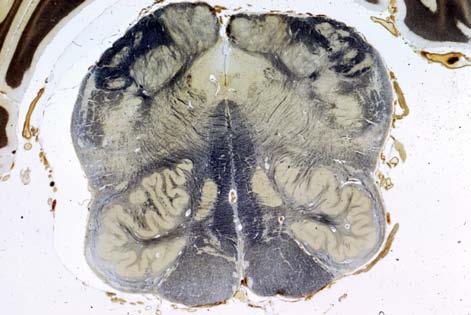
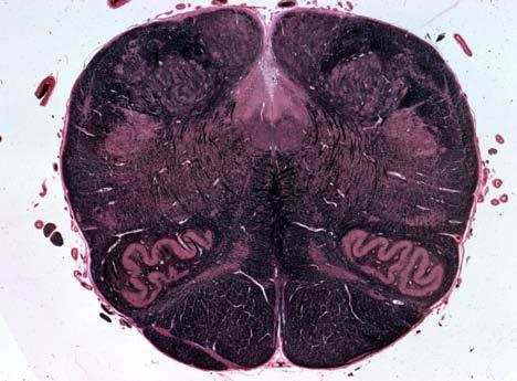
8 SACRAL CORD Suzanne S. Stensaas CERVICAL CORD Suzanne S. Stensaas


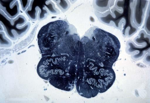
9 CAUDAL AND ROSTRAL MEDULLA Suzanne S. Stensaas


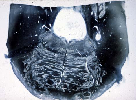
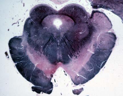
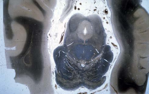
10 CAUDAL PONS Suzanne S. Stensaas MID-PONS Suzanne S. Stensaas


11 CAUDAL MIDBRAIN Suzanne S. Stensaas ROSTRAL MIDBRAIN AND THALAMUS Suzanne S. Stensaas
General Sensory Pathways of the Trunk and Limbs
 General Sensory Pathways of the Trunk and Limbs Lecture Objectives Describe gracile and cuneate tracts and pathways for conscious proprioception, touch, pressure and vibration from the limbs and trunk.
SOMATIC SENSATION PART I: ALS ANTEROLATERAL SYSTEM (or SPINOTHALAMIC SYSTEM) FOR PAIN AND TEMPERATURE
 FOR PAIN AND TEMPERATURE") Dental Neuroanatomy Thursday, February 3, 2011 Suzanne S. Stensaas, PhD SOMATIC SENSATION PART I: ALS ANTEROLATERAL SYSTEM (or SPINOTHALAMIC SYSTEM) FOR PAIN AND TEMPERATURE Reading: Waxman 26 th ed, :
Dental Neuroanatomy Thursday, February 3, 2011 Suzanne S. Stensaas, PhD SOMATIC SENSATION PART I: ALS ANTEROLATERAL SYSTEM (or SPINOTHALAMIC SYSTEM) FOR PAIN AND TEMPERATURE Reading: Waxman 26 th ed, :
Medical Neuroscience Tutorial
 Pain Pathways Medical Neuroscience Tutorial Pain Pathways MAP TO NEUROSCIENCE CORE CONCEPTS 1 NCC1. The brain is the body's most complex organ. NCC3. Genetically determined circuits are the foundation
SOMATOSENSORY SYSTEMS: Pain and Temperature Kimberle Jacobs, Ph.D.
 SOMATOSENSORY SYSTEMS: Pain and Temperature Kimberle Jacobs, Ph.D. Sensory systems are afferent, meaning that they are carrying information from the periphery TOWARD the central nervous system. The somatosensory
SOMATOSENSORY SYSTEMS: Conscious and Non-Conscious Proprioception Kimberle Jacobs, Ph.D.
 SOMATOSENSORY SYSTEMS: Conscious and Non-Conscious Proprioception Kimberle Jacobs, Ph.D. Divisions of Somatosensory Systems The pathways that convey sensory modalities from the body to consciousness are
Posterior White Column-Medial Lemniscal Pathway
 Posterior White Column-Medial Lemniscal Pathway Modality: Discriminative Touch Sensation (include Vibration) and Conscious Proprioception Receptor: Most receptors except free nerve endings Ist Neuron:
Our senses provide us with wonderful capabilities. If you had to lose one, which would it be?
 Our senses provide us with wonderful capabilities. If you had to lose one, which would it be? Neurological disorders take away sensation without a choice! http://neuroscience.uth.tmc.edu/s2/chapter04.html
SENSORY (ASCENDING) SPINAL TRACTS
 SPINAL TRACTS") SENSORY (ASCENDING) SPINAL TRACTS Dr. Jamila El-Medany Dr. Essam Eldin Salama OBJECTIVES By the end of the lecture, the student will be able to: Define the meaning of a tract. Distinguish between the different
SENSORY (ASCENDING) SPINAL TRACTS Dr. Jamila El-Medany Dr. Essam Eldin Salama OBJECTIVES By the end of the lecture, the student will be able to: Define the meaning of a tract. Distinguish between the different
Lecturer. Prof. Dr. Ali K. Al-Shalchy MBChB/ FIBMS/ MRCS/ FRCS 2014
 Lecturer Prof. Dr. Ali K. Al-Shalchy MBChB/ FIBMS/ MRCS/ FRCS 2014 Dorsal root: The dorsal root carries both myelinated and unmyelinated afferent fibers to the spinal cord. Posterior gray column: Long
Laurie L. Wellman Ph.D.
 Laurie L. Wellman Ph.D. Theodore Tzavaras MD2015 Eastern Virginia Medical School Dr. Craig Goodmurphy Anatomy Guy 1. Discuss -- what is pain? 2. Outline the Anterolateral Quadrant (ALQ) pathway 3. Locate
Somatosensory System. Steven McLoon Department of Neuroscience University of Minnesota
 Somatosensory System Steven McLoon Department of Neuroscience University of Minnesota 1 Course News Dr. Riedl s review session this week: Tuesday (Oct 10) 4-5pm in MCB 3-146B 2 Sensory Systems Sensory
The Nervous System: Sensory and Motor Tracts of the Spinal Cord
 15 The Nervous System: Sensory and Motor Tracts of the Spinal Cord PowerPoint Lecture Presentations prepared by Steven Bassett Southeast Community College Lincoln, Nebraska Introduction Millions of sensory
Non-cranial nerve nuclei
 Brainstem Non-cranial nerve nuclei Nucleus Gracile nucleus Cuneate nucleus Infeiro olivary nucleus Pontine nucleus inferior colliculus superior colliculus Red nucleus Substantia nigra Pretectal area Site
Pathways of proprioception
 The Autonomic Nervous Assess Prof. Fawzia Al-Rouq Department of Physiology College of Medicine King Saud University Pathways of proprioception System posterior column& Spinocerebellar Pathways https://www.youtube.com/watch?v=pmeropok6v8
Anatomical Substrates of Somatic Sensation
 Anatomical Substrates of Somatic Sensation John H. Martin, Ph.D. Center for Neurobiology & Behavior Columbia University CPS The 2 principal somatic sensory systems: 1) Dorsal column-medial lemniscal system
General Sensory Pathways of the Face Area, Taste Pathways and Hearing Pathways
 General Sensory Pathways of the Face Area, Taste Pathways and Hearing Pathways Lecture Objectives Describe pathways for general sensations (pain, temperature, touch and proprioception) from the face area.
Thalamus and Sensory Functions of Cerebral Cortex
 Thalamus and Sensory Functions of Cerebral Cortex I: To describe the functional divisions of thalamus. II: To state the functions of thalamus and the thalamic syndrome. III: To define the somatic sensory
By Dr. Saeed Vohra & Dr. Sanaa Alshaarawy
 By Dr. Saeed Vohra & Dr. Sanaa Alshaarawy 1 By the end of the lecture, students will be able to : Distinguish the internal structure of the components of the brain stem in different levels and the specific
UNIVERSITY OF JORDAN FACULTY OF MEDICINE DEPARTMENT OF PHYSIOLOGY & BIOCHEMISTRY NEUROPHYSIOLOGY (MEDICAL), SPRING 2014
, SPRING 2014") UNIVERSITY OF JORDAN FACULTY OF MEDICINE DEPARTMENT OF PHYSIOLOGY & BIOCHEMISTRY NEUROPHYSIOLOGY (MEDICAL), SPRING 2014 Textbook of Medical Physiology by: Guyton & Hall, 12 th edition 2011 Eman Al-Khateeb,
UNIVERSITY OF JORDAN FACULTY OF MEDICINE DEPARTMENT OF PHYSIOLOGY & BIOCHEMISTRY NEUROPHYSIOLOGY (MEDICAL), SPRING 2014 Textbook of Medical Physiology by: Guyton & Hall, 12 th edition 2011 Eman Al-Khateeb,
V1-ophthalmic. V2-maxillary. V3-mandibular. motor
 4. Trigeminal Nerve I. Objectives:. Understand the types of sensory information transmitted by the trigeminal system.. Describe the major peripheral divisions of the trigeminal nerve and how they innervate
Somatic Sensory System I. Background
 Somatic Sensory System I. Background A. Differences between somatic senses and other senses 1. Receptors are distributed throughout the body as opposed to being concentrated at small, specialized locations
Brainstem. Steven McLoon Department of Neuroscience University of Minnesota
 Brainstem Steven McLoon Department of Neuroscience University of Minnesota 1 Course News Change in Lab Sequence Week of Oct 2 Lab 5 Week of Oct 9 Lab 4 2 Goal Today Know the regions of the brainstem. Know
1. Somatosensory Pathways
 1. Somatosensory Pathways Objectives 1. Describe the general characteristics of sensory pathways 2. Understand the general organization and numbered areas of spinal cord gray matter 3. Understand dermatomes
Auditory and Vestibular Systems
 Auditory and Vestibular Systems Objective To learn the functional organization of the auditory and vestibular systems To understand how one can use changes in auditory function following injury to localize
Spinal Cord Organization. January 12, 2011
 Spinal Cord Organization January 12, 2011 Spinal Cord 31 segments terminates at L1-L2 special components - conus medullaris - cauda equina no input from the face Spinal Cord, Roots & Nerves Dorsal root
Biology 218 Human Anatomy
 Chapter 21 Adapted form Tortora 10 th ed. LECTURE OUTLINE A. Overview of Sensations (p. 652) 1. Sensation is the conscious or subconscious awareness of external or internal stimuli. 2. For a sensation
Unit VIII Problem 3 Neuroanatomy: Brain Stem, Cranial Nerves and Scalp
 Unit VIII Problem 3 Neuroanatomy: Brain Stem, Cranial Nerves and Scalp - Brain stem: It is connected to the cerebellum and cerebral hemispheres. Rostral end of brain stem: diencephalon is the area which
Fig Cervical spinal nerves. Cervical enlargement C7. Dural sheath. Subarachnoid space. Thoracic. Spinal cord Vertebra (cut) spinal nerves
 spinal nerves") Fig. 13.1 C1 Cervical enlargement C7 Cervical spinal nerves Dural sheath Subarachnoid space Thoracic spinal nerves Spinal cord Vertebra (cut) Lumbar enlargement Medullary cone T12 Spinal nerve Spinal nerve
Fig. 13.1 C1 Cervical enlargement C7 Cervical spinal nerves Dural sheath Subarachnoid space Thoracic spinal nerves Spinal cord Vertebra (cut) Lumbar enlargement Medullary cone T12 Spinal nerve Spinal nerve
lecture #2 Done by : Tyma'a Al-zaben
 lecture #2 Done by : Tyma'a Al-zaben ** Hello SERTONIN! note:: the slide included within the sheet but make sure back to slide for pictures in the previous lecture we talk about ascending tract and its
THE VISUAL PATHWAY FOR DENTAL STUDENTS
 Neuroanatomy Suzanne S. Stensaas, Ph.D. February 16, 2012 Objectives: THE VISUAL PATHWAY FOR DENTAL STUDENTS A. Draw the expected visual fields seen in classic lesions of the nerve, chiasm, thalamus, optic
SOMATOSENSORY SYSTEMS
 SOMATOSENSORY SYSTEMS Schematic diagram illustrating the neural pathways that convey somatosensory information to the cortex and, subsequently, to the motor system. Double arrows show reciprocal connections.
Fig.1: A, Sagittal 110x110 mm subimage close to the midline, passing through the cingulum. Note that the fibers of the corpus callosum run at a
 Fig.1 E Fig.1:, Sagittal 110x110 mm subimage close to the midline, passing through the cingulum. Note that the fibers of the corpus callosum run at a slight angle are through the plane (blue dots with
Unit VIII Problem 1 Physiology: Sensory Pathway
 Unit VIII Problem 1 Physiology: Sensory Pathway - Process of sensation: Sensory receptors: they are specialized cells considered as biologic signal transducers which can detect stimuli and convert them
Sheet lab 3. Page 8B Section1 of medulla at pyramidal {motor} decussation:
 Sheet lab 3 Page 8B Section1 of medulla at pyramidal {motor} decussation: This section is at lower third of medulla and is the most close part to spinal cord and it has some characteristic of spinal cord
C:\Documents and Settings\sstensaas\Desktop\dental visual 2010\VisualPath dental 2010.docVisualPath dental 2010.doc
 Neuroanatomy Suzanne Stensaas April 8, 2010, 10:00-12:00 p.m. Reading: Waxman Ch. 15, Computer Resources: HyperBrain Ch 7 THE VISUAL PATHWAY Objectives: 1. Describe the pathway of visual information from
Pain and Temperature Objectives
 Pain and Temperature Objectives 1. Describe the types of sensory receptors that transmit pain and temperature. 2. Understand how axon diameter relates to transmission of pain and temp information. 3. Describe
DEVELOPMENT OF BRAIN
 Ahmed Fathalla OBJECTIVES At the end of the lecture, students should: List the components of brain stem. Describe the site of brain stem. Describe the relations between components of brain stem & their
b. The groove between the two crests is called 2. The neural folds move toward each other & the fuse to create a
 Chapter 13: Brain and Cranial Nerves I. Development of the CNS A. The CNS begins as a flat plate called the B. The process proceeds as: 1. The lateral sides of the become elevated as waves called a. The
 Spinal Cord Tracts DESCENDING SPINAL TRACTS: Are concerned with somatic motor function, modification of ms. tone, visceral innervation, segmental reflexes. Main tracts arise form cerebral cortex and others
Anatomy of the Spinal Cord
 Spinal Cord Anatomy of the Spinal Cord Anatomy of the Spinal Cord Posterior spinal arteries Lateral corticospinal tract Dorsal column Spinothalamic tract Anterior spinal artery Anterior white commissure
Somatic Sensation (MCB160 Lecture by Mu-ming Poo, Friday March 9, 2007)
") Somatic Sensation (MCB160 Lecture by Mu-ming Poo, Friday March 9, 2007) Introduction Adrian s work on sensory coding Spinal cord and dorsal root ganglia Four somatic sense modalities Touch Mechanoreceptors
Somatic Sensation (MCB160 Lecture by Mu-ming Poo, Friday March 9, 2007) Introduction Adrian s work on sensory coding Spinal cord and dorsal root ganglia Four somatic sense modalities Touch Mechanoreceptors
Cortical Control of Movement
 Strick Lecture 2 March 24, 2006 Page 1 Cortical Control of Movement Four parts of this lecture: I) Anatomical Framework, II) Physiological Framework, III) Primary Motor Cortex Function and IV) Premotor
Skin types: hairy and glabrous (e.g. back vs. palm of hand)
") Lecture 19 revised 03/10 The Somatic Sensory System Skin- the largest sensory organ we have Also protects from evaporation, infection. Skin types: hairy and glabrous (e.g. back vs. palm of hand) 2 major
Lecture 19 revised 03/10 The Somatic Sensory System Skin- the largest sensory organ we have Also protects from evaporation, infection. Skin types: hairy and glabrous (e.g. back vs. palm of hand) 2 major
THE COCHLEA AND AUDITORY PATHWAY
 Dental Neuroanatomy Suzanne S. Stensaas, PhD February 23, 2012 Reading: Waxman, Chapter 16, Review pictures in a Histology book Computer Resources: http://www.cochlea.org/ - Promenade around the Cochlea
Lecture VIII. The Spinal Cord, Reflexes and Brain Pathways!
 Reflexes and Brain Bio 3411! Monday!! 1! Readings! NEUROSCIENCE 5 th ed: Review Chapter 1 pp. 11-21;!!Read Chapter 9 pp. 189-194, 198! THE BRAIN ATLAS 3 rd ed:! Read pp. 4-17 on class web site! Look at
Neural Integration I: Sensory Pathways and the Somatic Nervous System
 15 Neural Integration I: Sensory Pathways and the Somatic Nervous System PowerPoint Lecture Presentations prepared by Jason LaPres Lone Star College North Harris An Introduction to Sensory Pathways and
The Somatosensory System
 The Somatosensory System Reading: BCP Chapter 12 cerebrovortex.com Divisions of the Somatosensory System Somatosensory System Exteroceptive External stimuli Proprioceptive Body position Interoceptive Body
Nervous System. The Peripheral Nervous System Agenda Review of CNS v. PNS PNS Basics Cranial Nerves Spinal Nerves Reflexes Pathways
 Nervous System Agenda Review of CNS v. PNS PNS Basics Cranial Nerves Spinal Nerves Sensory Motor Review of CNS v. PNS Central nervous system (CNS) Brain Spinal cord Peripheral nervous system (PNS) All
I: To describe the pyramidal and extrapyramidal tracts. II: To discuss the functions of the descending tracts.
 Descending Tracts I: To describe the pyramidal and extrapyramidal tracts. II: To discuss the functions of the descending tracts. III: To define the upper and the lower motor neurons. 1. The corticonuclear
CHAPTER 10 THE SOMATOSENSORY SYSTEM
 CHAPTER 10 THE SOMATOSENSORY SYSTEM 10.1. SOMATOSENSORY MODALITIES "Somatosensory" is really a catch-all term to designate senses other than vision, hearing, balance, taste and smell. Receptors that could
Doctor Osama Asa ad Khader. Mohammad Alsalem
 6 Doctor 2015 Osama Asa ad Khader Mohammad Alsalem A quick revision for the spinal cord blood supply: Arterial Blood supply of spinal cord The spinal cord got its arterial supply by two ways: Longitudinal
Introduction and Basic structural organization of the nervous system
 Introduction and Basic structural organization of the nervous system **the slides are in bold and the book is in red Done by : razan krishan & marah marahleh INTRODUCTION The nervous system, along with
Cranial Nerves. Steven McLoon Department of Neuroscience University of Minnesota
 Cranial Nerves Steven McLoon Department of Neuroscience University of Minnesota 1 Course News Change in Lab Sequence Week of Oct 2 Lab 5 Week of Oct 9 Lab 4 2 Sensory and Motor Systems Sensory Systems:
Cortical Organization. Functionally, cortex is classically divided into 3 general types: 1. Primary cortex:. - receptive field:.
 Cortical Organization Functionally, cortex is classically divided into 3 general types: 1. Primary cortex:. - receptive field:. 2. Secondary cortex: located immediately adjacent to primary cortical areas,
Review or skim Ch 12 on the vascular supply of the brain. Just look at pictures and legends for the clinical part at the end.
 Dental Neuroanatomy January 20 and 27, 10-12, 2011 Suzanne S. Stensaas, Ph.D. Dear Students: Please print these notes and bring them with you. My style is to use a Tablet PC and I draw on either a Word
How strong is it? What is it? Where is it? What must sensory systems encode? 9/8/2010. Spatial Coding: Receptive Fields and Tactile Discrimination
 Spatial Coding: Receptive Fields and Tactile Discrimination What must sensory systems encode? How strong is it? What is it? Where is it? When the brain wants to keep certain types of information distinct,
Spatial Coding: Receptive Fields and Tactile Discrimination
 Spatial Coding: Receptive Fields and Tactile Discrimination What must sensory systems encode? How strong is it? What is it? Where is it? When the brain wants to keep certain types of information distinct,
*Anteriolateral spinothalamic tract (STT) : a sensory pathway that is positioned anteriorly and laterally in the spinal cord.
 : a sensory pathway that is positioned anteriorly and laterally in the spinal cord.") *somatic sensations : PAIN *Anteriolateral spinothalamic tract (STT) : a sensory pathway that is positioned anteriorly and laterally in the spinal cord. *This pathway carries a variety of sensory modalities:
*somatic sensations : PAIN *Anteriolateral spinothalamic tract (STT) : a sensory pathway that is positioned anteriorly and laterally in the spinal cord. *This pathway carries a variety of sensory modalities:
Vision II. Steven McLoon Department of Neuroscience University of Minnesota
 Vision II Steven McLoon Department of Neuroscience University of Minnesota 1 Ganglion Cells The axons of the retinal ganglion cells form the optic nerve and carry visual information into the brain. 2 Optic
OVERVIEW. Today. Sensory and Motor Neurons. Thursday. Parkinsons Disease. Administra7on. Exam One Bonus Points Slides Online
 OVERVIEW Today Sensory and Motor Neurons Thursday Parkinsons Disease Administra7on Exam One Bonus Points Slides Online 7 major descending motor control pathways from Cerebral Cortex or Brainstem
1) Drop off in the Bi 150 box outside Baxter 331 or to the head TA (jcolas).
 Drop off in the Bi 150 box outside Baxter 331 or to the head TA (jcolas).") Bi/CNS/NB 150 Problem Set 5 Due: Tuesday, Nov. 24, at 4:30 pm Instructions: 1) Drop off in the Bi 150 box outside Baxter 331 or e-mail to the head TA (jcolas). 2) Submit with this cover page. 3) Use a
Bi/CNS/NB 150 Problem Set 5 Due: Tuesday, Nov. 24, at 4:30 pm Instructions: 1) Drop off in the Bi 150 box outside Baxter 331 or e-mail to the head TA (jcolas). 2) Submit with this cover page. 3) Use a
M555 Medical Neuroscience Lab 1: Gross Anatomy of Brain, Crainal Nerves and Cerebral Blood Vessels
 M555 Medical Neuroscience Lab 1: Gross Anatomy of Brain, Crainal Nerves and Cerebral Blood Vessels Anatomical Directions Terms like dorsal, ventral, and posterior provide a means of locating structures
Lecture 4 The BRAINSTEM Medulla Oblongata
 Lecture 4 The BRAINSTEM Medulla Oblongata Introduction to brainstem 1- Medulla oblongata 2- Pons 3- Midbrain - - - occupies the posterior cranial fossa of the skull. connects the narrow spinal cord
PHYSIOLOHY OF BRAIN STEM
 PHYSIOLOHY OF BRAIN STEM Learning Objectives The brain stem is the lower part of the brain. It is adjoining and structurally continuous with the spinal cord. 1 Mid Brain 2 Pons 3 Medulla Oblongata The
Mechanosensation. Central Representation of Touch. Wilder Penfield. Somatotopic Organization
 Mechanosensation Central Representation of Touch Touch and tactile exploration Vibration and pressure sensations; important for clinical testing Limb position sense John H. Martin, Ph.D. Center for Neurobiology
Department of Neurology/Division of Anatomical Sciences
 Spinal Cord I Lecture Outline and Objectives CNS/Head and Neck Sequence TOPIC: FACULTY: THE SPINAL CORD AND SPINAL NERVES, Part I Department of Neurology/Division of Anatomical Sciences LECTURE: Monday,
CEREBRUM & CEREBRAL CORTEX
 CEREBRUM & CEREBRAL CORTEX Seonghan Kim Dept. of Anatomy Inje University, College of Medicine THE BRAIN ANATOMICAL REGIONS A. Cerebrum B. Diencephalon Thalamus Hypothalamus C. Brain Stem Midbrain Pons
1. Which part of the brain is responsible for planning and initiating movements?
 Section: Chapter 10: Multiple Choice 1. Which part of the brain is responsible for planning and initiating movements? p.358 frontal lobe hippocampus basal ganglia cerebellum 2. The prefrontal cortex is
Chapter 6. Gathering information; the sensory systems
 Chapter 6 Gathering information; the sensory systems Gathering information the sensory systems The parts of the nervous system that receive and process information are termed sensory systems. There are
IV. THE SPINAL CORD BLOOD SUPPLY
 IV. THE SPINAL CORD Spinal cord is covered by o Pia Mater Spinalis Film Teminale Denticulate Ligament ---------------------- Cordotomy o Arachnoid Membrane Subarachnoid Space ----------------------- Lumbar
Nervous System. Student Learning Objectives:
 Nervous System Student Learning Objectives: Identify the primary parts of the neuron Identify the major structures of the central nervous system Identify the major structures of the peripheral nervous
Brainstem. By Dr. Bhushan R. Kavimandan
 Brainstem By Dr. Bhushan R. Kavimandan Development Ventricles in brainstem Mesencephalon cerebral aqueduct Metencephalon 4 th ventricle Mylencephalon 4 th ventricle Corpus callosum Posterior commissure
Done by : Areej Al-Hadidi
 Brainstem &diencephalon Done by : Areej Al-Hadidi Brainstem Functions Ascending and descending tracts Reflex centers Cardiovascular and respiratory centers Coughing, sneezing, swallowing Nuclei of the
Brainstem. Amadi O. Ihunwo, PhD School of Anatomical Sciences
 Brainstem Amadi O. Ihunwo, PhD School of Anatomical Sciences Lecture Outline Constituents Basic general internal features of brainstem External and Internal features of Midbrain Pons Medulla Constituents
THE VESTIBULAR APPRATUS AND PATHWAY
 Dental Neuroanatomy February 23, 2012 Suzanne Stensaas, Ph.D. Reading: Waxman Chapter 17 Also pp 105-108 on control of eye movments Computer Resources: HyperBrain Ch. 8 Vestibulospinal Pathway Quiz http://library.med.utah.edu/kw/animations/hyperbrain/pathways/
Collin County Community College. BIOL 2401 : Anatomy/ Physiology PNS
 Collin County Community College BIOL 2401 : Anatomy/ Physiology PNS Peripheral Nervous System (PNS) PNS all neural structures outside the brain and spinal cord Includes sensory receptors, peripheral nerves,
Unit VIII Problem 5 Physiology: Cerebellum
 Unit VIII Problem 5 Physiology: Cerebellum - The word cerebellum means: the small brain. Note that the cerebellum is not completely separated into 2 hemispheres (they are not clearly demarcated) the vermis
The Spinal Cord. The Nervous System. The Spinal Cord. The Spinal Cord 1/2/2016. Continuation of CNS inferior to foramen magnum.
 The Nervous System Spinal Cord Continuation of CNS inferior to foramen magnum Simpler than the brain Conducts impulses to and from brain Two way conduction pathway Reflex actions Passes through vertebral
Chapter 8. Control of movement
 Chapter 8 Control of movement 1st Type: Skeletal Muscle Skeletal Muscle: Ones that moves us Muscles contract, limb flex Flexion: a movement of a limb that tends to bend its joints, contraction of a flexor
Overview of the Nervous System (some basic concepts) Steven McLoon Department of Neuroscience University of Minnesota
 Steven McLoon Department of Neuroscience University of Minnesota") Overview of the Nervous System (some basic concepts) Steven McLoon Department of Neuroscience University of Minnesota 1 Coffee Hour Tuesday (Sept 11) 10:00-11:00am Friday (Sept 14) 8:30-9:30am Surdyk s
Overview of the Nervous System (some basic concepts) Steven McLoon Department of Neuroscience University of Minnesota 1 Coffee Hour Tuesday (Sept 11) 10:00-11:00am Friday (Sept 14) 8:30-9:30am Surdyk s
Sensory information processing, somato-sensory systems
 mm? Sensory information processing, somato-sensory systems Recommended literature 1. Kandel ER, Schwartz JH, Jessel TM (2000) Principles of Neural Science, McGraw-Hill, Ch. xx. 2. Berne EM, Levy MN, Koeppen
Introduction to the Central Nervous System: Internal Structure
 Introduction to the Central Nervous System: Internal Structure Objective To understand, in general terms, the internal organization of the brain and spinal cord. To understand the 3-dimensional organization
Cranial Nerve VIII (The Vestibulo-Cochlear Nerve)
") Cranial Nerve VIII (The Vestibulo-Cochlear Nerve) Please view our Editing File before studying this lecture to check for any changes. Color Code Important Doctors Notes Notes/Extra explanation Objectives
Cranial Nerve VIII (The Vestibulo-Cochlear Nerve) Please view our Editing File before studying this lecture to check for any changes. Color Code Important Doctors Notes Notes/Extra explanation Objectives
Last time we talked about the descending pathways of pain and the ALS. Today we will continue talking about these descending pathways.
 Last time we talked about the descending pathways of pain and the ALS. Today we will continue talking about these descending pathways. Each higher level will control the level under It. In controlling
3/20/13. :: Slide 1 :: :: Slide 39 :: How Is the Nervous System Organized? Central Nervous System Peripheral Nervous System and Endocrine System
 :: Slide 1 :: :: Slide 39 :: How Is the Nervous System Organized? Central Nervous System Peripheral Nervous System and Endocrine System The nervous system is organized into several major branches, each
skilled pathways: distal somatic muscles (fingers, hands) (brainstem, cortex) are giving excitatory signals to the descending pathway
 (brainstem, cortex) are giving excitatory signals to the descending pathway") L15 - Motor Cortex General - descending pathways: how we control our body - motor = somatic muscles and movement (it is a descending motor output pathway) - two types of movement: goal-driven/voluntary
L15 - Motor Cortex General - descending pathways: how we control our body - motor = somatic muscles and movement (it is a descending motor output pathway) - two types of movement: goal-driven/voluntary
P. Hitchcock, Ph.D. Department of Cell and Developmental Biology Kellogg Eye Center. Wednesday, 16 March 2009, 1:00p.m. 2:00p.m.
 Normal CNS, Special Senses, Head and Neck TOPIC: CEREBRAL HEMISPHERES FACULTY: LECTURE: READING: P. Hitchcock, Ph.D. Department of Cell and Developmental Biology Kellogg Eye Center Wednesday, 16 March
The neurvous system senses, interprets, and responds to changes in the environment. Two types of cells makes this possible:
 NERVOUS SYSTEM The neurvous system senses, interprets, and responds to changes in the environment. Two types of cells makes this possible: the neuron and the supporting cells ("glial cells"). Neuron Neurons
Nsci 2100: Human Neuroanatomy 2017 Examination 3
 Name KEY Lab Section Nsci 2100: Human Neuroanatomy 2017 Examination 3 On this page, write your name and lab section. On your bubble answer sheet, enter your name (last name, space, first name), internet
Ch. 47 Somatic Sensations: Tactile and Position Senses (Reading Homework) - Somatic senses: three types (1) Mechanoreceptive somatic senses: tactile
 - Somatic senses: three types (1) Mechanoreceptive somatic senses: tactile") Ch. 47 Somatic Sensations: Tactile and Position Senses (Reading Homework) - Somatic senses: three types (1) Mechanoreceptive somatic senses: tactile and position sensations (2) Thermoreceptive senses:
Ch. 47 Somatic Sensations: Tactile and Position Senses (Reading Homework) - Somatic senses: three types (1) Mechanoreceptive somatic senses: tactile and position sensations (2) Thermoreceptive senses:
Brain and Cranial Nerves (Ch. 15) Human Anatomy lecture. caudal = toward the spinal cord)
 Human Anatomy lecture. caudal = toward the spinal cord)") Insight: Some cranial nerve disorders Brain and Cranial Nerves (Ch. 15) Human Anatomy lecture I. Overview (Directional terms: rostral = toward the forehead caudal = toward the spinal cord) A. 3 Major parts
Insight: Some cranial nerve disorders Brain and Cranial Nerves (Ch. 15) Human Anatomy lecture I. Overview (Directional terms: rostral = toward the forehead caudal = toward the spinal cord) A. 3 Major parts
Pain classifications slow and fast
 Pain classifications slow and fast Fast Pain Slow Pain Sharp, pricking (Aδ) fiber Short latency Well localized Short duration Dull, burning (C) fiber Slower onset Diffuse Long duration Less emotional Emotional,
The Nervous System. Divisions of the Nervous System. Branches of the Autonomic Nervous System. Central versus Peripheral
 The Nervous System Divisions of the Nervous System Central versus Peripheral Central Brain and spinal cord Peripheral Everything else Somatic versus Autonomic Somatic Nerves serving conscious sensations
THE COCHLEA AND AUDITORY PATHWAY
 Dental Neuroanatomy Suzanne S. Stensaas, PhD April 14, 2010 Reading: Waxman, Chapter 16, Review pictures in a Histology book Computer Resources: http://www.cochlea.org/ - Promenade around the Cochlea HyperBrain
The NIHSS score is 4 (considering 2 pts for the ataxia involving upper and lower limbs.
 Neuroscience case 5 1. Speech comprehension, ability to speak, and word use were normal in Mr. Washburn, indicating that aphasia (cortical language problem) was not involved. However, he did have a problem
Lecture - Chapter 13: Central Nervous System
 Lecture - Chapter 13: Central Nervous System 1. Describe the following structures of the brain, what is the general function of each: a. Cerebrum b. Diencephalon c. Brain Stem d. Cerebellum 2. What structures
Chapter 12b. Overview
 Chapter 12b Spinal Cord Overview Spinal cord gross anatomy Spinal meninges Sectional anatomy Sensory pathways Motor pathways Spinal cord pathologies 1 The Adult Spinal Cord About 18 inches (45 cm) long
Nervous System C H A P T E R 2
 Nervous System C H A P T E R 2 Input Output Neuron 3 Nerve cell Allows information to travel throughout the body to various destinations Receptive Segment Cell Body Dendrites: receive message Myelin sheath
Brain Stem and cortical control of motor function. Dr Z Akbari
 Brain Stem and cortical control of motor function Dr Z Akbari Brain stem control of movement BS nuclear groups give rise to descending motor tracts that influence motor neurons and their associated interneurons