Supplemental Information. A Labeled-Line Neural Circuit. for Pheromone-Mediated Sexual Behaviors in Mice
|
|
|
- Candace Powers
- 5 years ago
- Views:
Transcription
1 Neuron, Volume Supplemental Information A Labeled-Line Neural Circuit for Pheromone-Mediated Sexual Behaviors in Mice Kentaro K. Ishii, Takuya Osakada, Hiromi Mori, Nobuhiko Miyasaka, Yoshihiro Yoshihara, Kazunari Miyamichi, and Kazushige Touhara

2 0 Figure S. Whole brain analysis of anterograde mapping from VRp-expresing Sensory Neurons, related to Figure (A) Table showing brain regions analyzed for tdtomato+ cells in day, and. The intensity of anterograde labeling was determined by the % of animals with tdtomato+ cells in the area among all animals; blank: < 0%, +: 0%, ++: 0%, +++: 0%. Day, n =, day, n = 0, day, n =. (B) Schematic and representative coronal section showing tdtomato+ (shown in red) cells in day -sacrificed animals. DAPI staining is shown in blue. Scale bar, 0 μm. Abbreviations are as following. LSV, lateral septum nucleus ventral part. BNST, bed nucleus of stria terminalis. SHy, septohypothalamic nucleus. DEn, dorsal endopiriform nucleus. Pir, piriform cortex. StHy, striohypothalamic nuclesus. MPA, medial preoptic area. EA, sublenticular extend amygdala. MCPO, magnocell preoptic nucleus. VEn, ventral endopiriform nucleus. CxA, cortex-amygdala
3 0 transition area. VCl, ventral part of claustrum. PaXi, paraxiphoid nucleus of thalamus. VMH, ventromedial hypothalamic nucleus. Arc, arcuate nucleus. MeA, medial amygdala. CeA, central amygdala. BMA, basomedial amygdala. ACo, anterior cortical amygdala. PLCo, posterolateral cortical amygdala. BLV, basolateral amygdala ventral part. LaDL, lateral amygdala dorsolateral part. RMM, retromammillary nucleus medial part. PM, premammillary nucleus. DG, dentate gyrus. Or, oriens layer hippocampus. AHi, amygdalohippocampus. VS, ventral subiculum. PMCo, posteromedial cortical amygdala. BMP, basomedial amygdala posterior part. BLP, basolateral amygdala posterior part. RAPir, rostral amygdalopiriform. MnR, median raphe. GrDG, granular dentate gyrus. LMol, lacunosum moleculare. STr, subiculum transition area. Ent, entorhinal cortex.
 Number of cfos+ cells per mm in the MeApd and MeApv of mice exposed to ESP (black) or")
4 Figure S. Histochemical Characterization of ESP-responding Neurons and Projection Neurons in the MeA, related to Figure (A) Number of cfos+ cells per mm in the MeApd and MeApv of mice exposed to ESP (black) or the control (white) in female (left) and male (right) mice. n = for ESP and n = for control (females); n = each for ESP and control (males). Student s t-test with Bonferroni correction, *p < 0.0, **p < 0.0.
5 0 (B) Spatial organization of MeA sub-nuclei along the anterior-posterior axis. Eight typical coronal sections are shown that approximately correspond to the eight sections analyzed below. (C and D) Distribution of cfos+ cells in ESP-exposed female (B) or male (C) mice. Cells were counted from eight unilateral coronal sections from Bregma.0 mm to. mm. Female mice, control n = 0, ESP n =. Male mice, control n =, ESP n =. (E) Representative coronal sections showing cfos (green) and vglut+ or GAD+ (red) mrna expression in the MeApv following exposure to the ESP stimulus. Small panels represent higher magnification of each boxed area in merged and separated channels. (F) Proportion of cfos+ and marker+ (vglut+ or GAD+) cells among all cfos+ cells. n = animals each. (G) Representative coronal sections showing VMHd- or MPA-projectors labeled with red Retrobeads and vglut+ or GAD+ mrna-expressing cells. vglut+ and GAD+ were labeled with different RNA probes on the same section. Signals are pseudo-colored in green, and co-localization with Retrobeads is shown individually. (H) Number of vglut+, GAD+, or marker-negative cells among Retrobeads+ cells per MeApv section. Percentage of Retrobeads+ and vglut+ cells among total Retrobeads+ cells is shown in bar graphs. n = animal. Scale bar, 00 μm. Error bars, SEM.
 Quantitative analysis for normalization methods used in the axon mapping data of vglut+ MeApv")
6 0 Figure S. Additional Analysis on Axonal Arbor Distribution of MeApv vglut+ Neurons, related to Figure (A and B) Quantitative analysis for normalization methods used in the axon mapping data of vglut+ MeApv neurons (Figure ). We did not observe a significant correlation between mgfp+ area in CAV-injected brain regions (y-axis) and the number of source cells in the MeApv for VMHd- and MPA- projectors (A), suggesting that the axonal distributions are heterogeneous even among the same type of projectors. In contrast, we observed a significant correlation (p = ) between mgfp+ area in CAV-injected regions (y-axis) and total mgfp+ area in all counted regions (x-axis) (B). Thus, we used the proportion of mgfp+ area in a single brain region among the total mgfp+ area in all regions as a normalization strategy.
7 0 (C) Representative coronal section of the VMH showing mgfp (green in the left panel, black in the middle panel) and SypRuby (red in the left panel, black in the right panel) signals of VMHdprojectors in the MeApv. Scale bar, 00 μm. (D) Correlation between the number of SypRuby+ dots (y-axis) and the area of mgfp+ axons (xaxis). In this analysis, we pooled VMHd and MPA data from all VMHd-projectors, MPAprojectors, and pan-vglut labeling in male and female mice. We observed a positive correlation with R = 0.. (E) Correlation between the proportion of mgfp+ area ([VMHd]/[VMHd + MPA], x-axis) and the proportion of the number of SypRuby+ dots ([VMHd]/[VMHd + MPA], y-axis). We observed a strong positive correlation with R = 0..

8 0 0 Figure S. The VMHvl Is Necessary for Basal Lordosis in Female Mice, related to Figure (A) Schematic of the experimental setup and time line of procedures. AAV-FLEx-DTA or AAV- FLEx-GFP as a control was injected into the VMHvl of female Esr-Cre mice. (B) Representative coronal sections showing Esr mrna expression (red) in the VMHvl and in the arcuate nucleus (Arc) of AAV-FLEx-DTA-injected mice (Esr::DTA) and AAV-FLEx-GFPinjected mice (Esr::GFP). (C) Representative coronal sections showing SF mrna expression (red) in the VMHd of Esr::GFP and Esr::DTA mice. (D) Number of Esr+ cells in the VMHvl and Arc per unilateral section. Esr+ neurons were drastically reduced in the VMHvl, but not in the Arc. Esr::GFP, n = unilateral region. Esr::DTA, n =. (E) Number of SF+ cells in the VMHd per unilateral section. The number of SF+ neurons was also reduced by expressing DTA in Esr+ neurons, likely due to weak Cre activity in SF+ neurons in Esr-Cre mice. Esr::GFP, n =. Esr::DTA, n =. (F) Raster plots of behavior episodes during the mating assay. (G) Lordosis percentage of Esr::GFP and Esr::DTA mice exposed to ESP or the control stimulus. Note that addition of ESP to Esr::DTA mice did not rescue the basal lordosis defects, although we cannot exclude the possibility that this may be caused by non-specific ablation of a subset of SF+ neurons in the VMHd. Grey lines, trials from individual animals. Esr::GFP, n = animals. Esr::DTA, n =.
9 Error bars, SEM. Scale bar, 00 µm. (D) and (E) Unpaired Student s t-test, **p < 0.0. (G) Paired Student s t-test, **p < 0.0.
 Number of cfos-expressing VMHd-projectors in the MeApv per section")
10 0 Figure S. Additional Analysis of the Representation of Snake Signals and their Effect on Female Sexual Behaviors, related to Figures and (A) Number of cfos-expressing VMHd-projectors in the MeApv per section in female or male mice (left) and number of cfos-expressing MPA-projectors in the MeApv per section in female or male mice (right). Data for the control stimulus are the same as those used in Figure G. The number of animals used for each condition is shown below each bar. (B) Lordosis percentage of wild-type female mice pre-exposed to snakeskin or a control stimulus. Control, n = ; snakeskin n =. (C) Representative coronal sections from catfish analysis by using cfos coding (green) and intron (red) probes in the AOB. The stimulation conditions are shown in each image as in Figure A. Insets show high magnification images of the two boxed areas in each panel. Cells were counted from the glomerulus layer (GL) and the mitral cell layer (MCL).
11 0 (D) Proportion of dual-positive (nuclear and cytoplasmic cfos+) cells among total nuclear cfos+ expressing cells (A-index) in the GL and MCL. The number of animals used for each condition is shown below each bar. Control, Same, and Different conditions were collected as described in Figure B. (E) Representative coronal sections from catfish analysis in the MeApv by using NRa coding (green) and intron (red) probes. The stimulation conditions are shown in each image. Insets show high magnification images of the two boxed areas in each panel. (F) A-index in each condition of MeApv catfish in male and female mice as in panel D. D, dorsal, A, anterior, M, medial. Error bars, SEM. (A) and (B), Students t-test. (D) and (F), Oneway ANOVA on conditions with Bonferroni correction for female data set (D) and for data set of each sex (F). a vs. b, a vs. c, b vs. c, p < 0.0. Scale bars, 00 μm. 0
12
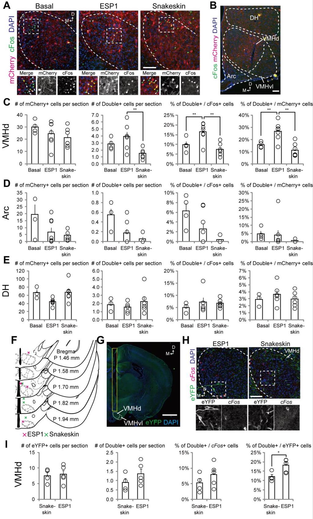
13 0 0 Figure S. Additional Analysis of TRAP in the VMHd, related to Figure (A) Representative images of coronal sections from ESP-, snakeskin- or basal activity-trap mice exposed to ESP. cfos+ cells (green) were detected by anti-cfos staining. Bottom panels show merged and separated channels for mcherry and cfos, with yellow arrowheads representing dual-positive, and white arrowheads representing cfos+ only cells. (B) Representative coronal section showing cfos+ and mcherry+ in the dorsal hypothalamus (DH) and arcuate nucleus (Arc). (C E) Number of mcherry+ and double+ (cfos+ mcherry+) cells per unilateral section, % of double+ cells among all mcherry+ cells or all cfos+ cells in VMHd (C), Arc (D) and DH (E). Basal, n = unilateral regions, ESP, n =, snakeskin, n =. (F) Distribution of fiber tips in the animals analyzed in Figures F and G. Red x, ESP-TRAP mice, green x, snakeskin-trap mice. P, posterior. (G) A representative image showing the location of optic fibers above the VMHd, and eyfp expression in the VMHd. (H) Representative coronal sections showing eyfp (green) and cfos mrna (red)-expressing cells from the ESP- or snakeskin-trap animals exposed to ESP. Bottom panels show separated channels of the areas indicated by white dashed boxes in the top images. (I) Number of eyfp+ and double+ (cfos+ eyfp+) cells per unilateral section, % of double+ cells among all cfos+ cells or all eyfp+ cells. ESP, n = unilateral VMHd, snakeskin, n =. Scale bar, 00 µm (A, B and H) or mm (G). One-way ANOVA with Bonferroni correction (C E) or Student s t-test (I), *p < 0.0, **p < 0.0.
 A representative coronal section showing mgfp and SypRuby expression in VMHd neurons following")
14 0 Figure S. SF+ VMHd Neurons Are Homogeneous with Respect to Their Axonal Projections, related to Figure (A) A representative coronal section showing mgfp and SypRuby expression in VMHd neurons following CAV-FLEx loxp -Flp injection into the PAGd and AAV-FLEx FRT -mgfp injection into the VMHd of female SF-Cre mice. Scale bar, 00 µm. (B) Fraction of source cells (defined by the expression of mgfp) in the VMHd for each injection condition. The number of animals for each condition is indicated at the bottom. Almost all source cells were restricted to the VMHd area. (C and D) Quantitative analysis for normalization methods. We observed a strong positive correlation (p = 0.00) between mgfp+ area in CAV-injected brain regions (y-axis) and the number of VMHd source cells (x-axis) for PAGd- or MPA-projectors (C). This was in contrast to the situation with MeApv source cells (Figure SA), suggesting that the axonal distribution of
15 0 SF+ VMHd neurons is highly homogeneous. We also observed a strong positive correlation (p = 0.00) between mgfp+ area in CAV-injected regions (y-axis) and the total mgfp+ area in all analyzed regions (x-axis) for PAGd- or MPA-projectors (D). Thus, for normalization, we divided the mgfp+ area in each region by the number of source cells and by the total mgfp+ area in all analyzed regions. (E) Representative images of binarized mgfp signals of a coronal section across eight brain regions collected from animals in which PAGd-projectors, MPA-projectors, or pan-vmhd projectors (CAV local injection) were labeled. (F and G) Proportion of mgfp+ area in each region normalized to the total mgfp+ area of brain regions (F), or mgfp+ area in each region normalized to the number of source cells in the VMHd (G). Error bars, SEM. Abbreviations, Sept, septum, Rapir, rostral amygdalopiriform area, lateral A, lateral amygdaloid nucleus, SN, substantia nigra, SubB, subbrachial nucleus, PAGd, dorsal periaqueductal gray. For other abbreviations, see Figures and.
 Representative coronal images showing traces of the cannula for CNO infusion above the PAGd (A) or MPA")
16 0 Figure S. Histological Verification of Cannula Locations in the MPA and PAGd, related to Figure (A and B) Representative coronal images showing traces of the cannula for CNO infusion above the PAGd (A) or MPA (B). Scale bar, mm. (C F) Schematics showing the locations of cannula tips from the coronal (C and E) and sagittal (D and F) views, in the PAGd (C and D) or MPA (E and F). Grey circles indicate individual cannula tips; yellow circular shadows indicate the estimated areas where the injected CNO may spread based on the data described previously (Stachniak et al., 0). The position of each cannula was judged to be far enough from the VMHd, and therefore, drugs do not affect the cell bodies of SF+ neurons.
Nature Neuroscience: doi: /nn Supplementary Figure 1. Diverse anorexigenic signals induce c-fos expression in CEl PKC-δ + neurons
 Supplementary Figure 1 Diverse anorexigenic signals induce c-fos expression in CEl PKC-δ + neurons a-c. Quantification of CEl c-fos expression in mice intraperitoneal injected with anorexigenic drugs (a),
Nature Neuroscience: doi: /nn Supplementary Figure 1. Splenic atrophy and leucopenia caused by T3 SCI.
 Supplementary Figure 1 Splenic atrophy and leucopenia caused by T3 SCI. (a) Gross anatomy of representative spleens from control and T3 SCI mice at 28 days post-injury. (b and c) Hematoxylin and eosin
Nature Neuroscience: doi: /nn Supplementary Figure 1. Distribution of starter cells for RV-mediated retrograde tracing.
 Supplementary Figure 1 Distribution of starter cells for RV-mediated retrograde tracing. Parcellation of cortical areas is based on Allen Mouse Brain Atlas and drawn to scale. Thick white curves, outlines
Zhu et al, page 1. Supplementary Figures
 Zhu et al, page 1 Supplementary Figures Supplementary Figure 1: Visual behavior and avoidance behavioral response in EPM trials. (a) Measures of visual behavior that performed the light avoidance behavior
Nature Neuroscience: doi: /nn.4642
 Supplementary Figure 1 Recording sites and example waveform clustering, as well as electrophysiological recordings of auditory CS and shock processing following overtraining. (a) Recording sites in LC
Hormonal gain control of a medial preoptic area social reward circuit
 CORRECTION NOTICE Nat. Neurosci. 20, 449 458 (2017) Hormonal gain control of a medial preoptic area social reward circuit Jenna A McHenry, James M Otis, Mark A Rossi, J Elliott Robinson, Oksana Kosyk,
ZNZ Advanced Course in Neuroscience Mon Limbic System II. David P. Wolfer MD
 ZNZ Advanced Course in Neuroscience Mon 05.05.2014 Limbic System II David P. Wolfer MD Institute of Anatomy, University of Zurich Institute for Human Movement Sciences and Sport, ETH Zurich http://www.dpwolfer.ch
Nature Neuroscience doi: /nn Supplementary Figure 1. Characterization of viral injections.
 Supplementary Figure 1 Characterization of viral injections. (a) Dorsal view of a mouse brain (dashed white outline) after receiving a large, unilateral thalamic injection (~100 nl); demonstrating that
Prss56, a novel marker of adult neurogenesis in the mouse brain. - Supplemental Figures 1 to 5- Brain Structure and Function
 Prss56, a novel marker of adult neurogenesis in the mouse brain - Supplemental Figures 1 to 5- Brain Structure and Function Alexandre Jourdon 1,2, Aurélie Gresset 1, Nathalie Spassky 1, Patrick Charnay
Supplementary Materials for
 advances.sciencemag.org/cgi/content/full/1/10/e1500775/dc1 Supplementary Materials for Structural-functional connectivity deficits of neocortical circuits in the Fmr1 /y mouse model of autism Matthias
SUPPLEMENTARY INFORMATION
 doi: 10.1038/nature06310 SUPPLEMENTARY INFORMATION www.nature.com/nature 1 www.nature.com/nature 2 www.nature.com/nature 3 Supplementary Figure S1 Spontaneous duration of wake, SWS and REM sleep (expressed
Nature Neuroscience: doi: /nn Supplementary Figure 1
 Supplementary Figure 1 Atlas representations of the midcingulate (MCC) region targeted in this study compared against the anterior cingulate (ACC) region commonly reported. Coronal sections are shown on
Nature Neuroscience: doi: /nn Supplementary Figure 1. MADM labeling of thalamic clones.
 Supplementary Figure 1 MADM labeling of thalamic clones. (a) Confocal images of an E12 Nestin-CreERT2;Ai9-tdTomato brain treated with TM at E10 and stained for BLBP (green), a radial glial progenitor-specific
Nature Neuroscience: doi: /nn Supplementary Figure 1. Visualization of AT1a-positive cells using AT1a lacz/+ mouse.
 Supplementary Figure 1 Visualization of AT1a-positive cells using AT1a lacz/+ mouse. (a f) Immunohistochemical detection of β-gal in the mouse brain. Coronal sections at the respective anteroposterior
ZBED6 expression pattern during embryogenesis and in the central nervous system
 ZBED6expressionpatternduringembryogenesisandin thecentralnervoussystem AxelEricsson 2010 DepartmentofNeuroscience DevelopmentalgeneticsUppsalaUniversity Supervisors:KlasKullanderandMartinLarhammar 1 P
Supplemental Information. Dorsal Raphe Dual Serotonin-Glutamate Neurons. Drive Reward by Establishing Excitatory Synapses
 Cell Reports, Volume 26 Supplemental Information Dorsal Raphe Dual Serotonin-Glutamate Neurons Drive Reward by Establishing Excitatory Synapses on VTA Mesoaccumbens Dopamine Neurons Hui-Ling Wang, Shiliang
Nature Neuroscience: doi: /nn Supplementary Figure 1. Trial structure for go/no-go behavior
 Supplementary Figure 1 Trial structure for go/no-go behavior a, Overall timeline of experiments. Day 1: A1 mapping, injection of AAV1-SYN-GCAMP6s, cranial window and headpost implantation. Water restriction
SOM Husse et al. Supplementary online material. Synaptotagmin10-Cre, a driver to disrupt clock genes in the SCN
 SOM Husse et al. Supplementary online material Synaptotagmin10-Cre, a driver to disrupt clock genes in the SCN Jana Husse, Xunlei Zhou, Anton Shostak, Henrik Oster and Gregor Eichele SOM Husse et al.,
Anatomy of the Hippocampus
 Anatomy of the Hippocampus Lecture 3.2 David S. Touretzky September, 2015 Human Hippocampus 2 Human Hippocampus 3 Hippocampus Means Seahorse Dissected human hippocampus next to a specimen of hippocampus
9.14 Class 32 Review. Limbic system
 9.14 Class 32 Review Limbic system 1 Lateral view Medial view Brainstem, sagittal section Sensory- Perceptual Motor Behavior Major functional modules of the CNS Motivation Courtesy of MIT Press. Used with
Supplemental Information. A Visual-Cue-Dependent Memory Circuit. for Place Navigation
 Neuron, Volume 99 Supplemental Information A Visual-Cue-Dependent Memory Circuit for Place Navigation Han Qin, Ling Fu, Bo Hu, Xiang Liao, Jian Lu, Wenjing He, Shanshan Liang, Kuan Zhang, Ruijie Li, Jiwei
Nature Neuroscience: doi: /nn Supplementary Figure 1
 Supplementary Figure 1 Drd1a-Cre driven ChR2 expression in the SCN. (a) Low-magnification image of a representative Drd1a-ChR2 coronal brain section (n = 2) showing endogenous tdtomato fluorescence (magenta).
SUPPLEMENTARY INFORMATION
 SUPPLEMENTARY INFORMATION doi:10.1038/nature12107 Supplementary Figure 1. CLARITY preserves GFP and TdTomato signals. (a) 3D rendering of a 1mm-thick Thy1-EGFP M line mouse brain block processed by CLARITY
Hypothalamus. To learn how the brain regulates neuroendocrine secretions NTA Ch 14, pgs Key Figs: 14-3; 14-4,
 Hypothalamus Objectives To learn the general organization of the hypothalamus and the functions of the major nuclei NTA Ch 14, pgs. 419-422 Key Figs: 14-2, 14-3 To learn how the brain regulates neuroendocrine
Nature Neuroscience: doi: /nn.4335
 Supplementary Figure 1 Cholinergic neurons projecting to the VTA are concentrated in the caudal mesopontine region. (a) Schematic showing the sites of retrograde tracer injections in the VTA: cholera toxin
Tuning properties of individual circuit components and stimulus-specificity of experience-driven changes.
 Supplementary Figure 1 Tuning properties of individual circuit components and stimulus-specificity of experience-driven changes. (a) Left, circuit schematic with the imaged component (L2/3 excitatory neurons)
Suppl. Information Supplementary Figure 1. Strategy/latency analysis of individual mice during maze learning. a,
 Goal-oriented searching mediated by ventral hippocampus early in trial-and-error learning Ruediger, S, Spirig, D., Donato, F., Caroni, P. Suppl. Information Supplementary Figure 1. Strategy/latency analysis
Supplementary Figure S1: Tanycytes are restricted to the central/posterior hypothalamus
 Supplementary Figure S1: Tanycytes are restricted to the central/posterior hypothalamus a: Expression of Vimentin, GFAP, Sox2 and Nestin in anterior, central and posterior hypothalamus. In the anterior
Supplementary Figure 1
 Supplementary Figure 1 Arcuate ChIEF-tdTomato neurons expressed TH These micrographs show that TH-Cre-ChIEF-tdTomato (magenta), expressed by AAV in a TH-Cre mouse, were immunostained with TH (green) in
Supplementary Figure 1. Nature Neuroscience: doi: /nn.4547
 Supplementary Figure 1 Characterization of the Microfetti mouse model. (a) Gating strategy for 8-color flow analysis of peripheral Ly-6C + monocytes from Microfetti mice 5-7 days after TAM treatment. Living
Chapter 3. Structure and Function of the Nervous System. Copyright (c) Allyn and Bacon 2004
 Allyn and Bacon 2004") Chapter 3 Structure and Function of the Nervous System 1 Basic Features of the Nervous System Neuraxis: An imaginary line drawn through the center of the length of the central nervous system, from the
Chapter 3 Structure and Function of the Nervous System 1 Basic Features of the Nervous System Neuraxis: An imaginary line drawn through the center of the length of the central nervous system, from the
Supplementary Figure 1 Information on transgenic mouse models and their recording and optogenetic equipment. (a) 108 (b-c) (d) (e) (f) (g)
 108 (b-c) (d) (e) (f) (g)") Supplementary Figure 1 Information on transgenic mouse models and their recording and optogenetic equipment. (a) In four mice, cre-dependent expression of the hyperpolarizing opsin Arch in pyramidal cells
Supplementary Figure 1 Information on transgenic mouse models and their recording and optogenetic equipment. (a) In four mice, cre-dependent expression of the hyperpolarizing opsin Arch in pyramidal cells
Structural basis for the role of inhibition in facilitating adult brain plasticity
 Structural basis for the role of inhibition in facilitating adult brain plasticity Jerry L. Chen, Walter C. Lin, Jae Won Cha, Peter T. So, Yoshiyuki Kubota & Elly Nedivi SUPPLEMENTARY FIGURES 1-6 a b M
Nature Neuroscience: doi: /nn Supplementary Figure 1. Large-scale calcium imaging in vivo.
 Supplementary Figure 1 Large-scale calcium imaging in vivo. (a) Schematic illustration of the in vivo camera imaging set-up for large-scale calcium imaging. (b) High-magnification two-photon image from
Nature Neuroscience: doi: /nn Supplementary Figure 1. Lick response during the delayed Go versus No-Go task.
 Supplementary Figure 1 Lick response during the delayed Go versus No-Go task. Trial-averaged lick rate was averaged across all mice used for pyramidal cell imaging (n = 9). Different colors denote different
Nature Neuroscience: doi: /nn Supplementary Figure 1
 Supplementary Figure 1 Subcellular segregation of VGluT2-IR and TH-IR within the same VGluT2-TH axon (wild type rats). (a-e) Serial sections of a dual VGluT2-TH labeled axon. This axon (blue outline) has
SUPPLEMENTARY INFORMATION
 SUPPLEMENTARY INFORMATION doi:10.1038/nature11306 Supplementary Figures Supplementary Figure 1. Basic characterization of GFP+ RGLs in the dentate gyrus of adult nestin-gfp mice. a, Sample confocal images
Age and sex differences in oxytocin and vasopressin V1a receptor binding densities in the rat brain: focus on the social decisionmaking
 Brain Struct Funct (2017) 222:981 1006 DOI 10.1007/s00429-016-1260-7 ORIGINAL ARTICLE Age and sex differences in oxytocin and vasopressin V1a receptor binding densities in the rat brain: focus on the social
Supplementary Figure 1) GABAergic enhancement by leptin hyperpolarizes POMC neurons A) Representative recording samples showing the membrane
 GABAergic enhancement by leptin hyperpolarizes POMC neurons A) Representative recording samples showing the membrane") Supplementary Figure 1) GABAergic enhancement by leptin hyperpolarizes POMC neurons A) Representative recording samples showing the membrane potential recorded from POMC neurons following treatment with
Supplementary Figure 1) GABAergic enhancement by leptin hyperpolarizes POMC neurons A) Representative recording samples showing the membrane potential recorded from POMC neurons following treatment with
Supplementary figure 1: LII/III GIN-cells show morphological characteristics of MC
 1 2 1 3 Supplementary figure 1: LII/III GIN-cells show morphological characteristics of MC 4 5 6 7 (a) Reconstructions of LII/III GIN-cells with somato-dendritic compartments in orange and axonal arborizations
Nature Neuroscience: doi: /nn Supplementary Figure 1. ACx plasticity is required for fear conditioning.
 Supplementary Figure 1 ACx plasticity is required for fear conditioning. (a) Freezing time of conditioned and control mice before CS presentation and during CS presentation in a new context. Student s
Supplementary Information
 Supplementary Information Title Degeneration and impaired regeneration of gray matter oligodendrocytes in amyotrophic lateral sclerosis Authors Shin H. Kang, Ying Li, Masahiro Fukaya, Ileana Lorenzini,
Chapter 6: Hippocampal Function In Cognition. From Mechanisms of Memory, second edition By J. David Sweatt, Ph.D.
 Chapter 6: Hippocampal Function In Cognition From Mechanisms of Memory, second edition By J. David Sweatt, Ph.D. Grid Cell The Hippocampus Serves a Role in Multimodal Information Processing and Memory
Broad Integration of Expression Maps and Co-Expression Networks Compassing Novel Gene Functions in the Brain
 Supplementary Information Broad Integration of Expression Maps and Co-Expression Networks Compassing Novel Gene Functions in the Brain Yuko Okamura-Oho a, b, *, Kazuro Shimokawa c, Masaomi Nishimura b,
Genesis of cerebellar interneurons and the prevention of neural DNA damage require XRCC1.
 Genesis of cerebellar interneurons and the prevention of neural DNA damage require XRCC1. Youngsoo Lee, Sachin Katyal, Yang Li, Sherif F. El-Khamisy, Helen R. Russell, Keith W. Caldecott and Peter J. McKinnon.
Brain Mechanisms of Emotion 1 of 6
 Brain Mechanisms of Emotion 1 of 6 I. WHAT IS AN EMOTION? A. Three components (Oately & Jenkins, 1996) 1. caused by conscious or unconscious evaluation of an event as relevant to a goal that is important
Brain anatomy and artificial intelligence. L. Andrew Coward Australian National University, Canberra, ACT 0200, Australia
 Brain anatomy and artificial intelligence L. Andrew Coward Australian National University, Canberra, ACT 0200, Australia The Fourth Conference on Artificial General Intelligence August 2011 Architectures
Supplemental Information. Evoked Axonal Oxytocin Release. in the Central Amygdala. Attenuates Fear Response
 Neuron, Volume 73 Supplemental Information Evoked Axonal Oxytocin Release in the Central Amygdala Attenuates Fear Response H. Sophie Knobloch, Alexandre Charlet, Lena C. Hoffmann, Marina Eliava, Sergey
2. Name and give the neurotransmitter for two of the three shown (Fig. 26.8) brainstem nuclei that control sleep and wakefulness.
 brainstem nuclei that control sleep and wakefulness.") Put your name here-> BL A-415 Nerve cell mechanisms in behavior - Prof. Stark BL A-615 Neural bases of behavior Final examination - Tuesday, Dec. 12, 2000 12 noon - 1:50 p.m. Keep "essays" brief. Pay close
Put your name here-> BL A-415 Nerve cell mechanisms in behavior - Prof. Stark BL A-615 Neural bases of behavior Final examination - Tuesday, Dec. 12, 2000 12 noon - 1:50 p.m. Keep "essays" brief. Pay close
Systems Neuroscience Dan Kiper. Today: Wolfger von der Behrens
 Systems Neuroscience Dan Kiper Today: Wolfger von der Behrens wolfger@ini.ethz.ch 18.9.2018 Neurons Pyramidal neuron by Santiago Ramón y Cajal (1852-1934, Nobel prize with Camillo Golgi in 1906) Neurons
SUPPLEMENTARY INFORMATION
 doi: 10.1038/nature05772 SUPPLEMENTARY INFORMATION Supplemental figure 1. Enrichment facilitates learning. a. Images showing a home cage and a cage used for environmental enrichment (EE). For EE up to
Ophthalmology, Radiation Oncology,
 Supporting Online Material Journal: Nature Neuroscience Article Title: Corresponding Author: All Authors: Affiliations: Tanycytes of the Hypothalamic Median Eminence Form a Diet- Responsive Neurogenic
Prof. Saeed Abuel Makarem & Dr.Sanaa Alshaarawy
 Prof. Saeed Abuel Makarem & Dr.Sanaa Alshaarawy 1 Objectives By the end of the lecture, you should be able to: Describe the anatomy and main functions of the thalamus. Name and identify different nuclei
Biological Bases of Behavior. 3: Structure of the Nervous System
 Biological Bases of Behavior 3: Structure of the Nervous System Neuroanatomy Terms The neuraxis is an imaginary line drawn through the spinal cord up to the front of the brain Anatomical directions are
Supplementary information for:
 Supplementary information for: Independent hypothalamic circuits for social and predator fear Bianca A. Silva, Camilla Mattucci, Piotr Krzywkowski, Emanuele Murana, Anna Illarionova, Valery Grinevich,
Orexin and Sleep. Team: A Little Bit of Leptin
 Orexin and Sleep Team: A Little Bit of Leptin Intro to Orexin 1997 -Scripps Research Institute gene expression in the hypothalamus Found gene clone 35 - expression limited to the lateral hypothalamus NTs
Research Paper. Christopher W. Butler, 1 Yvette M. Wilson, 1 Jenny M. Gunnersen, and Mark Murphy
 Research Paper Tracking the fear memory engram: discrete populations of neurons within amygdala, hypothalamus, and lateral septum are specifically activated by auditory fear conditioning Christopher W.
LIMBIC SYSTEM. Dr. Amani A. Elfaki Associate Professor Department of Anatomy
 LIMBIC SYSTEM Dr. Amani A. Elfaki Associate Professor Department of Anatomy Learning Objectives Define the limbic system Identify the parts of the limbic system Describe the circulation of the limbic system
Supplementary Figure 1. Microdialysis measurements of extracellular dopamine in ventral and dorsal striatum.
 Supplementary Figure 1 Microdialysis measurements of extracellular dopamine in ventral and dorsal striatum. A. Behavioural preparation. Mice are placed in an operant box where a spout containing the artificial
Introduction to the Central Nervous System: Internal Structure
 Introduction to the Central Nervous System: Internal Structure Objective To understand, in general terms, the internal organization of the brain and spinal cord. To understand the 3-dimensional organization
THE ORGANIZATION OF AMYGDALOPETAL PROJECTIONS FROM THE LATERAL HYPOTHALAMUS AND PREOPTIC AREA IN THE RAT
 ACTA NEUROBIOL. EXP. 1977, 37: 247-252 THE ORGANIZATION OF AMYGDALOPETAL PROJECTIONS FROM THE LATERAL HYPOTHALAMUS AND PREOPTIC AREA IN THE RAT Liliana NITECKA, Olgierd NARKIEWICZ and Czeslaw JAKIEL Department
Mechanosensation. Central Representation of Touch. Wilder Penfield. Somatotopic Organization
 Mechanosensation Central Representation of Touch Touch and tactile exploration Vibration and pressure sensations; important for clinical testing Limb position sense John H. Martin, Ph.D. Center for Neurobiology
Limbic system outline
 Limbic system outline 1 Introduction 4 The amygdala and emotion -history - theories of emotion - definition - fear and fear conditioning 2 Review of anatomy 5 The hippocampus - amygdaloid complex - septal
Nature Neuroscience: doi: /nn Supplementary Figure 1
 Supplementary Figure 1 Distribution of GlyT2::eGFP fibers in the mouse thalamus at three different coronal levels. Note the innervation centered in the rostral (CL, PC) and caudal (PF) nuclear groups of
Lack of GPR88 enhances medium spiny neuron activity and alters. motor- and cue- dependent behaviors
 Lack of GPR88 enhances medium spiny neuron activity and alters motor- and cue- dependent behaviors Albert Quintana, Elisenda Sanz, Wengang Wang, Granville P. Storey, Ali D. Güler Matthew J. Wanat, Bryan
Stress and Emotion. Stressors are things that challenge homeostasis -- these challenges may be real or merely anticipated
 Stress and Emotion 1 Stressors are things that challenge homeostasis -- these challenges may be real or merely anticipated Stress responses are what the body does about it 2 1 Two broad stressor categories
Collateral Pathways from the Ventromedial Hypothalamus Mediate Defensive Behaviors
 Article Collateral Pathways from the Ventromedial Hypothalamus Mediate Defensive Behaviors Highlights d Activating VMHdm/c SF1 cells induces defensive-like motor and autonomic responses d d d The VMH/PAG
Dopamine in Ube3a m-/p+ mice. Online Supplemental Material
 Online Supplemental Material S1 Supplemental Figure 1. Schematic of rate-dependent intracranial self-stimulation (ICSS) (A) Mice implanted with monopolar stimulating electrodes to the medial forebrain
Wenqin Hu, Cuiping Tian, Tun Li, Mingpo Yang, Han Hou & Yousheng Shu
 Distinct contributions of Na v 1.6 and Na v 1.2 in action potential initiation and backpropagation Wenqin Hu, Cuiping Tian, Tun Li, Mingpo Yang, Han Hou & Yousheng Shu Supplementary figure and legend Supplementary
Medical Neuroscience Tutorial
 Pain Pathways Medical Neuroscience Tutorial Pain Pathways MAP TO NEUROSCIENCE CORE CONCEPTS 1 NCC1. The brain is the body's most complex organ. NCC3. Genetically determined circuits are the foundation
Supplementary Figure 1
 Supplementary Figure 1 Localization of virus injections. (a) Schematic showing the approximate center of AAV-DIO-ChR2-YFP injection sites in the NAc of Dyn-cre mice (n=8 mice, 16 injections; caudate/putamen,
BMI risk SNPs associate with increased CADM1 and CADM2 expression in the cerebellum of human subjects.
 Supplementary Figure 1 BMI risk SNPs associate with increased CADM1 and CADM2 expression in the cerebellum of human subjects. Boxplots show the 25% and 75% quantiles of normalized mrna expression levels
Supplementary Figure 1
 Supplementary Figure 1 Miniature microdrive, spike sorting and sleep stage detection. a, A movable recording probe with 8-tetrodes (32-channels). It weighs ~1g. b, A mouse implanted with 8 tetrodes in
EFFERENT CONNECTIONS OF THE BASOLATERAL AMYGDALOID PART TO THE ARCHI-, PALEO-, AND NEOCORTEX IN DOGS
 ACTA NEUROBIOL. EXP. 1976, 36: 319-331 8 EFFERENT CONNECTIONS OF THE BASOLATERAL AMYGDALOID PART TO THE ARCHI-, PALEO-, AND NEOCORTEX IN DOGS Anna KOSMAL Department of Neurophysiology, Nencki Institute
Circuit Architecture of VTA Dopamine Neurons Revealed by Systematic Input-Output Mapping
 Article Circuit Architecture of VTA Dopamine Neurons Revealed by Systematic Input-Output Mapping Graphical Abstract Authors Kevin T. Beier, Elizabeth E. Steinberg, Katherine E. DeLoach,..., Eric J. Kremer,
Supplementary Figure 1: Signaling centers contain few proliferating cells, express p21, and
 Supplementary Figure 1: Signaling centers contain few proliferating cells, express p21, and exclude YAP from the nucleus. (a) Schematic diagram of an E10.5 mouse embryo. (b,c) Sections at B and C in (a)
The Role of Oxytocin in the Stress and Anxiety Response
 The Role of Oxytocin in the Stress and Anxiety Response by Rose C. Mantella BS, Allegheny College, 1998 Submitted to the Graduate Faculty of the School of Pharmacy in partial fulfillment of the requirements
SUPPLEMENTARY INFORMATION
 Supplementary Figure 1. Normal AMPAR-mediated fepsp input-output curve in CA3-Psen cdko mice. Input-output curves, which are plotted initial slopes of the evoked fepsp as function of the amplitude of the
Nature Neuroscience: doi: /nn Supplementary Figure 1
 Supplementary Figure 1 Reward rate affects the decision to begin work. (a) Latency distributions are bimodal, and depend on reward rate. Very short latencies (early peak) preferentially occur when a greater
Santulli G. et al. A microrna-based strategy to suppress restenosis while preserving endothelial function
 ONLINE DATA SUPPLEMENTS Santulli G. et al. A microrna-based strategy to suppress restenosis while preserving endothelial function Supplementary Figures Figure S1 Effect of Ad-p27-126TS on the expression
Nature Neuroscience: doi: /nn Supplementary Figure 1. Iliopsoas and quadratus lumborum motor neurons in the L2 spinal segment.
 Supplementary Figure 1 Iliopsoas and quadratus lumborum motor neurons in the L2 spinal segment. (A) IL and QL motor neurons were labeled after CTb-488 (green) muscle injections at birth. At P4, the L2
Supplementary Information
 1 Supplementary Information A role for primary cilia in glutamatergic synaptic integration of adult-orn neurons Natsuko Kumamoto 1,4,5, Yan Gu 1,4, Jia Wang 1,4, Stephen Janoschka 1,2, Ken-Ichi Takemaru
Social Stress in Hamsters: Defeat Activates Specific Neurocircuits within the Brain
 The Journal of Neuroscience, November 15, 1997, 17(22):8842 8855 Social Stress in Hamsters: Defeat Activates Specific Neurocircuits within the Brain S. Kollack-Walker, S. J. Watson, and H. Akil Mental
Developmental sequence of brain
 Cerebellum Developmental sequence of brain Fourth week Fifth week Location of cerebellum Lies above and behind the medullar and pons and occupies posterior cranial fossa Location of cerebellum External
Anatomical Substrates of Somatic Sensation
 Anatomical Substrates of Somatic Sensation John H. Martin, Ph.D. Center for Neurobiology & Behavior Columbia University CPS The 2 principal somatic sensory systems: 1) Dorsal column-medial lemniscal system
The Central Nervous System I. Chapter 12
 The Central Nervous System I Chapter 12 The Central Nervous System The Brain and Spinal Cord Contained within the Axial Skeleton Brain Regions and Organization Medical Scheme (4 regions) 1. Cerebral Hemispheres
CISC 3250 Systems Neuroscience
 CISC 3250 Systems Neuroscience Levels of organization Central Nervous System 1m 10 11 neurons Neural systems and neuroanatomy Systems 10cm Networks 1mm Neurons 100μm 10 8 neurons Professor Daniel Leeds
GFP/Iba1/GFAP. Brain. Liver. Kidney. Lung. Hoechst/Iba1/TLR9!
 Supplementary information a +KA Relative expression d! Tlr9 5!! 5! NSC Neuron Astrocyte Microglia! 5! Tlr7!!!! NSC Neuron Astrocyte! GFP/Sβ/! Iba/Hoechst Microglia e Hoechst/Iba/TLR9! GFP/Iba/GFAP f Brain
File name: Supplementary Information Description: Supplementary Figures, Supplementary Table and Supplementary References
 File name: Supplementary Information Description: Supplementary Figures, Supplementary Table and Supplementary References File name: Supplementary Data 1 Description: Summary datasheets showing the spatial
Introduction to Systems Neuroscience. Nov. 28, The limbic system. Daniel C. Kiper
 Introduction to Systems Neuroscience Nov. 28, 2017 The limbic system Daniel C. Kiper kiper@ini.phys.ethz.ch http: www.ini.unizh.ch/~kiper/system_neurosci.html LIMBIC SYSTEM The term limbic system mean
CHAPTER 7. RECEPTOR SELECTIVE CHANGES IN µ-, δ- AND κ- OPIOID RECEPTORS AFTER CHRONIC NALTREXONE TREATMENT IN MICE
 CHAPTER 7 RECEPTOR SELECTIVE CHANGES IN µ-, δ- AND κ- OPIOID RECEPTORS AFTER CHRONIC NALTREXONE TREATMENT IN MICE Heidi M.B. Lesscher, Alexis Bailey 1, J. Peter H. Burbach, Jan M. van Ree, Ian Kitchen
SUPPLEMENTARY INFORMATION
 Supplementary Figure 1. Behavioural effects of ketamine in non-stressed and stressed mice. Naive C57BL/6 adult male mice (n=10/group) were given a single dose of saline vehicle or ketamine (3.0 mg/kg,
Neocortex. Cortical Structures in the Brain. Neocortex Facts. Laminar Organization. Bark-like (cortical) structures: Shepherd (2004) Chapter 12
 structures: Shepherd (2004) Chapter 12") Neocortex Shepherd (2004) Chapter 12 Rodney Douglas, Henry Markram, and Kevan Martin Instructor: Yoonsuck Choe; CPSC 644 Cortical Networks Cortical Structures in the Brain Bark-like (cortical) structures:
Neocortex Shepherd (2004) Chapter 12 Rodney Douglas, Henry Markram, and Kevan Martin Instructor: Yoonsuck Choe; CPSC 644 Cortical Networks Cortical Structures in the Brain Bark-like (cortical) structures:
Overview of the Nervous System (some basic concepts) Steven McLoon Department of Neuroscience University of Minnesota
 Steven McLoon Department of Neuroscience University of Minnesota") Overview of the Nervous System (some basic concepts) Steven McLoon Department of Neuroscience University of Minnesota 1 Coffee Hour Tuesday (Sept 11) 10:00-11:00am Friday (Sept 14) 8:30-9:30am Surdyk s
Overview of the Nervous System (some basic concepts) Steven McLoon Department of Neuroscience University of Minnesota 1 Coffee Hour Tuesday (Sept 11) 10:00-11:00am Friday (Sept 14) 8:30-9:30am Surdyk s
UNIVERSITÀ DEGLI STUDI DI MILANO. Scuola di Dottorato in Scienze Biologiche e Molecolari. XXVI Ciclo
 UNIVERSITÀ DEGLI STUDI DI MILANO Scuola di Dottorato in Scienze Biologiche e Molecolari XXVI Ciclo Independent hypothalamic circuits for social and predator fear B. A. Silva PhD Thesis Scientific tutor:
Systems Neuroscience November 29, Memory
 Systems Neuroscience November 29, 2016 Memory Gabriela Michel http: www.ini.unizh.ch/~kiper/system_neurosci.html Forms of memory Different types of learning & memory rely on different brain structures
Nature Neuroscience: doi: /nn Supplementary Figure 1. Behavioral training.
 Supplementary Figure 1 Behavioral training. a, Mazes used for behavioral training. Asterisks indicate reward location. Only some example mazes are shown (for example, right choice and not left choice maze
Supplementary Figure 1. ACE robotic platform. A. Overview of the rig setup showing major hardware components of ACE (Automatic single Cell
 2 Supplementary Figure 1. ACE robotic platform. A. Overview of the rig setup showing major hardware components of ACE (Automatic single Cell Experimenter) including the MultiClamp 700B, Digidata 1440A,
Nature Neuroscience: doi: /nn Supplementary Figure 1. Confirmation that optogenetic inhibition of dopaminergic neurons affects choice
 Supplementary Figure 1 Confirmation that optogenetic inhibition of dopaminergic neurons affects choice (a) Sample behavioral trace as in Figure 1d, but with NpHR stimulation trials depicted as green blocks
Supplementary Figure 1: Kv7 currents in neonatal CA1 neurons measured with the classic M- current voltage-clamp protocol.
 Supplementary Figures 1-11 Supplementary Figure 1: Kv7 currents in neonatal CA1 neurons measured with the classic M- current voltage-clamp protocol. (a), Voltage-clamp recordings from CA1 pyramidal neurons