Spectrum of clinical presentations
|
|
|
- Martha Ford
- 5 years ago
- Views:
Transcription

1 Spectrum of clinical presentations
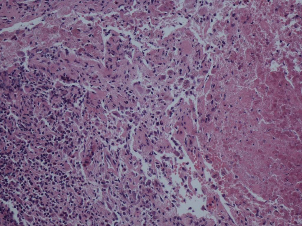
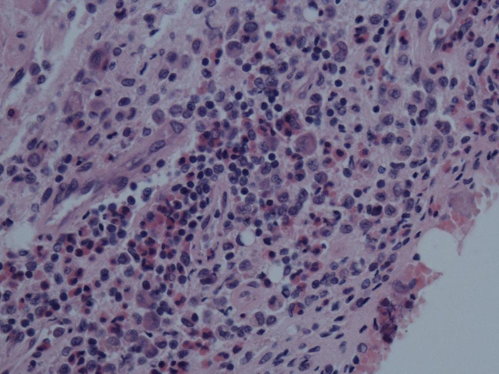
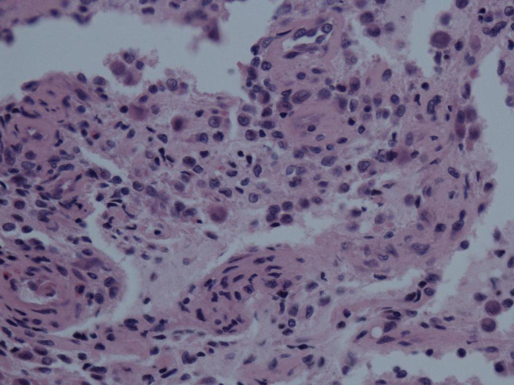
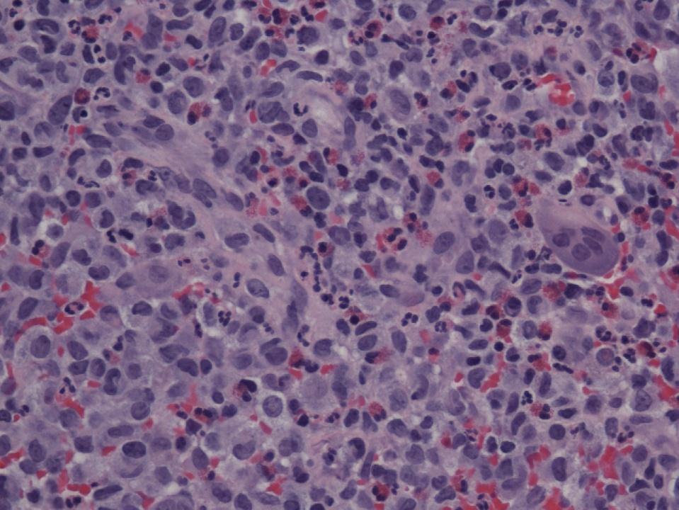
2 Case History A 7-day-old male patient born full-term via uncomplicated vaginal delivery was seen for multiple erythematous red-brown purpuric lesions that were present on the scalp, trunk and groin areas. The lesions resolved spontaneously within 3 months. Similar lesions appeared later at the age of 12 months. A 4 mm punch biopsy was performed on one of the representative papules on the chest and the biopsy showed:
3
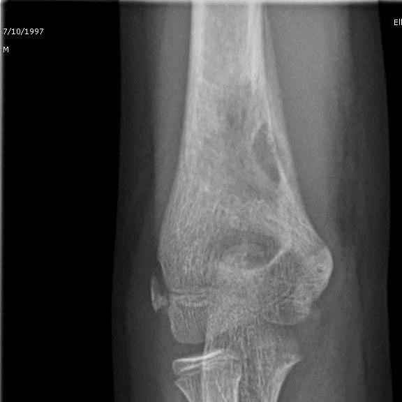
4

5 Bone lesion Screening of the body revealed few small radiolucent lesions in the skull and in the right femur

6

7 Bone lesion
8
9
10
11 Langerhans cell histiocytosis (LCH) Other names: Histiocytosis X eosinophilic granuloma Hand-Schüller-Christian disease Letterer-Siwe disease. A group of reactive conditions where Langerhans cells accumulate in tissues Any organ system may be involved: skin and bone.
12 Epidemiology Uncommon 2 10 cases per 1 million children <15 years. Median age at presentation is 30 months 2 cases per 1 million adults. Median age at presentation is 43.5 years True incidence is probably unknown because of underreporting and low prevalence/incidence
13 Pathogenesis and Etiology Pathogenesis are unknown Neoplastic disorder: LCH cells are monoclonal; rarely exhibit cytogenetic abnormalities (chromosome 7) Mutation of P53 occasionally seen Activating mutation of the BRAF oncogene detected in 57%. Cancer-based treatment Reactive Langerhans cells Differentiated cells Cytokine storm May regress spontaneously Granulomatous lesions resemble those in infection or foreign body reactions. Immune dysfunction and infectious agents (HHV6, cytomegalovirus, adenovirus, and parvovirus) have been implicated
14 Pathogenesis and Etiology T regulatory CD4+, CD25+, FoxP3+ cells appear to be in contact with LC cells in LCH lesions. T reg cells are elevated in LCH and return to baseline when LCH is in remission Marked elevation of FMS like tyrosine kinase 3 ligand (FLT 3) and M CSF often occur in LCH Levels can be directly correlated with extent of disease and decrease in response to treatment. M CSF and RANK L may allow in vitro transdifferentiation of immature DCs into osteoclasts. Increased M CSF may be the cause of osteolytic lesions in LCH. These proteins may be potential targets for treatment
, proliferate at a slow rate and present antigen poorly Upregulation of regulatory T cells, chemokines, and cytokines LC normally presents antigen to naïve T")
15 Pathogenesis and Etiology Cell of origin: LC from the skin or other histiocytes cells acquiring phenotype LCH cells possess LC markers (CD1a, CD207) and DC markers (CD80, CD86, and class II antigens), proliferate at a slow rate and present antigen poorly Upregulation of regulatory T cells, chemokines, and cytokines LC normally presents antigen to naïve T lymphs
16 Clinical presentation Presentation depending on the involved organ Dyspnea/tachypnea (lung), bone pain, polydipsia/polyuria (pituitary) Lymphadenopathy Anemia and thrombocytopenia Hypoalbuminemia, elevated liver enzymes and bilirubin Bone pain, pathologic fractures; Seen by imaging as lytic lesions
17 Langerhans cell histiocytosis of the skin Uncommon benign variant of LCH Newborn cutaneous disease that presents at birth or soon after delivery High rate of spontaneous resolution and failure of being recognized clinically: Congenital self-healing histiocytosis. May continue to grow after birth but tend to show signs of spontaneous self-resolution by 3 months.

18 Congenital self-healing histiocytosis Presents as asymptomatic, red, brown, or pink, crusted papulonodular and/or papulovesicular lesions with or without ulceration or erosion. Lesions typically ranging from 0.2 to 2.5 cm, can reach up to 8 cm in diameter.
19 Bone lesions Any bone can de affected. Skull bone and jaws in children <5 years age Long bones (femur and humerus) in older children Multiple bone lesions in 40% of cases
20 Classification of LCH Unifocal LCH: also called eosinophilic granuloma, slowly-progressing disease proliferation of Langerhans cells in various bones. It is a monostotic or polystotic disease with no extraskeletal involvement. Multifocal LCH: Seen mostly in children, characterized by fever, bone lesions and diffuse eruptions, usually on the scalp and in the ear canals. 50% of cases involve the pituitary stalk, leading to diabetes insipidus. The triad of diabetes insipidus, exopthalmos, and lytic bone lesions: Hand- Schüller-Christian triad. Multifocal multisystem LCH also called Letterer-Siwe disease, rapidly-progressing disease; prognosis is poor Involving many tissues. Mostly seen in children under age 2
21 Diagnosis Langerhans cell histiocytosis Clinical studies to exclude disseminated LCH. A biopsy of an affected organ is necessary to make the diagnosis of LCH
22 Disseminated disease 10-15% of children cases. More likely in children <2 years of age Systemic inflammatory response: fever, diarrhea, weight loss Systemic symptoms may relate to involvement of specific organ system Diabetes insipidus signifies involvement of the pituitary Poor prognosis


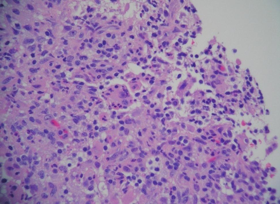
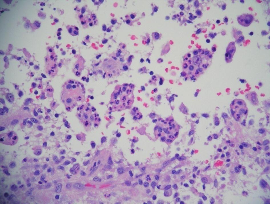
23 Histology of LCH Diffuse infiltration by pleomorphic histiocyte-like cells with ample eosinophilic granular, ground-glass or foamy cytoplasm and without dendritic processes Nuclei are characteristically indented or reniform. Background of inflammatory cells, particularly eosinophils Multinucleated giant cells may be present
24

25 CD1 a Immunohistochemistry Positivity for CD1a by IHC Transmembrane family of glycoproteins structurally related to the major histocompatibility complex (MHC) proteins form heterodimers with beta-2- microglobulin. CD1a proteins mediate the presentation of primarily lipid and glycolipid antigens of self or microbial origin to T cells.


26 Electron Microscopy Exhibit the characteristic intra-cytoplasmic Birbeck granules. Langerin, an antigen recently shown to colocalize with Birbeck granules, is now being used.
27 Differential diagnosis Non-Langerhans Cell Histiocytoses Diverse group of disorders defined by the accumulation of histiocytes that are not LC s
28

29 Juvenile xanthogranuloma Involves skin; least likely bone Fibrohistiocytic Xanthomatized histiocytes Touton giant cells Factor 13a positive
30 Xanthoma of bone May resemble LCH or JXG Xanthoma cells in non-ossifying fibroma, fibrous dysplasia and post-therapy changes Diagnosis of exclusion
31 Rosai-Dorfman disease Sinus histiocytosis with massive lymphadenopathy Prominent bilateral lymphadenopathy Can be extranodal with bone involvement Mixed inflammatory response; eosinophils are not prominent Macrophages with emperipolesis
32
33 Chronic recurrent multi-focal Osteomyelitis Inflammatory non-infective lesion Bone lesions widely distributed; upper anterior chest wall Can be inherited or associated with other lesions Neutrophils, chronic inflammatory cells, histiocytes Diagnosis of exclusion
34 LCH & Biologic Behavior May show extracutaneous involvement. Spontaneous regression in older children Potential for development into the disseminated form of LCH. 7 out of 18 patients with sole cutaneous involvement, had disease progression that required systemic treatment.
35
PHYSIOLOGY AND MANAGEMENT OF HISTIOCYTIC DISEASE. Brant Ward, MD, PhD Division of Rheumatology, Allergy, and Immunology
 PHYSIOLOGY AND MANAGEMENT OF HISTIOCYTIC DISEASE Brant Ward, MD, PhD Division of Rheumatology, Allergy, and Immunology What do histiocytes do? Apoptotic body removal Phagocytosis Antigen presentation Types
PET Imaging in Langerhans Cell Histiocytosis
 PET Imaging in Langerhans Cell Histiocytosis Christiane Franzius Bremen, Germany Histiocytosis Histiocytosis Idiopathic proliferation of histiocytes Two types of histiocytes macrophages: antigen processing
SWISS SOCIETY OF NEONATOLOGY. A newborn with a papulonodular rash at birth
 SWISS SOCIETY OF NEONATOLOGY A newborn with a papulonodular rash at birth June 2001 2 Glanzmann R, Neonatal Intensive Care Unit, UKKB Basel, Switzerland Swiss Society of Neonatology, Thomas M Berger, Webmaster
الفتوي الاصفر الحبيبوم = Xanthogranuloma_Juvenile JUVENILE XANTHOGRANULOMA 1 / 9
 JUVENILE XANTHOGRANULOMA 1 / 9 Clinical Findings CUTANEOUS LESIONS JXG is a benign, self-healing disorder that is characterized by asymptomatic yellowish papulonodular lesions of the skin and other organs
Arthritis and Rosai-Dorfman Disease of the Skin: A Diagnostic Dilemma
 Arthritis and Rosai-Dorfman Disease of the Skin: A Diagnostic Dilemma Introduction Pages with reference to book, From 280 To 282 Irshad Nabi Soomro ( Department of Pathology, The Aga Khan University Hospital,
Primary Cutaneous CD30-Positive T-cell Lymphoproliferative Disorders
 Primary Cutaneous CD30-Positive T-cell Lymphoproliferative Disorders Definition A spectrum of related conditions originating from transformed or activated CD30-positive T-lymphocytes May coexist in individual
Langerhans cell histiocytosis of the ear
 ABBR journal.. Vol. 1, No. 1 (2017) Archives of Biological and Biomedical Research An International Journal Langerhans cell histiocytosis of the ear Usama M Rashad 1, Al Hussein Awad Ali 2 and Mahmoud
Langerhans cell histiocytosis (LCH), formerly
, formerly") Langerhans cell histiocytosis of bone in children: a clinicopathologic study of 108 cases Jia Wang, Xv Wu, Zheng-Jun Xi Shanghai, China Background: Langerhans cell histiocytosis (LCH) is a rare disease
Langerhans cell histiocytosis of bone in children: a clinicopathologic study of 108 cases Jia Wang, Xv Wu, Zheng-Jun Xi Shanghai, China Background: Langerhans cell histiocytosis (LCH) is a rare disease
Burkitt lymphoma. Sporadic Endemic in Africa associated with EBV Translocations involving MYC gene on chromosome 8
 Heme 8 Burkitt lymphoma Sporadic Endemic in Africa associated with EBV Translocations involving MYC gene on chromosome 8 Most common is t(8;14) Believed to be the fastest growing tumor in humans!!!! Morphology
CASE REPORT. ACUTE DISSEMINATED LANGERHANS CELL DISEASE A RARE CASE REPORT Sunitha. M, R. Rajesh
 ACUTE DISSEMINATED LANGERHANS CELL DISEASE A RARE CASE REPORT Sunitha. M, R. Rajesh 1. Professor, Department of Oral Medicine & Radiology, Noorul Islam Dental College, Thiruvananthapuram, Kerala 2. Professor,
Histiocytic Neoplasms of the Dog and Cat
 Histiocytic Neoplasms of the Dog and Cat V.E. Valli DVM Histiocytic and Dendritic Cell Populations Both lineages are bone marrow derived. Macrophages are part of the innate immune system that are phagocytic
Langerhans Cell Histiocytosis: An Overview
 Langerhans Cell Histiocytosis: An Overview Milen Minkov International LCH Study Reference Center Children s Cancer Research Institute St. Anna Children s Hospital Vienna, Austria Achievements Case description
SLIDE SEMINAR NON NEOPLASTIC LYMPH NODE DISORDERS DR SHEILA NAIR CMC, VELLORE
 SLIDE SEMINAR NON NEOPLASTIC LYMPH NODE DISORDERS DR SHEILA NAIR CMC, VELLORE Case 1 34 year old male, mass right cervical region, for 4 years. No other significant findings. Grossly, the mass was well
Plasma cell myeloma (multiple myeloma)
") Plasma cell myeloma (multiple myeloma) Common lymphoid neoplasm, present at old age (70 years average) Remember: plasma cells are terminally differentiated B-lymphocytes that produces antibodies. B-cells
Plasma cell myeloma (multiple myeloma) Common lymphoid neoplasm, present at old age (70 years average) Remember: plasma cells are terminally differentiated B-lymphocytes that produces antibodies. B-cells
September is Histiocytosis Awareness Month
 IMMEDIATE RELEASE September is Histiocytosis Awareness Month Nationwide, September, 2011 September is Histiocytosis Awareness Month, an especially opportune time to raise visibility and funding for histiocytic
Case Report Xanthoma Disseminatum with Tumor-Like Lesion on Face
 Case Reports in Dermatological Medicine, Article ID 621798, 4 pages http://dx.doi.org/10.1155/2014/621798 Case Report Xanthoma Disseminatum with Tumor-Like Lesion on Face Habib Ansarin, 1 Hoda Berenji
المركب النموذج--- سبيتز وحمة = Type Spitz's Nevus, Compound SPITZ NEVUS 1 / 7
 SPITZ NEVUS 1 / 7 Epidemiology An annual incidence rate of 1.4 cases of Spitz nevus per 100,000 individuals has been estimated in Australia, compared with 25.4 per 100,000 individuals for cutaneous melanoma
ACCME/Disclosures. USCAP Specialty Evening Conference: Cytopathology New and recent Developments in Cytopathology: A case based approach
 USCAP Specialty Evening Conference: Cytopathology New and recent Developments in Cytopathology: A case based approach Sara E. Monaco, MD Associate Professor Program Director, UPMC Cytopathology Fellowship
ISPUB.COM. Vertebra Plana. R Chahal, S Acharya INTRODUCTION CASE HISTORY
 ISPUB.COM The Internet Journal of Spine Surgery Volume 4 Number 1 R Chahal, S Acharya Citation R Chahal, S Acharya.. The Internet Journal of Spine Surgery. 2006 Volume 4 Number 1. Abstract It is not uncommon
MULTISYSTEMIC LANGERHANS CELL HISTIOCYTOSIS IN ADULT AN UNCOMMON INCIDENCE POSING A DIAGNOSTIC CHALLENGE. Monal Trisal
 Pathology Case Report International Journal of Clinical And Diagnostic Research ISSN 2395-3403 Volume 4, Issue 4, July-Aug 2016. Glorigin Lifesciences Private Limited. MULTISYSTEMIC LANGERHANS CELL HISTIOCYTOSIS
NIH Public Access Author Manuscript J Cutan Pathol. Author manuscript; available in PMC 2013 April 08.
 NIH Public Access Author Manuscript Published in final edited form as: J Cutan Pathol. 2011 March ; 38(3): 280 285. doi:10.1111/j.1600-0560.2010.01650.x. Erdheim Chester disease presenting with cutaneous
TREATMENT AND SURVIVAL ANALYSIS FOR PEDIATRIC PATIENTS WITH LANGERHANS CELL HISTIOCYTOSIS A SINGLE INSTITUTION REVIEW
 TREATMENT AND SURVIVAL ANALYSIS FOR PEDIATRIC PATIENTS WITH LANGERHANS CELL HISTIOCYTOSIS A SINGLE INSTITUTION REVIEW HEMANT MENGHANI, MD; CORI MORRISON, MD PEDIATRIC HEMATOLOGY ONCOLOGY, LSUHSC AND CHILDREN
Question 1. Kupffer cells, microglial cells and osteoclasts are all examples of what type of immune system cell?
 Abbas Chapter 2: Sarah Spriet February 8, 2015 Question 1. Kupffer cells, microglial cells and osteoclasts are all examples of what type of immune system cell? a. Dendritic cells b. Macrophages c. Monocytes
Adult Orbital Xanthogranulomatous Disease
 Adult Orbital Xanthogranulomatous Disease Evening Specialty Conference: Ophthalmic Pathology Sunday, March 22, 2015 Lynn Schoenfield Associate Professor, Ohio State University Wexner Medical Center No
Adstock Science Club. Parvathy Elacode Harikumar. PhD student/research Assistant Clore Laboratory University of Buckingham
 Adstock Science Club Parvathy Elacode Harikumar PhD student/research Assistant Clore Laboratory University of Buckingham My Background Finished school education in India Completed A-Levels in London BSc
Differential Diagnosis of Oral Masses. Gingival Lesions
 Differential Diagnosis of Oral Masses Gingival Lesions Gingival/Alveolar Ridge Masses Parulis Periodontal Abscess Tori and Exostoses Reactive Proliferations Peripheral Odontogenic Cysts Peripheral Odontogenic
Canine Histiocytic Disorders DR. MEREDITH GAUTHIER, DVM DACVIM (ONCOLOGY) OCTOBER 29, 2015
 OCTOBER 29, 2015") Canine Histiocytic Disorders DR. MEREDITH GAUTHIER, DVM DACVIM (ONCOLOGY) OCTOBER 29, 2015 Canine Histiocytes! Cells derived from CD34+ stem cells and blood monocytes! Macrophages! Dendritic cells (DC)!
Canine Histiocytic Disorders DR. MEREDITH GAUTHIER, DVM DACVIM (ONCOLOGY) OCTOBER 29, 2015 Canine Histiocytes! Cells derived from CD34+ stem cells and blood monocytes! Macrophages! Dendritic cells (DC)!
Juvenile Xanthogranulomas in the First Two Decades of Life
 The American Journal of Surgical Pathology 27(5): 579 593, 2003 2003 Lippincott Williams & Wilkins, Inc., Philadelphia Juvenile Xanthogranulomas in the First Two Decades of Life A Clinicopathologic Study
BRAF V600E mutation in cutaneous lesions of patients with adult Langerhans cell histiocytosis
 BRAF V600E mutation in cutaneous lesions of patients with adult Langerhans cell histiocytosis Running head: adult Langerhans cell histiocytosis, skin, BRAF mutation Keywords: adult Langerhans cell histiocytosis
manifestations are uncommon. Initial descriptions of the disease (Rosai and Dorfman, 1969) specifically
 specifically") Postgraduate Medical Journal (July 1980) 56, 521-525 Diffuse cutaneous involvement and sinus histiocytosis with massive lymphadenopathy A. A. WOODCOCK B.Sc., M.B., Ch.B., M.R.C.P. Summary Severe skin involvement
Postgraduate Medical Journal (July 1980) 56, 521-525 Diffuse cutaneous involvement and sinus histiocytosis with massive lymphadenopathy A. A. WOODCOCK B.Sc., M.B., Ch.B., M.R.C.P. Summary Severe skin involvement
Bone pathologic fracture revealing an unusual association: coexistence of Langerhans cell histiocytosis with Rosai- Dorfman disease
 Efared et al. BMC Clinical Pathology (2017) 17:5 DOI 10.1186/s12907-017-0044-1 CASE REPORT Open Access Bone pathologic fracture revealing an unusual association: coexistence of Langerhans cell histiocytosis
Anaplastic Large Cell Lymphoma (of T cell lineage)
") Anaplastic Large Cell Lymphoma (of T cell lineage) Definition T-cell lymphoma comprised of large cells with abundant cytoplasm and pleomorphic, often horseshoe-shaped nuclei CD30+ Most express cytotoxic
Anaplastic Large Cell Lymphoma (of T cell lineage) Definition T-cell lymphoma comprised of large cells with abundant cytoplasm and pleomorphic, often horseshoe-shaped nuclei CD30+ Most express cytotoxic
Isolated Juvenile Xanthogranuloma in Thoracic Spine: Intraoperative Cytological Diagnosis of a Rare Presentation
 J Interdiscipl Histopathol 2014; 2(3): 158-162 ISSN: 2146-8362 Case Report Isolated Juvenile Xanthogranuloma in Thoracic Spine: Intraoperative Cytological Diagnosis of a Rare Presentation Shashi Singhvi,
, , 2011 HODGKIN LYMPHOMA
 European Federation of Cytology Societies 4tu Annual Tutorial in Cytopathology Trieste, June 6-10, 2011 HODGKIN LYMPHOMA Classification The World Health Organization Classification of Lymphomas (2001)
Cerebral Langerhans Cell Hystiocitosis: What the oncologist wants to know.
 Cerebral Langerhans Cell Hystiocitosis: What the oncologist wants to know. Poster No.: C-0383 Congress: ECR 2015 Type: Authors: Keywords: DOI: Educational Exhibit M. Diez Blanco, S. Sanchez Bernal, H.
Non-Hodgkin lymphomas (NHLs) Hodgkin lymphoma )HL)
 Hodgkin lymphoma )HL)") Non-Hodgkin lymphomas (NHLs) Hodgkin lymphoma )HL) Lymphoid Neoplasms: 1- non-hodgkin lymphomas (NHLs) 2- Hodgkin lymphoma 3- plasma cell neoplasms Non-Hodgkin lymphomas (NHLs) Acute Lymphoblastic Leukemia/Lymphoma
Non-Hodgkin lymphomas (NHLs) Hodgkin lymphoma )HL) Lymphoid Neoplasms: 1- non-hodgkin lymphomas (NHLs) 2- Hodgkin lymphoma 3- plasma cell neoplasms Non-Hodgkin lymphomas (NHLs) Acute Lymphoblastic Leukemia/Lymphoma
22 year old QH mare with regionally extensive alopecia and scaling on one front limb and ventral chest (Figure 1 and 2).
.") 22 year old QH mare with regionally extensive alopecia and scaling on one front limb and ventral chest (Figure 1 and 2). Which of the following is the most likely disease? a. Sterile granuloma complex
22 year old QH mare with regionally extensive alopecia and scaling on one front limb and ventral chest (Figure 1 and 2). Which of the following is the most likely disease? a. Sterile granuloma complex
Case Report. Introduction. H Ranu and SH Pang
 Hong Kong J. Dermatol. Venereol. (2009) 17, 204-208 Case Report Recalcitrant scalp and intertriginous rash in a patient with diabetes insipidus: diversity of cutaneous manifestations and pitfalls in diagnosis
Variant of Congenital Self-healing licelomstiocytosis: Solitary HasMmoto-Pritzker Disease
 DIAGNOSTIC DILEMMAS Pediatric Dermatology Vol. 3 No. 3 230-236 0736-8046/86/$2.00 Variant of Congenital Self-healing licelomstiocytosis: Solitary HasMmoto-Pritzker Disease Timothy G. Berger, M.D., Maj.,
Contents. vii. Preface... Acknowledgments... v xiii
 Contents Preface... Acknowledgments... v xiii SECTION I 1. Introduction... 3 Knowledge-Based Diagnosis... 4 Systematic Examination of the Lymph Node... 7 Cell Type Identification... 9 Cell Size and Cellularity...
Case Report Diagnosis of Langerhans Cell Histiocytosis on Fine Needle Aspiration Cytology: A Case Report and Review of the Cytology Literature
 SAGE-Hindawi Access to Research Pathology Research International Volume 2011, Article ID 439518, 5 pages doi:10.4061/2011/439518 Case Report Diagnosis of Langerhans Cell Histiocytosis on Fine Needle Aspiration
Cover Page. Author: Quispel, W.T. Title: Langerhans cell histiocytosis : genetic and immunologic fingerprinting Issue Date:
 Cover Page The handle http://hdl.handle.net/887/4387 holds various files of this Leiden University dissertation Author: Quispel, W.T. Title: Langerhans cell histiocytosis : genetic and immunologic fingerprinting
=ﻰﻤاﻤﺤﻠا ﺔﻴﻘﻠﺤﻠا ﺔذﺒاﻨﻠا
 1 / 15 Erythema Annulare Centrifugum and Other Figurate Erythemas The figurate erythemas include a variety of eruptions characterized by annular and polycyclic lesions. Classification of this group has
The Radiology Assistant : Bone tumor - well-defined osteolytic tumors and tumor-like lesions
 Bone tumor - well-defined osteolytic tumors and tumor-like lesions Henk Jan van der Woude and Robin Smithuis Radiology department of the Onze Lieve Vrouwe Gasthuis, Amsterdam and the Rijnland hospital,
CASE REPORT. Abstract. Introduction
 CSE REPORT Spontaneous Regression of Pulmonary Involvement after Smoking Reduction and Removal of and Radiation Therapy for Langerhans Cell Histiocytosis of the Sphenoid one: Which Comes First, the Chicken
Metabolic & Endocrine disorders of bone:
 Metabolic & Endocrine disorders of bone: Osteoporosis: Bone apposition < bone resorption Risk factors: Postmenopausal women Hyperthyroidism Hyperparathyroidism Cushing s syndrome bone quantity: thin cortex
Clinicopathologic Self- Assessment S003 AAD 2017
 Clinicopathologic Self- Assessment S003 AAD 2017 Clay J. Cockerell, M.D. Director, Cockerell Dermatopathology Director, Division of Dermatopathology UT Southwestern Medical Center July 2017 No relevant
Case Presentation. Maha Akkawi, MD, Fatima Obeidat, MD, Tariq Aladily, MD. Department of Pathology Jordan University Hospital Amman, Jordan
 Case Presentation Maha Akkawi, MD, Fatima Obeidat, MD, Tariq Aladily, MD Department of Pathology Jordan University Hospital Amman, Jordan The 25th Annual Congress of the ADIAP The 8/11/2013 1 5th International
3/24/2017 DENDRITIC CELL NEOPLASMS: HISTOLOGY, IMMUNOHISTOCHEMISTRY, AND MOLECULAR GENETICS. Disclosure of Relevant Financial Relationships
 DENDRITIC CELL NEOPLASMS: HISTOLOGY, IMMUNOHISTOCHEMISTRY, AND MOLECULAR GENETICS Jason L. Hornick, M.D., Ph.D. Director of Surgical Pathology and Immunohistochemistry Brigham and Women s Hospital Professor
Overview of Cutaneous Lymphomas: Diagnosis and Staging. Lauren C. Pinter-Brown MD, FACP Health Sciences Professor of Medicine and Dermatology
 Overview of Cutaneous Lymphomas: Diagnosis and Staging Lauren C. Pinter-Brown MD, FACP Health Sciences Professor of Medicine and Dermatology Definition of Lymphoma A cancer or malignancy that comes from
CHAPTER 20. Tumours of Undefined Neoplastic Nature
 CHAPTER 20 Tumours of Undefined Neoplastic Nature There are many conditions of bone that are generally considered non-neoplastic, but often constitute important lesions to be considered in the differential
5.1 Breast, Anatomy. 70
 Chapter 5 Breast 5.1 Breast, Anatomy Breasts, also called Mamma are mammary glands, subcutaneously placed on the ventral side of the trunk in mammalian species, and develop for the sole purpose of secreting
A Rare case of Tubercular Gingivitis Case Report
 Case Report A Rare case of Tubercular Gingivitis Case Report *Dr. Ansh Chugh 1, Dr. Firoz A Hakkim 2, Dr. Rajesh. V 3, Dr. Raghava Sharma 4 1: JUNIOR RESIDENT IN GENERAL MEDICINE 2: SENIOR RESIDENT IN
Classifications of lymphomas
 Classifications of lymphomas Lukes and Collins Kiel classification Working formulation REAL classification (1994) WHO classification (2000) WHO CLASSIFICATIONF OF NEOPLASMS HAEMATOPETIC AND LYMPHOID TISSUES
MECHANISMS OF HUMAN DISEASE: LABORATORY SESSIONS LYMPHOMA. April 16, 2008
 MECHANISMS OF HUMAN DISEASE: LABORATORY SESSIONS LYMPHOMA April 16, 2008 FACULTY COPY GOAL: Learn the appearance of normal peripheral blood elements and lymph nodes. Recognize abnormal peripheral blood
Lymphoma: What You Need to Know. Richard van der Jagt MD, FRCPC
 Lymphoma: What You Need to Know Richard van der Jagt MD, FRCPC Overview Concepts, classification, biology Epidemiology Clinical presentation Diagnosis Staging Three important types of lymphoma Conceptualizing
LANGERHANS CELL HISTIOCYTOSIS LCH-CHILDREN
 LANGERHANS CELL HISTIOCYTOSIS LCH-CHILDREN Histiocytosis UK Introduction Despite the misery it causes, Histiocytosis is too rare a disease to have generated substantial research in medical circles. Unfortunately,
Infections and nonmicrobial inflammatory stimuli can cause leukocytosis (as seen in Lab 1) as well as lymph node enlargement (lymphadenopathy).
 as well as lymph node enlargement (lymphadenopathy).") LAB 5: LYMPHOID TISSUE AND SKIN The focus of this week s lab will be pathology of the lymphoid tissue and skin. The lymphoid organs include the thymus, spleen, and lymph nodes. Abnormalities in the lymph
LAB 5: LYMPHOID TISSUE AND SKIN The focus of this week s lab will be pathology of the lymphoid tissue and skin. The lymphoid organs include the thymus, spleen, and lymph nodes. Abnormalities in the lymph
Evening specialty conference: Liver
 Evening specialty conference: Liver Joseph Misdraji, M.D. Disclosure of Relevant Financial Relationships Disclosure of Relevant Financial Relationships USCAP requires that all planners (Education Committee)
An overlooked case of mandibular histiocytosis: Case report and review of the literature
 Int J Case Rep Images 2018;9:100993Z01KI2019. CASE REPORT Imrani et al. 1 PEER REVIEWED OPEN ACCESS An overlooked case of mandibular histiocytosis: Case report and review of the literature Kaoutar Imrani,
Langerhans cell histiocytosis of skull: a retrospective study of 18 cases
 Original Article Langerhans cell histiocytosis of skull: a retrospective study of 18 cases Xiang-Heng Zhang, Ji Zhang, Zheng-He Chen, Ke Sai, Yin-Sheng Chen, Jian Wang, Chao Ke, Chen-Chen Guo, Zhong-Ping
Differential diagnosis of hematolymphoid tumors composed of medium-sized cells. Brian Skinnider B.C. Cancer Agency, Vancouver General Hospital
 Differential diagnosis of hematolymphoid tumors composed of medium-sized cells Brian Skinnider B.C. Cancer Agency, Vancouver General Hospital Lymphoma classification Lymphoma diagnosis starts with morphologic
CPC. Chutika Srisuttiyakorn, M.D. Kobkul Aunhachoke, M.D. Phramongkutklao Hospital Bangkok, Thailand
 CPC Chutika Srisuttiyakorn, M.D. Kobkul Aunhachoke, M.D. Phramongkutklao Hospital Bangkok, Thailand A 53 year-old woman with fever, facial swelling and rashes on face, trunk and upper extremities for 3
Incidence. Bimodal age incidence 15-40, >55 years Childhood form (0-14) more common in developing countries M:F=1.5:1; in all subtypes except NS
 more common in developing countries M:F=1.5:1; in all subtypes except NS") Hodgkin Lymphoma Hodgkin Lymphoma 30% of all lymphomas Absolute incidence unchanged Arise in lymph node, cervical region Neoplastic tissues usually contain a small number of tumor cells Incidence Bimodal
Hodgkin Lymphoma Hodgkin Lymphoma 30% of all lymphomas Absolute incidence unchanged Arise in lymph node, cervical region Neoplastic tissues usually contain a small number of tumor cells Incidence Bimodal
21/07/2017. Hobnail endothelial cells are not the same as epithelioid endothelial cells
 UPDATE IN CUTANEOUS VASCULAR S DERMATOPATHOLOGY SESSION BELFAST PATHOLOGY JUNE 21/2017 Dr E Calonje St John s Institute of Dermatology, London, United Kingdom THE FAMILY OF VASCULAR S WITH EPITHELIOID
Desmoplastic Melanoma R/O BCC. Clinical Information. 74 y.o. man with lesion on left side of neck r/o BCC
 R/O BCC Sabine Kohler, M.D. Professor of Pathology and Dermatology Dermatopathology Service Stanford University School of Medicine Clinical Information 74 y.o. man with lesion on left side of neck r/o
REVIEW Uncommon Histiocytic Disorders: The Non-Langerhans Cell Histiocytoses
 Pediatr Blood Cancer 2005;45:256 264 REVIEW Uncommon Histiocytic Disorders: The Non-Langerhans Cell Histiocytoses Sheila Weitzman, MB 1,2 * and Ronald Jaffe, MB, BCh 3 Background. Histiocytic disorders
LANGERHANS CELL HISTIOCYTOSIS LCH-CHILDREN
 LANGERHANS CELL HISTIOCYTOSIS LCH-CHILDREN Histiocytosis UK Introduction Despite the misery it causes, Histiocytosis is too rare a disease to have generated substantial research in medical circles. Unfortunately,
LANGERHANS CELL HISTIOCYTOSIS LCH-ADULTS
 LANGERHANS CELL HISTIOCYTOSIS LCH-ADULTS Histiocytosis UK Introduction Despite the misery it causes, Histiocytosis is too rare a disease to have generated substantial research in medical circles. Unfortunately,
2/18/19. Case 1. Question
 Case 1 Which of the following can present with granulomatous inflammation? A. Sarcoidosis B. Necrobiotic xanthogranulma C. Atypical mycobacterial infection D. Foreign Body Reaction E. All of the above
Craniofacial Manifestations Of Eosinophilic Granuloma
 ISPUB.COM The Internet Journal of Plastic Surgery Volume 3 Number 1 B Tümerdem Ulug, A Ar?nc?,? Ermi? Citation B Tümerdem Ulug, A Ar?nc?,? Ermi?.. The Internet Journal of Plastic Surgery. 2006 Volume 3
David H. Aguirre P. Last Year MS University of Chile Gillian Lieberman MD Harvard Medical School
 David H. Aguirre P. Last Year MS University of Chile Gillian Lieberman MD Harvard Medical School OCTOBER 19, 2009 I. INTRODUCTION CASE REPORT OUR PATIENT: PRESENTATION 23y/o M without significant PMHX.
Childhood Histiocytoses: A Review of Twenty Two Cases
 INDIAN PEDIATRICS VOLUME 35-FEBRAURY 1998 REFERENCES 1. Pickering H. Social and environmental factors associated with diarrhea and growth in young children: Child health in urban Africa. Soc Sci Med 1985;
CHRONIC INFLAMMATION
 CHRONIC INFLAMMATION Chronic inflammation is an inflammatory response of prolonged duration often for months, years or even indefinitely. Its prolonged course is proved by persistence of the causative
Erdheim Chester Disease Mark Heaney MD PhD
 Erdheim Chester Disease 2017 Mark Heaney MD PhD Erdheim-Chester Disease Described as Lipid Granulomatosis by Jakob Erdheim and William Chester in 1930 Lipid-laden macrophages and Touton giant cells CD
A 40-year old male with follicular papule and pustule at central face area for 3 months
 A 40-year old male with follicular papule and pustule at central face area for 3 months GMS- Neg AFB-Neg Fite stain - neg HISTOPATHOLOGICAL DIFFERENTIAL DIAGNOSIS CASEOUS GRANULOMA INFECTION -MYCOBACTERIUM
A case of primary cutaneous Langerhans cell sarcoma clinically mimicking pyogenic granuloma
 Hong Kong J. Dermatol. Venereol. (2017) 25, 187-191 Case Report A case of primary cutaneous Langerhans cell sarcoma clinically mimicking pyogenic granuloma O Kwon, KD Park, H Chung, JB Park, JY Park, DH
Superficial Granulomatous Pyoderma of the Face: A Case Report and Review of the Literature
 Superficial Granulomatous Pyoderma of the Face: A Case Report and Review of the Literature Sarah M. Persing, MPH, a and Donald Laub Jr, MD, FACS a,b a University of Vermont College of Medicine, Burlington;
Benign Fibro-osseous Lesions
 Benign Fibro-osseous Lesions Plus Vision is the art of seeing things invisible. Jonathan Swift 1667-1745 Steven R. Singer, DDS srs2@columbia.edu 212.305.5674 Benign Fibro-osseous Lesions A group of lesions
Biological and clinical significance of somatic mutations in Langerhans cell histiocytosis and related histiocytic neoplastic disorders
 RECENT ADVANCES IN HISTIOCYTIC NEOPLASMS Biological and clinical significance of somatic mutations in Langerhans cell histiocytosis and related histiocytic neoplastic disorders Carl E. Allen 1-3 and D.
CELL AND TISSUE INJURY COURSE-II PATHOLOGY LABORATORY
 CELL AND TISSUE INJURY COURSE-II PATHOLOGY LABORATORY PATHOLOGY of INFECTIOUS DISEASES MICROSCOPY Rengin Ahıskalı Macroscopy samples are shown in the macroscopy presentations of the first two courses.
MICHAEL P. GLOTZBECKER, 1 DAVID F. CARPENTIERI, M.D., 2 AND JOHN P. DORMANS, M.D. 3
 The University of Pennsylvania Orthopaedic Journal 15: 67 73, 2002 2002 The University of Pennsylvania Orthopaedic Journal Langerhans Cell Histiocytosis: Clinical Presentation, Pathogenesis, and Treatment
CASE year old male with a PET avid nodule in the left adrenal gland
 CASE 1 55 year old male with a PET avid nodule in the left adrenal gland Case 1 Adrenal gland parenchyma partly replaced by a spindle cell tumour with mild nuclear pleomorphism Atypical mitoses present
This is the second learning component (Learning Component 2) in our first learning module (Learning Module 1). In this component we review a very
 in our first learning module (Learning Module 1). In this component we review a very") This is the second learning component (Learning Component 2) in our first learning module (Learning Module 1). In this component we review a very basic response to injury inflammation. We ll look at examples
This is the second learning component (Learning Component 2) in our first learning module (Learning Module 1). In this component we review a very basic response to injury inflammation. We ll look at examples
WBCs Disorders 1. Dr. Nabila Hamdi MD, PhD
 WBCs Disorders 1 Dr. Nabila Hamdi MD, PhD ILOs Compare and contrast ALL, AML, CLL, CML in terms of age distribution, cytogenetics, morphology, immunophenotyping, laboratory diagnosis clinical features
Benign, Reactive and Inflammatory Lesions of the Breast
 Benign, Reactive and Inflammatory Lesions of the Breast Marilin Rosa, MD Associate Member Section Head of Breast Pathology Department of Anatomic Pathology Program Director, Breast Pathology Fellowship
Intrapelvic Bulky Tumor as an Unusual Presentation of Erdheim-Chester Disease
 CASE REPORT Intrapelvic Bulky Tumor as an Unusual Presentation of Erdheim-Chester Disease Satoru Taguchi 1,2, Yukiko Kishida 3, Koichi Tamura 3, Yorito Nose 2, Toshikazu Sato 1,2, Akira Ishikawa 1,2, Yukio
Mast Cell Tumors in Dogs
 Mast Cell Tumors in Dogs 803-808-7387 www.gracepets.com These notes are provided to help you understand the diagnosis or possible diagnosis of cancer in your pet. For general information on cancer in pets
Primary bone tumors > metastases from other sites Primary bone tumors widely range -from benign to malignant. Classified according to the normal cell
 Primary bone tumors > metastases from other sites Primary bone tumors widely range -from benign to malignant. Classified according to the normal cell counterpart and line of differentiation. Among the
T cell-mediated immunity
 T cell-mediated immunity Overview For microbes within phagosomes in phagocytes.cd4+ T lymphocytes (TH1) Activate phagocyte by cytokines studies on Listeria monocytogenes For microbes infecting and replicating
BSD Self Assessment Workshop 7 th July 2013 CASE 27 RAC6123
 BSD Self Assessment Workshop 7 th July 2013 CASE 27 RAC6123 M55. 4/7 tender lesions on knee, legs and arms. Also iritis/ weight loss/headache, synovitis.?vasculitis. Sarcoidosis. Biopsy from left elbow
9/25/2017. Disclosure. I have nothing to disclose. Young S. Kim MD Dept. of Pathology
 Disclosure MAST CELLNEOPLASM I have nothing to disclose. Young S. Kim MD Dept. of Pathology 1 Objectives What is mast cell lineage? Changes in updated WHO 2016 mastocytosis Issues of Mastocytosis CD30
5/21/2018. Objectives
 2018 Pathology CME Dermatopathology Symposium Napa CA May 19 th Pediatric Histiocytoses What s new in the WHO World? Alejandro A. Gru, M.D. Assistant Professor of Pathology & Dermatology Dermatopathology
SESSION 1: GENERAL (BASIC) PATHOLOGY CONCEPTS Thursday, October 16, :30am - 11:30am FACULTY COPY
 PATHOLOGY CONCEPTS Thursday, October 16, :30am - 11:30am FACULTY COPY") SESSION 1: GENERAL (BASIC) PATHOLOGY CONCEPTS Thursday, October 16, 2008 9:30am - 11:30am FACULTY COPY GOAL: Describe the basic morphologic (structural) changes which occur in various pathologic conditions.
SESSION 1: GENERAL (BASIC) PATHOLOGY CONCEPTS Thursday, October 16, 2008 9:30am - 11:30am FACULTY COPY GOAL: Describe the basic morphologic (structural) changes which occur in various pathologic conditions.
Pathology of Hematopoietic and Lymphoid tissue
 CONTENTS Pathology of Hematopoietic and Lymphoid tissue White blood cells and lymph nodes Quantitative disorder of white blood cells Reactive lymphadenopathies Infectious lymphadenitis Tumor metastasis
( Rosai Dorfman disease)
") Sinus histiocytosis with massive lymphadenopathy ( Rosai Dorfman disease) BIBI SHAIN SHAMSIAN Rosai Dorfman disease Rosai-Dorfman Disease (RDD) or Sinus Histiocytosis with massive lymhadenopathy (SHML):
Sinus histiocytosis with massive lymphadenopathy ( Rosai Dorfman disease) BIBI SHAIN SHAMSIAN Rosai Dorfman disease Rosai-Dorfman Disease (RDD) or Sinus Histiocytosis with massive lymhadenopathy (SHML):
Pathology of the Hematopoietic System. Case studies
 Pathology of the Hematopoietic System Case studies Shannon Martinson, September 2015 Signalment: 9 yr-old MC cat Case Study 1 History: Cat had been anorexic and developed bleeding in the eyes Physical
V. VALENTINI', V. TERENZI', A. CASSONI', S. BOSC02, E. BRAUNER I, J. SHAHINAS I
 EUROPEAN JOURNAL OF INFLAMMATION Vol. 10, no. I, 159-164 (2012) DENTISTRYSECTION GIANT CELL LESION OR LANGERHANS' CELL HISTIOCYTOSIS OF THE MANDIBLE? A CASE REPORT V. VALENTINI', V. TERENZI', A. CASSONI',
Done By : WESSEN ADNAN BUTHAINAH AL-MASAEED
 Done By : WESSEN ADNAN BUTHAINAH AL-MASAEED Acute Myeloid Leukemia Firstly we ll start with this introduction then enter the title of the lecture, so be ready and let s begin by the name of Allah : We
Spinal LCH Joseph Junewick, MD FACR
 Spinal LCH Joseph Junewick, MD FACR 05/16/2009 History 16 year old female with multiply recurrent Langerhans Cell Histiocytosis now with severe left sided neck pain. Diagnosis Langerhans Cell Histiocytosis