Pediatric Hepatobiliary, Pancreatic & Splenic US
|
|
|
- Madlyn Lindsey
- 6 years ago
- Views:
Transcription
1 Pediatric Hepatobiliary, Pancreatic & Splenic US Susan J. Back, MD Department of Radiology, The Children s Hospital of Philadelphia
2 No Disclosures
3 Objectives Normal Abnormal: cases and US advances
4 Objectives Normal Abnormal: cases and US advances

5 Hepatobiliary Ultrasound Size Echotexture Duct dilation Gallbladder Calculi

6 Hepatobiliary: Normal Liver Size Term 307 children 5 days to 16 years Genders equal Height best correlate
7 Hepatobiliary: Normal Liver Size Term 307 children 5 days to 16 years Genders equal Height best correlate Preterm 498 infants wk GA Girls smaller Weight best correlate
8 Hepatobiliary: Normal Gallbladder Mean length Mean Diameter Birth 1y: 2.5 cm 1 cm y: 6 cm 2 cm Wall thickness < 3 mm Radiology 1982; 144:

9 Hepatobiliary: Normal CHD Common hepatic duct little variation with age Always < 4 mm Radiology 1982; 144:

10 Hepatobiliary: Echotexture Homogeneous No sound attenuation deep lobe Portal vein/duct wall interfaces

11 Hepatobiliary: Echotexture Homogeneous No sound attenuation deep lobe Portal vein/duct wall interfaces Echogenicity > right kidney
12 Spleen Ultrasound Shape: cleft, lobules Location: wandering Radiographics 1999; 19: Number: polysplenia or asplenia Size: splenomegaly or atrophy

13 Spleen Ultrasound Lesions Solitary Multiple Diffuse Radiographics 1999; 19:
14 Spleen: Normal Size Term 512 children 1 day to 17 years Genders equal Height best correlate* *Others weight best AJR 1991; 157: AJR 1993; 160:

15 Spleen: Normal Size Term 512 children 1 day to 17 years Genders equal Height best correlate Preterm 498 infants wk GA Girls smaller Weight best correlate


16 Spleen Echotexture Homogeneous Echogenicity > liver Echogenicity >> left kidney Convex surface smooth Concave surface nodular

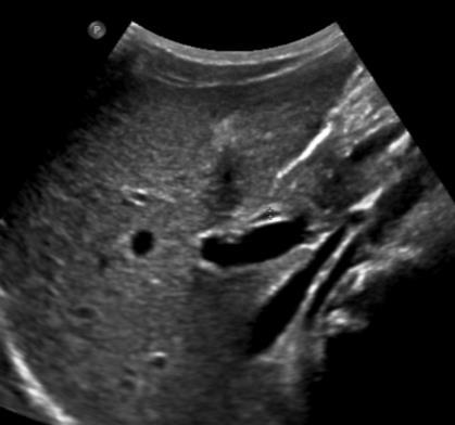
17 Pancreas Ultrasound Size Echotexture Duct dilation Fluid collection
18 Pancreas US Technique Sonographic window Ultrasound Clin 2013;8: Left hepatic lobe Left kidney or spleen Stomach with water ingestion

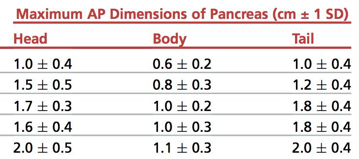
19 Pancreas: Normal Size Ultrasound Clin 2013; 8:


20 Pancreas: Normal Duct Size Mean / mm J Ultrasound Med 2000; 19:


21 J Ultrasound Med 2000; 19: Pediatr Radiol 1990; 20: Pancreas: Echotexture 1 day old Echogenicity > liver Newborn hyperechoic Preterm > term Smooth or slightly lobulated 7 month old
22 Objectives Normal Abnormal: cases and US advances
23 Inherited and Congenital Fibropolycystic Disease Biliary Atresia Hyperinsulinism Cystic Fibrosis Sickle cell disease
24 3 yo girl with progressive abdominal distension, thrombocytopenia 10.2 cm (95%) Size: large Echotexture: coarse Duct dilation: variable
 Size: large Echotexture: coarse Duct dilation: variable Duct Size Fibropolycystic disease Small Medium Large CHF, biliary hamartoma ADPLD")
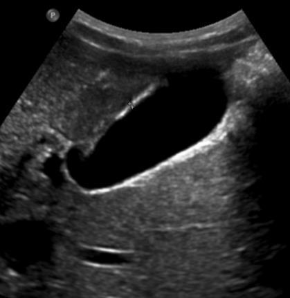
25 Congenital Hepatic Fibrosis (CHF) 10.2 cm (95%) Size: large Echotexture: coarse Duct dilation: variable Duct Size Fibropolycystic disease Small Medium Large CHF, biliary hamartoma ADPLD Choledochal cyst, Caroli disease Fibrous enlargement of bile ducts and portal tracts Ducts are present, not paucity
 Size: large Echotexture: coarse Duct dilation: variable Additional Cirrhotic")
26 Congenital Hepatic Fibrosis (CHF) 10.2 cm (95%) Size: large Echotexture: coarse Duct dilation: variable Additional Cirrhotic morphology Portal hypertension Splenomegaly 11.4 cm
 Size: large Echotexture: coarse Duct dilation: variable Mean 2.")
27 Congenital Hepatic Fibrosis (CHF) 10.2 cm (95%) Size: large Echotexture: coarse Duct dilation: variable Mean 2.58 m/s Elastography: quantify stiffness/fibrosis
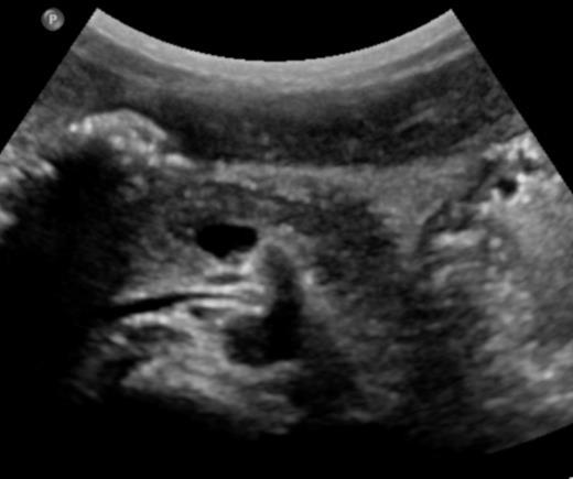
28 4 week old girl with conjugated hyperbilirubinemia
29 Biliary atresia Gallbladder Triangular cord Triangular cord sign (TCS) Absent CBD Abnormal GB (small) No GB change with feed
30 Biliary atresia Meta-analysis 23 studies US in BA Gallbladder US Sign Sensitivity Specificity Triangular cord GB abnormal 0.85 ( ) 0.92( ) TCS 0.74 ( ) 0.97 ( ) Triangular cord sign (TCS) Absent CBD Abnormal GB (small) No GB change with feed GB abn +TCS 0.95 ( ) 0.89 ( ) AJR 2016: 206:W73-82
 No GB change with feed Images at 1, 4, 6.")
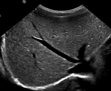
31 Biliary atresia Triangular cord Triangular cord sign (TCS) Absent CBD Abnormal GB (small) No GB change with feed Images at 1, 4, 6.5 hours no bowel activity, GB not visualized
 liver disease Increased ratio HA to portal vein Pediatr Radiol (2015) 45:366 375")
32 Biliary atresia Gallbladder Triangular cord Triangular cord sign (TCS) Absent CBD Abnormal GB (small) No GB change with feed Additional Enlarged Elastography: hepatic BA artery vs other (HA) liver disease Increased ratio HA to portal vein Pediatr Radiol (2015) 45:
33 1 month old girl hyperinsulinemic hypoglycemia 18F-DOPA PET/CT


34 Congenital Hyperinsulinism 18F-DOPA PET/CT Hyperfunctioning β cells Focal or diffuse Unregulated release of insulin
35 Congenital Hyperinsulinism sagittal Focal can be difficult to find in OR Intraoperative US assistance Hypoechoic Variable homogeneity


36 Congenital Hyperinsulinism 15 MHz 50 MHz sagittal Focal can be difficult to find in OR Intraoperative US assistance Hypoechoic Variable homogeneity 4.2 x 4.2 mm 0.3 mm duct
37 12 yo boy with Cystic Fibrosis


38 Cystic Fibrosis liver disease Echotexture: Hyperechoic, homogeneous Additional Heterogeneous Cirrhotic morphology Portal hypertension
39 Cystic Fibrosis Liver Disease Network (CFLD NET) Prediction by US Risk of Hepatic Cirrhosis (PUSH) 719 children, 3-12 yo No known cirrhosis J Pediatr 2015; 167:

40 Cystic Fibrosis Liver Disease Network (CFLD NET) Prediction by US Risk of Hepatic Cirrhosis (PUSH) 719 children, 3-12 yo No known cirrhosis US detected: 3.3% cirrhosis 8.9% heterogeneous J Pediatr 2015; 167:
41 Cystic Fibrosis liver disease Focal biliary cirrhosis ARFI elastography Radiol med 2012; 117:
42 Cystic Fibrosis- Pancreas Size: normal or atrophy Echotexture: increased Calcifications, cysts, cystosis

43 Cystic Fibrosis- Pancreas ARFI Elastography Softer in pancreatic insufficiency Insufficiency 0.88 m/s No insufficiency 1.07 m/s Normal 1.22 m/s * Size: normal or atrophy Echotexture: increased Calcifications, cysts, cystosis J Cystic Fibrosis 2013;12: *Eur J Radiol 2011;80:e226 30
44 8 yo boy Hb SS and abdominal pain
45 8 yo boy Hb SS and abdominal pain Number: present, one Size: small Echotexture: increased Lesions: multiple Previously autosplenectomy by 5y Transfusion & Hydroxyurea Splenomegaly Echogenic parenchyma Nodules Hemosiderosis Regenerative nodules Extramedullary hematopoiesis J Pediatr : J Ultrasound Med 2016; 35:


46 8 yo boy Hb SS and abdominal pain Number: present, one Size: small Echotexture: increased Lesions: multiple Additional Absent Heterogeneous echotexture Calcification Abscess
47 Summary Normal Abnormal: cases and US advances
IT 의료융합 1 차임상세미나 복부질환초음파 이재영
 IT 의료융합 1 차임상세미나 2013-4-3 복부질환초음파 이재영 나는오늘누구를위하여 종을울리나? 전통적의료 의사 공학설계자 의사 최첨단진단장비들 USG, CT, MRI 환자 환자 현대의료 사용자중심의사고 US in the Abdomen Detection DDx Look Behavior Response by external stimuli Guiding Tool
CT 101 :Pancreas and Spleen
 CT 101 :Pancreas and Spleen Shikha Khullar,, MD, MPH Division of Radiology University of South Alabama The Pancreas Normal Pancreas 3 Phase Pancreatic CT Non contrast Arterial phase : 30-35 35 second
Guidelines, Policies and Statements D5 Statement on Abdominal Scanning
 Guidelines, Policies and Statements D5 Statement on Abdominal Scanning Disclaimer and Copyright The ASUM Standards of Practice Board have made every effort to ensure that this Guideline/Policy/Statement
Radiology of hepatobiliary diseases
 GI cycle - Lecture 14 436 Teams Radiology of hepatobiliary diseases Objectives 1. To Interpret plan x-ray radiograph of abdomen with common pathologies. 2. To know the common pathologies presentation.
Elastography in the. technically difficult patient. EPIQ ultrasound system. Ultrasound
 Ultrasound Elastography in the technically difficult patient EPIQ ultrasound system Chairman Department of Diagnostic Radiology Allegheny General Hospital Pittsburgh, PA, USA You can offer more information
Imaging of common diseases of hepatobiliary and GI system
 Imaging of common diseases of hepatobiliary and GI system Natthaporn Tanpowpong, M.D. Diagnostic radiology Faculty of Medicine, Chulalongkorn University Normal plain radiograph A = Common bile duct
Normal Sonographic Anatomy
 hapter 2:The Liver DUNSTAN ABRAHAM Normal Sonographic Anatomy Homogeneous, echogenic texture (Figure 2-1) Measures approximately 15 cm in length and 10 12.5 cm anterior to posterior; measurement taken
Cystic Fibrosis in Children and Young Adults: Findings on Routine Abdominal Sonography
 bdominal Sonography in Cystic Fibrosis bdominal Imaging Pictorial Essay Downloaded from www.ajronline.org by 37.44.206.10 on 01/08/18 from IP address 37.44.206.10. Copyright RRS. For personal use only;
My Patient Has Abdominal Pain PoCUS of the Biliary Tract and the Urinary Tract
 My Patient Has Abdominal Pain PoCUS of the Biliary Tract and the Urinary Tract Objectives PoCUS for Biliary Disease PoCUS for Renal Colic PoCUS for Urinary Retention Biliary Disease A patient presents
Biliary Atresia. Karen F. Murray, MD Professor of Pediatrics Director, Hepatobiliary Program Seattle Children s
 Biliary Atresia Karen F. Murray, MD Professor of Pediatrics Director, Hepatobiliary Program Seattle Children s Biliary Atresia Incidence: 1/8,000-15,000 live births Girls > boys 1.5:1 The most common cause
Hepatic Imaging: What Every Practitioner Should Know
 Hepatic Imaging: What Every Practitioner Should Know Shuchi K. Rodgers, MD Section Chief, Abdominal Imaging Director of Ultrasound Department of Radiology Einstein Medical Center rodgerss@einstein.edu
Appendix 5. EFSUMB Newsletter. Gastroenterological Ultrasound
 EFSUMB Newsletter 87 Examinations should encompass the full range of pathological conditions listed below A log book listing the types of examinations undertaken should be kept Training should usually
GENERAL ABDOMINAL IMAGING PERITONEAL SPACE, PANCREAS, & SPLEEN. VMB 960 March 25, 2013
 GENERAL ABDOMINAL IMAGING PERITONEAL SPACE, PANCREAS, & SPLEEN VMB 960 March 25, 2013 REFERENCE Chapters 35-36 Pages 650-678 Chapter 37 Pages 694-701 Chapter 3 Pages 38-49 OBJECTIVES Radiography and Ultrasound
Abdominal Manifestations of Cystic Fibrosis in Children: Report of 50 cases
 Abdominal Manifestations of Cystic Fibrosis in Children: Report of 50 cases Poster No.: C-2070 Congress: ECR 2013 Type: Scientific Exhibit Authors: B. Iturre Salinas 1, A. Gozalo García 2, N. Martinez
Cystic Biliary Atresia: Why Is It Important to Distinguish this from Congenital Choledochal Cyst?
 Bahrain Medical Bulletin, Vol. 36, No. 2, June 2014 Cystic Biliary Atresia: Why Is It Important to Distinguish this from Congenital Choledochal Cyst? Hussein Ahmed Mohammed Hamdy, MRCSEd, FEBPS* Hind Mustafa
Abdominal Ultrasound. Diane Hallinen, MD. Bloodroot
 Abdominal Ultrasound Diane Hallinen, MD Bloodroot Abdominal Ultrasound Vasculature Hepatobiliary Spleen Kidney Bladder Bowel Where to put the probe? Vasculature We are going to talk about Celiac Trunk
Anatomy Jessica Ferguson Ashley Dobos May 31, 2006 LIVER
 Anatomy Jessica Ferguson Ashley Dobos May 31, 2006 LIVER 1) Other Names: Reidel s Lobe normal anatomic variant; projection of the right lobe that can extend as far as the iliac crest (Tempkin, p.54, Anatomy).
Prolonged Neonatal Jaundice
 Prolonged Neonatal Jaundice Ahmed Laving KPA Annual Scientific Conference 2018 Prolonged Jaundice? >6 months >3 months >2 weeks >4 weeks Prolonged Jaundice? >6 months >3 months >2 weeks >4 weeks Case Presentation
Policies, Standards, and Guidelines. Guidelines for Abdominal Ultrasound Examination
 Policies, Standards, and Guidelines Guidelines for Abdominal Ultrasound Examination Approved by Council Feb 2018 Disclaimer and Copyright The ASUM Standards of Practice Board have made every effort to
Abdomen and Retroperitoneum Ultrasound Protocols
 Abdomen and Retroperitoneum Ultrasound Protocols Reviewed By: Anna Ellermeier, MD Last Reviewed: March 2018 Contact: (866) 761-4200, Option 1 **NOTE for all examinations: 1. If documenting possible flow
Case Study: #3: Gallbladder Carcinoma?
 Case Study: #3: Gallbladder Carcinoma? By: Megan Wyatt K. SON Wyatt 225 2B1 RDMS, RVT Patient: Male 85 YOA Caucasian Indication: Elevated Alkaline Phosphatase History Annual physical showed elevated alkaline
Evaluation of Diffuse Liver Diseases Using Conventional Ultrasound
 IOSR Journal of Dental and Medical Sciences (IOSR-JDMS) e-issn: 2279-0853, p-issn: 2279-0861.Volume 16, Issue 6 Ver. VII (June. 2017), PP 70-74 www.iosrjournals.org Evaluation of Diffuse Liver Diseases
US LI-RADS v2017 CORE
 US LI-RADS v2017 CORE Screening or surveillance US in patient at high risk for HCC US category US-1 US-2 US-3 Negative Subthreshold Positive Category Concept Definition US-1 Negative US-2 Subthreshold
Imaging of liver and pancreas
 Imaging of liver and pancreas.. Disease of the liver Focal liver disease Diffusion liver disease Focal liver disease Benign Cyst Abscess Hemangioma FNH Hepatic adenoma HCC Malignant Fibrolamellar carcinoma
US in non-traumatic acute abdomen. Lalita, M.D. Radiologist Department of radiology Faculty of Medicine ChiangMai university
 US in non-traumatic acute abdomen Lalita, M.D. Radiologist Department of radiology Faculty of Medicine ChiangMai university Sagittal Orientation Transverse (Axial) Orientation Coronal Orientation Intercostal
Abdominal ultrasound:
 Abdominal ultrasound: Non-traumatic acute abdomen Wittanee Na-ChiangMai, MD Department of Radiology ChiangMai University 26/04/2017 Contents Technique of examination Normal anatomy Emergency conditions
DIAGNOSTIC IMAGING: LIVER DISEASE
 Vet Times The website for the veterinary profession https://www.vettimes.co.uk DIAGNOSTIC IMAGING: LIVER DISEASE Author : Abby Caine Categories : Vets Date : February 1, 2010 ABBY CAINE reviews both established
Malignant Focal Liver Lesions
 Malignant Focal Liver Lesions Other Than HCC Pablo R. Ros, MD, MPH, PhD Departments of Radiology and Pathology University Hospitals Cleveland Medical Center Case Western Reserve University Pablo.Ros@UHhospitals.org
Alice Fung, MD Oregon Health and Science University
 Alice Fung, MD Oregon Health and Science University Disclosure Comments The speaker Alice Fung, MD Has relevant financial relationships to disclose. Received honorarium from (Guerbet). This individual
Hilar cholangiocarcinoma. Frank Wessels, Maarten van Leeuwen, UMCU utrecht
 Hilar cholangiocarcinoma Frank Wessels, Maarten van Leeuwen, UMCU utrecht Content Anatomy Biliary strictures (Hilar) Cholangiocarcinoom Staging Biliary tract 1 st order Ductus hepatica dextra Ductus hepaticus
Extraosseous myeloma: imaging features
 Extraosseous myeloma: imaging features C. Santos Montón, R. Corrales, J. M. Bastida Bermejo, M. Villanueva Delgado, R. E. Correa Soto, J. M. Alonso Sánchez; Salamanca/ES Learning objectives -To review
DR NICKY WIESELTHALER RADIOLOGY CONSULTANT RED CROSS CHILDREN S HOSPITAL
 A CASE BASED APPROACH TO ULTRASOUND IN GIT DR NICKY WIESELTHALER RADIOLOGY CONSULTANT RED CROSS CHILDREN S HOSPITAL Ultrasound= No Radiation!!! 1 CXR= 0.02 msv ( effective dose that calculates dose absorbed)
HEPATO-BILIARY IMAGING
 HEPATO-BILIARY IMAGING BY MAMDOUH MAHFOUZ MD PROF.OF RADIOLOGY CAIRO UNIVERSITY mamdouh.m5@gmail.com www.ssregypt.com CT ABDOMEN Indications Patient preparation Patient position Scanogram Fasting 4-6 hours
Body MRI from the Liver to the Bladder
 Body MRI from the Liver to the Bladder I Want You! Audience Participation Methodist Hospital Continuing Education Seminar Jordan Swensson, MD November 7, 2015 Objectives Observe the uses of MRI for organs
Jaundice in children - Imaging features
 Jaundice in children - Imaging features Poster No.: C-2340 Congress: ECR 2011 Type: Educational Exhibit Authors: M. D. R. Matos, A. T. Ferreira, A. V. Nunes ; Lisbon/PT, 1 2 2 1 1 Oeiras/PT Keywords: Ultrasound,
Jaundice in children - Imaging features
 Jaundice in children - Imaging features Poster No.: C-2340 Congress: ECR 2011 Type: Educational Exhibit Authors: M. D. R. Matos, A. T. Ferreira, A. V. Nunes ; Lisboa/PT, 1 2 2 3 1 3 Oeiras/PT, 1800-348/PT
Evaluation of Liver Mass Lesions. American College of Gastroenterology 2013 Regional Postgraduate Course
 Evaluation of Liver Mass Lesions American College of Gastroenterology 2013 Regional Postgraduate Course Lewis R. Roberts, MB ChB, PhD Division of Gastroenterology and Hepatology Mayo Clinic College of
4/9/2018 OBJECTIVES PANCREAOTO BILIARY ULTRASOUND: BEYOND CHOLECYSTITIS
 PANCREAOTO BILIARY ULTRASOUND: BEYOND CHOLECYSTITIS Jean Yves Sewah Kaiser Permanente West Los Angeles 1 OBJECTIVES Discuss the role of ultrasound in the evaluation of the gallbladder, biliary tree and
Financial Disclosure
 Benign Liver Masses Adil Abdalla, MBBS Creighton University-CHI Health August 25, 2018 Financial Disclosure Nothing to disclose Financial Disclosure 1 Objectives To assess patients with benign liver tumors
Interesting Cases from Liver Tumor Board. Jeffrey C. Weinreb, M.D.,FACR Yale University School of Medicine
 Interesting Cases from Liver Tumor Board Jeffrey C. Weinreb, M.D.,FACR Yale University School of Medicine jeffrey.weinreb@yale.edu Common Liver Diseases Hemangioma Cyst FNH Focal Fat/Sparing THID Non-Cirrhotic
LIVER IMAGING TIPS IN VARIOUS MODALITIES. M.Vlychou, MD, PhD Assoc. Professor of Radiology University of Thessaly
 LIVER IMAGING TIPS IN VARIOUS MODALITIES M.Vlychou, MD, PhD Assoc. Professor of Radiology University of Thessaly Hepatocellular carcinoma is a common malignancy for which prevention, screening, diagnosis,
Personal Profile. Name: 劉 XX Gender: Female Age: 53-y/o Past history. Hepatitis B carrier
 Personal Profile Name: 劉 XX Gender: Female Age: 53-y/o Past history Hepatitis B carrier Chief complaint Fever on and off for 2 days Present illness 94.10.14 Sudden onset of epigastric pain 94.10.15 Fever
Congenital Pediatric Anomalies: A Collection of Abdominal Scintigraphy Findings: An Imaging Atlas
 ISPUB.COM The Internet Journal of Nuclear Medicine Volume 5 Number 1 Congenital Pediatric Anomalies: A Collection of Abdominal Scintigraphy Findings: An Imaging Atlas V Vijayakumar, T Nishino Citation
Gallbladder & Pancreas Ultrasonography
 복부초음파 : 담낭과췌장 Gallbladder & Pancreas Ultrasonography 김정훈 Department of Radiology 1 Interaction of sound with matter (1) 반사 (Reflection) (2) 굴절 (Refraction) (3) 흡수 (Absorption) (4) 산란 (Scattering) 음향저항
Classification of choledochal cyst with MR cholangiopancreatography in children and infants: special reference to type Ic and type IVa cyst
 Classification of choledochal cyst with MR cholangiopancreatography in children and infants: special reference to type Ic and type IVa cyst Poster No.: C-1333 Congress: ECR 2011 Type: Educational Exhibit
X-Ray Corner. Imaging Approach to Cystic Liver Lesions. Pantongrag-Brown L. Solitary cystic liver lesions. Hepatic simple cyst (Figure 1)
") THAI J 136 Imaging Approach to Cystic Liver Lesions GASTROENTEROL 2013 X-Ray Corner Imaging Approach to Cystic Liver Lesions Pantongrag-Brown L Cystic liver lesions are common findings in daily practice
THAI J 136 Imaging Approach to Cystic Liver Lesions GASTROENTEROL 2013 X-Ray Corner Imaging Approach to Cystic Liver Lesions Pantongrag-Brown L Cystic liver lesions are common findings in daily practice
Biliary tree dilation - and now what?
 Biliary tree dilation - and now what? Poster No.: C-1767 Congress: ECR 2012 Type: Educational Exhibit Authors: I. Ferreira, A. B. Ramos, S. Magalhães, M. Certo; Porto/PT Keywords: Pathology, Diagnostic
Summary and conclusions
 Summary and conclusions 7 Chapter 7 68 Summary and conclusions Chapter 1 provides a general introduction to this thesis focused on the use of ultrasound (US) in children with abdominal problems. The literature
GASTROINTESTINAL IMAGING STUDY GUIDE
 GASTROINTESTINAL IMAGING STUDY GUIDE Pharynx Diverticula Foreign bodies Trauma o Motility Disorders Esophagus Diverticula Trauma Esophagitis Barrett esophagus Rings, webs, and strictures Varices Benign
Vascular Imaging in the Pediatric Abdomen. Jonathan Swanson, MD
 Vascular Imaging in the Pediatric Abdomen Jonathan Swanson, MD Goals and Objectives To understand the imaging approach, appearance, and clinical manifestations of the common pediatric abdominal vascular
The Focal Hepatic Lesion: Radiologic Assessment
 The Focal Hepatic Lesion: Radiologic Assessment Kevin Kuo, Harvard Medical School Year III Our Patient: PS 67 y/o female w/ long history of alcohol use Drinking since age 18, up to one bottle of wine/day
Contrast Enhanced Ultrasound of Parenchymal Masses in Children
 Contrast Enhanced Ultrasound of Parenchymal Masses in Children Sue C Kaste, DO On behalf of Beth McCarville, MD St. Jude Children s Research Hospital Memphis, TN Overview Share St. Jude experience with
Evaluation of Suspected Pancreatic Cancer
 Evaluation of Suspected Pancreatic Cancer October 15, 2015 If you experience technical difficulty during the presentation: Contact WebEx Technical Support directly at: US Toll Free: 1-866-779-3239 Toll
Biliary Tree Ultrasound - In a nutshell. Pamela Parker Lead Sonographer
 Biliary Tree Ultrasound - In a nutshell Pamela Parker Lead Sonographer Aims Review what we know about the biliary system Common pathologies Pitfalls Reporting tips The Nutshell Background Biliary examinations
FOR YOUR EYES ONLY: A Guide to Accurate Detection of Diffuse Infiltrators in the Liver Eric C. Ehman, MD 1
 FOR YOUR EYES ONLY: A Guide to Accurate Detection of Diffuse Infiltrators in the Liver Eric C. Ehman, MD 1 INFILTRATOR Brian T. Welch, MD 1 Naoki Takahashi, MD 1 Christine O. Menias, MD 2 Ajit H. Goenka,
Liver Ultrasound - Beyond the Basics. Pamela Parker Lead Sonographer
 Liver Ultrasound - Beyond the Basics Pamela Parker Lead Sonographer Aims Review what we know about the liver Reasons for imaging Focal lesions Diffuse disease Can we do more? The Liver The Liver The Liver
Congenital Lung Malformations: Radiologic-Pathologic Correlation
 Acta Radiológica Portuguesa, Vol.XVIII, nº 70, pág. 51-60, Abr.-Jun., 2006 Congenital Lung Malformations: Radiologic-Pathologic Correlation Marilyn J. Siegel Mallinckrodt Institute of Radiology, Washington
Monosegmental Hepatobiliary Fibropolycystic Disease Mimicking a Mass: Report of Three Cases
 Case Report Gastrointestinal Imaging http://dx.doi.org/10.3348/kjr.2014.15.1.54 pissn 1229-6929 eissn 2005-8330 Korean J Radiol 2014;15(1):54-60 Monosegmental Hepatobiliary Fibropolycystic Disease Mimicking
Essentials of Clinical MR, 2 nd edition. 65. Benign Hepatic Masses
 65. Benign Hepatic Masses Pulse sequences acquired for abdominal MRI typically consist of fast acquisition schemes such as single-shot turbo spin echo (i.e. HASTE) and gradient echo schemes such as FLASH
Biliary Tree Ultrasound - In a nutshell. Pamela Parker Lead Sonographer
 Biliary Tree Ultrasound - In a nutshell Pamela Parker Lead Sonographer Aims Review what we know about the biliary system Common pathologies Pitfalls Reporting tips The Nutshell Background Biliary examinations
Accessory Glands of Digestive System
 Accessory Glands of Digestive System The liver The liver is soft and pliable and occupies the upper part of the abdominal cavity just beneath the diaphragm. The greater part of the liver is situated under
Evaluation of liver and spleen stiffness using a ultrasound guided method: Accuracy of ARFI(R) measurements in liver disease patients
 measurements in liver disease patients") Evaluation of liver and spleen stiffness using a ultrasound guided method: Accuracy of ARFI(R) measurements in liver disease patients Poster No.: C-3242 Congress: ECR 2010 Type: Topic: Authors: Keywords:
Evaluation of liver and spleen stiffness using a ultrasound guided method: Accuracy of ARFI(R) measurements in liver disease patients Poster No.: C-3242 Congress: ECR 2010 Type: Topic: Authors: Keywords:
Case 7729 Child with choledochal cyst presenting with episodes of vomitting and jaundice
 Case 7729 Child with choledochal cyst presenting with episodes of vomitting and jaundice dos Santos R 1, Almeida J 1, Mendes PP 2, Pereira S 3, Borges C 3, Soares E 4. 1) Radiology resident, 2) Radiology
Prospective evaluation of the diagnostic accuracy of liver ultrasonography
 Gut, 1981, 22, 130-135 Prospective evaluation of the diagnostic accuracy of liver ultrasonography J C DEBONGNIE,* C PAULS, M FIEVEZ, AND E WIBIN From the Departments of Internal Medicine, Radiology, and
Appendix 9: Endoscopic Ultrasound in Gastroenterology
 Appendix 9: Endoscopic Ultrasound in Gastroenterology This curriculum is intended for clinicians who perform endoscopic ultrasonography (EUS) in gastroenterology. It includes standards for theoretical
Independent Ultrasound Reporting. BMUS UCD Study day 2018
 Independent Ultrasound Reporting BMUS UCD Study day 2018 Pamela Parker Consultant Sonographer Hull & East Yorkshire Hospitals Colin Griffin Lead Sonographer Royal Liverpool University Hospital Report To
Liver transplant for biliary atresia
 Jean de Ville de Goyet ISMETT Director of the Department for the Treatment and Study of Pediatric Abdominal Diseases and Abdominal Transplantation The first human liver transplant was performed on a pediatric
Case-based discussion:
 Case-based discussion: Pailin Kongmebhol, M.D. Department of Radiology Faculty of Medicine Chiang Mai University There are many guidelines for managing thyroid nodules Two important guidelines: 2015 American
CTA/MRA of Pediatric Hepatic Masses Radiology-Pathology Correlation
 Acta Radiológica Portuguesa, Vol.XVIII, nº70, pág. 41-50, Abr.-Jun., 2006 CTA/MRA of Pediatric Hepatic Masses Radiology-Pathology Correlation Marilyn J. Siegel Mallinckrodt Institute of Radiology, Washington
State of the Art Imaging for Hepatic Malignancy: My Assignment
 State of the Art Imaging for Hepatic Malignancy: My Assignment CT vs MR vs MRCP Which one to choose for HCC vs Cholangiocarcinoma What special protocols to use for liver tumors Role of PET and Duplex US
Use of Ultrasound in NAFLD
 Institute for Liver and Digestive Health Use of Ultrasound in NAFLD Dr. Davide Roccarina Specialist in General Medicine Specialist Doctor in Clinical Ultrasound and non-invasive liver assessment Hepatology
GENERAL ABDOMINAL IMAGING PERITONEAL SPACE, PANCREAS, & SPLEEN
 GENERAL ABDOMINAL IMAGING PERITONEAL SPACE, PANCREAS, & SPLEEN VMB 960 March 25, 2013 REFERENCE Chapters 35-36 Pages 650-678 Chapter 37 Pages 694-701 Chapter 3 Pages 38-49 OBJECTIVES Radiography and Ultrasound
The Spleen. Dr Fahad Ullah
 The Spleen BY Dr Fahad Ullah Spleen The spleen is an largest lymphoid organ shaped like a shoe that lies relative to the 9th and 11th ribs and is located in the left hypochondrium. Thus, the spleen is
iu22 Liver Shear Wave ElastPQ
 iu22 Liver Shear Wave ElastPQ Clinical Case Study Lucy Wang Clinical Application Specialist ASEAN Case Study: History: 58-year-old male patient, hepatitis B virus (HBV) carrier, with non clinical symptoms
Hepatobiliary Ultrasound Rimon Bengiamin, MD, RDMS Assistant Clinical Professor Director of Emergency Ultrasound UCSF Fresno. Objectives. Why?
 Hepatobiliary Ultrasound Rimon Bengiamin, MD, RDMS Assistant Clinical Professor Director of Emergency Ultrasound UCSF Fresno Objectives Discuss the goals of point-of-care biliary ultrasound Review the
Sonographic imaging of pediatric thyroid disorders in childhood. Experiences and report in 150 cases
 Sonographic imaging of pediatric thyroid disorders in childhood. Experiences and report in 150 cases M. Mearadji International Foundation for Pediatric Imaging Aid Sonographic technique. Use of high frequency
Congenital Disorders of the Canine and Feline Biliary Tree
 Congenital Disorders of the Canine and Feline Biliary Tree John M. Cullen VMD PhD DACVP FIATP 1 Annual Seminar of the French Society of Veterinary Pathology 2 Embryonic Liver 3 Cytokeratin 7 Ductal Plate
Gastrointestinal tract
 Gastrointestinal tract Colloidal liver-spleen imaging Presented by: Jehad Felemban Introduction: To obtain better anatomic display of liver and spleen architecture, we use (CT Ultrasound). (Radionuclide
Basic of Ultrasound Physics E FAST & Renal Examination. Dr Muhammad Umer Ihsan MBBS,MD, DCH CCPU,DDU1,FACEM
 Basic of Ultrasound Physics E FAST & Renal Examination Dr Muhammad Umer Ihsan MBBS,MD, DCH CCPU,DDU1,FACEM What is Sound? Sound is Mechanical pressure waves What is Ultrasound? Ultrasounds are sound waves
Biliary Ultrasonography Kathleen O Brien MD MPH RDMS Kaiser Permanente South Sacramento
 Biliary Ultrasonography Kathleen O Brien MD MPH RDMS Kaiser Permanente South Sacramento https://www.google.com/search?sa=g&hl=en&q=public+disclosure&tbm=isch&tbs=simg:caqsigeahwelekju2aqaaawlelcmpwgaygpgcamskpib_1qnza7ai
Sonography of soft-tissue vascular lesions
 Sonography of soft-tissue vascular lesions Oscar M. Navarro Associate Professor, University of Toronto Dept. of Diagnostic Imaging, The Hospital for Sick Children Toronto, Canada Declaration of Disclosure
Nuclear medicine in gastrointestinal system. Assoc. prof. V. Marković, MD, PhD Assoc. prof. A. Punda, MD, PhD A. Barić, MD, nucl. med. spec.
 Nuclear medicine in gastrointestinal system Assoc. prof. V. Marković, MD, PhD Assoc. prof. A. Punda, MD, PhD A. Barić, MD, nucl. med. spec. Hepatobiliary imaging Hepatobiliary imaging is nuclear medicine
Evaluation of liver and spleen stiffness using a ultrasound guided method: Accuracy of ARFI(R) measurements in liver disease patients
 measurements in liver disease patients") Evaluation of liver and spleen stiffness using a ultrasound guided method: Accuracy of ARFI(R) measurements in liver disease patients Poster No.: C-3242 Congress: ECR 2010 Type: Topic: Authors: Keywords:
Evaluation of liver and spleen stiffness using a ultrasound guided method: Accuracy of ARFI(R) measurements in liver disease patients Poster No.: C-3242 Congress: ECR 2010 Type: Topic: Authors: Keywords:
Nasogastric tube. Stomach. Pylorus. Duodenum 1. Duodenum 2. Duodenum 3. Duodenum 4
 Esophagus Barium Swallow Stomach and Duodenum 4 year old Upper GI Nasogastric tube Stomach and Duodenum 4 year old Upper GI Nasogastric tube Stomach Pylorus Duodenum 1 Duodenum 2 Duodenum 3 Duodenum 4
Introduction. Splenic pathologies. Polysplenia syndrome. Anatomical variants 21/08/2017. Ultrasound of the forgotten organ of the abdomen: the spleen
 Ultrasound of the forgotten organ of the abdomen: the spleen Olivier Lucidarme Pitié Salpêtrière UPMC/Sorbonne University Paris France Malformations Introduction must look for other malformations frequently
Dr Claire Smith, Consultant Radiologist St James University Hospital Leeds
 Dr Claire Smith, Consultant Radiologist St James University Hospital Leeds Imaging in jaundice and 2ww pathway Image protocol Staging Limitations Pancreatic cancer 1.2.4 Refer people using a suspected
Langerhans cell histiocytosis as a cause of periportal abnormal signal intensity on MRI. Case 1. Case 2
 Abdom Imaging 24:373 377 (1999) Abdominal Imaging Springer-Verlag New York Inc. 1999 Langerhans cell histiocytosis as a cause of periportal abnormal signal intensity on MRI M. Kim, 1 C. Lyu, 2 Y. Jin,
Ultrasonographic Triangular Cord Sign and Gallbladder Abnormality in Diagnosis of Biliary Atresia
 Iranian Journal of Neonatology 14 Ultrasonographic Triangular Cord Sign and Gallbladder Abnormality in Diagnosis of Biliary Atresia Seyed Ali Jafari*, MD,1 Mehrzad Mehdizadeh, MD,2 Fatemeh Farahmand, MD,
Hematologic Malignancies of the Liver : Spectrum of Disease. Zhou Jian
 Hematologic Malignancies of the Liver : Spectrum of Disease Zhou Jian 2015-7-8 Hematologic malignancies include a wide spectrum of lymphoproliferative and myeloproliferative disorders with nodal and extranodal
Lab Monitor Images Dissection of the Abdominal Vasculature + Lower Digestive System
 Lab Monitor Images Dissection of the Abdominal Vasculature + Lower Digestive System Stomach & Duodenum Frontal (AP) View Nasogastric tube 2 1 3 4 Stomach Pylorus Duodenum 1 Duodenum 2 Duodenum 3 Duodenum
Liver, Pancreas and Biliary System. Wirana Angthong, M.D.
 Liver, Pancreas and Biliary System Wirana Angthong, M.D. Objectives Outline Anatomy Imaging Techniques Common Diseases Outline Anatomy Imaging Techniques Common Diseases Liver anatomy Morphological anatomy:
AACE/ACE Advanced Endocrine Neck Ultrasound Training Course 2016
 AACE/ACE Advanced Endocrine Neck Ultrasound Training Course 2016 This 9mm left inferior nodule should remind us all why we re here! There is no absolute number of images required for documentation
3/17/2015. Importance of clinical correlation. Liver Review. Abdominal Sonography Registry Review. Purpose of the Lecture
 Purpose of the Lecture Abdominal Sonography Registry Review Steven M. Penny, M.A., RT(R), RDMS Johnston Community College NCUS 2015 Annual Symposium Recount rational facts about various organs and systems
Newcastle HPB MDM updated radiology imaging protocol recommendations. Author Dr John Scott. Consultant Radiologist Freeman Hospital
 Newcastle HPB MDM updated radiology imaging protocol recommendations Author Dr John Scott. Consultant Radiologist Freeman Hospital This document is intended as a guide to aid radiologists and clinicians
Current Management of Biliary Atresia. Janeen Jordan, MD PGY5 Surgery Grand Rounds November 19, 2007
 Current Management of Biliary Atresia Janeen Jordan, MD PGY5 Surgery Grand Rounds November 19, 2007 Overview Etiology and diagnosis Work-up and management History of Kasai Portoenterostomy Studies Advances
Acknowledgements. Update of Focal Liver Lesions Goals. Focal Liver Lesions. Imaging Choices For Liver Lesions. Focal Liver Lesions
 Acknowledgements Update of Focal Liver Lesions 2012 Giles Boland Massachusetts General Hospital Harvard Medical School No disclosures Dushyant Sahani Mukesh Harisinghani Goals Focal liver lesions Imaging
Jaundice. Agnieszka Dobrowolska- Zachwieja, MD, PhD
 Jaundice Agnieszka Dobrowolska- Zachwieja, MD, PhD Jaundice definition Jaundice, as in the French jaune, refers to the yellow discoloration of the skin. It arises from the abnormal accumulation of bilirubin
Basic Abdominal Sonography
 24S Basic Abdominal Sonography Procedural Overview JOHN FATCHETT II, RDMS is provided. Patient preparation (i.e., fasting) scanning techniques, spleen, transducer. evaluation of abdominal anatomy in the
6 th August 2018 Day 1 - Gallbladder & Bile duct Topic
 Venue: Sterling Hospital Auditorium, Sterling Hospitals, Gurukul Road Ahmedabad, Gujarat 6 th August 2018 Day 1 - Gallbladder & Bile duct Registration(8:00am-8:15am) Inauguration(8:15am-8:30am) Welcome
Various kinds of cystic lesions in the Liver: Case-based analysis
 Various kinds of cystic lesions in the Liver: Case-based analysis Poster No.: R-0018 Congress: RANZCR-AOCR 2012 Type: Educational Exhibit Authors: J.-H. Yoon, S.-H. Kim, Y.-J. Lim, J.-H. Ryu, H.-D. Kim