Neonatal intestinal obstruction: how to make etiological diagnosis?
|
|
|
- Tracy Holland
- 5 years ago
- Views:
Transcription
1 Neonatal intestinal obstruction: how to make etiological diagnosis? Poster No.: C-1414 Congress: ECR 2013 Type: Educational Exhibit Authors: W. Mnari, M. Zguidi, A. Zrig, M. Maatouk, B. Hmida, R. Salem, M. Golli; Monastir/TN Keywords: Abdomen, Paediatric, Gastrointestinal tract, Conventional radiography, Ultrasound, Contrast agent-other, Surgery, Acute, Obstruction / Occlusion, Volvulus DOI: /ecr2013/C-1414 Any information contained in this pdf file is automatically generated from digital material submitted to EPOS by third parties in the form of scientific presentations. References to any names, marks, products, or services of third parties or hypertext links to thirdparty sites or information are provided solely as a convenience to you and do not in any way constitute or imply ECR's endorsement, sponsorship or recommendation of the third party, information, product or service. ECR is not responsible for the content of these pages and does not make any representations regarding the content or accuracy of material in this file. As per copyright regulations, any unauthorised use of the material or parts thereof as well as commercial reproduction or multiple distribution by any traditional or electronically based reproduction/publication method ist strictly prohibited. You agree to defend, indemnify, and hold ECR harmless from and against any and all claims, damages, costs, and expenses, including attorneys' fees, arising from or related to your use of these pages. Please note: Links to movies, ppt slideshows and any other multimedia files are not available in the pdf version of presentations. Page 1 of 30
2 Learning objectives To report and illustrate the contribution of different imaging modalities in diagnosis of neonatal digestive obstruction. To develop an approach to the management of bowel neonatal obstruction involving clinical and imaging data. Page 2 of 30
3 Background Background Bowel obstruction is one of the most common surgical emergencies in newborns. The diagnosis can be evoked in prenatal period using fetal ultrasonography or within the first 30 days of life based on clinical signs of occlusion. It is due to either an anatomical lesion (eg:atresia) or functional disorders such as Hirschsprung's disease. Diagnosis : Prenatally: By ultrasonography (polyhydramnios, intestinal expansion ) Family history (cystic fibrosis ) Aassociated abnormalities Signs of bowel obstruction can include Vomiting with or without bile stained material Gastric residuals before feedings Failure to pas meconium in the first 24 hours of life Abdominal distension The diagnosis is based on history and physical examination The role of imaging is to confirm the obstruction syndrome and to clarify the level and the nature of the obstacle. Imaging is based on the triad: plain X-Ray,water-soluble contrast enema and ultrasonographie with doppler. After birth, air progresses through the gastrointestinal truct: - H1: stomach and duodenum - H6: cæcum - H12: rectum Normally, the same timeline is followed on abdominal radiograph. False-positives bowel obstruction on abdominal radiograph: Page 3 of 30
4 Infection : entérocolitis. Mask ventilation. Incessant crying. Page 4 of 30
5 Imaging findings OR Procedure details Etiological diagnosis A. Organic intraluminal obstruction Gastrointestinal atresia : Atresia types : (fig.1) Type I = mucosal web Type II = fibrous cord Type III = complete separation Duodenal atresia Bilious vomiting in 90% cases. The abdomen is flat except in the epigastric region. Failure to pass meconium. Associated abnormalities. X-Ray usually shows a characteristic «double-bubble» appearance (fig.2 and fig.3). Contrast enema is useless and even dangerous. Other parts of abdomen do not contain gas in complete form of duodenal atresia. Antro-pyloric atresia Exceptional Isolated gastric dilatation Early feedinf vomiting (H1) Respiratory distress: inhalation Plain X-ray: gastric distension without air in the other parts of abdomen (single bubble image) (fig.4) Page 5 of 30
6 Ultrasonography : associated abnormalities Small intestinal atresia Small intestinal atresia is usually caused by the loss of blood supply at the part of the intestine that closes off. The diagnosis is established later than for duodenal atrasia. Bilious vomiting with early abdominal distention. Failure to pas meconium. Plain X-ray : variable distension depending on the seat of atresia with air-fluid levels without gaz in large intestine area (fig.5). Water-soluble contrast enema is usually useless, if it is done it shows a non-functional micro colon with an interrupted small intestine (fig.6). Calcifications on the plain X-ray can be related to a meconium peritonitis. Large intestinal atresia Rare congenital conditions of the lower gastro-intestinal tract It is due to a mucosal web. Image from a barium enema study (fig.7) demonstrates microcolon with complete obstruction to the retrograde flow of barium in the transverse portion of the colon. 2. Gastrointestinal duplications (fig.8) Rarely obstructive in the neonatal period. They may be spherical or tubular in appearance. Sonography revealed an anechoic double-walled cystic lesion. B. Organic extraluminal obstruction Malrotation of the intestine occurs when the two-part rotational process does not proceed normally. The duodenojejunal junction remains right of midline and the cecum remains in the upper left abdomen. The mesentery still attaches to the retroperitoneum. The attachment is very narrow. This «omega» configuration predisposes postnatal rotation about the narrow vascular mesenteric axis, leading to midgut volvulus with subsequent intestinal ischemia and necrosis. Page 6 of 30
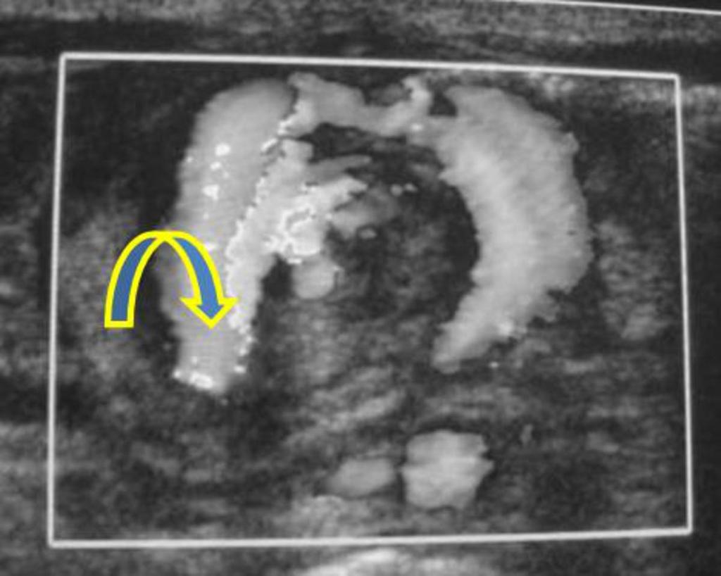
7 Ladd's bands is a fibrous stalk of peritoneal tissue that attaches the cecum to the abdominal wall. This condition is found in malrotation of the intestine. (fig.9 and 10). Intestinal malrotation may be complicated by volvulus and intestinal necrosis. Small intestinal volvulus Initially flat abdomen with vomiting, then abdominal distension and signs of severity in relation to intestinal distress. Plain X-ray:Gastroduodenal distension, rare gaz underlying (fig.11) Ultrasonography: Reversal of mesenteric pedicle (fig.12), «Whirlpool sign» (fig.13) Upper gastro-intestinal truct contrast: Turns of twist (fig.14), pre duodenal bar, topographic abnormality C. Functional obstruction 1. Abnormal intestinal contents Meconium Ileus Cystic fibrosis ++ Failure to pas meconium in the first 24 hours of life Abdominal distension Vomiting without then with bile stained material Plain X-ray: multiple dilated bowel loops The classic finding on water-soluble contrast enema (fig.15) is a small colon filled with pellet-like meconium when contrast material goes back into last part of the small intestine. Carefully repeated enema can sometimes raise the intra luminal obstruction. Left microcolon (fig.16) Left colon obstruction in the neonate results from meconium plug syndrome in which there is a failure to pass meconium in the first 24 hours of life, caused by blockage of the distal colon by meconium Flat abdomen immediately after birth, then obstruction syndrome. Digital rectal examination can sometimes eliminate a long meconium plug. Page 7 of 30
8 Plain X-ray: small intestine, right and transverse colon distension. Enema is useful for the diagnosis and for treatment. It shows a left colon with a reduced size. Always think of Hirschsprung's disease (biopsy) and cystic fibrosis. 2. Abnormal peristaltis Hirschsprung's Disease Hirschsprung's disease is a blockage of the large intestine due to improper muscle movement in the bowel. It is a congenital condition. The nerves are missing from a part of the bowel. Areas without such nerves cannot push material through. This causes a blockage. Intestinal contents build up behind the blockage, causing the bowel and abdomen to become swollen. - Rectal or rectosigmoid form (80% of cases) - Long form involving the left colon - Total form reaching the last ileal loop (poor prognosis) - Short form juxta-anal. Hirschsprung's disease is revealed in 90% in the neonatal period. Its diagnosis is histological. Symptoms are variable with complete or incomplete obstruction or failure to pas meconium. Water-soluble contrast enema (fig.17 and 18): abrupt transition++ D. Necrotising enterocolitis Necrosis, ulceration and parietal pneumatosis of the large and small intestine. Mechanisms: ischemia and infection, severe prematurity, perinatal distress. Possible complications: Perforation and peritonitis (early), Bowel obstruction (late). Imaging : diffuse bowel distension, parietal and portal pneumatosis, pneumoperitoneum DIAGNOSTIC STRATEGY AND CHOICE OF RADIOLOGICAL INVESTIGATIONS The choice of imaging explorations depends primarily on the clinical presentation (expanded or flat abdomen, bilious or feeding vomiting...). The goal is to: Page 8 of 30
9 Confirm the diagnosis of occlusion Specify the seat (high or low) Type (complete or incomplete) Start the etiological survey (associated abnormalities, family background, mother's diseases...) A. Plain X-ray is the first examination to be conducted While standing: air-fluid levels While lying down: Complete obstruction: no gaz downstream Incomplete obstruction: gaz downstream (rectum particularly) Questions : Is there an obstacle? If yes, area? Complet ou incomplet? Is there air out of the gastrointestinal tract? Is there calcifications? Systematic analysis the bone? B. Contrast enema Complete obstacle: No indication++ (surgery immediately) Incomplete obstacle : water-soluble upper gastro intestinal contrast Doubt between high and low obstruction :lower intestinal tract enema C. Doppler ultrasound Modality of choice in: Obstruction by volvulus (Whirlpool sign) Intestinal rotation abnormalities (abnormal position of the mesenteric vessels) Intestinal duplications D. Diagrams: Page 9 of 30
10 High neonatal bowel obstruction (fig.19) Lower GI tract obstruction (fig.20) Page 10 of 30

11 Images for this section: Fig. 6 Page 11 of 30

12 Fig. 1 Page 12 of 30

13 Fig. 2: Incomplete duodenal atresia Page 13 of 30

14 Fig. 3: Complete duodenal atresia Page 14 of 30

15 Fig. 4: Antro-pyloric atresia Page 15 of 30

16 Fig. 5: Small intestine atresia : small intestine distension without gaz in large intestine area Page 16 of 30

17 Fig. 7: Small intestine atresia : non functional micro colon with small intestine distension Page 17 of 30
 responsible for intussusception (open arrow) and")

18 Fig. 8: Obstruction in the neonatal period : duplication of the distal small intestine (arrow) responsible for intussusception (open arrow) and bowel obstruction upstream. Fig. 9: Normal rotation of the intestine: note the normal appearance of the root of the mesentery (blue) Page 18 of 30
19 Fig. 10: Malrotation of the intestine: short mesentery leading to volvulus Page 19 of 30

20 Fig. 11 Page 20 of 30

21 Fig. 12 Page 21 of 30

22 Fig. 13 Fig. 14 Page 22 of 30

23 Fig. 15: Meconium ileus : a small colon filled with pellet-like meconium Page 23 of 30

24 Fig. 16: Left microcolon: enema and laparotomy findings Page 24 of 30

25 Fig. 17 Page 25 of 30

26 Fig. 18 Page 26 of 30

27 Fig. 19 Page 27 of 30

28 Fig. 20 Page 28 of 30
29 Conclusion Imaging plays a major role in neonatal bowel obstruction. It is helpful, with clinical symptoms, to establish a diagnosis. Successful management depends on timely diagnosis and appropriate intervention. Page 29 of 30
30 References Juan D, Snyder CL. Neonatalbowelobstruction. SurgClinNorthAm ;92(3): Page 30 of 30
Neonatal intestinal obstruction: how to make etiological diagnosis?
 Neonatal intestinal obstruction: how to make etiological diagnosis? Poster No.: C-1414 Congress: ECR 2013 Type: Educational Exhibit Authors: W. MNARI, M. Zguidi, A. Zrig, M. MAATOUK, B. Hmida, R. Salem,
The "whirl sign". Diagnostic accuracy for intestinal volvulus.
 The "whirl sign". Diagnostic accuracy for intestinal volvulus. Poster No.: C-0670 Congress: ECR 2014 Type: Scientific Exhibit Authors: M. Pire, M. Marti, A. Borobia, A. Verón; Madrid/ES Keywords: Abdomen,
Imaging findings in complications of bariatric surgery.
 Imaging findings in complications of bariatric surgery. Poster No.: C-1791 Congress: ECR 2012 Type: Educational Exhibit Authors: A. Fernandez Alfonso, G. Anguita Martinez, D. C. Olivares Morello, C. García
Hirschprung s. Meconium plug R/S >1 R/S <1
 NEONATAL ABDOMINAL EMERGENCIES LOW OBSTRUCTION HIGH OBSTRUCTION INTESTINAL OBSTRUCTION High obstruction - proximal to mid-ileumileum Few dilated, air filled bowel loops Complete obstruction diagnosed by
CT evaluation of small bowel carcinoid tumors
 CT evaluation of small bowel carcinoid tumors Poster No.: C-0060 Congress: ECR 2015 Type: Educational Exhibit Authors: N. V. V. P. Costa, L. Nascimento, T. Bilhim ; Estoril/PT, PT, 1 2 3 1 2 3 Lisbon/PT
Volvulus of the Gastrointestinal Tract: x-ray and CT imaging
 Volvulus of the Gastrointestinal Tract: x-ray and CT imaging Poster No.: C-0076 Congress: ECR 2013 Type: Educational Exhibit Authors: E. Papadaki, S. Paschalidou, S. GIANNOU ; Rethymno, CR/ 1 2 2 3 1 3
Influence of pulsed fluoroscopy and special radiation risk training on the radiation dose in pneumatic reduction of ileocoecal intussusceptions.
 Influence of pulsed fluoroscopy and special radiation risk training on the radiation dose in pneumatic reduction of ileocoecal intussusceptions. Poster No.: C-0599 Congress: ECR 2013 Type: Authors: Keywords:
Excavated pulmonary nodule: steps to diagnosis?
 Excavated pulmonary nodule: steps to diagnosis? Poster No.: C-1044 Congress: ECR 2014 Type: Authors: Keywords: DOI: Educational Exhibit W. Mnari, M. MAATOUK, A. Zrig, B. Hmida, M. GOLLI; Monastir/ TN Metastases,
Biliary tree dilation - and now what?
 Biliary tree dilation - and now what? Poster No.: C-1767 Congress: ECR 2012 Type: Educational Exhibit Authors: I. Ferreira, A. B. Ramos, S. Magalhães, M. Certo; Porto/PT Keywords: Pathology, Diagnostic
64-MDCT imaging of the pancreas: Scan protocol optimisation by different scan delay regimes
 64-MDCT imaging of the pancreas: Scan protocol optimisation by different scan delay regimes Poster No.: C-051 Congress: ECR 2009 Type: Scientific Exhibit Topic: Abdominal and Gastrointestinal Authors:
Pneumatosis intestinalis, not always a surgical emergency
 Pneumatosis intestinalis, not always a surgical emergency Poster No.: C-2233 Congress: ECR 2012 Type: Educational Exhibit Authors: E. Vanhoutte, M. Lefere, R. Vanslembrouck, D. Bielen, G. De 1 1 2 1 1
Scientific Exhibit Authors: V. Moustakas, E. Karallas, K. Koutsopoulos ; Rodos/GR, 2
 Diagnosis of Acute Appendicitis: the role of Color Doppler Ultrasound as first-line imaging method and evaluation of the higher diagnostic performances of CT against its disadvantages. Poster No.: C-0708
Computed tomography (CT) imaging review of small bowel obstruction
 imaging review of small bowel obstruction") Computed tomography (CT) imaging review of small bowel obstruction Poster No.: C-1602 Congress: ECR 2010 Type: Educational Exhibit Topic: GI Tract - Small Bowel Authors: A. Vousough, D. S. Prasad ; Aberdeen/UK,
Computed tomography (CT) imaging review of small bowel obstruction Poster No.: C-1602 Congress: ECR 2010 Type: Educational Exhibit Topic: GI Tract - Small Bowel Authors: A. Vousough, D. S. Prasad ; Aberdeen/UK,
Computed tomography (CT) imaging review of small bowel obstruction
 imaging review of small bowel obstruction") Computed tomography (CT) imaging review of small bowel obstruction Poster No.: C-1602 Congress: ECR 2010 Type: Educational Exhibit Topic: GI Tract Authors: A. Vousough, D. S. Prasad ; Aberdeen/UK, Leeds/UK
Computed tomography (CT) imaging review of small bowel obstruction Poster No.: C-1602 Congress: ECR 2010 Type: Educational Exhibit Topic: GI Tract Authors: A. Vousough, D. S. Prasad ; Aberdeen/UK, Leeds/UK
Whirlpool sign of testis, a sonographic sign of incomplete torsion
 Whirlpool sign of testis, a sonographic sign of incomplete torsion Poster No.: C-0965 Congress: ECR 2011 Type: Educational Exhibit Authors: F. Esposito, P. Sgambati, M. Di Serafino, D. Noviello, G. 1 3
Hyperechoic breast lesions can be malignant.
 Hyperechoic breast lesions can be malignant. Poster No.: C-0041 Congress: ECR 2015 Type: Educational Exhibit Authors: G. Babu, R. bradley; Edinburgh/UK Keywords: Breast, Ultrasound, Biopsy, Cancer DOI:
Pneumo-esophageal 64-MDCT technique for gastric cancer evaluation
 Pneumo-esophageal 64-MDCT technique for gastric cancer evaluation Poster No.: C-1627 Congress: ECR 2010 Type: Scientific Exhibit Topic: GI Tract Authors: M. Ulla, E. Gentile, E. Levy, D. Cavadas, J. Ithurralde
Small-bowel obstruction due to bezoar: CT diagnosis and characterization
 Small-bowel obstruction due to bezoar: CT diagnosis and characterization Poster No.: C-1450 Congress: ECR 2013 Type: Scientific Exhibit Authors: I. lópez blasco, S. Paz Maya, R. Dosdá Muñoz, D. Soriano
Long bones manifestations of congenital syphilis
 Long bones manifestations of congenital syphilis Poster No.: C-0139 Congress: ECR 2011 Type: Educational Exhibit Authors: T. F. de Souza 1, P. P. Collier 1, E. J. M. Bronzatto 1, G. L. P. Keywords: DOI:
Cognitive target MRI-TRUS fusion biopsies of MRI detected PIRADS 4 and 5 lesions
 Cognitive target MRI-TRUS fusion biopsies of MRI detected PIRADS 4 and 5 lesions Poster No.: B-0704 Congress: ECR 2015 Type: Scientific Paper Authors: P. P. van Westerveld, J. Vriesema, J. H. W. van den
Imaging of gastrointestinal perforation: Is there a place for plain radiography?
 Imaging of gastrointestinal perforation: Is there a place for plain radiography? Poster No.: R-0243 Congress: Type: Authors: 2014 CSM Scientific Exhibit M. L. Chan 1, A. Steward 2, M. Schneider 3 ; 1 BOX
Ultrasonic evaluation of superior mesenteric vein in cancer of the pancreatic head
 Ultrasonic evaluation of superior mesenteric vein in cancer of the pancreatic head Poster No.: C-1430 Congress: ECR 2012 Type: Authors: Keywords: DOI: Scientific Exhibit E. Fisenko, N. Vetsheva, E. Pershina;
Purpose. Methods and Materials. Results
 Prevalence and significance of hypoattenuating hepatic lesions deemed too small to characterise: How are we following up these lesions and what are the outcomes? Poster No.: C-014 Congress: ECR 2009 Type:
BI-RADS 3, 4 and 5 lesions on US: Five categories and their diagnostic efficacy and pitfalls in interpretation
 BI-RADS 3, 4 and 5 lesions on US: Five categories and their diagnostic efficacy and pitfalls in interpretation e-poster: C-118 Congress: ECR 2008 Type: Educational Exhibit Topic: Breast / Ultrasound Authors:
Popliteal pterygium syndrome
 Popliteal pterygium syndrome Poster No.: C-1816 Congress: ECR 2011 Type: Educational Exhibit Authors: L. B. S. Santos, J. L. D. O. Schiavon, O. O. Guimaraes Neto, 1 1 2 3 1 1 C. A. P. Braga, R. S. LEMOS,
Abdominal compartment syndrome: radiological signs
 Abdominal compartment syndrome: radiological signs Poster No.: C-0903 Congress: ECR 2011 Type: Scientific Exhibit Authors: R. Ignarra, C. Acampora, R. MAZZEO, C. muzj, L. Romano ; 1 1 2 2 3 3 1 4 4 napoli/it,
Role of ultrasound in the evaluation of the ileocecal valve
 Role of ultrasound in the evaluation of the ileocecal valve Poster No.: C-1581 Congress: ECR 2010 Type: Scientific Exhibit Topic: GI Tract Authors: M. Mohammed, M. Hussain, U. Momin, S. Lakhtakia, N. D.
"Ultrasound measurements of the lateral ventricles in neonates: A comparison of multiple measurements methods."
 "Ultrasound measurements of the lateral ventricles in neonates: A comparison of multiple measurements methods." Poster No.: C-1557 Congress: ECR 2014 Type: Authors: Keywords: DOI: Educational Exhibit I.
Ultrasound (US) evaluation of peritoneal thickness in children and young patients on peritoneal dialysis (PD): A single centre experience
 evaluation of peritoneal thickness in children and young patients on peritoneal dialysis (PD): A single centre experience") Ultrasound (US) evaluation of peritoneal thickness in children and young patients on peritoneal dialysis (PD): A single centre experience Poster No.: C-2812 Congress: ECR 2010 Type: Scientific Exhibit
Ultrasound (US) evaluation of peritoneal thickness in children and young patients on peritoneal dialysis (PD): A single centre experience Poster No.: C-2812 Congress: ECR 2010 Type: Scientific Exhibit
Single cold nodule in Graves' disease: benign vs malignant
 Single cold nodule in Graves' disease: benign vs malignant Poster No.: C-0073 Congress: ECR 2011 Type: Authors: Keywords: DOI: Scientific Paper L. I. Sonoda, M. Halim, K. Balan; Cambridge/UK Head and neck,
Intraluminal gas in non-perforated acute appendicitis: a CT sign of gangrenous appendicitis
 Intraluminal gas in non-perforated acute appendicitis: a CT sign of gangrenous appendicitis Poster No.: C-978 Congress: ECR 202 Type: Scientific Exhibit Authors: D. Plata Ariza, E. MARTINEZ CHAMORRO, J.
Valsalva-manoeuvre or prone belly position for computed tomography (CT) scan when an orbita varix is suspected: a single-case study.
 scan when an orbita varix is suspected: a single-case study.") Valsalva-manoeuvre or prone belly position for computed tomography (CT) scan when an orbita varix is suspected: a single-case study. Poster No.: C-0512 Congress: ECR 2012 Type: Authors: Keywords: DOI:
Valsalva-manoeuvre or prone belly position for computed tomography (CT) scan when an orbita varix is suspected: a single-case study. Poster No.: C-0512 Congress: ECR 2012 Type: Authors: Keywords: DOI:
Identification and numbering of lumbar vertebrae using various anatomical landmarks on MRI of lumbosacral spine
 Identification and numbering of lumbar vertebrae using various anatomical landmarks on MRI of lumbosacral spine Poster No.: C-2125 Congress: ECR 2015 Type: Authors: Scientific Exhibit S. patil 1, A. M.
Pelvic inflammatory disease - spectrum of tomodensitometric findings
 Pelvic inflammatory disease - spectrum of tomodensitometric findings Poster No.: C-2451 Congress: ECR 2015 Type: Educational Exhibit Authors: E. Matos, A. T. Almeida, D. Castelo; Vila Nova de Gaia/PT Keywords:
Essure Permanent Birth Control Device: Radiological followup results at our center
 Essure Permanent Birth Control Device: Radiological followup results at our center Poster No.: C-0212 Congress: ECR 2013 Type: Scientific Exhibit Authors: R. Díaz Aguilera, A. M. Higuera Higuera, V. Palomo
A novel plain abdominal radiograph sign to diagnose malrotation with volvulus
 A novel plain abdominal radiograph sign to diagnose malrotation with volvulus Nataraja RM 1, Mahomed AA 1* 1. Department of Paediatric Surgery, Royal Alexandra Hospital for Sick Children, Brighton,UK *
Ultra-low dose CT of the acute abdomen: Spectrum of imaging findings
 Ultra-low dose CT of the acute abdomen: Spectrum of imaging findings Poster No.: C-1452 Congress: ECR 2010 Type: Educational Exhibit Topic: GI Tract Authors: P. A. Vlachou, C. Kloeters, S. Kandel, P. Hein,
CT findings of gastric and intestinal anisakiasis as cause of acute abdominal pain
 CT findings of gastric and intestinal anisakiasis as cause of acute abdominal pain Poster No.: C-2258 Congress: ECR 2015 Type: Educational Exhibit Authors: S. Marcos 1, J. Gonzalez 1, L. Sarria Octavio
Treatment options for endoleaks: stents, embolizations and conversions
 Treatment options for endoleaks: stents, embolizations and conversions Poster No.: C-0861 Congress: ECR 2012 Type: Authors: Keywords: DOI: Scientific Exhibit G. Lombardi; napoli/it Arteries / Aorta, Abdomen,
Quantitative imaging of hepatic cirrhosis on abdominal CT images
 Quantitative imaging of hepatic cirrhosis on abdominal CT images Poster No.: C-0556 Congress: ECR 2013 Type: Authors: Keywords: DOI: Scientific Exhibit S. Kido, A. Nakamura, Y. Hirano; Ube/JP Cirrhosis,
Intestinal Malrotation
 Intestinal Malrotation Poster No.: C-1368 Congress: ECR 2015 Type: Educational Exhibit Authors: K. Carpentier, B. De Foer, F. Deckers, P. Leyman, M. 1 1 1 2 1 1 1 2 Pouillon, P. M. Parizel ; Wilrijk/BE,
Shear Wave Elastography in diagnostics of supraspinatus tendon.
 Shear Wave Elastography in diagnostics of supraspinatus tendon. Poster No.: C-2168 Congress: ECR 2013 Type: Authors: Keywords: DOI: Scientific Exhibit V. Saltykova; Moscow/RU Musculoskeletal joint, Musculoskeletal
A pictorial review of normal anatomical appearences of Pericardial recesses on multislice Computed Tomography.
 A pictorial review of normal anatomical appearences of Pericardial recesses on multislice Computed Tomography. Poster No.: C-1787 Congress: ECR 2012 Type: Educational Exhibit Authors: N. Ahmed 1, G. Avery
Curious case of Misty Mesentery
 Curious case of Misty Mesentery Poster No.: C-1385 Congress: ECR 2015 Type: Authors: Keywords: DOI: Educational Exhibit T. Simelane 1, H. Khosa 2, N. Ramesh 2 ; 1 Dublin/IE, 2 Portlaoise/IE Abdomen, Anatomy,
Computed tomography and Modified RECIST criteria for assessment of response in malignant pleural mesothelioma
 Computed tomography and Modified RECIST criteria for assessment of response in malignant pleural mesothelioma Poster No.: C-0729 Congress: ECR 2013 Type: Scientific Exhibit Authors: A. Marin, I. Pozek,
Intra-abdominal abscesses radiology diagnostic
 Intra-abdominal abscesses radiology diagnostic Poster No.: C-2320 Congress: ECR 2012 Type: Scientific Exhibit Authors: K. Viksna; Riga/LV Keywords: Abscess, Computer Applications-Detection, diagnosis,
Radiological features of Legionella Pneumophila Pneumonia
 Radiological features of Legionella Pneumophila Pneumonia Poster No.: E-0048 Congress: ESTI 2012 Type: Scientific Exhibit Authors: M. Vinciguerra, L. Stefanetti, E. Teti, G. Argentieri, L. G. 1 1 1 1 1
Lesions of the pancreaticoduodenal groove, a pictorial review
 Lesions of the pancreaticoduodenal groove, a pictorial review Poster No.: C-2131 Congress: ECR 2013 Type: Educational Exhibit Authors: E. Ni Mhurchu, L. Lavelle, I. Murphy, S. Skehan ; IE, Dublin/ IE Keywords:
CT imaging of chronic radiation enteritis in surgical and non surgical patients
 CT imaging of chronic radiation enteritis in surgical and non surgical patients Poster No.: C-0334 Congress: ECR 2017 Type: Educational Exhibit Authors: M. Zappa, S. Kemel, C. Bertin, M. Ronot, D. Cazals-Hatem,
Seemingly isolated greater trochanter fractures do not exist
 Seemingly isolated greater trochanter fractures do not exist Poster No.: B-0950 Congress: ECR 2012 Type: Scientific Paper Authors: D. Dunker, J. H. Göthlin, M. Geijer ; Gothenburg/SE, Lund/SE Keywords:
Volvulus characterization in radiology: A review
 Volvulus characterization in radiology: A review Poster No.: C-1677 Congress: ECR 2010 Type: Topic: Educational Exhibit GI Tract Authors: C. Antunes, M. Seco, A. Canelas, C. Ruivo, C. Paulino, F. Cruz,
Imaging characterization of renal clear cell carcinoma
 Imaging characterization of renal clear cell carcinoma Poster No.: C-0327 Congress: ECR 2011 Type: Educational Exhibit Authors: S. Ballester 1, A. Gaser 2, M. Dotta 1, M. F. CAPPA 1, F. Hammar 1 ; 1 2
Artifact in Head CT Images Due to Air Bubbles in X-Ray Tube Oil
 Artifact in Head CT Images Due to Air Bubbles in X-Ray Tube Oil Poster No.: C-0671 Congress: ECR 2016 Type: Educational Exhibit Authors: H. Patel 1, W. Liu 2, J. DeSanto 2, S. Meagher 2, M. Zagardo 2,
CT Enteroclysis in the Diagnosis of Crohn's Disease (CD)
") CT Enteroclysis in the Diagnosis of Crohn's Disease (CD) Poster No.: C-2291 Congress: ECR 2012 Type: Scientific Exhibit Authors: I. Kiss, A. Rosztóczy, F. Nagy, T. Wittmann, A. Palko; Szeged/HU Keywords:
CT Enteroclysis in the Diagnosis of Crohn's Disease (CD) Poster No.: C-2291 Congress: ECR 2012 Type: Scientific Exhibit Authors: I. Kiss, A. Rosztóczy, F. Nagy, T. Wittmann, A. Palko; Szeged/HU Keywords:
Tubes and lines in neonatal chest radiograph
 Tubes and lines in neonatal chest radiograph Poster No.: C-1008 Congress: ECR 2014 Type: Educational Exhibit Authors: R. TUMMA, N. AHMED, V. Prasad ; Hyderabad/IN, 1 2 1 1 2 HYDERABAD, ANDHRA PRADESH/IN
Home FAQ Archives ABP Topics NeoReviews.org My Bookmarks CME Information Help. Print this Page Add to my Bookmarks Page 3 of 10
 Welcome Kristin Ingstrup [ Logout ] SEARCH Home FAQ Archives ABP Topics NeoReviews.org My Bookmarks CME Information Help Overview Editorial Board My Learning Plan January February March May June July August
Doppler Ultrasound findings in Children with Cow's Milk Allergy
 Doppler Ultrasound findings in Children with Cow's Milk Allergy Poster No.: C-0174 Congress: ECR 2012 Type: Authors: Keywords: DOI: Educational Exhibit M. Baldisserotto; Porto Alegre/BR Acceptance testing,
Chronic knee pain in adults - a multimodality approach or which modality to choose and when?
 Chronic knee pain in adults - a multimodality approach or which modality to choose and when? Poster No.: P-0157 Congress: ESSR 2013 Type: Authors: Keywords: DOI: Scientific Exhibit E. Ilieva, V. Tasseva,
AFib is the most common cardiac arrhythmia and its prevalence and incidence increases with age (Fuster V. et al. Circulation 2006).
.") Feasibility, image quality and radiation dose of coronary CT angiography (CCTA) in patients with atrial fibrillation using a new generation 256 multi-detector CT (MDCT) Poster No.: C-2378 Congress: ECR
Feasibility, image quality and radiation dose of coronary CT angiography (CCTA) in patients with atrial fibrillation using a new generation 256 multi-detector CT (MDCT) Poster No.: C-2378 Congress: ECR
Extrapulmonary Manifestations of Tuberculosis: A Radiologic Review
 Extrapulmonary Manifestations of Tuberculosis: A Radiologic Review Poster No.: C-1958 Congress: ECR 2014 Type: Authors: Educational Exhibit J. Isern 1, S. Llaverias Borrell 1, A. Olarte 1, E. Grive 1,
Thoracic causes of pneumoperitoneum - it is not all about perforation
 Thoracic causes of pneumoperitoneum - it is not all about perforation Poster No.: C-2590 Congress: ECR 2013 Type: Educational Exhibit Authors: E. Ilieva; Sofia/BG Keywords: Education, Plain radiographic
Computed tomographic dacryocystography as compared with X-ray dacryocystography in patients with dacryostenosis
 Computed tomographic dacryocystography as compared with X-ray dacryocystography in patients with dacryostenosis Poster No.: C-1887 Congress: ECR 2016 Type: Authors: Keywords: DOI: Educational Exhibit M.
High density thrombi of pulmonary embolism on precontrast CT scan: Is it dangerous?
 High density thrombi of pulmonary embolism on precontrast CT scan: Is it dangerous? Poster No.: C-1753 Congress: ECR 2013 Type: Authors: Keywords: DOI: Scientific Exhibit B. Y. Lee, H. R. KIM, J. I. Jung,
Slowly growing malignant nodules and rapidly growing benign nodules: Evaluation of the value of volume doubling time
 Slowly growing malignant nodules and rapidly growing benign nodules: Evaluation of the value of volume doubling time Poster No.: C-208 Congress: ECR 2009 Type: Educational Exhibit Topic: Chest Authors:
Using diffusion-tensor imaging and tractography (DTT) to study biological characteristics of glyoma in brain stem for neurosurgical planning
 to study biological characteristics of glyoma in brain stem for neurosurgical planning") Using diffusion-tensor imaging and tractography (DTT) to study biological characteristics of glyoma in brain stem for neurosurgical planning Poster No.: C-0016 Congress: ECR 2014 Type: Authors: Keywords:
Using diffusion-tensor imaging and tractography (DTT) to study biological characteristics of glyoma in brain stem for neurosurgical planning Poster No.: C-0016 Congress: ECR 2014 Type: Authors: Keywords:
The Abdominal plain film: A justified 21st century imaging investigation?
 The Abdominal plain film: A justified 21st century imaging investigation? Poster No.: C-0877 Congress: ECR 2012 Type: Authors: Keywords: DOI: Scientific Exhibit Z. J. Hussain 1, H. F. D'Costa 2 ; 1 Oxford/UK,
Feasibility of magnetic resonance elastography using myofascial phantom model
 Feasibility of magnetic resonance elastography using myofascial phantom model Poster No.: C-0971 Congress: ECR 2013 Type: Scientific Exhibit Authors: H. J. Kang, J.-S. Yoon, S.-J. Hong, C.-H. Oh, S. H.
Clearing the mind before the "caliber change": Diagnostic algorithm for small bowel obstruction.
 Clearing the mind before the "caliber change": Diagnostic algorithm for small bowel obstruction. Poster No.: C-0255 Congress: ECR 2014 Type: Educational Exhibit Authors: C. Santos Montón, D. Oquillas Izquierdo,
MDCT signs differentiating retroperitoneal and intraperitoneal lesions- diagnostic pearls
 MDCT signs differentiating retroperitoneal and intraperitoneal lesions- diagnostic pearls Poster No.: C-0987 Congress: ECR 2015 Type: Educational Exhibit Authors: D. V. Bhargavi, R. Avantsa, P. Kala; Bangalore/IN
The Role of Radionuclide Lymphoscintigraphy in the Diagnosis of Lymphedema of the Extremities
 The Role of Radionuclide Lymphoscintigraphy in the Diagnosis of Lymphedema of the Extremities Poster No.: C-1229 Congress: ECR 2014 Type: Authors: Keywords: DOI: Scientific Exhibit M. Osher 1, A. Pallas
Follow-up after Whipple operation by CT: techniques for the improvement of the afferent jejunal loop visualization and patterns of recurrence
 Follow-up after Whipple operation by CT: techniques for the improvement of the afferent jejunal loop visualization and patterns of recurrence Poster No.: C-1971 Congress: ECR 2012 Type: Educational Exhibit
Sonographic and Mammographic Features of Phyllodes Tumours of the Breast: Correlation with Histological Grade
 Sonographic and Mammographic Features of Phyllodes Tumours of the Breast: Correlation with Histological Grade Poster No.: C-0046 Congress: ECR 2014 Type: Authors: Keywords: DOI: Scientific Exhibit C. Y.
CT imaging findings of acute mesenteric ischemia and ischemic colitis. A brief pictorial essay.
 CT imaging findings of acute mesenteric ischemia and ischemic colitis. A brief pictorial essay. Poster No.: C-0750 Congress: ECR 2011 Type: Educational Exhibit Authors: Y. Arias Morales, J. P. Giraldo
The role of abdominal CT and MRI in detection of complications after transplantations of liver, kidney and pancreas.
 The role of abdominal CT and MRI in detection of complications after transplantations of liver, kidney and pancreas. Poster No.: C-1319 Congress: ECR 2015 Type: Educational Exhibit Authors: R. Muslimov,
Acute abdominal venous thromboses- the hyperdense noncontrast CT sign
 Acute abdominal venous thromboses- the hyperdense noncontrast CT sign Poster No.: C-1095 Congress: ECR 2011 Type: Educational Exhibit Authors: M. Goldstein, K. Jhaveri; Toronto, ON/CA Keywords: Abdomen,
Diffusion-weighted MRI (DWI) "claw sign" is useful in differentiation of infectious from degenerative Modic I signal changes of the spine
 claw sign is useful in differentiation of infectious from degenerative Modic I signal changes of the spine") Diffusion-weighted MRI (DWI) "claw sign" is useful in differentiation of infectious from degenerative Modic I signal changes of the spine Poster No.: C-0894 Congress: ECR 2012 Type: Scientific Exhibit
Diffusion-weighted MRI (DWI) "claw sign" is useful in differentiation of infectious from degenerative Modic I signal changes of the spine Poster No.: C-0894 Congress: ECR 2012 Type: Scientific Exhibit
The Radiologic Features of Xanthogranulomatous Cholecystitis: An Important Mimic of Gallbladder Carcinoma
 The Radiologic Features of Xanthogranulomatous Cholecystitis: An Important Mimic of Gallbladder Carcinoma Poster No.: C-0691 Congress: ECR 2014 Type: Authors: Keywords: DOI: Educational Exhibit H. L. khosa
Comparison of MRI and ultrasound based liver volumetry in iron overload diseases
 Comparison of MRI and ultrasound based liver volumetry in iron overload diseases Poster No.: C-0913 Congress: ECR 2015 Type: Scientific Exhibit Authors: S. Keller, B. P. Schönnagel, P. Nielsen, M. T. Brehmer,
Adenomyosis by myometrial Invasion of endometriosis: Comparison with typical adenomyosis
 Adenomyosis by myometrial Invasion of endometriosis: Comparison with typical adenomyosis Poster No.: C-1294 Congress: ECR 2010 Type: Scientific Exhibit Topic: Genitourinary Authors: S. Moon, H. K. Lim,
Psoriatic arthritis: early ultrasound findings
 Psoriatic arthritis: early ultrasound findings Poster No.: C-0399 Congress: ECR 2014 Type: Educational Exhibit Authors: R. Persechino 1, L. Cristiano 1, A. Bartoloni 1, C. Cantone 2, A. Keywords: DOI:
Neonatal Spinal Ultrasound Imaging - A Pictorial Review from The Royal Liverpool Children Hospital, Alder Hey, Liverpool
 Neonatal Spinal Ultrasound Imaging - A Pictorial Review from The Royal Liverpool Children Hospital, Alder Hey, Liverpool Poster No.: C-0081 Congress: ECR 2012 Type: Educational Exhibit Authors: K. Chetcuti,
Diffuse high-attenuation within mediastinal lymph nodes on non-enhanced CT scan: Usefulness in the prediction of benignancy
 Diffuse high-attenuation within mediastinal lymph nodes on non-enhanced CT scan: Usefulness in the prediction of benignancy Poster No.: C-1785 Congress: ECR 2012 Type: Authors: Keywords: DOI: Scientific
Detection of prostate cancer by MR-ultrasound fusion guided biopsy
 Detection of prostate cancer by MR-ultrasound fusion guided biopsy Poster No.: C-0761 Congress: ECR 2014 Type: Scientific Exhibit Authors: T. Durmus, C. Stephan, T. Slowinski, A. Thomas, A. Maxeiner, B.
Reliability of the pronator quadratus fat pad sign to predict the severity of distal radius fractures
 Reliability of the pronator quadratus fat pad sign to predict the severity of distal radius fractures Poster No.: C-0669 Congress: ECR 2014 Type: Scientific Exhibit Authors: J. Tonak, I. Wobbe, R. L. Duschka,
Single ventricle on cardiac MRI
 Single ventricle on cardiac MRI Poster No.: C-0414 Congress: ECR 2011 Type: Scientific Exhibit Authors: F. Secchi, V. G. Nardella, A. Giardino, G. Di Leo, F. 1 2 1 1 1 1 2 Sardanelli ; Milano/IT, Milan/IT
Acute pelvic pain in female patient: Clinical and Radiological evaluation
 Acute pelvic pain in female patient: Clinical and Radiological evaluation Poster No.: C-0909 Congress: ECR 2014 Type: Authors: Keywords: DOI: Educational Exhibit N. Ramesh 1, T. Simelane 2 ; 1 Portlaoise/IE,
Acute pelvic pain in female patient: Clinical and Radiological evaluation
 Acute pelvic pain in female patient: Clinical and Radiological evaluation Poster No.: C-0909 Congress: ECR 2014 Type: Authors: Keywords: DOI: Educational Exhibit N. Ramesh 1, T. Simelane 2 ; 1 Portlaoise/IE,
MRI in staging of rectal carcinoma
 MRI in staging of rectal carcinoma Poster No.: C-0152 Congress: ECR 2015 Type: Scientific Exhibit Authors: J. R. Ramos Rodriguez, M. Atencia Ballesteros, M. D. M. Muñoz Ruiz, A. J. Márquez Moreno, M. D.
How to plan a Zenith AAA stent-graft from a CTA: Basic measurements and concepts explained
 How to plan a Zenith AAA stent-graft from a CTA: Basic measurements and concepts explained Poster No.: C-3077 Congress: ECR 2010 Type: Educational Exhibit Topic: Vascular Authors: D. V. Thomas; Northampton/UK
Clinical impact of double reading of abdominal CT scans of surgical patients
 Clinical impact of double reading of abdominal CT scans of surgical patients Poster No.: B-0037 Congress: ECR 2015 Type: Scientific Paper Authors: P. Lauritzen, J.-G. Andersen, M. V. Stokke, A. L. Tennstrand,
CT staging in sigmoid diverticulitis
 CT staging in sigmoid diverticulitis Poster No.: C-1503 Congress: ECR 2012 Type: Scientific Paper Authors: M. Buchberger, B. von Rahden, J. Schmid, W. Kenn, C.-T. Germer, D. Hahn; Würzburg/DE Keywords:
The forgotten Upper gastrointestinal series. When and how I do it?
 The forgotten Upper gastrointestinal series. When and how I do it? Poster No.: C-0617 Congress: ECR 2015 Type: Educational Exhibit Authors: W. Mnari, K. Bouslama, M. Maatouk, A. Zrig, B. Hmida, R. Salem,
Audit of Micturating Cystourethrograms performed over 1 year in a Children's Hospital
 Audit of Micturating Cystourethrograms performed over 1 year in a Children's Hospital Poster No.: C-1773 Congress: ECR 2012 Type: Scientific Exhibit Authors: K. Lyons, J. Sorensen, E. L. Twomey, V. Donoghue,
Esophagus: Spectrum of pathologies on Barium Swallow
 Esophagus: Spectrum of pathologies on Barium Swallow Poster No.: C-1426 Congress: ECR 2013 Type: Authors: Keywords: DOI: Educational Exhibit E. Dhamija 1, D. Chandan 1, D. Srivastava 2 ; 1 New Delhi/IN,
Gastrointestinal Angiodysplasia: CT Findings
 Gastrointestinal Angiodysplasia: CT Findings Poster No.: C-1792 Congress: ECR 2012 Type: Authors: Keywords: DOI: Educational Exhibit G. Anguita Martinez, A. Fernandez Alfonso, D. C. Olivares Morello, J.
Ethanol ablation of benign thyroid cysts and predominantly cystic thyroid nodules: factors that predict outcome.
 Ethanol ablation of benign thyroid cysts and predominantly cystic thyroid nodules: factors that predict outcome. Poster No.: C-0322 Congress: ECR 2014 Type: Authors: Keywords: DOI: Scientific Exhibit J.
Bolus administration of esmolol allows for safe and effective heart rate control during coronary computed tomography angiography
 Bolus administration of esmolol allows for safe and effective heart rate control during coronary computed tomography angiography Poster No.: C-1342 Congress: ECR 2013 Type: Scientific Exhibit Authors:
Classification of choledochal cyst with MR cholangiopancreatography in children and infants: special reference to type Ic and type IVa cyst
 Classification of choledochal cyst with MR cholangiopancreatography in children and infants: special reference to type Ic and type IVa cyst Poster No.: C-1333 Congress: ECR 2011 Type: Educational Exhibit
Intracystic papillary carcinoma of the breast
 Intracystic papillary carcinoma of the breast Poster No.: C-1932 Congress: ECR 2011 Type: Educational Exhibit Authors: V. Dimarelos, F. TZIKOS, N. Kotziamani, G. Rodokalakis, 1 2 3 1 1 1 2 T. MALKOTSI
Spectrum of Cranio-facial anomalies during 2 Ultrasound. trimester on
 Spectrum of Cranio-facial anomalies during 2 Ultrasound nd trimester on Poster No.: C-0378 Congress: ECR 2015 Type: Scientific Exhibit Authors: K. Dave, S. Solanki; Ahmedabad/IN Keywords: Obstetrics (Pregnancy