Biliary Ultrasonography Kathleen O Brien MD MPH RDMS Kaiser Permanente South Sacramento
|
|
|
- MargaretMargaret Sutton
- 6 years ago
- Views:
Transcription


1 Biliary Ultrasonography Kathleen O Brien MD MPH RDMS Kaiser Permanente South Sacramento 9gObEoAK8wH1A5gGzT2sPb4_1rT3RPas9oj3TPdA9gj0aMKH8NOYEFXq-bLiqT1dZVwE0H7ZToFj_1o1v8lT5SxLIe14QK-_1Ecx3m3snDE4-4zCSADDAsQjq7-CBoKCggIARIEz6gJwAw&ved=0ahUKEwim9uWFrMPLAhUQ0GMKHazFD5IQwg4IGigA&biw=1347&bih=592 NONE 2 Objectives: Discuss clinical indications and questions answered by RUQUS Review of pertinent RUQ anatomy Share techniques & scanning tips Literature to support use of RUQUS Scope of the problem: Abdominal pain accounts for 5-10% of ED visits in US 1/3 of our abdominal pain patients in ED have GB etiologies for their pain 20M Americans have gallstones; ½ M undergo cholecystectomy each year 2 stones SEE MORE PATIENTS! DISPO THEM FASTER! SPEND LESS MONEY! BUT DON T COMPROMISE QUALITY OF CARE. 3 4 *Abdominal pain in the ED: stability and change over 20 years. Powers RD, Guertler AT.Am J Emerg Med. 1995;13(3):301. 1


2 One solution: Current imaging options for AC: HIDA: -highest diagnostic accuracy in older studies - sensitivity 96%, specificity 90% Ultrasound: -sensitivity 88-90%, specificity 80-88% -NPV 95-98% CT: MRI: -helpful for detecting complications - sensitivity 73-99%, specificity 42-74% -similar to u/s test characteristics - MRCP helpful if choledocholithiasis suspected overview 5 nutrition aha moments/ 6 But isn t that why we have radiologists? ED performed RUQ ultrasound shown to be as sensitive and specific for radiology performed RUQ ultrasound for acute cholecystitis! ED physicians often not formally trained in RUQ u/s and test characteristics still acceptable Advantage: increased efficiency, decreased time to diagnosis and disposition Purpose of RUQUS: Evaluate for: Cholelithiasis Acute cholecystitis Obvious liver/biliary pathology Indications: RUQ pain Flank/shoulder/ epigastric pain Ascites Hepatomegaly Jaundice Pancreatitis Sepsis 7 8 2


 and the portal vein")


3 Anatomy Gallbladder is located at the inferior surface of the liver; consists of the fundus, the body and the neck Anatomy Liver The neck of the gallbladder drains into the cystic duct which joins the hepatic duct to form the common bile duct () The portal triad consists of the hepatic artery, common bile duct () and the portal vein The and the hepatic artery lie anterior to the portal vein Gallbladder Duodenum Pancreatic duct Hepatic duct Cystic duct Pancreas Common bile duct Techniques 101: Probe selection Use MHz low frequency abdominal probe


4 13 Techniques 101: SUBcostal approach The probe is placed below the rib cage, lateral to epigastrium Good for avoiding Rib shadows Reliable Sono Murphy s Probe marker to head/r Shoulder hold probe at shallow angle 14 Techniques 101: INTERcostal approach Probe placed in the right anterior axillary line over the lower rib spaces, marker facing to right shoulder/head Slow sweep across the ribs Use the liver as an acoustic window Anchor your hand for stability Aka X minus 7 Techniques 101: Positioning in Left Lateral Decubitus Techniques 101: Can place probe subcostal or intercostal GB should move anteriorly Use the liver as acoustic window Slow sweep along costal margin -Always scan the entire gallbladder in two planes: Longitudinal Transverse -Slowly fan through entire gallbladder in these two planes


5 What should you see: GB long Gallbladder anterior anterior Portal vein head feet supine head feet posterior Left lateral decubitus 18 posterior anterior What should you see: GB short anterior head QuickTime and a Animation decompressor are needed to see this picture. feet supine right left posterior Normal Gallbladder in long axis 20 Left lateral decubitus posterior 5


6 anterior Normal variants of the Gallbladder Pharyngian cap: The fundus is folded onto the body right left Septate GB: thin septa inside gallbladder posterior Normal Gallbladder in short axis 22 The highly elusive Common Bile Duct... Exclamation point sign lies anterior to portal vein and next to hepatic artery -Color Doppler can help identify vascular structures Normal <7mm. - dilates with increasing age and after cholecystectomy! -PEARL: measure from inner wall to inner wall Portal vein Hepatic artery Find the Gallbladder in the longest axis, follow the main lobar fissure from the neck of the gallbladder to the porta hepatis. forms the point of the exclamation mark, anterior to the portal vein. 24 6


7 Mickey Mouse sign Portal vein Hepatic artery Inferior vena cava Again, anterior to portal vein and hepatic artery. does not show flow; helps to identify the Great news perhaps finding the doesn t really matter?! What am I looking for exactly? Question Mark 7
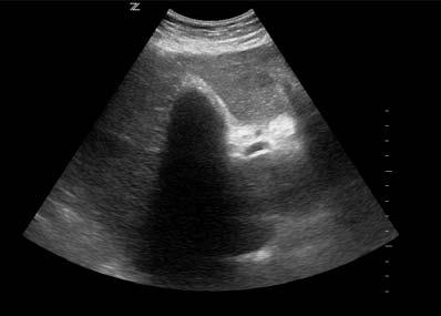
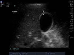
 Is there a sonographic murphy s sign? 5) +/- Is the dilated? 29 30 When looking for stones, keep in mind Stones: hyperechoic, cast a shadow.")
: When GB is filled multiple stones or one giant stone you just see wall, then bright reflex and then shadow.")
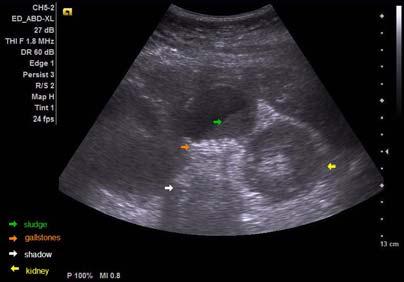
8 Look for Acute cholecystitis by asking: Acute cholecystitis: 1) Are there gallstones present? AND 2) Is there pericholecystic fluid present? 3) Is there GB wall thickening? 4) Is there a sonographic murphy s sign? 5) +/- Is the dilated? When looking for stones, keep in mind Stones: hyperechoic, cast a shadow. Stones are often mobile; scan patients in different positions. ALWAYS convince yourself there is no stone in GB neck. Wall-echo-complex (WES): When GB is filled multiple stones or one giant stone you just see wall, then bright reflex and then shadow. Sludge: biliary sand/microlithiasis: Echoes within depending part of GB without shadowing (resettles in dependent parts > scan patients in different positions) 31 8

9 Posterior acoustic enhancement


10 37 38 WES sign Wall Echo Shadow 39 Gallbladder filled completely with stone 40 10


11 1) Is there a stone? Patient supine, stones in the neck Stones vs polyps or tumors: -stones are mobile and can be moved by changing the position of the patient, not adhered to wall. -Polyps do not shadow. Pat. rolled to left lateral decubitus, stones in body Life just got easier Brief mention: stones Dilated intrahepatic ducts Stone in Shadow cast by stone stones: round echogenic lesion with posterior shadowing. Most stones are impacted in the distal duct at the papilla. 11

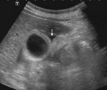
12 #2) Is there pericholecystic fluid? #2) Is there pericholecystic fluid? #2) Is there pericholecystic fluid? 3) Is there GB wall thickening? PEARL: Measure anterior wall because resolution is better


13 RUQUS and GB wall thickening: 4) Is there a Sono Murphys sign? -NONSPECIFIC finding! -DDx include: CHF Renal failure Hypoalbuminemia Hepatitis Cirrhosis Pancreatitis Carcinoma. maximal abdominal tenderness from pressure of the ultrasound probe over the visualised gallbladder SMS is a sign of local inflammation around the gallbladder along with right upper quadrant pain, tenderness or mass 49 list.aspx?cat= murphys sign and sonographic.html 5) Is the dilated? <=6mm is normal Add 1 mm as normal dilatation for every decade above 60 years old dilated in pts s/p cholecystecomy Measure INNER wall to inner wall FYI: Cholangitis Fever, RUQ pain, Jaundice. ~85% of cases associated with stones. On ultrasound: Dilation of biliary tree Choledocholithiasis and possibly sludge Bile duct wall thickening Hepatic abscess with thickened wall Stone in Shadow cast by stone 51 13
14 Again, ask yourself: 1) Are there gallstones present? AND 2) Is there pericholecystic fluid present? 3) Is there GB wall thickening? 4) Is there a sonographic murphy s sign? 5) +/- Is the dilated? Take home points: Always scan through the GB in both longitudinal and transverse planes. Scan through GB neck to ensure no obstructing stone Use color Doppler to help distinguish nonvascular from vascular structures. Be aware of normal variants (folds). Measure the anterior wall of the gallbladder. Normal GB wall <4mm Normal <7mm Position for success: left lateral decubitus Can t see the GB? Ask pt to take a deep breath in Stones are mobile and shadow; polyps do not. Ultrasound findings must ALWAYS be interpreted in the context of the clinical presentation. 53 Further reading Hepatobiliary disease: a comparative evaluation by ultrasound and computed tomography, Raskin MM. Gastrointest Radiol Aug 31;3(3): Thank you! Questions? Kathleen.OBrien@kp.org, katieoinaz@gmail.com Role of ultrasonography for acute cholecystic conditions in the emergency room. Golea et al. Med Ultrason 2010 Dec;12 (4):271-9 Performance and interpretation of focused right upper quadrant ultrasound by emergency physicians Kendall et al. J Emerg Med 2001 A prospective evaluation of emergency department bedside ultrasonography for the detection of acute cholecystitis, Shane et al. 14

15 Gallbladder Portal vein QuickTime and a Animation decompressor are needed to see this picture. Hepatic artery Inferior vena cava Exclamation point sign 15
Hepatobiliary Ultrasound Rimon Bengiamin, MD, RDMS Assistant Clinical Professor Director of Emergency Ultrasound UCSF Fresno. Objectives. Why?
 Hepatobiliary Ultrasound Rimon Bengiamin, MD, RDMS Assistant Clinical Professor Director of Emergency Ultrasound UCSF Fresno Objectives Discuss the goals of point-of-care biliary ultrasound Review the
Bedside RUQ Ultrasound. Replace Formal ULS? Why Bedside ULS RUQ? RUQ Ultrasound. Bedside ULS is Limited, Goal-Directed
 Bedside RUQ Ultrasound RUQ Ultrasound Why do it How to do it Elizabeth Kwan UCSF Emergency Ultrasound Fellow Why Bedside ULS RUQ? Dx or Rule Out Acute Cholecystitis Cholelithiasis, Choledocolithiasis Earlier
Objectives. Hepatobiliary Ultrasound: Anatomy, Technique, Pathology. RUQ: Normal Anatomy. Emergency Ultrasound: Gallbladder Location
 Hepatobiliary Ultrasound: Anatomy, Technique, Pathology Laleh Gharahbaghian, MD FAAEM Associate Director, EM Ultrasound Co-Director, EM Ultrasound Fellowship Stanford University Medical Center Seric Cusick,
My Patient Has Abdominal Pain PoCUS of the Biliary Tract and the Urinary Tract
 My Patient Has Abdominal Pain PoCUS of the Biliary Tract and the Urinary Tract Objectives PoCUS for Biliary Disease PoCUS for Renal Colic PoCUS for Urinary Retention Biliary Disease A patient presents
Abdominal ultrasound:
 Abdominal ultrasound: Non-traumatic acute abdomen Wittanee Na-ChiangMai, MD Department of Radiology ChiangMai University 26/04/2017 Contents Technique of examination Normal anatomy Emergency conditions
Abdominal Ultrasound. Diane Hallinen, MD. Bloodroot
 Abdominal Ultrasound Diane Hallinen, MD Bloodroot Abdominal Ultrasound Vasculature Hepatobiliary Spleen Kidney Bladder Bowel Where to put the probe? Vasculature We are going to talk about Celiac Trunk
Basic Abdominal Sonography
 24S Basic Abdominal Sonography Procedural Overview JOHN FATCHETT II, RDMS is provided. Patient preparation (i.e., fasting) scanning techniques, spleen, transducer. evaluation of abdominal anatomy in the
4/9/2018 OBJECTIVES PANCREAOTO BILIARY ULTRASOUND: BEYOND CHOLECYSTITIS
 PANCREAOTO BILIARY ULTRASOUND: BEYOND CHOLECYSTITIS Jean Yves Sewah Kaiser Permanente West Los Angeles 1 OBJECTIVES Discuss the role of ultrasound in the evaluation of the gallbladder, biliary tree and
Radiology of hepatobiliary diseases
 GI cycle - Lecture 14 436 Teams Radiology of hepatobiliary diseases Objectives 1. To Interpret plan x-ray radiograph of abdomen with common pathologies. 2. To know the common pathologies presentation.
Certificate in Clinician Performed Ultrasound (CCPU) Syllabus. Biliary
 Syllabus. Biliary") Certificate in Clinician Performed Ultrasound (CCPU) Syllabus Biliary Page 1 of 6 12/18 Biliary Syllabus Purpose: This unit is designed to cover the theoretical and practical curriculum for basic ultrasound
Certificate in Clinician Performed Ultrasound (CCPU) Syllabus Biliary Page 1 of 6 12/18 Biliary Syllabus Purpose: This unit is designed to cover the theoretical and practical curriculum for basic ultrasound
Anatomy Jessica Ferguson Ashley Dobos May 31, 2006 LIVER
 Anatomy Jessica Ferguson Ashley Dobos May 31, 2006 LIVER 1) Other Names: Reidel s Lobe normal anatomic variant; projection of the right lobe that can extend as far as the iliac crest (Tempkin, p.54, Anatomy).
Biliary Tree Ultrasound - In a nutshell. Pamela Parker Lead Sonographer
 Biliary Tree Ultrasound - In a nutshell Pamela Parker Lead Sonographer Aims Review what we know about the biliary system Common pathologies Pitfalls Reporting tips The Nutshell Background Biliary examinations
Abdomen and Retroperitoneum Ultrasound Protocols
 Abdomen and Retroperitoneum Ultrasound Protocols Reviewed By: Anna Ellermeier, MD Last Reviewed: March 2018 Contact: (866) 761-4200, Option 1 **NOTE for all examinations: 1. If documenting possible flow
Biliary Tree Ultrasound - In a nutshell. Pamela Parker Lead Sonographer
 Biliary Tree Ultrasound - In a nutshell Pamela Parker Lead Sonographer Aims Review what we know about the biliary system Common pathologies Pitfalls Reporting tips The Nutshell Background Biliary examinations
US Applications. Case Based Wrap-Up 1. Case 1 E-FAST. Case presentations E-FAST Abdominal. Pearls for each indication
 Case Based Wrap-Up 1 Stephanie J. Doniger MD RDMS FAAP FACEP Associate Director, Pediatric Emergency Ultrasound Stanford University Medical Center US Applications Case presentations E-FAST Abdominal Aorta
Guidelines, Policies and Statements D5 Statement on Abdominal Scanning
 Guidelines, Policies and Statements D5 Statement on Abdominal Scanning Disclaimer and Copyright The ASUM Standards of Practice Board have made every effort to ensure that this Guideline/Policy/Statement
Accessory Glands of Digestive System
 Accessory Glands of Digestive System The liver The liver is soft and pliable and occupies the upper part of the abdominal cavity just beneath the diaphragm. The greater part of the liver is situated under
For more information about how to cite these materials visit
 Project: Ghana Emergency Medicine Collaborative Document Title: Right Upper Quadrant Ultrasound Author(s): Jeff Holmes License: Unless otherwise noted, this material is made available under the terms of
Imaging of common diseases of hepatobiliary and GI system
 Imaging of common diseases of hepatobiliary and GI system Natthaporn Tanpowpong, M.D. Diagnostic radiology Faculty of Medicine, Chulalongkorn University Normal plain radiograph A = Common bile duct
US in non-traumatic acute abdomen. Lalita, M.D. Radiologist Department of radiology Faculty of Medicine ChiangMai university
 US in non-traumatic acute abdomen Lalita, M.D. Radiologist Department of radiology Faculty of Medicine ChiangMai university Sagittal Orientation Transverse (Axial) Orientation Coronal Orientation Intercostal
Case Study: #3: Gallbladder Carcinoma?
 Case Study: #3: Gallbladder Carcinoma? By: Megan Wyatt K. SON Wyatt 225 2B1 RDMS, RVT Patient: Male 85 YOA Caucasian Indication: Elevated Alkaline Phosphatase History Annual physical showed elevated alkaline
Normal Sonographic Anatomy
 hapter 2:The Liver DUNSTAN ABRAHAM Normal Sonographic Anatomy Homogeneous, echogenic texture (Figure 2-1) Measures approximately 15 cm in length and 10 12.5 cm anterior to posterior; measurement taken
L o o k L i s t e n F e e l S c a n. Your Pocus Cards For Your Every Day Scanning.
 L o o k L i s t e n F e e l S c a n Your Pocus Cards For Your Every Day Scanning E-FAST Extended Focused Assessment by Sonography in Trauma Subcostal Heart View Pleural Sliding on M-mode (Sea-shore sign)
Cholelithiasis & cholecystitis
 1 Cholelithiasis & cholecystitis Dr. Muhammad Shamim FCPS (Pak), FACS (USA), FICS (USA) Assistant Professor, Dept. of Surgery College of Medicine, Prince Sattam bin Abdulaziz University Email: surgeon.shamim@gmail.com
Cholecystitis is defined as nonspecific inflammation of the gallbladder with or without cholelithiasis. Types: calculous and acalculous.
 Cholecystitis is defined as nonspecific inflammation of the gallbladder with or without cholelithiasis. Types: calculous and acalculous. Anatomy of the gallbladder The gallbladder, a pear-shaped reservoir
Gallbladder & Pancreas Ultrasonography
 복부초음파 : 담낭과췌장 Gallbladder & Pancreas Ultrasonography 김정훈 Department of Radiology 1 Interaction of sound with matter (1) 반사 (Reflection) (2) 굴절 (Refraction) (3) 흡수 (Absorption) (4) 산란 (Scattering) 음향저항
Intrahepatic Cholangiocarcinoma (ICC) Detected by Sonography
 Detected by Sonography") 661245JDMXXX10.1177/8756479316661245Journal of Diagnostic Medical SonographyHamer research-article2016 Case Study Intrahepatic Cholangiocarcinoma (ICC) Detected by Sonography Journal of Diagnostic Medical
661245JDMXXX10.1177/8756479316661245Journal of Diagnostic Medical SonographyHamer research-article2016 Case Study Intrahepatic Cholangiocarcinoma (ICC) Detected by Sonography Journal of Diagnostic Medical
Focused Assessment Sonography of Trauma (FAST) Scanning Protocol
 Scanning Protocol") Focused Assessment Sonography of Trauma (FAST) Scanning Protocol Romolo Gaspari CHAPTER 3 GOAL OF THE FAST EXAM Demonstrate free fluid in abdomen, pleural space, or pericardial space. EMERGENCY ULTRASOUND
Focused Assessment Sonography of Trauma (FAST) Scanning Protocol Romolo Gaspari CHAPTER 3 GOAL OF THE FAST EXAM Demonstrate free fluid in abdomen, pleural space, or pericardial space. EMERGENCY ULTRASOUND
Pancreas & Biliary System. Dr. Vohra & Dr. Jamila
 Pancreas & Biliary System Dr. Vohra & Dr. Jamila 1 Objectives At the end of the lecture, the student should be able to describe the: Location, surface anatomy, parts, relations & peritoneal reflection
Management of Gallbladder Disease. Cory Buschmann, MD PGY-5 11/28/2017
 Management of Gallbladder Disease Cory Buschmann, MD PGY-5 11/28/2017 Financial disclosures None Content Scope of gallbladder diseases Evaluation H&P Labs Imaging Cholecystectomy vs cholecystostomy Ancillary
Elastography in the. technically difficult patient. EPIQ ultrasound system. Ultrasound
 Ultrasound Elastography in the technically difficult patient EPIQ ultrasound system Chairman Department of Diagnostic Radiology Allegheny General Hospital Pittsburgh, PA, USA You can offer more information
Imaging of Biliary Tract Emergencies in Jorge A. Soto, MD Professor of Radiology Boston University Medical Center.
 Imaging of Biliary Tract Emergencies in 2011 Jorge A. Soto, MD Professor of Radiology Boston University Medical Center Introduction Biliary emergencies are: Common Come in many flavors Deceiving: frequent
What Are Gallstones? GALLSTONES. Gallstones are pieces of hard, solid matter that form over time in. the gallbladder of some people.
 What Are Gallstones? Gallstones are pieces of hard, solid matter that form over time in the gallbladder of some people. The gallbladder sits under the liver and stores bile (a key digestive juice ). Gallstones
Abdominal Ultrasound
 Abdominal Ultrasound Imaging Control Buttons Depth The organ imaged should take up 3/4 of the screen Frequency = Penetration Use high frequencies (harmonics) for fluid filled and superficial structures
Abdominal Ultrasonography
 Abdominal Ultrasonography David A. Masneri, DO, FACEP, FAAEM Assistant Professor of Emergency Medicine Assistant Director, Emergency Medicine Residency Medical Director, Operational Medicine Division Center
Sonography of Gall Bladder
 Sonography of Gall Bladder Vikram Dogra,MD Professor of Radiology, Urology and BME Director of Ultrasound Associate Chair of Education and Research University of Rochester, NY Objectives Describe the Congenital
Personal Profile. Name: 劉 XX Gender: Female Age: 53-y/o Past history. Hepatitis B carrier
 Personal Profile Name: 劉 XX Gender: Female Age: 53-y/o Past history Hepatitis B carrier Chief complaint Fever on and off for 2 days Present illness 94.10.14 Sudden onset of epigastric pain 94.10.15 Fever
Imaging of liver and pancreas
 Imaging of liver and pancreas.. Disease of the liver Focal liver disease Diffusion liver disease Focal liver disease Benign Cyst Abscess Hemangioma FNH Hepatic adenoma HCC Malignant Fibrolamellar carcinoma
IT 의료융합 1 차임상세미나 복부질환초음파 이재영
 IT 의료융합 1 차임상세미나 2013-4-3 복부질환초음파 이재영 나는오늘누구를위하여 종을울리나? 전통적의료 의사 공학설계자 의사 최첨단진단장비들 USG, CT, MRI 환자 환자 현대의료 사용자중심의사고 US in the Abdomen Detection DDx Look Behavior Response by external stimuli Guiding Tool
Pancreas and Biliary System
 Pancreas and Biliary System Please view our Editing File before studying this lecture to check for any changes. Color Code Important Doctors Notes Notes/Extra explanation Objectives At the end of the lecture,
Policies, Standards, and Guidelines. Guidelines for Abdominal Ultrasound Examination
 Policies, Standards, and Guidelines Guidelines for Abdominal Ultrasound Examination Approved by Council Feb 2018 Disclaimer and Copyright The ASUM Standards of Practice Board have made every effort to
Alice Fung, MD Oregon Health and Science University
 Alice Fung, MD Oregon Health and Science University Disclosure Comments The speaker Alice Fung, MD Has relevant financial relationships to disclose. Received honorarium from (Guerbet). This individual
Abdominal Imaging. Gallbladder perforation: color Doppler findings
 Abdom Imaging 27:47 50 (2002) DOI: 10.1007/s00261-001-0048-1 Abdominal Imaging Springer-Verlag New York Inc. 2002 Gallbladder perforation: color Doppler findings K. Konno, 1 H. Ishida, 1 M. Sato, 1 H.
Surface Anatomy. Location Shape Weight Role of Five Surfaces Borders Fissures Lobes Peritoneal Lig
 The Liver Functions Bile production and secretion Detoxification Storage of glycogen Protein synthesis Production of heparin and bile pigments Erythropoiesis (in fetus) Surface Anatomy Location Shape Weight
Navigating the Biliary Tract with CT & MR: An Imaging Approach to Bile Duct Obstruction
 Navigating the Biliary Tract with CT & MR: An Imaging Approach to Bile Duct Obstruction Ann S. Fulcher, MD Medical College of Virginia Virginia Commonwealth University Richmond, Virginia Objectives To
MRI Abdomen Protocol Pancreas/MRCP with Contrast
 MRI Abdomen Protocol Pancreas/MRCP with Contrast Reviewed By: Brett Mollard, MD; Anna Ellermeier, MD Last Reviewed: July 2018 Contact: (866) 761-4200 Standard uses: 1. Characterization of cystic and solid
FHS Appendicitis US Protocol
 FHS Appendicitis US Protocol Reviewed By: Shireen Khan, MD; Sarah Farley, MD; Anna Ellermeier, MD Last Reviewed: May 2018 Contact: (866) 761-4200 **NOTE for all examinations: 1. If documenting possible
Management of Gallbladder Disease
 Management of Gallbladder Disease Steven B. Johnson, MD, FACS, FCCM Professor and Chairman, Department of Surgery Program Director, Phoenix Integrated Surgical Residency University of Arizona College of
The Radiologic Features of Xanthogranulomatous Cholecystitis: An Important Mimic of Gallbladder Carcinoma
 The Radiologic Features of Xanthogranulomatous Cholecystitis: An Important Mimic of Gallbladder Carcinoma Poster No.: C-0691 Congress: ECR 2014 Type: Authors: Keywords: DOI: Educational Exhibit H. L. khosa
Biliary cancers: imaging diagnosis. Study of 30 cases
 Biliary cancers: imaging diagnosis. Study of 30 cases N Hammoune, S Semlali, M Eddarai, T. Amil, M Zentar, S. El Kandri,, M Benameur,, S Chaouir. Radiology Department. Mohamed V Military Hospital. Rabat-
Original Contributions
 PII S0736-4679(01)00329-8 The Journal of Emergency Medicine, Vol. 21, No. 1, pp. 7 13, 2001 Copyright 2001 Elsevier Science Inc. Printed in the USA. All rights reserved 0736-4679/01 $ see front matter
Clinical Anatomy of the Biliary Apparatus: Relations & Variations
 Clinical Anatomy of the Biliary Apparatus: Relations & Variations Handout download: http://www.oucom.ohiou.edu/dbms-witmer/gs-rpac.htm 27 March 2007 Lawrence M. Witmer, PhD Professor of Anatomy Department
A CASE REPORT OF SPONTANEOUS BILOMA - AN ENIGMATIC SURGICAL PROBLEM
 A CASE REPORT OF SPONTANEOUS BILOMA - AN ENIGMATIC SURGICAL PROBLEM *Sumanta Kumar Ghosh and Biswajit Mukherjee ESIC Medical College, Joka, Kolkata, India *Author for Correspondence ABSTRACT Occurrence
REFERRAL GUIDELINES: GALLSTONES
 REFERRAL GUIDELINES: GALLSTONES Document Purpose To ensure patients with gallstones disease are managed appropriately in primary/ secondary care Oxford Radcliffe Hospital Surgical Department Surgical Registrar
The role for contrast-enhanced ultrasonography outside of focal liver lesions
 The role for contrast-enhanced ultrasonography outside of focal liver lesions Paul S. Sidhu King s College Hospital, London, UK Introduction Contrast-enhanced ultrasonography (US) of focal liver lesions
To describe the liver. To list main structures in porta hepatis.
 GI anatomy Lecture: 6 د. عصام طارق Objectives: To describe the liver. To list main structures in porta hepatis. To define portal system & portosystemic anastomosis. To list parts of biliary system. To
Emergent Right Upper Quadrant Sonography
 Image Presentation Emergent Right Upper Quadrant Sonography Susanna C. Spence, MD, Davis Teichgraeber, MD, Chitra Chandrasekhar, MD Objective. The purpose of this presentation is to review the sonographic
Duodenum retroperitoneal
 Duodenum retroperitoneal C shaped Initial region out of stomach into small intestine RETROperitoneal viscus Superior 1 st part duodenal cap ; moves upwards and backwards to lie on the R crura medial to
Biliary tree dilation - and now what?
 Biliary tree dilation - and now what? Poster No.: C-1767 Congress: ECR 2012 Type: Educational Exhibit Authors: I. Ferreira, A. B. Ramos, S. Magalhães, M. Certo; Porto/PT Keywords: Pathology, Diagnostic
-12. -Renad Habahbeh. -Dr Mohammad mohtasib
 -12 -Renad Habahbeh - -Dr Mohammad mohtasib The Gallbladder -The gallbladder has a body, a fundus (a rounded end), a neck, Hartmann s pouch before the neck and a cystic duct that meets the common hepatic
Hilar cholangiocarcinoma. Frank Wessels, Maarten van Leeuwen, UMCU utrecht
 Hilar cholangiocarcinoma Frank Wessels, Maarten van Leeuwen, UMCU utrecht Content Anatomy Biliary strictures (Hilar) Cholangiocarcinoom Staging Biliary tract 1 st order Ductus hepatica dextra Ductus hepaticus
ACEP Policy. Statement. Emergency Ultrasound Imaging Criteria Compendium
 Statement Approved by ACEP Board of Directors April 2006 This compendium contains the following criteria: Aorta Biliary Echocardiography Pelvic Ultrasound Renal Trauma Ultrasound-Guided Procedures Venous
Cholangiocarcinoma (Bile Duct Cancer)
") Cholangiocarcinoma (Bile Duct Cancer) The Bile Duct System (Biliary Tract) A network of bile ducts (tubes) connects the liver and the gallbladder to the small intestine. This network begins in the liver
Cholangiocarcinoma (Bile Duct Cancer) The Bile Duct System (Biliary Tract) A network of bile ducts (tubes) connects the liver and the gallbladder to the small intestine. This network begins in the liver
A patient with an unusual congenital anomaly of the pancreaticobiliary tree
 A patient with an unusual congenital anomaly of the pancreaticobiliary tree Thomas Hocker, HMS IV BIDMC Core Radiology Case Presentation September 17, 2007 Review of Normal Pancreaticobiliary Tract Anatomy
DIAGNOSTIC IMAGING: LIVER DISEASE
 Vet Times The website for the veterinary profession https://www.vettimes.co.uk DIAGNOSTIC IMAGING: LIVER DISEASE Author : Abby Caine Categories : Vets Date : February 1, 2010 ABBY CAINE reviews both established
Lab Monitor Images Dissection of the Abdominal Vasculature + Lower Digestive System
 Lab Monitor Images Dissection of the Abdominal Vasculature + Lower Digestive System Stomach & Duodenum Frontal (AP) View Nasogastric tube 2 1 3 4 Stomach Pylorus Duodenum 1 Duodenum 2 Duodenum 3 Duodenum
Reporting Initiative: Motivations and Approach
 The RSNA Structured Reporting Initiative: Motivations and Approach Curtis P. Langlotz, MD, PhD Chair, RSNA Structured Reporting Committee Vice Chair for Informatics, Department of Radiology Professor of
In this edition we will take a look at Cholelithiasis diagnoses and illustrate the increased specificity under the ICD-10-CM nomenclature.
 On October 1, 2015, the ICD-9-CM code set that is used to report medical diagnoses in the United States will be replaced with the ICD-10-CM code set. The new code set provides more than 68,000 codes, compared
State of the Art Imaging for Hepatic Malignancy: My Assignment
 State of the Art Imaging for Hepatic Malignancy: My Assignment CT vs MR vs MRCP Which one to choose for HCC vs Cholangiocarcinoma What special protocols to use for liver tumors Role of PET and Duplex US
Background. RUQ Ultrasound Normal, Recommend Clinical Correlation. Sohail R. Shah, MD, MSHA, FACS, FAAP Texas Children s Hosptial
 RUQ Ultrasound Normal, Recommend Clinical Correlation Sohail R. Shah, MD, MSHA, FACS, FAAP Texas Children s Hosptial Background Incidence of pediatric gallbladder disease continues to rise U.S. Pediatric
Disclosures. Overview. Case 1. Common Bile Duct Sizes 10/14/2016. General GI + Advanced Endoscopy: NAFLD/Stones/Pancreatitis
 Disclosures General GI + Advanced Endoscopy: NAFLD/Stones/Pancreatitis 123 Blank Blank, LLC Aldo Maspons, MD Assistant Professor Director of Endoscopy Department of Pediatrics Texas Tech University Health
USMLE and COMLEX II. CE / CK Review. General Surgery. 1. Northwestern Medical Review
 USMLE and COMLEX II CE / CK Review General Surgery 1. Northwestern Medical Review Northwestern Medical Review www.northwesternmedicalreview.com Lansing, Michigan 2014-2015 Acute Abdomen 1. Your patient
Clinics in Diagnostic Imaging (79)
") Singapore Med J 2002 Vol 43(11) : 591-596 M e d i c a l E d u c a t i o n Clinics in Diagnostic Imaging (79) P Lerttumnongtum, M Muttarak, K Wasanavijit 1a Fig. 1a Subcostal US scan taken at the porta
Singapore Med J 2002 Vol 43(11) : 591-596 M e d i c a l E d u c a t i o n Clinics in Diagnostic Imaging (79) P Lerttumnongtum, M Muttarak, K Wasanavijit 1a Fig. 1a Subcostal US scan taken at the porta
Biliary MRI w Eovist
 Biliary MRI w Eovist Is there any added value? Elmar M. Merkle, MD Director of MR Imaging Duke University Medical Center elmar.merkle@duke.edu Declaration of Conflict of Interest or Relationship Research
ASSESSING THE PLAIN ABDOMINAL RADIOGRAPH M A A M E F O S U A A M P O F O
 ASSESSING THE PLAIN ABDOMINAL RADIOGRAPH M A A M E F O S U A A M P O F O Introduction The abdomen (less formally called the belly, stomach, is that part of the body between the thorax (chest) and pelvis,
Abdominal Examination Benchmarks
 Abdominal Examination Benchmarks Preparation and Positioning: Stand on the right side of the patient. The patient should be supine and double draped so only the abdomen is exposed o To relax the abdominal
Case Report Two Congenital Anomalies in One: An Ectopic Gallbladder with Phrygian Cap Deformity
 Case Reports in Radiology, Article ID 246476, 4 pages http://dx.doi.org/10.1155/2014/246476 Case Report Two Congenital Anomalies in One: An Ectopic Gallbladder with Phrygian Cap Deformity Vasileios Rafailidis,
Diffuse Gallbladder Wall Thickening: Differential Diagnosis
 van reda Vriesman et al. Diffuse Gallbladder Wall Thickening Hepatobiliary Imaging Pictorial Essay driaan C. van reda Vriesman 1 Marc R. Engelbrecht 2 Robin H. M. Smithuis 1 Julien. C. M. Puylaert 3 van
Jaundice. Agnieszka Dobrowolska- Zachwieja, MD, PhD
 Jaundice Agnieszka Dobrowolska- Zachwieja, MD, PhD Jaundice definition Jaundice, as in the French jaune, refers to the yellow discoloration of the skin. It arises from the abnormal accumulation of bilirubin
The abdominal Esophagus, Stomach and the Duodenum. Prof. Oluwadiya KS
 The abdominal Esophagus, Stomach and the Duodenum Prof. Oluwadiya KS www.oluwadiya.com Viscera of the abdomen Abdominal esophagus: Terminal part of the esophagus The stomach Intestines: Small and Large
LAPAROSCOPIC GALLBLADDER SURGERY
 LAPAROSCOPIC GALLBLADDER SURGERY Treating Gallbladder Problems with Laparoscopy A Common Problem If you ve had an attack of painful gallbladder symptoms, you re not alone. Gallbladder disease is very common.
Clinical Application and Value of Ultrasound in Diagnosis of. Acute Abdomen A Single Center Experience
 Original Paper Research in Health Science ISSN 2470-6205 (Print) ISSN 2470-6213 (Online) Vol. 3, No. 1, 2018 www.scholink.org/ojs/index.php/rhs Clinical Application and Value of Ultrasound in Diagnosis
Pocket-sized versus standard ultrasound machines in abdominal imaging
 Singapore Med J 2014; 55(6): 325-333 doi: 10.11622/smedj.2014078 CMEArticle Pocket-sized versus standard ultrasound machines in abdominal imaging Ka Hei Tse 1, MBChB, Wing Hang Luk 1, FRCR, FHKAM, Mau
Anatomical and Functional MRI of the Pancreas
 Anatomical and Functional MRI of the Pancreas MA Bali, MD, T Metens, PhD Erasme Hospital Free University of Brussels Belgium mbali@ulb.ac.be Introduction The use of MRI to investigate the pancreas has
Roaa M.Hussein Professor, Department of Physics, College of Science, Ramadi, Iraq. Abstract:
 Abstract: Ultrasound Waves Employment in the Medical Diagnostic for Lıver and Gallbladder Faik H. Antar Professor, Department of Physics, College of Science, AL- Anbar University, Ramadi, Iraq. Ultrasound
Point-of-Care Ultrasound Guide for Landmarks, Recording, and Report Content. TJUH/MHD EM Ultrasound Division 2012
 Point-of-Care Ultrasound Guide for Landmarks, Recording, and Report Content TJUH/MHD EM Ultrasound Division 2012 Table of Contents 1 - Objectives 2 - Procedural 3 - AAA 4 - Abdominal OB 5 - Transvaginal
In The Name of God. Advanced Concept of Nursing- II UNIT- V Advance Nursing Management of GIT diseases. Cholecystitis.
 In The Name of God (A PROJECT OF NEW LIFE HEALTH CARE SOCIETY, KARACHI) Advanced Concept of Nursing- II UNIT- V Advance Nursing Management of GIT diseases. Cholecystitis. Shahzad Bashir RN, BScN, DCHN,MScN
Approach to the Patient with Liver Disease
 Approach to the Patient with Liver Disease Diagnosis of liver disease Careful history taking Physical examination Laboratory tests Radiologic examination and imaging studies Liver biopsy Liver diseases
NATIONAL INSTITUTE FOR HEALTH AND CLINICAL EXCELLENCE SCOPE
 Appendix B: Scope NATIONAL INSTITUTE FOR HEALTH AND CLINICAL EXCELLENCE SCOPE Post publication note: The title of this guideline changed during development. This scope was published before the guideline
Q129. Which of the following is NOT true about lymph node?
 Q129. Which of the following is NOT true about lymph node? (1). Normal lymph node is not seen in the ultrasound image (2). It is general that high frequency probe is used due to normal lymph node is located
Contrast enhanced ultrasound (CEUS) in gallbladder and bile duct pathology: technique, interpretation and clinical applications
 in gallbladder and bile duct pathology: technique, interpretation and clinical applications") Contrast enhanced ultrasound (CEUS) in gallbladder and bile duct pathology: technique, interpretation and clinical applications Poster No.: C-2099 Congress: ECR 2011 Type: Scientific Exhibit Authors: E.
Contrast enhanced ultrasound (CEUS) in gallbladder and bile duct pathology: technique, interpretation and clinical applications Poster No.: C-2099 Congress: ECR 2011 Type: Scientific Exhibit Authors: E.
Ultrasonography of the Neck as an Adjunct to FNA. Nicole Massoll M.D.
 Ultrasonography of the Neck as an Adjunct to FNA Nicole Massoll M.D. Basic Features of Head and Neck Ultrasound and Anatomy Nicole Massoll M.D. University of Arkansas for Medical Sciences, Little Rock
Newcastle HPB MDM updated radiology imaging protocol recommendations. Author Dr John Scott. Consultant Radiologist Freeman Hospital
 Newcastle HPB MDM updated radiology imaging protocol recommendations Author Dr John Scott. Consultant Radiologist Freeman Hospital This document is intended as a guide to aid radiologists and clinicians
Abdominal Imaging - 9 Topics in 90 min
 Abdominal Imaging 9 topics in 90 min Antonio C. Westphalen, MD PhD Departments of Radiology and Biomedical Imaging, and Urology Liver Biliary tree Gallbladder Pancreas Kidneys Small bowel Colon Abscess?
Pediatric Hepatobiliary, Pancreatic & Splenic US
 Pediatric Hepatobiliary, Pancreatic & Splenic US Susan J. Back, MD Department of Radiology, The Children s Hospital of Philadelphia No Disclosures Objectives Normal Abnormal: cases and US advances Objectives
Study of post cholecystectomy biliary leakage and its management
 Original Research Article Study of post cholecystectomy biliary leakage and its management P. Krishna Kishore 1*, B. Manju Sruthi 2, G. Obulesu 3 1 Assistant Professor, Departmentment of General Surgery,
Liver, Pancreas and Biliary System. Wirana Angthong, M.D.
 Liver, Pancreas and Biliary System Wirana Angthong, M.D. Objectives Outline Anatomy Imaging Techniques Common Diseases Outline Anatomy Imaging Techniques Common Diseases Liver anatomy Morphological anatomy:
Hepatobiliary investigations
 Hepatobiliary investigations Hepatobiliary Services Information for patients Liver i Stomach Pancreas Gall bladder Introduction You have been referred to the Hepatobiliary Unit. We specialise in procedures
Nasogastric tube. Stomach. Pylorus. Duodenum 1. Duodenum 2. Duodenum 3. Duodenum 4
 Esophagus Barium Swallow Stomach and Duodenum 4 year old Upper GI Nasogastric tube Stomach and Duodenum 4 year old Upper GI Nasogastric tube Stomach Pylorus Duodenum 1 Duodenum 2 Duodenum 3 Duodenum 4