The Physics of Ultrasound. The Physics of Ultrasound. Claus G. Roehrborn. Professor and Chairman. Ultrasound Physics
|
|
|
- Anabel Edwards
- 5 years ago
- Views:
Transcription

 range The Physics of Ultrasound The most commonly used transducers range from 3.")
1 The Physics of Ultrasound Pipe Organ Emission Dog Man Spectrum Bat 10, ,000 Porpoise ,000 Claus G. Roehrborn Professor and Chairman Cycles per second Reception 100 Dog 15-50,000 Man 20-20, K Bat , K 500K 3 M Moth ,000 Porpoise ,000 Ultrasound 3 MHz-12 MHz The Physics of Ultrasound Ultrasound waves are mechanical waves Like other mechanical waves, ultrasound waves need a medium to be transmitted The ultrasound waves used for medical imaging are in the frequency range of 1 million cycles per second or in the megahertz (MHz) range The Physics of Ultrasound The most commonly used transducers range from 3.5 MHz to 10 MHz depending on the application Ultrasound waves are created by applying alternating current to a piezoelectric crystal For CME Use Only 1

 Direction of Travel For CME Use")
2 Piezoelectric Effect Transducer CONTRACTED THICKNESS A B C NORMAL THICKNESS EXPANDED THICKNESS Transverse Waves Longitudinal Waves (A) Looking down on surface (A) Source Particle Motion (B) Particle Motion Direction of travel Particle motion in and out of paper plane Looking across surface (B) Direction of Travel For CME Use Only 2
3 Characteristics of Sound Wave Wave length Wave length Distance The Physics of Ultrasound The physical description of ultrasound waves follows the standard nomenclature of all naturally occurring waves. Frequency = number of cycles per second 1 cycle per second = 1 Hertz 1,000,000 cycles per second = 1 MHz Amplitude is the maximal excursion in the + and - direction Period is the time it takes for one complete cycle ( peak and valley ) The Physics of Ultrasound Velocity is a constant, and thus as frequency changes, wavelength must change Velocity = frequency x wavelength This has important consequences regarding choice of transducer for medical imaging The Physics of Ultrasound The transducer (from latin transducere = to convert) in medical imaging has a dual function as a sender and receiver Electrical energy is converted into mechanical sound wave energy Sound waves are at least partially reflected For CME Use Only 3
4 The Physics of Ultrasound Reflected mechanical sound waves are received by the transducer and converted back into electrical energy Pulse duration or pulse ON time Pulsed-wave Output The electrical energy is converted into a picture on the screen Listen time or pulse OFF time TIME The scan head acts as receiver > 99% of the time Total cycle time or pulse repetition period Diagram of Ultrasound Imaging Pulse generator Clock Mechanisms of Attenuation Attenuation refers to the weakening of ultrasound waves as they travel through the body Object Transducer / receiver Monitor Image memory/ scan converter Time-gain compensation Amplifier Attenuation is due to the following interactions: reflection interference absorption (conversion to heat) scattering divergence For CME Use Only 4
5 Reflection Reflection and Refraction Sono Source Mechanisms of Attenuation Incident wave Incident Reflected Transmitted Medium 1 Θ i Θr Reflected wave Medium 1 Medium 2 Medium 2 Θ t Transmitted and refracted wave Reflection and Refraction Critical Angle Refraction Simple Refraction Critical Angle Total Reflection Edging.avi For CME Use Only 5
6 Reflection and Impedance Reflection Impedance 1 x 0.5 Impedance 2 x 1.5 Source Source intensity Incident intensity Transmitted intensity Received intensity Reflected intensity 1.5 cm soft tissue Tissue boundary Received intensity is 6.3% of source intensity Interference Scattering Sound is reinforced Scattering particles Sound is diminished Sound is cancelled Spherical scatter waves For CME Use Only 6


7 Absorption Conversion of mechanical energy to heat Directly proportional to frequency Most pronounced at interfaces with large impedance differences HIFU High-intensity focused ultrasound Ultrasound Ranging TIME=0 TIME=1/2 t Transmitted TIME=t Reflected 1 cm For CME Use Only 7
8 = Distance to Reflector (cm) round trip burst travel time (µs) 13 Velocity = 1 cm / 13 µs Axial Resolution Axial resolution refers to the ability to identify (as separate) two objects in the direction of the traveling sound wave Axial resolution is dependent on the frequency of sound waves The higher the frequency, the better the axial resolution Increase in frequency and decrease in wavelength limit penetration of tissue planes due to greater loss of energy (attenuation) A B Axial Resolution Unresolved Resolved LF - Poor HF - Good Lateral Resolution Lateral resolution refers to the ability to identify (as separate) objects which are equidistant from the transducer but spaced apart Lateral resolution is a function of the focused width of the sound wave beam The more focused the beam, the better the lateral resolution (i.e. even closely spaced objects can be differentiated) Most transducers have a focal point (producing the best lateral resolution) and a focal range (producing adequate resolution) A narrow focal range limits the ability to image large organs For CME Use Only 8
9 Lateral Resolution Clinical Importance of Focal Range A Poor Focal Range Focal Point Good B Effective Focal Range vs Frequency 3 mhz 7 mhz 10 mhz Transducer Frequency vs Range Maximum Range in cm For CME Use Only 9
 Propagation velocity")
 0103053 Edging.")
10 Artifacts in Ultrasound Reverberation Edging artifact Axial distortion (refraction artifact) Propagation velocity artifact Reverberation Artifact Testicular Prosthesis Edging Artifact (Refraction) Edging.avi For CME Use Only 10
11 Refraction Artifact Axial Distortion Transducer Image Transducer Image Target Refracted beam Image of target... Correct location of target Low velocity Linear Structure Distorted linear structure Tissue Density and Impedance Differences in impedance refer to differences in the mechanical and acoustical properties of tissue The boundaries between different tissues in the body can be seen because of impedance differences. If the difference in impedance is large, significant amount of ultrasound energy will be reflected back and not through-transmitted Tissue Density and Impedance If the difference in impedance is very large, all ultrasound energy will be reflected, and no through-transmission will occur (shadowing behind the object with high or low impedance, loss of any imaging capability) In general, the relatively small difference in impedance between soft tissues allows tissue differentiation For CME Use Only 11
12 Porcine Kidney Tissue Density and Impedance Density Impedance Air & other gases Water & other clear liquids Avg of soft tissues Muscle Liver Fat tissue Bone & other calcified objects Tissue Appearance The appearance of tissue in the body is a consequence of: the tissue composition the various mechanisms of attenuation the impedance difference between the target tissue and the surrounding tissues Tissue Appearance The liver is used as a benchmark for echogenicity Hypoechoic = darker and black Hyperechoic = bright and white Isoechoic = similar to reference point of liver High water content makes tissue appear hypoechoic High fat content makes tissue appear hyperechoic For CME Use Only 12

13 Description of Ultrasound Images Description of Ultrasound Images Description of Ultrasound Images Description of Ultrasound Images For CME Use Only 13
Preamble (disclaimer)
") Preamble (disclaimer) PHYSICS AND PRINCIPLES OF HEAD/NECK ULTRASOUND Joseph C. Sniezek, MD FACS LTC, MC, USA Otolaryngology/H&N Surgery Tripler Army Medical Center 1. I am not a physicist 2. ACS has recommended
Preamble (disclaimer) PHYSICS AND PRINCIPLES OF HEAD/NECK ULTRASOUND Joseph C. Sniezek, MD FACS LTC, MC, USA Otolaryngology/H&N Surgery Tripler Army Medical Center 1. I am not a physicist 2. ACS has recommended
Principles of Ultrasound. Cara C. Prideaux, M.D. University of Utah PM&R Sports Medicine Fellow March 14, 2012
 Principles of Ultrasound Cara C. Prideaux, M.D. University of Utah PM&R Sports Medicine Fellow March 14, 2012 None Disclosures Outline Introduction Benefits and Limitations of US Ultrasound (US) Physics
Lesson 03: Sound Wave Propagation and Reflection. This lesson contains 15 slides plus 14 multiple-choice questions.
 Lesson 03: Sound Wave Propagation and Reflection This lesson contains 15 slides plus 14 multiple-choice questions. Accompanying text for the slides in this lesson can be found on pages 8 through 14 in
Basic Physics of Ultrasound and Knobology
 WELCOME TO UTMB Basic Physics of Ultrasound and Knobology By Daneshvari Solanki, FRCA Laura B. McDaniel Distinguished Professor Anesthesiology and Pain Medicine University of Texas Medical Branch Galveston,
Basic of Ultrasound Physics E FAST & Renal Examination. Dr Muhammad Umer Ihsan MBBS,MD, DCH CCPU,DDU1,FACEM
 Basic of Ultrasound Physics E FAST & Renal Examination Dr Muhammad Umer Ihsan MBBS,MD, DCH CCPU,DDU1,FACEM What is Sound? Sound is Mechanical pressure waves What is Ultrasound? Ultrasounds are sound waves
Terminology Tissue Appearance
 By Marc Nielsen, MD Advantages/Disadvantages Generation of Image Ultrasound Machine/Transducer selection Modes of Ultrasound Terminology Tissue Appearance Scanning Technique Real-time Portable No ionizing
Ultrasound Physics & Terminology
 Ultrasound Physics & Terminology This module includes the following: Basic physics terms Basic principles of ultrasound Ultrasound terminology and terms Common artifacts seen Doppler principles Terms for
Dr Emma Chung. Safety first - Physical principles for excellent imaging
 Safety first - Physical principles for excellent imaging Dr Emma Chung Lecturer in Medical Physics, University of Leicester Clinical Scientist, University Hospitals of Leicester NHS Trust Thanks to Caroline
Ultrasonic Testing Level I:
 Ultrasonic Testing Level I: 1- Sound Wave - Introduction - ASNT Level I - Sound Wave Propagation - Velocity / Frequency / Wave Length - Acoustic Impedance - Energy / Intensity 2- Ultrasound Wave Modes
Ultrasound Physics and Knobology Alan Macfarlane. Consultant Anaesthetist Glasgow Royal Infirmary
 Ultrasound Physics and Knobology Alan Macfarlane Consultant Anaesthetist Glasgow Royal Infirmary RAPM 2009; 34: 40-46 Ultrasound Proficiency Understanding US image generation and device operation Image
Ultrasound Principles cycle Frequency Wavelength Period Velocity
 ! Teresa S. Wu, MD, FACEP Director, EM Ultrasound Program & Fellowship Co-Director, Simulation Based Training Program & Fellowship Associate Program Director, EM Residency Program Maricopa Medical Center
Ultrasound Physics & Doppler
 Ultrasound Physics & Doppler Endocrine University 2018 Mark Lupo, MD, FACE, ECNU Objectives Review the essential components of ultrasound physics in neck sonography Demonstrate the importance of ultrasound
1 Fundamentals. Basic Definitions and Physics Principles. Fundamentals
 1 To become versed in the language of ultrasonography, it is necessary to review some of the basic principles of physics. The wave physics principles of ordinary (i.e., audible) sound apply to ultrasound
Ultrasound. Principles of Medical Imaging. Contents. Prof. Dr. Philippe Cattin. MIAC, University of Basel. Oct 17th, 2016
 Ultrasound Principles of Medical Imaging Prof. Dr. Philippe Cattin MIAC, University of Basel Contents Abstract 1 Image Generation Echography A-Mode B-Mode M-Mode 2.5D Ultrasound 3D Ultrasound 4D Ultrasound
Diagnostic Ultrasound. Sutiporn Khampunnip, M.D.
 Diagnostic Ultrasound Sutiporn Khampunnip, M.D. Definition of Ultrasound Ultrasound is simply sound waves, like audible sound. High-frequency sound and refers to mechanical vibrations above 20 khz. Human
ULTRASOUND IMAGING EE 472 F2018. Prof. Yasser Mostafa Kadah
 ULTRASOUND IMAGING EE 472 F2018 Prof. Yasser Mostafa Kadah www.k-space.org Recommended Textbook Diagnostic Ultrasound: Physics and Equipment, 2nd ed., by Peter R. Hoskins (Editor), Kevin Martin (Editor),
What is Ultrasound? What is Ultrasound? B A. Basic Principles of Ultrasound. Basic Principles of Ultrasound. Basic Principles of Ultrasound
 Introduction to Ultrasound Principles Mani Montazemi, RDMS Baylor College of Medicine Division of Maternal-Fetal Medicine Department of Obstetrics and Gynecology Manager, Maternal Fetal Center Imaging
Physical Principles of Ultrasound
 Physical Principles of Ultrasound Grateful appreciation to Richard A. Lopchinsky, MD, FACS and Nancy H. Van Name, RDMS, RTR, and MarleneKattaron, RDMS 2000 UIC All Rights Reserved. Course Objectives Identify
Ultrasound Knobology
 Ultrasound Knobology Raj Dasgupta MD, FACP, FCCP, FASSM Assistant Professor of Clinical Medicine Pulmonary / Critical Care / Sleep Medicine University of Southern California (USC) Objectives Physics of
ULTRASOUND. OB/Gyn (Core) Ultrasound PIEZOELECTRIC EFFECT. Principles of Ultrasound Physics and Instrumentation. Nathan Pinkney, BS, CDOS
 Ultrasound PIEZOELECTRIC EFFECT. Principles of Ultrasound Physics and Instrumentation. Nathan Pinkney, BS, CDOS") 1 OB/Gyn (Core) Ultrasound Principles of Ultrasound Physics and Instrumentation Nathan Pinkney, BS, CDOS Philadelphia College of Osteopathic Medicine 2016 ULTRASOUND CATEGORIES OF SOUND INFRASOUND = below
1 OB/Gyn (Core) Ultrasound Principles of Ultrasound Physics and Instrumentation Nathan Pinkney, BS, CDOS Philadelphia College of Osteopathic Medicine 2016 ULTRASOUND CATEGORIES OF SOUND INFRASOUND = below
DIGITAL IMAGE PROCESSING IN ULTRASOUND IMAGES
 DIGITAL IMAGE PROCESSING IN ULTRASOUND IMAGES Kamaljeet Kaur Computer Science & Engineering Department Guru Nanak Dev Engg. College, Ludhiana. Punjab-India meetk.89@gmail.com ABSTRACT-- Image processing
Sound in medicine. CH.12. Dr.Rajaa أ.م.د. رجاء سهيل جنم جامعة تكريت كلية طب االسنان. General Properties of Sound
 CH.12. Dr.Rajaa Sound in medicine أ.م.د. رجاء سهيل جنم جامعة تكريت كلية Sound : It is the audible waves of frequency between 20 Hz and 20 khz. Infrasound : refers to the sound of frequency below the normal
Introduction & Physics of ED Ultrasound. Objectives. What? - Limited Studies. Who? - ED Docs
 Introduction & Physics of ED Ultrasound Martine Sargent, MD Ultrasound Director, Assistant Professor UCSF Department of Emergency Medicine San Francisco General Hospital & Trauma Center Objectives Who?
Introduction to Ultrasound Guided Region Anesthesia
 Introduction to Ultrasound Guided Region Anesthesia Brian D. Sites, MD Dept of Anesthesiology Dartmouth-Hitchcock Medical Center INTRODUCTION Welcome to Introduction to Ultrasound Guided Regional Anesthesia.
Physical Principles of Ultrasound
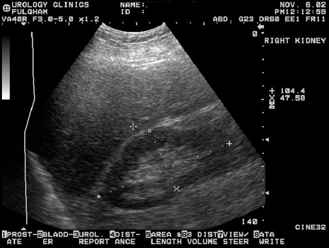
 Physical Principles of Ultrasound Pat F. Fulgham 2 Introduction The use of ultrasound is fundamental to the practice of urology. In order for urologists to best use this technology on behalf of their patients,
CONTENTS. Test Number cpd Tanya Reynolds (Nat. Dip. Diag. Rad., B. Tech. Diag. Rad., B. Tech. Ultrasound)
") CONTENTS page 1-15 page 16 BASIC 2-DIMENSIONAL ULTRASOUND PRINCIPLES Multiple Choice Test Test Number cpd 41640 Tanya Reynolds (Nat. Dip. Diag. Rad., B. Tech. Diag. Rad., B. Tech. Ultrasound) Tanya is
CONTENTS page 1-15 page 16 BASIC 2-DIMENSIONAL ULTRASOUND PRINCIPLES Multiple Choice Test Test Number cpd 41640 Tanya Reynolds (Nat. Dip. Diag. Rad., B. Tech. Diag. Rad., B. Tech. Ultrasound) Tanya is
Point-of-Care Ultrasound: An Introduction
 Point-of-Care Ultrasound: An Introduction Delegation Teaching Package for Registered Respiratory Therapists and Anesthesia Assistants Developed by: Rob Bryan RRT, AA Edited by: Kelly Hassall RRT, FCSRT,
Chapter 14. Imaging Artifacts
 Chapter 14 Image Artifacts The complex physical interactions that occur between an ultrasound beam and human anatomy and the intricate and sophisticated technological components of a sonographic imaging
WELCOME! Introduction to Bedside Ultrasound
 WELCOME! Introduction to Bedside Ultrasound TEACHERS University of California-Irvine School of Medicine Nathan Molina nathan.d.molina@gmail.com Trevor Plescia taplescia90@gmail.com Jack Silva jpsilva42@gmail.com
Basic Physics of Ultrasound in Transesophageal Echocardiography
 SPECIAL ARTICLE IJUTPC Basic Physics of Ultrasound in Transesophageal Echocardiography Basic Physics of Ultrasound in Transesophageal Echocardiography 1 Mary Korula, 2 Ravi Hebballi 1 Senior Consultant,
Supplement (videos)
") Supplement (videos) Ruben s tube (sound): http://www.youtube.com/watch?v=gpcquuwqayw Doppler US (diagnostic use): http://www.youtube.com/watch?v=fgxzg-j_hfw http://www.youtube.com/watch?v=upsmenyoju8 High
Supplement (videos) Ruben s tube (sound): http://www.youtube.com/watch?v=gpcquuwqayw Doppler US (diagnostic use): http://www.youtube.com/watch?v=fgxzg-j_hfw http://www.youtube.com/watch?v=upsmenyoju8 High
1. Fig. 1 shows data for the intensity of a parallel beam of X-rays after penetration through varying thicknesses of a material
 1. Fig. 1 shows data for the intensity of a parallel beam of X-rays after penetration through varying thicknesses of a material. intensity / MW m 2 thickness / mm 0.91 0.40 0.69 0.80 0.52 1.20 0.40 1.60
Ultrasound Measurements and Non-destructive Testing Educational Laboratory
 Session 3548 Ultrasound Measurements and Non-destructive Testing Educational Laboratory Vladimir Genis, Horacio Sosa Goodwin College of Professional Studies, Drexel University, Philadelphia, 19104 Emil
Introduction to Biomedical Imaging
 Alejandro Frangi, PhD Computational Imaging Lab Department of Information & Communication Technology Pompeu Fabra University www.cilab.upf.edu Basic principles. Comparison to X-rays Ultrasound > 20kHz
Diploma of Medical Ultrasonography (DMU) Physical Principles of Ultrasound and Instrumentation Syllabus
 Physical Principles of Ultrasound and Instrumentation Syllabus") Diploma of Medical Ultrasonography (DMU) Physical Principles of Ultrasound and Instrumentation Syllabus Page 1 of 7 11/18 Candidates are expected to cover all of the content of this syllabus when preparing
Diploma of Medical Ultrasonography (DMU) Physical Principles of Ultrasound and Instrumentation Syllabus Page 1 of 7 11/18 Candidates are expected to cover all of the content of this syllabus when preparing
Application of Phased Array Radar Theory to Ultrasonic Linear Array Medical Imaging System
 Application of Phased Array Radar Theory to Ultrasonic Linear Array Medical Imaging System R. K. Saha, S. Karmakar, S. Saha, M. Roy, S. Sarkar and S.K. Sen Microelectronics Division, Saha Institute of
ULTRASOUND NOMENCLATURE
 Chapter 1: Ultrasound Nomenclature, Image Orientation, and Basic Instrumentation CYNTHIA SIKOWSKI Ultrasound waves are sound waves that have a frequency exceeding 20,000 Hz. When sound waves are transmitted
What is Ultrasound? Resolution Image production Attenuation Imaging modes Ultrasound artifacts... 7
 What is Ultrasound?... 1 Resolution... 3 Image production... 3 Attenuation... 4 Imaging modes... 5 Ultrasound artifacts... 7 0 What is Ultrasound? High frequency sound of frequencies 2-50 MHz is used in
Chapter 1. Principles of medical ultrasound. Overview. Background history: first steps to the piezo-electric effect.
 Chapter 1 Principles of medical ultrasound GRAHAM ARTHURS, PATRICK HILL AND TREVOR FRANKEL Overview This chapter provides an introduction to the ultrasound process for trainees in anesthesia and other
Basic Ultrasound Physics Board Review Questions
 Basic Ultrasound Physics Board Review Questions Sidney K. Edelman, PhD ESP Ultrasound The Woodlands, TX Question 1 What is the wavelength of 2 MHz sound in soft tissue? 1. 1.54 mm 2. 0.75 mm 3. 0.75 cm
The table below shows the density and velocity of waves in two different substances. Density / kg m 3 Velocity / m s 1
 Q1.(a) When ultrasound is incident at an interface between two different media some energy is transmitted and some is reflected. The ratio of the reflected energy intensity I r to the incident energy intensity
Ultrasound guidance in regional anesthesia has
 Ultrasound and Regional Anesthesia Artifacts and Pitfall Errors Associated With Ultrasound-Guided Regional Anesthesia. Part I: Understanding the Basic Principles of Ultrasound Physics and Machine Operations
Chapter 3 Physical Principles of Ultrasound of the Male Genitalia
 Chapter 3 Physical Principles of Ultrasound of the Male Genitalia Bruce R. Gilbert and Pat Fox Fulgham Introduction The value of ultrasound evaluation of the male genitalia depends, in large part, on the
Chapter 2 Pitfalls in Musculoskeletal Ultrasound
 Chapter 2 Pitfalls in Musculoskeletal Ultrasound Violeta Maria Vlad MD, PhD Introduction Taking a good ultrasound (US) picture is an art. Interpreting it is a science. This is in fact everything US is
Table of contents. Foreword. Preface. 1 Introduction Historical Perspective 00
 Table of contents Foreword Preface 1 Introduction 00 1.1 Historical Perspective 00 2 Fundamentals of musculoskeletal ultrasound 00 2.1 Frequency and wavelength 00 2.2 Generating ultrasound waves 00 2.3
Breast Imaging Essentials
 Breast Imaging Essentials Module 9 Transcript 2016 ASRT. All rights reserved. Breast Imaging Essentials Module 9 Breast Ultrasound 1. ASRT Animation 2. Welcome Welcome to Module 9 of Breast Imaging Essentials
Emergency Medicine Interest Group (EMIG) 2016
 2016") Emergency Medicine Interest Group (EMIG) 2016 Welcome to the flipped classroom (learning objectives summary) for the 2016 Emergency Medicine Interest Group (EMIG) Procedures Workshop. Overview - Tuesday
Emergency Medicine Interest Group (EMIG) 2016 Welcome to the flipped classroom (learning objectives summary) for the 2016 Emergency Medicine Interest Group (EMIG) Procedures Workshop. Overview - Tuesday
Ultrasound: Past and Present. Lecturer: Dr. John M Hudson, PhD
 Ultrasound: Past and Present Lecturer: Dr. John M Hudson, PhD Disclosures 2 No conflicts of interest to declare Course Outline 3 1. Survey of ultrasound physics & applications 2. (Sep 21) 3. (Sep 28) 4.
Ultrasound in Anesthesia: Applying Scientific Principles to Clinical Practice
 AANA Journal Course Update for Nurse Anesthetists 3 6 CE Credits* Ultrasound in Anesthesia: Applying Scientific Principles to Clinical Practice Christian R. Falyar, CRNA, DNAP The use of ultrasound as
Thyroid Ultrasound Physics and Doppler
 Thyroid Ultrasound Physics and Doppler Advanced AACE-ACE US training course 2017 Dev Abraham MD, MRCP(UK), ECNU, FACE Professor of Medicine, University of Utah No Disclosures Natural Ability to see with
4.17. RESEARCHING MODELS WITH AN ULTRASONIC ECHOSCOPE
 4.17. RESEARCHING MODELS WITH AN ULTRASONIC ECHOSCOPE Purpose of experiment Determine the main characteristics of ultrasound waves, and the distances and positions of models using an ultrasonic echoscope.
The Essentials Tissue Characterization and Knobology
 The Essentials Tissue Characterization and Knobology Randy E. Moore, DC, RDMS RMSK No relevant financial relationships Ultrasound The New Standard of Care Musculoskeletal sonography has become the standard
Development of Ultrasound Based Techniques for Measuring Skeletal Muscle Motion
 Development of Ultrasound Based Techniques for Measuring Skeletal Muscle Motion Jason Silver August 26, 2009 Presentation Outline Introduction Thesis Objectives Mathematical Model and Principles Methods
Physics. Norman McDicken Tom Anderson CHAPTER ULTRASOUND. Ultrasound Propagation
 CHPTER 2 Physics Norman McDicken Tom nderson This chapter provides an introduction to the physics of medical ultrasound (US). Several books exist that can be consulted to extend the material presented
4.17. RESEARCHING MODELS WITH AN ULTRASONIC ECHOSCOPE
 4.17. RESEARCHING MODELS WITH AN ULTRASONIC ECHOSCOPE Purpose of experiment Determine the main characteristics of ultrasound waves, and the distances and positions of models using an ultrasonic echoscope.
Ultrasonic Testing. Basic Principles
 Ultrasonic Testing Ultrasonic Testing (UT) uses high frequency sound waves (typically in the range between 0.5 and 15 MHz) to conduct examinations and make measurements. Besides its wide use in engineering
The 2 nd Cambridge Advanced Emergency Ultrasound Course
 The 2 nd Cambridge Advanced Emergency Ultrasound Course Addenbrooke s Hospital Cambridge Sept 2008 1 2 Faculty! UK! USA! Australia! Toshiba! Emergency Medicine! Radiology 3 Programme! Day 1 Introduction
Ultrasonography of the Neck as an Adjunct to FNA. Nicole Massoll M.D.
 Ultrasonography of the Neck as an Adjunct to FNA Nicole Massoll M.D. Basic Features of Head and Neck Ultrasound and Anatomy Nicole Massoll M.D. University of Arkansas for Medical Sciences, Little Rock
Descriptions of NDT Projects Fall 2004 October 31, 2004
 Descriptions of NDT Projects Fall 2004 October 31, 2004 Introduction There are two separate NDT labs in Magister: ULTRA for ultrasound and EDDY for eddy current. Both labs are equipped with mechanical
Tissue Strain Analytics Virtual Touch Tissue Imaging and Quantification
 Whitepaper Tissue Strain Analytics Virtual Touch Tissue Imaging and Quantification ACUSON S2000 Ultrasound System Answers for life. Page 1 Tissue Strain Analytics: Virtual Touch Tissue Imaging and Quantification
Roaa M.Hussein Professor, Department of Physics, College of Science, Ramadi, Iraq. Abstract:
 Abstract: Ultrasound Waves Employment in the Medical Diagnostic for Lıver and Gallbladder Faik H. Antar Professor, Department of Physics, College of Science, AL- Anbar University, Ramadi, Iraq. Ultrasound
Basic Training Programme. 16 Februrary 2018, ROTTERDAM. Pre and Post-Course Test Answers
 Basic Training Programme 16 Februrary 2018, ROTTERDAM Pre and Post-Course Test Answers Your details: Name: Conference registration number/ BT delegate number: Email address: Are you already performing
3/20/2017. Disclosures. Ultrasound Fundamentals. Ultrasound Fundamentals. Bone Anatomy. Tissue Characteristics
 Disclosures Images of ultrasound equipment in this presentation are not an endorsement Fundamentals of Musculoskeletal Ultrasound Physics and Knobology Shane A. Shapiro, M.D. Assistant Professor Orthopedic
Evaluation of the Quality of Thick Fibre Composites Using Immersion and Air- Coupled Ultrasonic Techniques
 ECNDT 2006 - We.1.6.4 Evaluation of the Quality of Thick Fibre Composites Using Immersion and Air- Coupled Ultrasonic Techniques Kaj K. BORUM, Risø National Laboratory, Materials Research Department, Roskilde,
CSB 046 Complementary Imaging Techniques
 CSB 046 Complementary Imaging Techniques - Quizzes are only ultrasound, final includes nuc med and ultrasound Week 1 Intro to Ultrasound Physics - Uses 1 to 20 MHz frequencies, which is way above the sound
Concepts of Imaging and Knobology
 Concepts of Imaging and Knobology Pravin Patil, MD FACC FASE Associate Professor of Medicine Director, Cardiovascular Disease Training Program Lewis Katz School of Medicine at Temple University Disclosures
Underwater Acoustic Measurements in Megahertz Frequency Range.
 Underwater Acoustic Measurements in Megahertz Frequency Range. Current State and Prospects of Development in Russia Alexander M. Enyakov,, Many medical applications of underwater acoustic measurements
INTRODUCTION. Getting the best scan. Choosing a probe. Choosing the frequency
 Getting the best scan Choosing a probe Select the most appropriate probe for the particular scan required. s vary in their: operating frequency range higher ultrasound frequencies provide better discrimination
An Overview of Ultrasound Testing For Lesion Detection in Human Kidney
 Journal of Tomography System & Sensors Application Vol.1, Issue 1, June 2018 An Overview of Ultrasound Testing For Lesion Detection in Human Kidney Aina Fadhilah Abd Rahim 1, Zawin Najah Abd Halim 1, Jaysuman
Ultrasonic arrays are now widely used in underwater sonar
 Ultrasonics NDT FUNDAMENTALS Part 12. Fundamentals of ultrasonic phased arrays S Cochran Ultrasonic arrays are now widely used in underwater sonar and in more than 25% of medical scans but their use in
Non-Destructive Inspection of Composite Wrapped Thick-Wall Cylinders
 Non-Destructive Inspection of Composite Wrapped Thick-Wall Cylinders Jikai Du, John Feldhacker, Christopher Jerred and Fereidoon Delfanian May 17-19, 2010 Joint Armaments Conference, Exhibition and Firing
US Artifacts 1 EDUCATION EXHIBIT. Myra K. Feldman, MD Sanjeev Katyal, MD Margaret S. Blackwood, MS
 Note: This copy is for your personal, non-commercial use only. To order presentation-ready copies for distribution to your colleagues, use the RadioGraphics Reprints form at the end of this article. EDUCATION
FAST Focused Assessment with Sonography in Trauma
 FAST Focused Assessment with Sonography in Trauma Wilma Rodriguez Mojica,MD,FACR Professor of Radiology UPR School of Medicine Ultrasound Section - Radiological Sciences Department OBJECTIVES Understand
Pulse-Echo Ultrasound Imaging. Resolution in Ultrasound Imaging. Doppler Ultrasound. Resolution vs Penetration. Medical Imaging (EL582/BE620/GA4426)
") Medical Imaging (EL582/BE620/GA4426) Pulse-Echo Ultrasound Imaging Ultrasound Imaging Lecture 2 Daniel (Dan) Turnbull, Ph.D. Skirball Institute and Dept of Radiology NYU School of Medicine (daniel.turnbull@med.nyu.edu)
Medical Imaging (EL582/BE620/GA4426) Pulse-Echo Ultrasound Imaging Ultrasound Imaging Lecture 2 Daniel (Dan) Turnbull, Ph.D. Skirball Institute and Dept of Radiology NYU School of Medicine (daniel.turnbull@med.nyu.edu)
1. SCOPE ELIGIBILITY EXAMINATION CONTENT RENEWAL & RECERTIFICATION PROCEDURE ESSENTIAL READING...
 Certification Services Division Newton Building, St George s Avenue Northampton, NN2 6JB United Kingdom Tel: +44(0)1604-893-811. Fax: +44(0)1604-893-868. E-mail: pcn@bindt.org PCN/GEN ISO 20807 Appendix
Flip Chips and Acoustic Micro Imaging: An Overview of Past Applications, Present Status, And Roadmap for the Future
 Flip Chips and Acoustic Micro Imaging: An Overview of Past Applications, Present Status, And Roadmap for the Future Janet E. Semmens Sonoscan, Inc. 2149 E. Pratt Boulevard Elk Grove Village, IL 60007 USA
Flaw Assessment Using Shear wave Phased array Ultrasonic Transducer
 18th World Conference on Nondestructive Testing, 16-20 April 2012, Durban, South Africa Flaw Assessment Using Shear wave Phased array Ultrasonic Transducer Byungsik YOON AUTHOR 1, Hee-Jong LEE CO-AUTHOR
Performance of phased array and conventional ultrasonic probes on the new ISO reference block
 Performance of phased array and conventional ultrasonic probes on the new ISO 19675 reference block C. Udell, D. Chai 1 and F. Gattiker Proceq S.A., Ringstrasse 2, Schwerzenbach, Switzerland. More info
COURSE DESCRIPTION FOR NONDESTRUCTIVE TESTING
 COURSE DESCRIPTION FOR NONDESTRUCTIVE TESTING TULSA, OKLAHOMA INTRODUCTION TO NONDESTRUCTIVE TESTING QCT1817 COURSE DESCRIPTION: In this course students will learn about materials and processes, find basic
BASIC PRINCIPLES OF DIAGNOSTIC ULTRASONOGRAPHY
 EXTENSION ARTICLE Pakistan Vet. J.. 17 (3): 1997 BASIC PRINCIPLES OF DIAGNOSTIC ULTRASONOGRAPHY Nazir Ahmad Department of Animal Reproduction, University of Agriculture, Faisalabad-38040, Pakistan INTRODUCTION
HSC Physics. Module 9.6. Medical Physics
 HSC Physics Module 9.6 Medical Physics Contextual Outline 9.6 Medical Physics (28 indicative hours) The use of other advances in technology, developed from our understanding of the electromagnetic spectrum,
Ultrasound in Medicine
 Ultrasound in Medicine Experimental Equipment for Medical Education Universities Colleges Medical Schools Medical and Med-Technical Training Education can befun! WELCOME TO GAMPT Devices and accessories
Can you believe that the ultrasonic waves are used for cleaning purpose???
 Introduction (Ultrasonic Cleaning Unit) Can you believe that the ultrasonic waves are used for cleaning purpose??? Learning Objectives On completion of this chapter you will be able to: 1. Describe the
Ultrasound Evaluation of the Posterior Segment of the Eye A Ready Reckoner
 180 Kerala Journal of Ophthalmology Vol. XX, No. 2 OPHTHALMIC INSTRUMENTATION Ultrasound Evaluation of the Posterior Segment of the Eye A Ready Reckoner Dr. Mahesh G. MS DO DNB FRCSEd., Dr. A. Giridhar
Strain Assessment in Echo
 Strain Assessment in Echo Joe M. Moody, Jr, MD UTHSCSA and STVHCS 2010 Acknowledging many illustrations from Weyman s text and others. Echo-Doppler Basic Principles Background: Ultrasound physics (resolution,
Shcherbakov O.N., Petrov A.E., Polevoy A.G., Annenkov A.S. ULTES LLC, Moscow, Russia
 METHODS AND MEANS OF ULTRASONIC INSPECTION (USI) OF WELDED JOINTS WITH ACOUSTIC CONTACT LEVEL TRACKING AND EVALUATION OF THE NATURE AND ACTUAL CHARACTERISTICS OF FLAWS Shcherbakov O.N., Petrov A.E., Polevoy
Linear Ultrasonic Wave Propagation in Biological Tissues
 Indian Journal of Biomechanics: Special Issue (NCBM 7-8 March 29) Linear Ultrasonic Wave Propagation in Biological Tissues Narendra D Londhe R. S. Anand 2, 2 Electrical Engineering Department, IIT Roorkee,
Chapter 3. Sonographic Image Interpretation
 Chapter 3 Sonographic Image Interpretation Sonograms are two-dimensional gray-scale images that allow assessment and diagnosis of many anatomic and pathologic changes that can occur in the human body.
Exam Practice Guide. Units 1 & 2 Physics: Detailed Study 5 - Investigations: Medical physics Examination Questions
 Exam Practice Guide Units 1 & 2 Physics: Detailed Study 5 - Investigations: Medical physics Examination Questions Key Features: 22 original examination style questions on all examinable topics. Full solutions
PART 1c: Time of Flight Diffraction Ultrasonic Inspector (TOFD) of Welds in Ferritic and Non-Ferritic Materials, Levels 1, 2 and 3
 of Welds in Ferritic and Non-Ferritic Materials, Levels 1, 2 and 3") CERTIFICATION SCHEME FOR PERSONNEL DOCUMENT No. CSWIP-ISO-NDT-11/93-R Requirements for the Certification of Personnel Engaged in Non- Destructive Testing in accordance with the requirement of BS EN ISO
CERTIFICATION SCHEME FOR PERSONNEL DOCUMENT No. CSWIP-ISO-NDT-11/93-R Requirements for the Certification of Personnel Engaged in Non- Destructive Testing in accordance with the requirement of BS EN ISO
Medical Imaging. Ultrasound Imaging
 Medical Imaging Ultrasound Imaging Prof. Ed X. Wu Overview History Physics of Ultrasound wave propagation, attenuation, scattering, and reflection Generation and Detection of Ultrasound Piezoelectric Transducers
VISUALIZATION OF TRANSDUCER-PRODUCED SOUND FIELDS IN SOLIDS
 VISUALIZATION OF TRANSDUCER-PRODUCED SOUND FIELDS IN SOLIDS Wolfgang Sachse* Department of Theoretical and Applied Mechanics Cornell University, Ithaca, New York - 14853 ABSTRACT Broadband ultrasonic pulses
Q1. The diagram shows an ultrasound monitor being used to scan a fetus.
 Q1. The diagram shows an ultrasound monitor being used to scan a fetus. The table shows the velocity of ultrasound waves in different tissues of the fetus. Tissue Amniotic fluid (liquid surrounding fetus)
Employer s Unit of Competence Ultrasonic testing of materials, products and plant
 Employer s Unit of Competence Ultrasonic testing of materials, products and plant Document: AA064 Issue 2 May 2016 Image - if cover page required Supported by lead employer Overview This standard identifies
ADVANCED PHASED ARRAY TECHNOLOGIES
 IRNDT 2016 3rd Iranian International NDT Conference ADVANCED PHASED ARRAY TECHNOLOGIES Wolfram A. Karl Deutsch Karl Deutsch Pruef- und Messgeraetebau GmbH + Co KG, Wuppertal, Germany, E-Mail: info@karldeutsch.de
Confident and Conclusive Diagnosis with Flexible Solutions
 Internal Medicine Confident and Conclusive Diagnosis with Flexible Solutions ALPINION MEDICAL SYSTEMS We are Ultrasound Professionals Ultrasound is widely used as the gold-standard for internal medicine
DOW-RAD, DOW DIAGNOSTIC COMPLEX, DUHS TRAINING PROGRAM HANDBOOK 2013
 DOW-RAD, DOW DIAGNOSTIC COMPLEX, DUHS TRAINING PROGRAM HANDBOOK 2013 CERTIFICATE COURSE INVASCULAR/DOPPLER ULTRASOUND: Introduction: Ultrasound is an evolving technology with wide spectrum application
Learning Objectives. Ultrasound for the Primary Care Provider. Portable Ultrasound: Laptops, Tablets, Plug-in Probes, and Pocket devices
 Learning Objectives Ultrasound for the Primary Care Provider Richard Hoppmann, MD, FACP University of South Carolina School of Medicine Assess the main components and functions of a portable ultrasound
High resolution ultrasound scanner for skin imaging
 High resolution ultrasound scanner for skin imaging Christine Turlat Sales Director Atys medical 17 Parc d Arbora 69510 SOUCIEU EN JARREST Atys company Principle of ultrasound imaging DERMCUP Normal image
Ultrasonic Testing courses at DGZfP Education and Training Ltd
 ECNDT 2006 - Poster 29 Ultrasonic Testing courses at DGZfP Education and Training Ltd A few topics on ultrasonic testing training courses level 1 to 3 at a glance Wolf-Dieter JANKE, DGZfP, Berlin, Germany