Basics of nuclear medicine
|
|
|
- Stephany Brianna Cummings
- 6 years ago
- Views:
Transcription

1 Basics of nuclear medicine Prof. dr. Davor Eterović Prof. dr. Vinko Marković
2 Radioisotopes are used both in diagnostics and in therapy Diagnostics gamma emitters are used since gamma rays can penetrate the body and be detected outside Therapy beta emitters; short range => local delivery of high doses
3 Diagnostic activities correspond to sub-physiological masses Activities used: 370 KBq-740 MBq (10µCi- 20 mci) Corresponding masses <1 g => No physiological effects, but enough for registration and diagnostic information
4 Tc-99m- the most used radioisotope in medicine Offspring of a nuclear reactor product Mo-99, which by beta-minus decay becomes Tc-99m, the meta-stable (long living) gamma emitter ADVANTAGES: 1. pure gamma emitter- no beta radiation 2. optimal photon energy- sufficient to penetrate body, not too large for efficient detection/protection 3. half life long enough to perform the investigation, with acceptable patient radiation dose 4. great chemical affinity 5. relatively cheap.

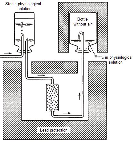
5 Technetium generator
6 Scintillation detectors: gamma rays are detected after conversion to visible light When some materials absorb ionizing radiation the part of absorbed energy is used to excite the atoms to higher energy states, the subsequent de-excitations produce flashes of visible light These flashes are called scintillations, and the materials scinillators => SCINTIGRAMS
7 Sodium iodide chrystal-the commonest gamma ray detector 1. High sensitivity of detection (owing to high density and atomic number of iodine) 2. Relatively high efficiency of conversion of gamma rays to visible light (around 10 %) 3. Short scintillation time (short dead-time) enables high registration rate (over 10 4 counts/sec)
8 Scintillation detectors can measure the energy of gamma rays The output electrical pulse is proportional to energy of the absorbed radiation, enabling: 1. using the detector as absorbed dose-meter (besides counting) 2. selective counting according to energy of photon (pulse height)- spectral analysis

9 Scintillator and photomultiplier tube comprise the scintillation counter
10 SCINTIGRAM image of organ function Accumulation of radiotracer in part of a body depends only on chemical affinity and physical properties of the tracer to which the radioisotope is bound. Nuclear medicine image reflects the physiological function and is created by gamma rays that leave the body.

11 Scintigraphy Image of distribution of radioisotope that is attached to a molecule that accumulates in the desired region, tissue or organ, to extent that depends on cellular function


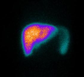
12 99m-Tc-pertechnetatescintigram of normal thyroid distribution (left) 201-Tl-chloride- scintigram of normal heart muscle (down)
13

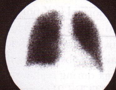
14 I-131 scintigraphy of toxic nodular goitter
")

")
15 Scanner (middle) Gamma camera PET scanner (down) Imaging equipment
16 Registration of gamma radiation requires massive detector Two purposes of radiactive measurements: 1. measurements of concentration in a sample 2. imaging distribution within the body Due to large energy and consequent penetrability in both cases one needs massive detector, which limits resolution and sensitivity of registration

17 Well counter

18 Pinhole collimator: imaging of small volumes
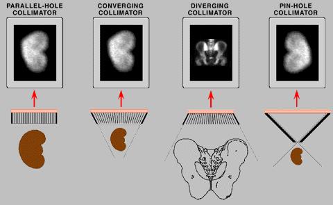
19
20 Gamma camera creates live scintigrams Parallel collimator lead plate with many narrow parallel holes (channels) at right angle to plate and crystal bases => gamma ray hitting the crystal originates from the area directly bellow the place of absorption gamma camera is always sensitive to the toal area beneath the crystal
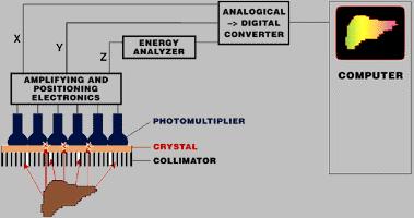
21 Gamma camera components
22
23 Two ways to present the gamma camera image: 1. analog image: the position of scintillation is projected on the screen of an oscilloscope, which illuminates the photograpic film 2. digital image: position and rate of scintillations are stored in computer memory as a digital matrix (64x64; 128x128;..)

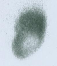
24 Liver scintigraphy with Tc-99m-colloid in case of trauma causing tissue rupture analog image digital image

25 The effect of matrix size
26 Distinctive scintigram features: functionality and quantitative aspect functionality: radiotracer accumulation mirrors the organ(s) function quantitative aspect: scintigram is basically a table of numbers
27 Factors of scintigram (dis)advantages 1. Large energy of a gamma ray: requires massive detector, but enables registration of small, deep sources of minute activities 2. spreadness/wide distribution of gamma ray sources: requires collimation of radiation, but enables functional organ images => Minute amounts of radioindicator, in short time, create diagnostically useful images
28 Lesion detectability increases with its size, but primarily depends on its contrast against the neighboring tissue Resolution-important for small lesions Digital image can manipulated on a computer to enhance visual effects (caution: possibility of artifacts!)

29 Hot lesions are better seen than cold ones


30 Radiohistogram are analyzed with computer aid to diagnose and quantify various parameters
 scintigrams or time-activity")
31 Radihistogram: time-activity curve computer programs: algorithm codes to analyze (series of) scintigrams or time-activity curves

32 Typical mechanisms of ratiotracer localization Mechanism Organ Radiotracer active transport thyroid J-131 active transport kidney ortho-i-131-hypurric acid active transport myocardiu Tl-201 m capillary blockade lungs Tc-99m-macroaggregate filtration kidney Tc-99m-diethilene-triaminepentaacetic acid dilution blood Tc-99m-human serum albumin phagocytosis liver Tc-99m-sumphor colloid sequestration spleen Tc-99m- erythrocytes (damaged by heating)
33 Effective half-life: determinant of absorbed dose due to radioacitivity introduced into the body constant of biological elimination ( B ) biological half-life (T 1/2 ) B = ln(2)/ B constant of effective elimination: EF = B + F (constant of radiactive decay) effective half-life (T 1/2 ) EF = ln(2)/ EF
 = A 0")
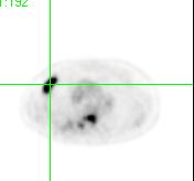
34 => The inital radioacitivity in body decreases in time according to: A(t) = A 0 exp(- EF t)
35 131 99m I Tc Absorbed doses in common procedures Procedure Radiotracer Activity Dose in critical organ (Gy) a Brain scintigraphy Liver scintigraphy pertechnetate sulphor colloid 500 MBq (~ 15 mci) Intestines, 0.02 Dose in gonads (mgy) 150 MBq (~ 4 mci) Liver, Lung scintigraphy Bone scintigraphy macroaggregate pyrophosphate 100 MBq (~ 3 mci) Lungs, MBq (~ 15 mci) Bladder, Renography Thyroid function Thyroid scintigraphy hyppuric acid sodium iodide pertechnetate Bq (~ 200 μci) 300 kbq (~ 8 μci) 150 MBq (~ 4 mci) a critical organ has the largest absorbed dose Bladder, Thyroid, Intestines,
36 Imaging of body slices= tomography SPECT = single-photon emission computed tomography PET = positron emission tomography
37 Imaging from many directions enables slice isolation SPECT: gamma camera rotates around body, the series of scintigrams (projections) thus obtained are raw dana from which computer reconstructs slices

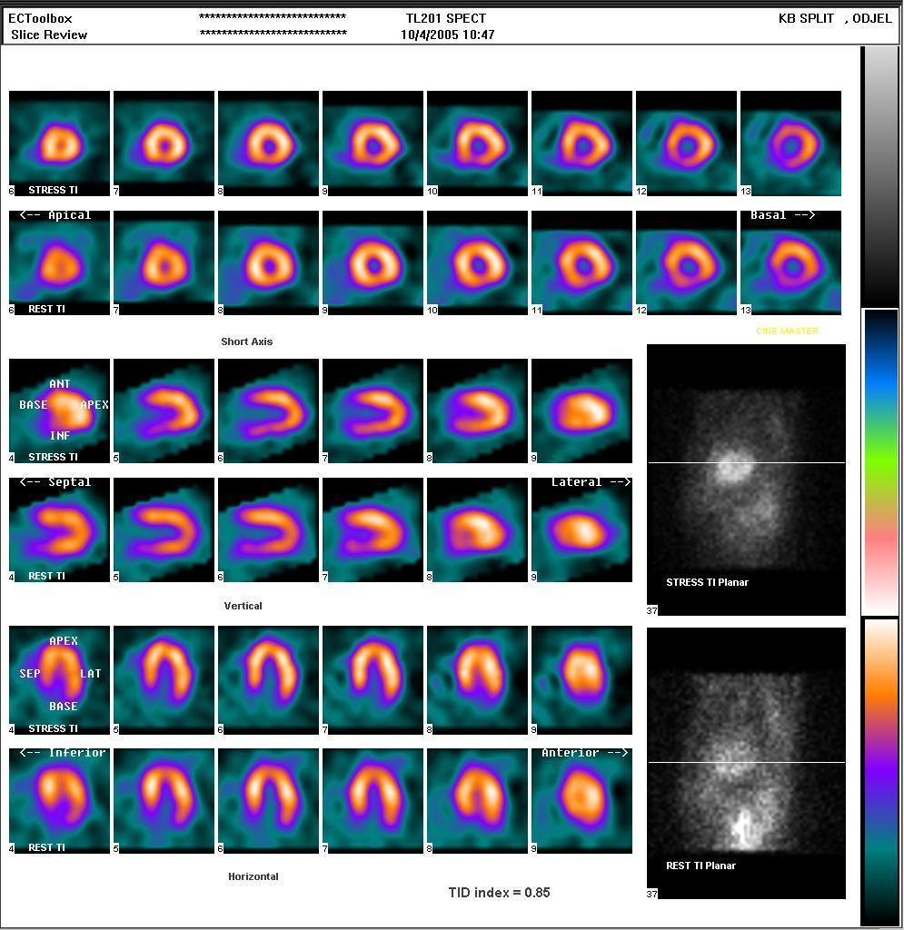
38 SPECT
39 Tomograms have inferior resolution compared to planar images Tomogram, being derived from series of planar images (projections) by non-perfect algorithm (absorption of radation in the body can be accounted for only approximately) has inferior resolution than the original (raw) images Nevertheless, due to increased contrasts, the overall diagnostic information is greater
, or mollecules attached to them. The images reflect: tissue perfusion, the metabolism of oxygen, carbohydrates, fatty acids, amino acids, as well as neuroreceptors.")
40 Positron emissiion tomography- PET PET is used to detect the (pato)physiological and biochemical processes by virtue of kinetics of positron emitters (F-18, C-11, O-15, N-13), or mollecules attached to them. The images reflect: tissue perfusion, the metabolism of oxygen, carbohydrates, fatty acids, amino acids, as well as neuroreceptors.
41 Positron emitters display metabolic processes: Radionuclide Half-life Tracer Use (min) Carbon Nitric acid Clinical research Nitrogen Cardiology Cardiology Oxygen H 2 O, CO, CO 2 Fluorine FDG and F- dopamine Rubidium Potassium analogs Clinical research Oncology, cardiology, neurology Cardiology

42 Creation of PET image The positions of sources (A,B) are reconstructed as sections of straight lines passing through detector pairs


43 Principle of coincident detection No collimation needed: resolution, sensitivity

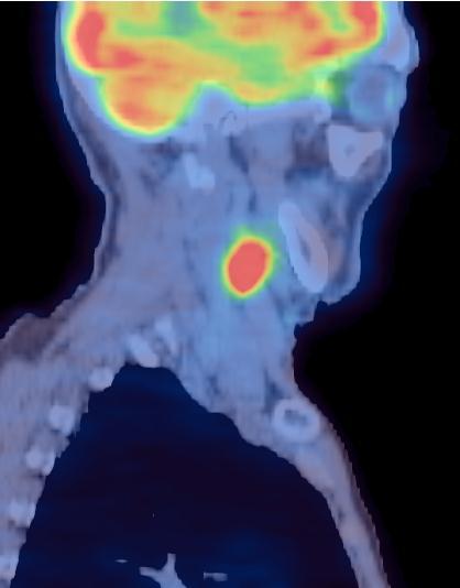
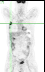
44 PET image of a patient with liver metastasis

45 PET: pathologic focus in shoulder region


46 CT: liver metastasis PET: liver metastasis

47 Image fusion: PET-CT

48 CT PET Lung metastasis

49 PET-CT fusion (lung metastasis)
50

 can be")


51 Therapy Radiotracers containg particle emitters (mostly beta-emitters) can be used for selective tissue destruction, by virtue of their small range and consequent local energy absorption
52 Nuclear medicine perspective The future of nuclear medicine depends on development of new radiotracers: mollecular nuclear medicine THE END
Medical Use of Radioisotopes
 Medical Use of Radioisotopes Therapy Radioisotopes prove to be useful in the application of brachytherapy, the procedure for using temporary irradiation close to the area of disease (i.e. cancer) 10% Medical
45 Hr PET Registry Review Course
 45 HR PET/CT REGISTRY REVIEW COURSE Course Control Document Timothy K. Marshel, MBA, R.T. (R), (N)(CT)(MR)(NCT)(PET)(CNMT) The PET/CT Training Institute, Inc. SNMMI-TS 028600-028632 45hr CEH s Voice Credits
Austin Radiological Association Nuclear Medicine Procedure WHITE BLOOD CELL MIGRATION STUDY (In-111-WBCs, Tc-99m-HMPAO-WBCs)
") Austin Radiological Association Nuclear Medicine Procedure WHITE BLOOD CELL MIGRATION STUDY (In-111-WBCs, Tc-99m-HMPAO-WBCs) Overview Indications The White Blood Cell Migration Study demonstrates the distribution
Austin Radiological Association Nuclear Medicine Procedure WHITE BLOOD CELL MIGRATION STUDY (In-111-WBCs, Tc-99m-HMPAO-WBCs) Overview Indications The White Blood Cell Migration Study demonstrates the distribution
CEREBRAL BLOOD FLOW AND METABOLISM
 Supported by: HURO/0901/069/2.3.1 HU-RO-DOCS CEREBRAL BLOOD FLOW AND METABOLISM Part 3 Modern imaging methods SPECT, PET, nmri History of Nuclear Medicine Starts with the invention of the X-ray 1946: radioactive
PHYSICS 2: HSC COURSE 2 nd edition (Andriessen et al) CHAPTER 20 Radioactivity as a diagnostic tool (pages 394-5)
 CHAPTER 20 Radioactivity as a diagnostic tool (pages 394-5)") PHYSICS 2: HSC COURSE 2 nd edition (Andriessen et al) CHAPTER 20 Radioactivity as a diagnostic tool (pages 394-5) 1. (a) A radioisotope is an isotope that is unstable and will emit particles from the nucleus
PHYSICS 2: HSC COURSE 2 nd edition (Andriessen et al) CHAPTER 20 Radioactivity as a diagnostic tool (pages 394-5) 1. (a) A radioisotope is an isotope that is unstable and will emit particles from the nucleus
Nuclear Medicine and PET. D. J. McMahon rev cewood
 Nuclear Medicine and PET D. J. McMahon 150504 rev cewood 2018-02-15 Key Points Nuclear Medicine and PET: Imaging: Understand how Nuc Med & PET differ from Radiography & CT by the source of radiation. Be
Option D: Medicinal Chemistry
 Option D: Medicinal Chemistry Basics - unstable radioactive nuclei emit radiation in the form of smaller particles alpha, beta, positron, proton, neutron, & gamma are all used in nuclear medicine unstable
Itroduction to the Nuclear Medicine: biophysics and basic principles. Zámbó Katalin Department of Nuclear Medicine
 Itroduction to the Nuclear Medicine: biophysics and basic principles Zámbó Katalin Department of Nuclear Medicine NUCLEAR MEDICINE Application of the radioactive isotopes in the diagnostics and in the
Radionuclides in Medical Imaging. Danielle Wilson
 Radionuclides in Medical Imaging Danielle Wilson Outline Definitions History and development Radionuclide applications & techniques in imaging Conclusion Definition #1 : Radionuclide An unstable nucleus
Radiopharmacy. Prof. Dr. Çetin ÖNSEL. CTF Nükleer Tıp Anabilim Dalı
 Prof. Dr. Çetin ÖNSEL CTF Nükleer Tıp Anabilim Dalı What is Nuclear Medicine? Nuclear Medicine is the branch of medicine concerned with the use of radionuclides in the study and the diagnosis of diseases.
KEYWORDS: nuclear medicine; gamma camera; radiopharmaceutical activities.
 Radiopharmaceutical Activities Administered for Diagnostic Procedures in Nuclear Medicine in the First Six Months of the Gamma Camera Use in the Clinical Center of Montenegro - Podgorica Nevenka Antovic
A Snapshot on Nuclear Cardiac Imaging
 Editorial A Snapshot on Nuclear Cardiac Imaging Khalil, M. Department of Physics, Faculty of Science, Helwan University. There is no doubt that nuclear medicine scanning devices are essential tool in the
Radiation Detection and Measurement
 Radiation Detection and Measurement Range of charged particles (e.g.,!: µm; ": mm) Range of high energy photons (cm) Two main types of interactions of high energy photons Compton scatter Photoelectric
Gastrointestinal tract
 Gastrointestinal tract Colloidal liver-spleen imaging Presented by: Jehad Felemban Introduction: To obtain better anatomic display of liver and spleen architecture, we use (CT Ultrasound). (Radionuclide
Radiation physics and radiation protection. University of Szeged Department of Nuclear Medicine
 Radiation physics and radiation protection University of Szeged Department of Nuclear Medicine Radiation doses to the population 1 Radiation doses to the population 2 Sources of radiation 1 Radiation we
OTHER NON-CARDIAC USES OF Tc-99m CARDIAC AGENTS Tc-99m Sestamibi for parathyroid imaging, breast tumor imaging, and imaging of other malignant tumors.
 DEFINITION OF CARDIAC RADIOPHARMACEUTICAL: A radioactive drug which, when administered for purpose of diagnosis of heart disease, typically elicits no physiological response from the patient. Even though
General Nuclear Medicine
 General Nuclear Medicine What is General Nuclear Medicine? What are some common uses of the procedure? How should I prepare? What does the equipment look like? How does the procedure work? How is the procedure
Nuclear Medicine Head and Neck Region. Bán Zsuzsanna, MD University of Pécs, Department of Nuclear Medicine
 Nuclear Medicine Head and Neck Region Bán Zsuzsanna, MD University of Pécs, Department of Nuclear Medicine Thyroid scintigraphy Parathyroid scintigraphy F18-FDG PET examinations in head and neck cancer
Molecular Imaging and the Brain
 Molecular imaging technologies are playing an important role in neuroimaging, a branch of medical imaging, by providing a window into the living brain. Where CT and conventional MR imaging provide important
Applications of radioactivity in medicine
 Lec.3 Applications of radioactivity in medicine -Nuclear medicine (N.M) -Applications of radioactive material in medicine Nuclear Medicine:- The clinical uses of radioactivity for the diagnosis of disease.
Department of Nuclear Medicine with Positron Emission Tomography
 (PET) Unit [1] Contact information: Registration: +48 41 367 4850 Main office: +48 41 367 4860 Fax: +48 41 367 4887 e-mail: zmnsco@onkol.kielce.pl [2] Head of the Department: Professor Janusz Braziewicz
Positron Emission Tomography Computed Tomography (PET/CT)
") Positron Emission Tomography Computed Tomography (PET/CT) What is Positron Emission Tomography Computed Tomography (PET/CT) Scanning? What are some common uses of the procedure? How should I prepare for
Positron Emission Tomography Computed Tomography (PET/CT) What is Positron Emission Tomography Computed Tomography (PET/CT) Scanning? What are some common uses of the procedure? How should I prepare for
Medical Physics 4 I3 Radiation in Medicine
 Name: Date: 1. This question is about radiation dosimetry. Medical Physics 4 I3 Radiation in Medicine Define exposure. A patient is injected with a gamma ray emitter. The radiation from the source creates
Internal Dosimetry of Human Brain for 99m tc and 131 I Using Nuclear Imaging in Bangladesh
 Sri Lankan Journal of Physics, Vol. 6 (2005) 33-41 Institute of Physics - Sri Lanka Internal Dosimetry of Human Brain for 99m tc and 131 I Using Nuclear Imaging in Bangladesh M. M. Alam a, M. I. Kabir
Medical imaging. Medical imaging uses electromagnetic radiation, sound or ingestion of radioactive substances. 10/6/2011 Medical imaging 1
 Medical imaging Medical imaging uses electromagnetic radiation, sound or ingestion of radioactive substances 10/6/2011 Medical imaging 1 0 Ultrasound Imaging Transducer Reflector Use high-frequency sound
Fundamentals of Nuclear Cardiology. Terrence Ruddy, MD, FRCPC, FACC
 Fundamentals of Nuclear Cardiology Terrence Ruddy, MD, FRCPC, FACC Objectives To understand the Principles of Nuclear Cardiac Imaging Radiotracers Image acquisition and processing Stress protocols To appreciate
Introduction to the Course and the Techniques. Jeffry R. Alger, PhD Ahmanson-Lovelace Brain Mapping Center Department of Neurology
 Introduction to the Course and the Techniques Jeffry R. Alger, PhD Ahmanson-Lovelace Brain Mapping Center Department of Neurology (jralger@ucla.edu) CTSI Neuroimaging April 2014 Rationale for the Course
Austin Radiological Association Ga-68 NETSPOT (Ga-68 dotatate)
") Austin Radiological Association Ga-68 NETSPOT (Ga-68 dotatate) Overview Ga-68 dotatate binds to somatostatin receptors, with highest affinity for subtype 2 receptors (sstr2). It binds to cells that express
Austin Radiological Association Ga-68 NETSPOT (Ga-68 dotatate) Overview Ga-68 dotatate binds to somatostatin receptors, with highest affinity for subtype 2 receptors (sstr2). It binds to cells that express
Austin Radiological Association Nuclear Medicine Procedure THYROID UPTAKE MEASUREMENT (I-123 or I-131 as Sodium Iodide)
") Austin Radiological Association Nuclear Medicine Procedure THYROID UPTAKE MEASUREMENT (I-123 or I-131 as Sodium Iodide) Overview Indications The Thyroid Uptake Measurement measures the metabolic activity
Austin Radiological Association Nuclear Medicine Procedure THYROID UPTAKE MEASUREMENT (I-123 or I-131 as Sodium Iodide) Overview Indications The Thyroid Uptake Measurement measures the metabolic activity
Nuclear medicine studies of the digestiv system. Zámbó Katalin Department of Nuclear Medicine
 Nuclear medicine studies of the digestiv system Zámbó Katalin Department of Nuclear Medicine Imaging tehniques Anatomy Physiology Metabolism Molecular Rtg. / CT PET / SPECT MRI MR spectroscopy fmri Ultrasound
Physics of MBI (~10 slides)
") Physics of MBI (~10 slides) Molecular Breast Imaging (MBI) physics and performance testing JW Hugg, BR Simrak, PD Smith, BE Patt Gamma Medica, Inc., Northridge, CA Molecular Breast Imaging (MBI) is an
Physics of MBI (~10 slides) Molecular Breast Imaging (MBI) physics and performance testing JW Hugg, BR Simrak, PD Smith, BE Patt Gamma Medica, Inc., Northridge, CA Molecular Breast Imaging (MBI) is an
AN INTRODUCTION TO NUCLEAR MEDICINE
 AN INTRODUCTION TO NUCLEAR MEDICINE WITH RESPECT TO THYROID DISORDERS By: B.Shafiei MD Nuclear Physician Taleghani Medical Center Radioactive: An element with Unstable Nucleus (Excess Energy), can achieve
THYROID IMAGING STUDY (Tc-99m as Sodium Pertechnetate)
") THYROID IMAGING STUDY (Tc-99m as Sodium Pertechnetate) Overview Indications The Thyroid Imaging Study with Tc-99m-pertechnetate demonstrates the distribution of tissues that take up anions. Such tissues
THYROID IMAGING STUDY (Tc-99m as Sodium Pertechnetate) Overview Indications The Thyroid Imaging Study with Tc-99m-pertechnetate demonstrates the distribution of tissues that take up anions. Such tissues
Recent initiatives of the FANC. Michel Biernaux Health Protection Service Health and Environment Department
 Recent initiatives of the FANC Michel Biernaux Michel.biernaux@fanc.fgov.be Health Protection Service Health and Environment Department Reminder objectives of the national survey : 1.Review the average
Molecular Imaging and Breast Cancer
 Molecular Imaging and Breast Cancer Breast cancer forms in tissues of the breast usually in the ducts, tubes that carry milk to the nipple, and lobules, the glands that make milk. It occurs in both men
An investigation of the effect of ionising radiation on nurses and their patients during dialysis
 International Scholars Journals African Journal of Nursing and Midwifery ISSN 2198-4638 Vol. 2 (7), pp. 548-552, September, 2015. Available online at www.internationalscholarsjournals.org International
I. Cancer staging problem
 Instrumentation Lab (Craig Levin) Angela Craig Peter Jin Frezghi Guillem Billie Garry Research interests (by imaging modality) I. High resolution radionuclide imaging : positron emission tomography (PET)
Radio-isotopes in Clinical Medicine
 Radio-isotopes in Clinical Medicine Radiactive isotopes, whether naturally occurring or artificially produced, have a number of different uses in clinical medicine. These include: (1) Diagnostic and research
ADVANCES IN RADIATION TECHNOLOGIES IN THE TREATMENT OF CANCER
 ADVANCES IN RADIATION TECHNOLOGIES IN THE TREATMENT OF CANCER Bro. Dr. Collie Miller IARC/WHO Based on trends in the incidence of cancer, the International Agency for Research on Cancer (IARC) and WHO
Austin Radiological Association Nuclear Medicine Procedure BONE MINERAL STUDY (Tc-99m-MDP, Tc-99m-HMDP)
") Austin Radiological Association Nuclear Medicine Procedure BONE MINERAL STUDY (Tc-99m-MDP, Tc-99m-HMDP) Overview The Bone Mineral Study, with either Tc-99m-MDP or Tc-99m-HMDP, depicts the distribution
Austin Radiological Association Nuclear Medicine Procedure BONE MINERAL STUDY (Tc-99m-MDP, Tc-99m-HMDP) Overview The Bone Mineral Study, with either Tc-99m-MDP or Tc-99m-HMDP, depicts the distribution
Medical imaging X-ray, CT, MRI, scintigraphy, SPECT, PET Györgyi Műzes
 Medical imaging X-ray, CT, MRI, scintigraphy, SPECT, PET Györgyi Műzes Semmelweis University, 2nd Dept. of Medicine Medical imaging: definition technical process of creating visual representations about
Radiologic Units: What You Need to Know
 Radiologic Units: What You Need to Know TODD VAN AUKEN M.ED. RT (R)(MR) Agenda Greys, Sieverts, Coulombs per kg, & Becquerel's Conventional Units Other Concepts (LET, Q-Factor, Effective Dose, NCRP Report
COMENIUS-Project: SM&CLIL Radiation & Medicine
 Medical imaging refers to the techniques and processes used to create images of the human body (or parts thereof) for clinical purposes. Thanks to modern mathematics and computer technology, medical imaging
Physical Bases : Which Isotopes?
 Physical Bases : Which Isotopes? S. Gnesin Institute of Radiation Physics, Lausanne University Hospital, Lausanne, Switzerland 1/53 Theranostic Bruxelles, 2 Octobrer 2017 Theranostic : use of diagnostic
Cardiac Nuclear Medicine
 Cardiac Nuclear Medicine What is Cardiac Nuclear Medicine? What are some common uses of the procedure? How should I prepare? What does the equipment look like? How does the procedure work? How is the procedure
E LT O N A. M O S M A N OUTLINE
 34 NUCLEAR MEDICINE E LT O N A. M O S M A N OUTLINE Principles of nuclear medicine, 388 Historical development, 389 Comparison with other modalities, 390 Physical principles of nuclear medicine, 392 Radiation
Chapter 1. Introduction
 Chapter 1 Introduction 1.1 PRINCIPLES OF NUCLEAR MEDICINE Nuclear medicine techniques use radioactive tracers and imaging devices, mainly to provide diagnostic information, but in some cases also for therapeutic
Positron Emission Tomography - Computed Tomography (PET/CT)
") Scan for mobile link. Positron Emission Tomography - Computed Tomography (PET/CT) Positron emission tomography (PET) uses small amounts of radioactive materials called radiotracers, a special camera and
Scan for mobile link. Positron Emission Tomography - Computed Tomography (PET/CT) Positron emission tomography (PET) uses small amounts of radioactive materials called radiotracers, a special camera and
György HEVESY ( ) 1943 Nobel Laureate in Chemistry for his work on the use of isotopes as tracers in the study of chemical processes
 1943 Nobel Laureate in Chemistry for his work on the use of isotopes as tracers in the study of chemical processes") József Varga Introduction to UCLEAR MEDICIE Department of uclear Medicine University of Debrecen 2011. UCLEAR MEDICIE uclear Medicine: Manuals Required reading: Taylor A., Alazraki., and Schuster D.M.:
József Varga Introduction to UCLEAR MEDICIE Department of uclear Medicine University of Debrecen 2011. UCLEAR MEDICIE uclear Medicine: Manuals Required reading: Taylor A., Alazraki., and Schuster D.M.:
PHYS 383: Applications of physics in medicine (offered at the University of Waterloo from Jan 2015)
") PHYS 383: Applications of physics in medicine (offered at the University of Waterloo from Jan 2015) Course Description: This course is an introduction to physics in medicine and is intended to introduce
PHYS 383: Applications of physics in medicine (offered at the University of Waterloo from Jan 2015) Course Description: This course is an introduction to physics in medicine and is intended to introduce
Nuclear Medicine: Manuals. Nuclear Medicine. Nuclear imaging. Emission imaging: study types. Bone scintigraphy - technique
 Nuclear Medicine - Unsealed radioactive preparations the tracer mixes with the patients body fluids on a molecular level (e.g. after intravenous injection) - 3 main fields: - In vitro : measuring concentrations
Children's (Pediatric) Nuclear Medicine
 Nuclear Medicine") Scan for mobile link. Children's (Pediatric) Nuclear Medicine Children s (pediatric) nuclear medicine imaging uses small amounts of radioactive materials called radiotracers, a special camera and a computer
Scan for mobile link. Children's (Pediatric) Nuclear Medicine Children s (pediatric) nuclear medicine imaging uses small amounts of radioactive materials called radiotracers, a special camera and a computer
CHARACTERIZATION OF A SMALL FIELD GAMMA CAMERA FOR THYROID SCINTISCANNING
 CHARACTERIZATION OF A SMALL FIELD GAMMA CAMERA FOR THYROID SCINTISCANNING E. Richetta Supervisor: M. I. Ferrero Co-examiner: G. Scielzo Examiner: G. Perno Aim Thyroid scintiscanning is a diagnostic methodology
First Clinical Experience with
 First Clinical Experience with Discovery MI Digital PET/CT Martin Huellner Department of Nuclear Medicine University Hospital Zurich / University of Zurich Switzerland Agenda BelNuc GE Symposium 1. Digital
Radioactivity. Alpha particles (α) :
 :") Radioactivity It is the property of an element that causes it to emit radiation Discovered by Becquerel (1896) Radiation comes from the nucleus of the atom There are three types of radiation : alpha particles
Radioactivity It is the property of an element that causes it to emit radiation Discovered by Becquerel (1896) Radiation comes from the nucleus of the atom There are three types of radiation : alpha particles
SPECT IMAGING AND MAIN MEDICAL APPLICATIONS
 SPECT IMAGING AND MAIN MEDICAL APPLICATIONS Teresa Alonso Ubago Raúl Gijón Villanova María Ramos Ontiveros Medical image and instrumentation. UGR Course 2015-2016 Index 1.Introduction: History 2.What is
ITEM WRITERS' GUIDELINES. for the PET SPECIALTY EXAM. offered by the NUCLEAR MEDICINE TECHNOLOGY CERTIFICATION BOARD
 ITEM WRITERS' GUIDELINES for the PET SPECIALTY EXAM offered by the NUCLEAR MEDICINE TECHNOLOGY CERTIFICATION BOARD The purpose of this guide is to assist item writers in developing test questions (items)
Nuclear Medicine Diagnosis
 Anatomy : MRI Fonction : Nucl Med Nuclear Medicine Diagnosis Functional imaging = distribution of a (radio)- tracer in organs Each tracer is a SPY of a function, usually through a metabolic pathway flow
Typical PET Image. Elevated uptake of FDG (related to metabolism) Lung cancer example: But where exactly is it located?
 Lung cancer example: But where exactly is it located?") Typical PET Image Elevated uptake of FDG (related to metabolism) Lung cancer example: But where exactly is it located? PET/CT Oncology Imaging Anatometabolic fusion images are useful in the management
Typical PET Image Elevated uptake of FDG (related to metabolism) Lung cancer example: But where exactly is it located? PET/CT Oncology Imaging Anatometabolic fusion images are useful in the management
Austin Radiological Association Nuclear Medicine Procedure GASTRIC EMPTYING STUDY (Tc-99m-Sulfur Colloid in Egg)
") Austin Radiological Association Nuclear Medicine Procedure GASTRIC EMPTYING STUDY (Tc-99m-Sulfur Colloid in Egg) Overview Indications The Gastric Emptying Study demonstrates the movement of an ingested
Austin Radiological Association Nuclear Medicine Procedure GASTRIC EMPTYING STUDY (Tc-99m-Sulfur Colloid in Egg) Overview Indications The Gastric Emptying Study demonstrates the movement of an ingested
Chapter 4: Single Photon Emission Computed. Tomography (SPECT)
") Chapter 4: Single Photon Emission Computed Tomography (SPECT) NPRE 435, Principles of Imaging with Ionizing Radiation, Fall 2018 Contents Emission Tomography (ET) for Nuclear Medicine Applications Introduction
Chapter 4: Single Photon Emission Computed Tomography (SPECT) NPRE 435, Principles of Imaging with Ionizing Radiation, Fall 2018 Contents Emission Tomography (ET) for Nuclear Medicine Applications Introduction
2. RADIOPHARMACEUTICALS UTILIZED
 1. OVERVIEW AND INDICATIONS (adapted from Society of Nuclear Medicine Procedure Guideline for Thyroid Uptake Measurement, Version 3.0; reprinted from http://snmmi.files.cmsplus.com/docs/thyroid%20uptake%20measure%20v3%200.pdf,
Chapter 10. Summary, conclusions and future perspectives
 Chapter 10 Summary, conclusions and future perspectives 10.1 SUMMARY In this thesis, a new tumor imaging tracer in nuclear medicine is studied. This 123 tracer, L-3-[ I]Iodo-alpha-methyl-tyrosine (IMT),
Nuclear Medicine in the Diabetic Foot
 26.11.2015, Uniklinik Balgrist Nuclear Medicine in the Diabetic Foot Martin Hüllner Nuklearmedizin und Neuroradiologie, USZ / UZH Outline A. Imaging modalities brief technical overview B. Nuclear medicine
Molecular Imaging and Cancer
 Molecular Imaging and Cancer Cancer causes one in every four deaths in the United States, second only to heart disease. According to the U.S. Department of Health and Human Services, more than 512,000
Radionuclide & Radiopharmaceuticals
 Radionuclide & Radiopharmaceuticals 1. Generator & Reactors 2. Cyclotrons & PET tracer 3. Quality control 4. Renal 5. GIT 6. CNS & Psychiatrics 7. Tumor Diagnosis & Treatment 8. Bones & joints 9. Thyroid
I-123 Thyroid Scintigraphy
 APPROVED BY: Director of Radiology Page 1 of 6 I-123 Thyroid Scintigraphy Primary Indications: Thyroid scintigraphy with I-123 is indicated to evaluate thyroid morphology and global and/or regional function
Isotopes in Functional Cancer Imaging
 Seeing and Believing: i Medical Isotopes in Functional Cancer Imaging François Bénard, MD, FRCPC BCCancer Cancer Agency and University of British Columbia Nuclear Medicine 101 A radioactive atom is produced
Nuclear pulmonology. Katalin Zámbó Department of Nuclear Medicine
 Nuclear pulmonology Katalin Zámbó Department of Nuclear Medicine Imaging techniques Morphology Physiology Metabolism Molecules X-ray / CT MRI NM - SPECT/ PET MR spectroscopy fmri Ultrasound Hybrid imaging:
Journal of Radiation Research and Applied Sciences 8 (2015) 317e322. Available online at ScienceDirect
 317e322. Available online at ScienceDirect") Journal of Radiation Research and Applied Sciences 8 (2015) 317e322 HOSTED BY Available online at www.sciencedirect.com ScienceDirect Journal of Radiation Research and Applied Sciences journal homepage:
Journal of Radiation Research and Applied Sciences 8 (2015) 317e322 HOSTED BY Available online at www.sciencedirect.com ScienceDirect Journal of Radiation Research and Applied Sciences journal homepage:
Radiation Exposure to Staff Using PET/CT Facility
 Egyptian J. Nucl. Med., Vol. 8, No. 2, December 2013 1 Editorial Radiation Exposure to Staff Using PET/CT Facility Taalab, Kh; and Mohsen, Z Department of Nuclear Medicine, International Medical Center;
PET-CT for radiotherapy planning in lung cancer: current recommendations and future directions
 PET-CT for radiotherapy planning in lung cancer: current recommendations and future directions Gerry Hanna Centre for Cancer Research and Cell Biology Queen s University of Belfast @gerryhanna Talk Outline
SCINTIGRAPHY OF THE CENTRAL NERVOUS SYSTEM Part 1: Introduction and BBB studies
 SCINTIGRAPHY OF THE CENTRAL NERVOUS SYSTEM Part 1: Introduction and BBB studies George N. Sfakianakis MD Professor of Radiology and Pediatrics Director, Division of Nuclear Medicine October 2009 FIRST
Calculation methods in Hermes Medical Solutions dosimetry software
 Calculation methods in Hermes Medical Solutions dosimetry software Helena McMeekin MSc. Clinical Applications Scientist, Hermes Medical Solutions MRTDosimetry Scientific Workshop The Principals and Clinical
Society of Nuclear Medicine Procedure Guideline for Tumor Imaging Using F-18 FDG
 Society of Nuclear Medicine Procedure Guideline for Tumor Imaging Using F-18 FDG version 2.0, approved February 7, 1999 Authors: Heinrich R. Schelbert, MD, PhD (UCLA School of Medicine, Los Angeles, CA);
Radiopharmaceutical Activities Administered for Diagnostic and Therapeutic Procedures in Nuclear Medicine in Argentine: Results of a National Survey
 Radiopharmaceutical Activities Administered for Diagnostic and Therapeutic Procedures in Nuclear Medicine in Argentine: Results of a National Survey A. M. Bomben, C. A. Chiliutti Autoridad Regulatoria
Quantitative Theranostics in Nuclear Medicine
 Quantitative Theranostics in Nuclear Medicine M. Lassmann Klinik und Poliklinik für Nuklearmedizin Direktor: Prof. Dr. A. Buck Contents What is Theranostics? Potential Targets Basic Principles of Quantitative
NUCLEAR MEDICINE Molecular Imaging + Endo-Radiotherapy
 NUCLEAR MEDICINE Molecular Imaging + Endo-Radiotherapy Istvan Szilvási Dept. of Nuclear Medicine Semmelweis University and HDF Medical Centre 2016 DEFINITION OF NUCLEAR MEDICINE Medical applications of
Measurement of organ dose in abdomen-pelvis CT exam as a function of ma, KV and scanner type by Monte Carlo method
 Iran. J. Radiat. Res., 2004; 1(4): 187-194 Measurement of organ dose in abdomen-pelvis CT exam as a function of ma, KV and scanner type by Monte Carlo method M.R. Ay 1, M. Shahriari 2, S. Sarkar 3, P.
Martin Law, PhD, DABSNM, DABMP Physicist ic Radiology/QMH
 A practical measure in personnel dose reduction for 90 Y-micropsheres liverdirected radioembolization: from radiology department to patient ward Martin Law, PhD, DABSNM, DABMP Physicist ic Radiology/QMH
Austin Radiological Association Nuclear Medicine Procedure SPHINCTER OF ODDI STUDY (Tc-99m-Mebrofenin)
") Austin Radiological Association Nuclear Medicine Procedure SPHINCTER OF ODDI STUDY (Tc-99m-Mebrofenin) Overview Indications The successively demonstrates hepatic perfusion, hepatocyte clearance, hepatic
Austin Radiological Association Nuclear Medicine Procedure SPHINCTER OF ODDI STUDY (Tc-99m-Mebrofenin) Overview Indications The successively demonstrates hepatic perfusion, hepatocyte clearance, hepatic
Molecular Imaging: - SPECT agents under development - Imaging challenges
 Molecular Imaging: - SPECT agents under development - Imaging challenges Jody Garrard, CNMT Gamma Camera Product Manager Philips Nuclear Medicine jody.garrard@philips.com When you reach turning points
Arteriogram An X-ray of an artery after the injection of dye.
 A Abscess A localized collection of pus in any part of the body, usually surrounded by inflamed tissue. Anesthetic An agent that causes loss of sensation with or without the loss of consciousness. Angiography,
Update in Nuclear Cardiology: Patient-Centered Imaging Radiation Dose Reduction
 Update in Nuclear Cardiology: Patient-Centered Imaging Radiation Dose Reduction E. Gordon DePuey, M.D. Icahn School of Medicine ant Mt. Sinai New York, NY Disclosures: Grant Support: Michael J. Fox Foundation
Bone PET/MRI : Diagnostic yield in bone metastases and malignant primitive bone tumors
 Bone PET/MRI : Diagnostic yield in bone metastases and malignant primitive bone tumors Lars Stegger, Benjamin Noto Department of Nuclear Medicine University Hospital Münster, Germany Content From PET to
CARDIAC PET PERFUSION IMAGING with RUBIDIUM-82
 CARDIAC PET PERFUSION IMAGING with RUBIDIUM-82 Pr Denis AGOSTINI Président du Groupe de Cardiologie Nucléaire et IRM CHU Caen Bordeaux 2006 Cardiac Perfusion-Metabolism Mismatch with PET Cumulative Survival
Principles of nuclear metabolic imaging. Prof. Dr. Alex Maes AZ Groeninge Kortrijk and KULeuven Belgium
 Principles of nuclear metabolic imaging Prof. Dr. Alex Maes AZ Groeninge Kortrijk and KULeuven Belgium I. Molecular imaging probes A. Introduction - Chemical disturbances will precede anatomical abnormalities
Austin Radiological Association Nuclear Medicine Procedure LYMPHOSCINTIGRAPHY (Tc-99m-Sulfur Colloid [Filtered])
![Austin Radiological Association Nuclear Medicine Procedure LYMPHOSCINTIGRAPHY (Tc-99m-Sulfur Colloid [Filtered])](/thumbs/87/95258373.jpg "Austin Radiological Association Nuclear Medicine Procedure LYMPHOSCINTIGRAPHY (Tc-99m-Sulfur Colloid [Filtered])") Austin Radiological Association Nuclear Medicine Procedure LYMPHOSCINTIGRAPHY (Tc-99m-Sulfur Colloid [Filtered]) Overview Indications The Lymphoscintigraphy Study demonstrates the flow of lymph from the
Austin Radiological Association Nuclear Medicine Procedure LYMPHOSCINTIGRAPHY (Tc-99m-Sulfur Colloid [Filtered]) Overview Indications The Lymphoscintigraphy Study demonstrates the flow of lymph from the
Molecular Imaging Guided Therapy: The Perfect Storm. David M Schuster, MD Emory University Department of Radiology Atlanta, GA
 Molecular Imaging Guided Therapy: The Perfect Storm David M Schuster, MD Emory University Department of Radiology Atlanta, GA Talk can be found at radiology.emory.edu Let s start with a case 74 year
EMPHISIS ON PHYSIOLOGY PHYSIOLOGY REQUIRES TIME QUALITATIVE vs. QUANTITATIVE ISOTOPES TO RADIOPHARMACEUTICALS
 1926 Herman Blumgard used solutions of radon gas to measure what he called velocity of the circulation. 1927 Blumberg and Soma Weiss wrote article in (Journal of Clinical Investigation) 1929 Werner Forssmann
INDICATIONS AND USAGE
 1. INDICATIONS AND USAGE a) Axumin is indicated for positron emission tomography (PET) in men with suspected prostate cancer recurrence based on elevated blood prostate specific antigen (PSA) levels following
Contrast Agents and Radiopharmaceuticals 2017
 Contrast Agents and Radiopharmaceuticals 207 Covered: Code Code Description Allow with Code(s) Code Description Max Units A464 Radiopharmaceutical, diagnostic, not otherwise classified n/a Invoice Req'd
Cardiac PET. John Buscombe
 Cardiac PET John Buscombe Why PET? Improved resolution-not really required in cardiology Improved sensitivity this may be important-financially as reduced acquisition time Improved attenuation correction-good
Nuclear Medicine - Hepatobiliary
 Scan for mobile link. Nuclear Medicine - Hepatobiliary Hepatobiliary nuclear medicine imaging helps evaluate the parts of the biliary system, including the liver, gallbladder and bile ducts, using small
María del Pilar Garrido Ruiz Teresa Mendoza Dobaño Cristian Jesús Lucena Morales
 María del Pilar Garrido Ruiz Teresa Mendoza Dobaño Cristian Jesús Lucena Morales Index 1. Introduction. 2. Physical principles: annihilation reaction. 3. PET image creation. 4. Advantages of PET use. 5.
Water Any Inulin IV Normal renal function Inulin IV Abnormal renal function Glucose IV Normal Carfentanil IV 17.
 Table 1: Effective Dose Equivalent (HE) values for common procedures involving the use of radiopharmaceuticals. Listings (last column) are averaged over both sexes, and are computed for adult subjects.
MRI and CT of the CNS
 MRI and CT of the CNS Dr.Maha ELBeltagy Assistant Professor of Anatomy Faculty of Medicine The University of Jordan 2018 Computed Tomography CT is used for the detection of intracranial lesions. CT relies
Radiologic Imaging Magnetic Resonance Imaging (MRI)
") Radiologic Imaging X-ray has always been the golden rule in diagnosing and treating podiatric patients. Unfortunately, for some patients the diagnosis is not as evident. That is when we need to utilize
Radiologic Imaging X-ray has always been the golden rule in diagnosing and treating podiatric patients. Unfortunately, for some patients the diagnosis is not as evident. That is when we need to utilize
Cardiac Imaging Tests
 Cardiac Imaging Tests http://www.medpagetoday.com/upload/2010/11/15/23347.jpg Standard imaging tests include echocardiography, chest x-ray, CT, MRI, and various radionuclide techniques. Standard CT and
HSC Physics. Module 9.6. Medical Physics
 HSC Physics Module 9.6 Medical Physics Contextual Outline 9.6 Medical Physics (28 indicative hours) The use of other advances in technology, developed from our understanding of the electromagnetic spectrum,