Pathology of the Respiratory System 5: Lung and Thoracic Cavity
|
|
|
- Josephine Andrews
- 5 years ago
- Views:
Transcription

1 Pathology of the Respiratory System 5: Lung and Thoracic Cavity Shannon Martinson, Jan VPM 222 Systemic Pathology

2 DISORDERS OF THE LUNG Congenital Pigmentary deposition Circulatory Disturbances Inflation disturbances Pneumonia Neoplasia


3 LUNG PNEUMONIA Examples of Diseases that Cause Granulomatous Pneumonia in Domestic Animals Granulomas / Nodular texture


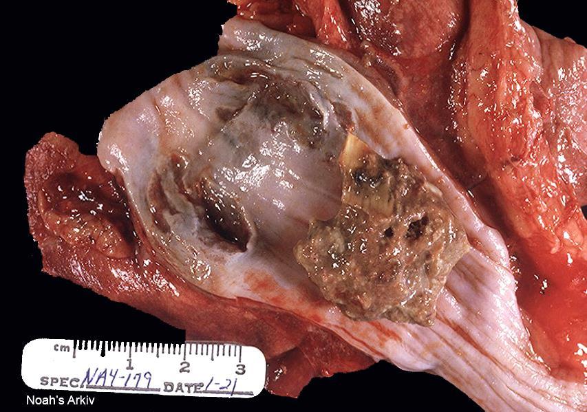
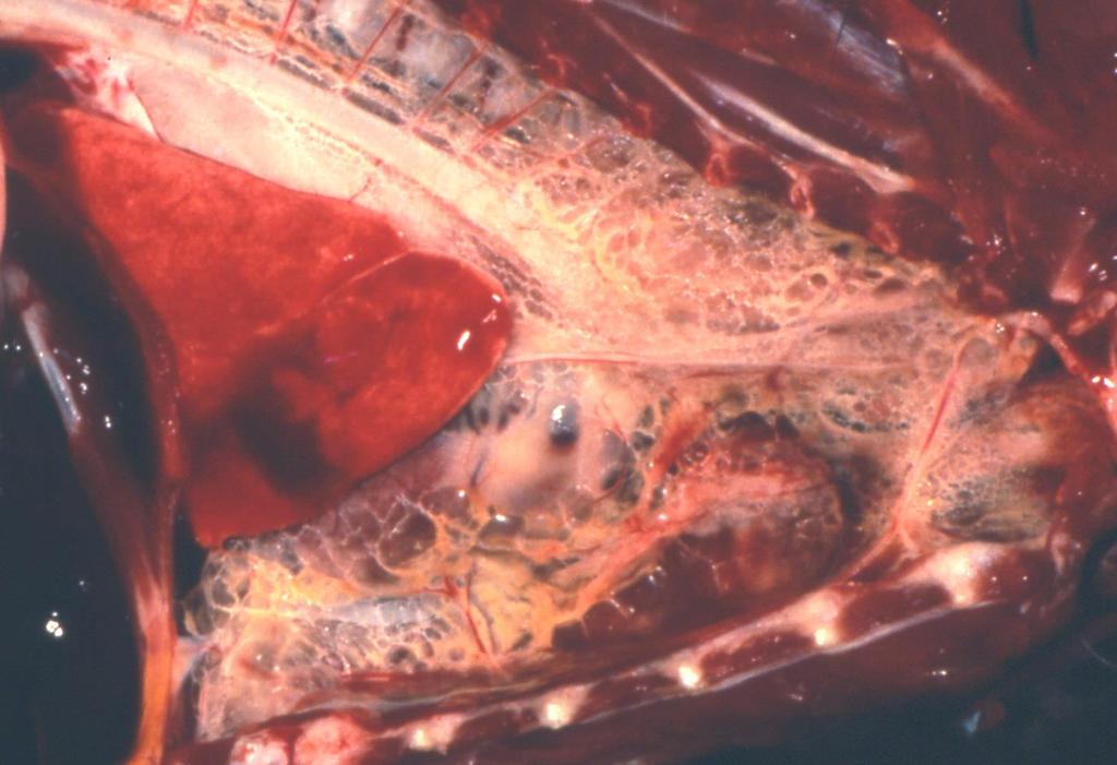
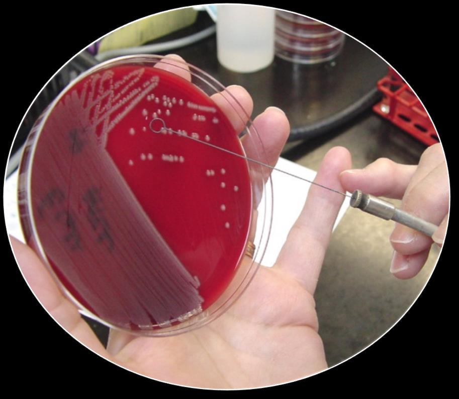
4 LUNG GRANULOMATOUS PNEUMONIA HORSE Rhodococcosis Etiology: Rhodococcus equi Respiratory Intestinal Bacteriologic culture


5 LUNG GRANULOMATOUS PNEUMONIA CAT/DOGS Systemic mycoses Histoplasmosis Blastomyces dermatitides Cryptococcus neoformans / gatti Histoplasma capsulatum Coccidiodes immitis Blastomycosis

")

6 LUNG GRANULOMATOUS PNEUMONIA CAT/DOGS Feline Infectious Peritonitis (FIP) Etiology: Mutated Feline enteric coronavirus


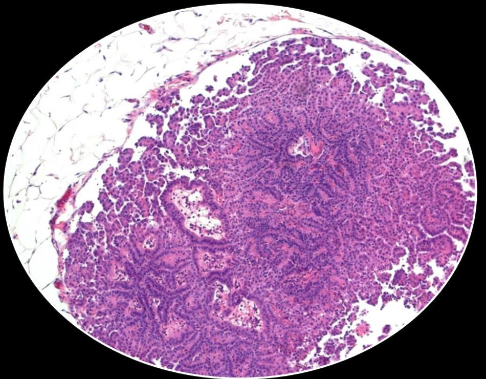
7 LUNG TUMOURS 1 lung tumours are relatively uncommon 2 tumours (metastases) in the lung are common Epithelial Round cell Mesenchymal Carcinoma > Adenoma


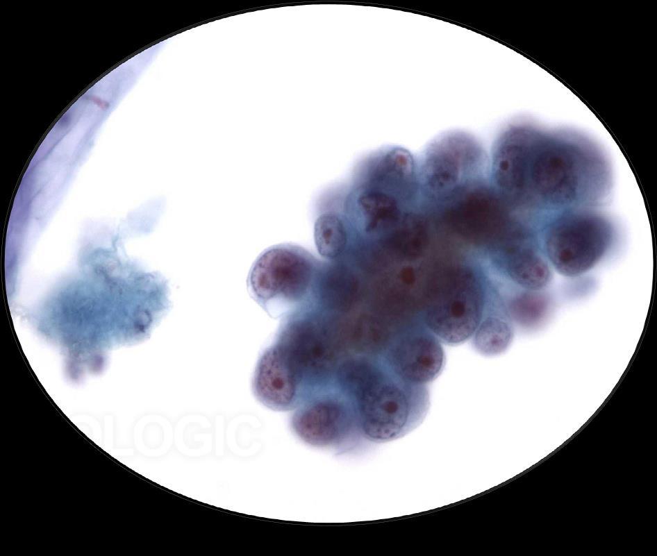


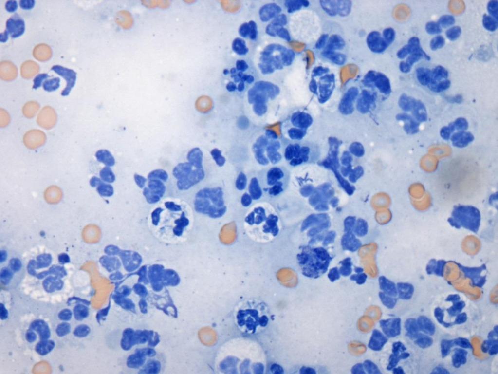
8 LUNG TUMOURS Diagnosis FNA Biopsy
9 LUNG TUMOURS Pulmonary Carcinoma (primary) Clinical signs: Weight loss Lethargy Dyspnea (effusion) Tachypnea Non productive cough Hemoptysis Wheezing Biopsy
 In cats")

10 LUNG TUMOURS Pulmonary Carcinoma (primary) In cats pulmonary carcinoma commonly metastasize to the digits* Biopsy

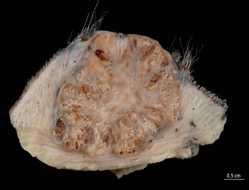
11 LUNG TUMOURS Ovine Pulmonary Adenocarcinoma (Pulmonary Adenomatosis) (Jaagsietke) Etiology: Jaagsietke sheep retrovirus (JRSV) Transmissible retrovirus induced neoplasia of sheep Clinical signs: Weight loss, coughing, respiratory distress Nasal discharge Copious when tilted ** Dr. Marcelo de las Heras, Zaragoza
 (Jaagsietke)")
 May look like bronchopneumonia")

12 LUNG TUMOURS Ovine Pulmonary Adenocarcinoma (Pulmonary Adenomatosis) (Jaagsietke) Etiology: Jaagsietke sheep retrovirus (JRSV) May look like bronchopneumonia grossly Need histology Histology papillary growths of epithelial cells Dr. Marcelo de las Heras, Zaragoza


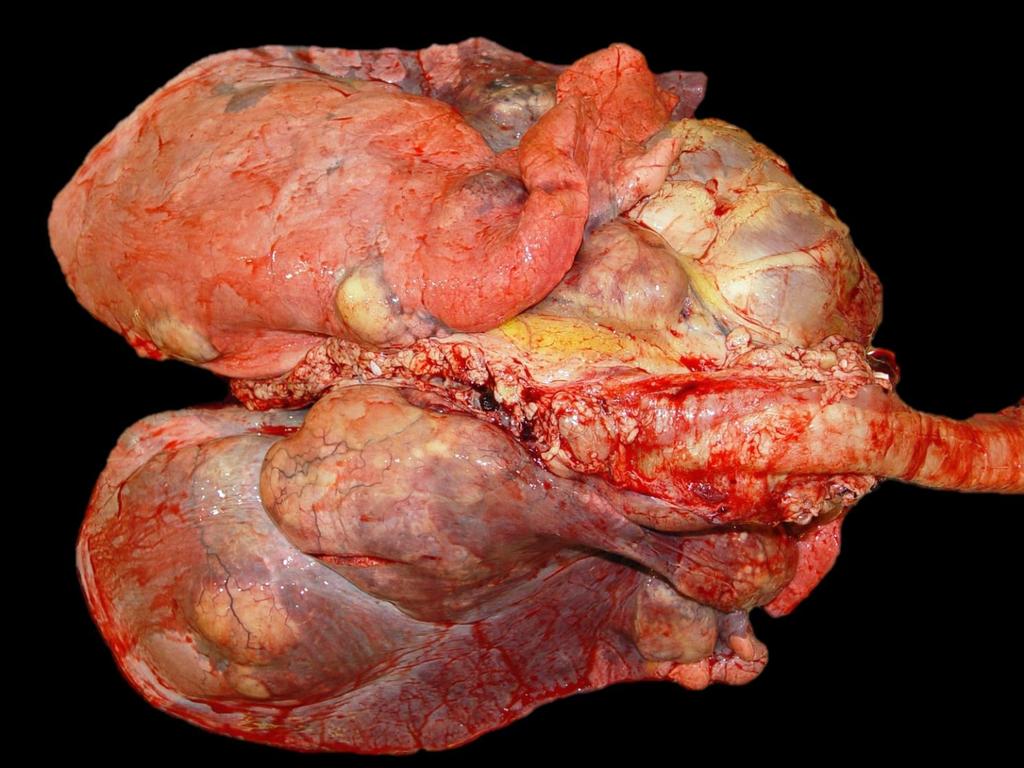
13 LUNG TUMOURS Lung Metastases Mammary carcinoma UNAM


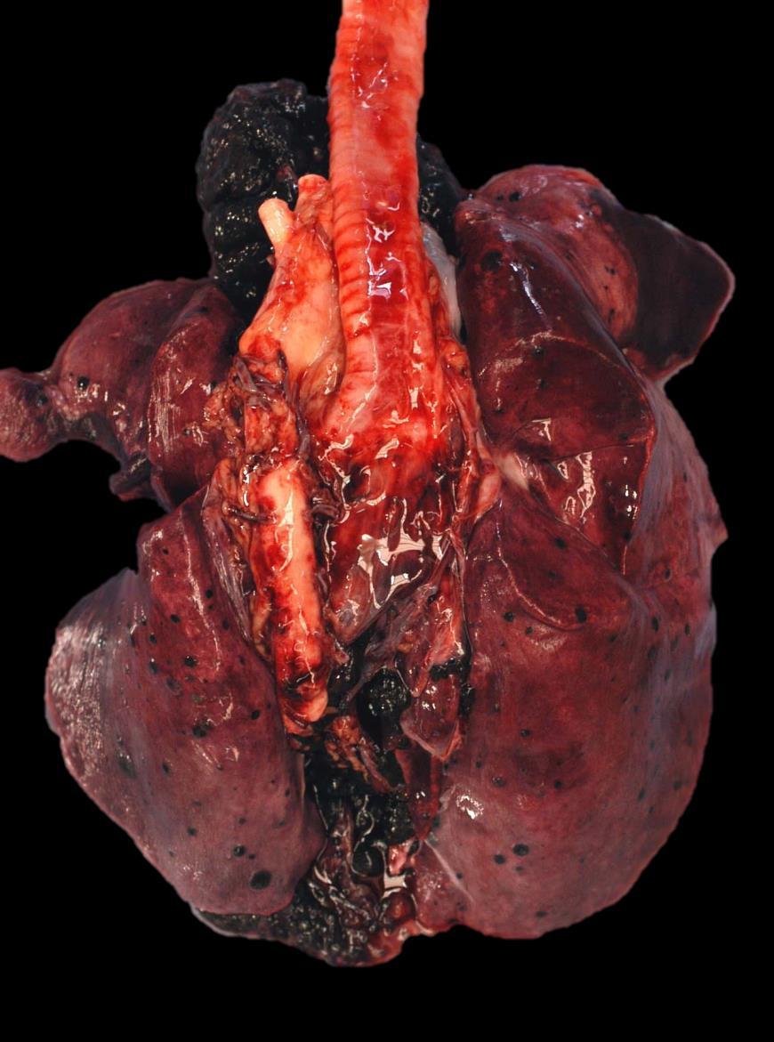
14 LUNG TUMOURS Lung Metastases Dark masses in the lung 2 main differentials Hemangiosarcoma UNAM-FMZ PATOLOGIA Melanoma


15 LUNG TUMOURS Lung Metastases Dark masses in the lung 2 main differentials Hemangiosarcoma

16 THE THORACIC CAVITY Pneumothorax Pleural effusions Neoplasia

17 THORACIC CAVITY


18 THORACIC CAVITY

19 Pneumothorax = Air in the thoracic cavity Lesion atelectasis


20 Pneumothorax Etiology Punctured lung (rib fracture) Penetrating wound Ruptured emphysematous bullae Ruptured esophagus Idiopathic
21 Pneumothorax Pneumomediastinum

22 Pleural Effusions Transudate Lymph Blood Exudate


23 Pleural Effusions Handbook of Veterinary Cytology, Rebar AH. Purina Thoracocentesis Transudate Hydrothorax Clear fluid, low cellularity and low protein Lymph Chylothorax Milky fluid, lymphocytes and lipid Blood Hemothorax RBCs and protein Exudate Pleuritis Turbid fluid, high cellularity and high protein

24 Pleural Effusions Transudate Hydrothorax Clear fluid, low cellularity and low protein quizlet.com Lesions: Clear, straw coloured fluid in thorax +/- Atelectasis +/- Pleural fibrosis Chronic pleural irritation


25 Pleural Effusions Transudate Hydrothorax Clear fluid, low cellularity and low protein Underlying Causes: Congestive heart Hypoproteinemia: failure Starvation Lymphatic obstruction Renal disease Intestinal disease Liver failure quizlet.com


26 Pleural Effusions Gunshot wound Blood Hemothorax RBCs and protein Birthing trauma Hit by car Lesion: Blood in the thorax Most common causes: Trauma Aneurysm Coagulopathy


27 Pleural Effusions Blood Hemothorax RBCs and protein Cytology Ruptured aortic aneurysm Lesion: Blood in the thorax Most common causes: Trauma Aneurysm Coagulopathy


28 Pleural Effusions Lymph Chylothorax Milky fluid, lymphocytes and lipid Lesions: Milky fluid in the thorax Often has fat droplets


29 Pleural Effusions Lymph Chylothorax Milky fluid, lymphocytes and lipid Underlying cause: Rupture/leaking from a major lymphatic vessel Trauma Iatrogenic (surgery) Neoplasia Idiopathic Increased pressure in the vena cava Heart failure


30 Pleural Effusions Lymph Chylothorax Milky fluid, lymphocytes and lipid Cytology Image: Dr C Gilroy Image: Dr C Gilroy Triglyceride analysis
 = Pyothorax Fibrinous Granulomatous Lesions: Cloudy to opaque yellow tinged fluid/viscous exudate in the thorax Often")
31 Pleural Effusions Exudate Pyothorax/Pleuritis Turbid fluid, high cellularity and high protein Alone or in combination with pneumonia According to exudate: Purulent (suppurative) = Pyothorax Fibrinous Granulomatous Lesions: Cloudy to opaque yellow tinged fluid/viscous exudate in the thorax Often smells bad

32 Pleural Effusions Exudate Pyothorax/Pleuritis Turbid fluid, high cellularity and high protein Underlying cause: Penetrating trauma with implantation Rupture of the esophagus Rupture of a pulmonary abscess Hematogenous dissemination / Sepsis


33 Pleural Effusions Exudate Pyothorax/Pleuritis Turbid fluid, high cellularity and high protein Cattle: Mannheimia hemolytica Histophilus somni Cats Pasteurella multocida


34 Pleural Effusions Dogs and Cats Exudate Pyothorax/Pleuritis Turbid fluid, high cellularity and high protein tomato soup pyothorax


35 Pleural Effusions Exudate Pyothorax/Pleuritis Turbid fluid, high cellularity and high protein Pigs

36 Pleural Effusions Exudate Pyothorax/Pleuritis Turbid fluid, high cellularity and high protein Horses


37 Pleural Effusions Exudate Pyothorax/Pleuritis Turbid fluid, high cellularity and high protein Videos Courtesy of Dr. Marco Lores, AVC VTH


38 Pleural Effusions Exudate Pyothorax/Pleuritis Cytology Turbid fluid, high cellularity and high protein


39 Pleural Effusions Exudate Pyothorax/Pleuritis Turbid fluid, high cellularity and high protein Chronic pleuritis typically results in extensive pleural adhesions


40 Mesothelioma Neoplasia Rare tumour of serosal membranes Malignant tumour Disseminates by implantation Asbestos in humans FMVZ-UNAM

41
like humans, have well-developed mediastinal separation between the left and right hemithorax, thus unilateral changes can occur. On the other hand,
 Tutorial Module 6 Thoracic Cavity and Tumors of Lung and Pleura Alfonso López Atlantic Veterinary College University of Prince Edward Island Canada 2009 Enero 3 Thoracic Cavity There are significant anatomical
Thoracic Cavity and Tumors of Lung and Pleura
 Tutorial Module 6 Thoracic Cavity and Tumors of Lung and Pleura Alfonso López Atlantic Veterinary College University of Prince Edward Island Canada Sept 28, 2014 Thoracic Cavity There are anatomical differences
Respiratory Pathology Lab 2: Lung. Shannon Martinson,
 Respiratory Pathology Lab 2: Lung Shannon Martinson, 2017 http://people.upei.ca/smartinson/ Case 1 Signalment: 9 month old DSH cat History: Poor doer with stunted growth One month of lethargy one day the
Pathology of the Respiratory System 4: Pneumonia
 Pathology of the Respiratory System 4: Pneumonia Shannon Martinson, March 2016 http://people.upei.ca/smartinson/ VPM 222 Systemic Pathology LUNG PNEUMONIA Review Classification of Pneumonia Diffuse LUNG
Pathology of the Alimentary Tract
 Pathology of the Alimentary Tract Lab 2: Lower alimentary tract SI, LI, cecum, and peritoneum GIST in the cecum of a dog Shannon Martinson: http://people.upei.ca/smartinson VPM 221: November, 2011 3 year
Pathology of the Liver and Biliary Tract 5 Diseases of the Biliary Tract. Shannon Martinson, March 2017
 Pathology of the Liver and Biliary Tract 5 Diseases of the Biliary Tract Shannon Martinson, March 2017 http://people.upei.ca/smartinson/ OUTLINE Normal anatomy & function Hepatobiliary injury and responses
Proceedings of the World Small Animal Veterinary Association Sydney, Australia 2007
 Proceedings of the World Small Animal Sydney, Australia 2007 Hosted by: Next WSAVA Congress THE LAST GASP II: LUNGS AND THORAX David Holt, BVSc, Diplomate ACVS University of Pennsylvania School of Veterinary
Chylothorax Basics OVERVIEW GENETICS SIGNALMENT/DESCRIPTION OF PET
 Chylothorax Basics OVERVIEW Chylo- refers to chyle; thorax refers to the chest Chyle is a milky to slightly yellow fluid composed of lymph and fats (rich in triglycerides) taken up from the intestines
Dr. A.Torossian, M.D., Ph. D. Department of Respiratory Diseases
 Pleural effusions Dr. A.Torossian, M.D., Ph. D. Department of Respiratory Diseases A pleural effusion is an abnormal collection of fluid in the pleural space resulting from excess fluid production or decreased
Firm Texture. (chronic) Cut surface: purulent exudate in bronchi Sequels: Abscesses,
 Cut surface: purulent exudate in bronchi Sequels: Abscesses,") 2008 Classification of Pneumonias in Domestic Animals There is no universal classification! Based on texture, distribution of lesions and type of exudate, pneumonias in domestic animals are currently classified
2008 Classification of Pneumonias in Domestic Animals There is no universal classification! Based on texture, distribution of lesions and type of exudate, pneumonias in domestic animals are currently classified
Management of Pleural Effusion
 Management of Pleural Effusion Development of Pleural Effusion pulmonary capillary pressure (CHF) capillary permeability (Pneumonia) intrapleural pressure (atelectasis) plasma oncotic pressure (hypoalbuminemia)
PLEURAL EFFUSION. Prof. G. Zuliani
 PLEURAL EFFUSION Prof. G. Zuliani Anatomy of pleural membrane and pleural space Pleural membrane consists of parietal pleura and visceral pleura A space situated between parietal and visceral pleura is
Causes of pleural effusion and its imaging approach in pediatrics. M. Mearadji International Foundation for Pediatric Imaging Aid
 Causes of pleural effusion and its imaging approach in pediatrics M. Mearadji International Foundation for Pediatric Imaging Aid Pleural fluid A tiny amount of fluid in the pleural cavity is physiological.
Respiratory Interactive Session. Elaine Borg
 Respiratory Interactive Session Elaine Borg Case 1 Respiratory Cytology 55 year old gentleman Anterior mediastinal mass EBUS FNA Case 1 Respiratory Cytology 55 year old gentleman with anterior mediastinal
Inflammation Laboratory 2. Shannon Martinson: VPM 152: March 2012
 Inflammation Laboratory 2 Shannon Martinson: http://people.upei.ca/smartinson VPM 152: March 2012 Reminder - Creating a Morphologic Diagnosis for Inflammatory Lesions Organ and Process Exudate Distribution
Post Mortal Approach to the Respiratory System Part 1
 Post Mortal Approach to the Respiratory System Part 1 System examination Before the carcass is opened examination of the nasal openings is carried out. Observe for any evidence of nasal discharge or nasal
Respiratory Pathology Lab 1: Upper Respiratory Tract. Shannon Martinson,
 Respiratory Pathology Lab 1: Upper Respiratory Tract Shannon Martinson, 2017 http://people.upei.ca/smartinson/ Case 1 Signalment: 5 year old dog History: 2 month history of nasal discharge Decreased airflow
Pulmonary Morning Report. Ashley Schmehl D.O. PGY-3 January,
 Pulmonary Morning Report Ashley Schmehl D.O. PGY-3 January, 8 2015 Pleural Effusion Unilateral versus Bilateral Associated symptoms Transudate versus Exudate Light s Criteria: Pleural protein: Serum protein
Respiratory Diseases and Disorders
 Chapter 9 Respiratory Diseases and Disorders Anatomy and Physiology Chest, lungs, and conducting airways Two parts: Upper respiratory system consists of nose, mouth, sinuses, pharynx, and larynx Lower
Bone Injury and Inflammatory Diseases of Bone
 Bone Injury and Inflammatory Diseases of Bone Module 3 Alfonso López Atlantic Veterinary College January 10, 2014 Bone Necrosis / Cross Section Necrotic bone is often difficult to detect grossly but it
Exam 2 Respiratory Disorders
 Exam 2 Respiratory Disorders Common Cold Common Cold Pathology Common Cold Consequences Rhinosinusitis Rhinosinusitis Pathology Rhinosinusitis ostia can close due to Influenza (Flu) Influenza Pathology
UNDERSTANDING CHYLE IN CATS
 Vet Times The website for the veterinary profession https://www.vettimes.co.uk UNDERSTANDING CHYLE IN CATS Author : DAN FORSTER Categories : Vets Date : February 11, 2008 DAN FORSTER discusses diagnosis
*according to content of fluid we can divide pleural effusion to 2 main types
 Pleural lesion and lesion of the Done by: Upper respiratory tract Saef Bassam ma'adat **Lets start with pleural lesion there is a little differet between pleural effustion and empyema accumulation of fluid
PLEURAL DISEASES. (Pleural effusion & empyema) Menaldi Rasmin
 Menaldi Rasmin") PLEURAL DISEASES (Pleural effusion & empyema) Menaldi Rasmin Department of Pulmonology & Respiratory Medicine Faculty of Medicine, University of Indonesia Introduction Pleural effusion is the most common
PLEURAL DISEASES (Pleural effusion & empyema) Menaldi Rasmin Department of Pulmonology & Respiratory Medicine Faculty of Medicine, University of Indonesia Introduction Pleural effusion is the most common
Pleural Diseases. Dr Matthew J Knight Consultant Respiratory Physician
 Pleural Diseases Dr Matthew J Knight Consultant Respiratory Physician What do you need to know? What do you need to know? Pleura- normal anatomy and physiology Pleural effusions Causes and investigations
Element of Competency V6:23 Carry out post mortem examinations, interpret findings and initiate and interpret results of further investigations.
 Performance Criterion d.: Recognize artifacts Range Indicators: Common to all species groups Bile imbibition Chicken fat & post mortem clots Freezing artifact Hemoglobin imbibition Hypostatic congestion
INFLAMMATION & REPAIR
 INFLAMMATION & REPAIR Histopath Laboratory 1 Winter 2013 Chelsea Martin Special thanks to Drs. Hanna and Forzan Goals: Examine Tissue and Identify the Organ Describe the lesion, grossly and histologically
Inflammation Laboratory 1
 Inflammation Laboratory 1 Lab1 Emphasis: The exudates of acute inflammation Descriptions Morphologic Diagnoses Shannon Martinson: http://people.upei.ca/smartinson VPM 152: February 2012 Describing Lesions
Pleural Effusion. Exudative pleural effusion - Involve an increase in capillary permeability and impaired pleural fluid resorption
 Pleural Effusion Definition of pleural effusion Accumulation of fluid between the pleural layers Epidemiology of pleural effusion Estimated prevalence of pleural effusion is 320 cases per 100,000 people
Pathology of the Liver and Biliary Tract 5 Diseases of the Biliary Tract. Shannon Martinson, April 2016
 Pathology of the Liver and Biliary Tract 5 Diseases of the Biliary Tract Shannon Martinson, April 2016 http://people.upei.ca/smartinson/ OUTLINE Normal anatomy & function Hepatobiliary Injury and responses
BELLWORK page 343. Apnea Dyspnea Hypoxia pneumo pulmonary Remember the structures of the respiratory system 1
 BELLWORK page 343 Apnea Dyspnea Hypoxia pneumo pulmonary respiratory system 1 STANDARDS 42) Review case studies that involve persons with respiratory disorders, diseases, or syndromes. Citing information
Respiratory Pathology. Kristine Krafts, M.D.
 Respiratory Pathology Kristine Krafts, M.D. Normal lung: alveolar spaces Respiratory Pathology Outline Acute respiratory distress syndrome Obstructive lung diseases Restrictive lung diseases Vascular
Inflammation Laboratory 1
 Inflammation Laboratory 1 Lab1 Emphasis: The exudates of acute inflammation Descriptions Morphologic Diagnoses Shannon Martinson: http://people.upei.ca/smartinson VPM 152: March 2013 Describing Lesions
Table of Contents. Preface xi. Acknowledgments xiii. Part I Overview of the Diagnostic Process 1. 1 Overview of Grading and Staging 3
 Table of Contents Preface xi Acknowledgments xiii Part I Overview of the Diagnostic Process 1 1 Overview of Grading and Staging 3 Identification of the process 3 Identification of tumor types 5 Grading
What s Your Diagnosis? Signalment: Species: Canine Breed: Golden Retriever Sex: Female (spayed) Date of Birth: 04/01/99
 Date of Birth: 04/01/99") What s Your Diagnosis? Signalment: Species: Canine Breed: Golden Retriever Sex: Female (spayed) Date of Birth: 04/01/99 Presenting Complaint: Acute onset of lethargy Vomited twice (partially digested food)
What s Your Diagnosis? Signalment: Species: Canine Breed: Golden Retriever Sex: Female (spayed) Date of Birth: 04/01/99 Presenting Complaint: Acute onset of lethargy Vomited twice (partially digested food)
The Thorax Excluding the Heart and Pulmonary Patterns
 The Thorax Excluding the Heart and Pulmonary Patterns Lisa G. Britt, DVM, MS, Diplomate American College of Veterinary Radiology, Clinical Assistant Professor @ University of Missouri s College of Veterinary
Concepts in Small Animal Thoracic Radiology Thoracic Radiology
 Concepts in Small Animal Thoracic Radiology + Radiology of the Pleural Space VMB 960 2/21/2011 Optimizing Image Quality Inherent subject contrast Thorax has high inherent subject contrast c/f abdomen Primarily
How to Analyse Difficult Chest CT
 How to Analyse Difficult Chest CT Complex diseases are:- - Large lesion - Unusual or atypical pattern - Multiple discordant findings Diffuse diseases are:- - Numerous findings in both sides 3 basic steps
Case of the Day Chest
 Case of the Day Chest Darin White MDCM FRCPC Department of Radiology, Mayo Clinic 76 th Annual Scientific Meeting Canadian Association of Radiologists Montreal, QC April 26, 2013 2013 MFMER slide-1 Disclosures
Bone necrosis. Cross section.
 Bone necrosis. Cross section. Note the large area of necrosis (N) seen as pale discolored bone (beneath physeal cartilage). The texture of the necrotic bone is also changed. Necrotic bone becomes friable
Cardiovascular Pathology Lab. Shannon Martinson,
 Cardiovascular Pathology Lab Shannon Martinson, 2017 http://people.upei.ca/smartinson/ Case 1 Signalment: 10 year old MC DSH Cat History Heart murmur detected on PE recommended cardiac US Blood work was
Pathology of the Hematopoietic System - Lab.
 Pathology of the Hematopoietic System - Lab http://people.upei.ca/smartinson/ Shannon Martinson, September 2015 Case #1 Signalment: 96 kg gilt History: Pig from minimal disease herd. Sudden death Case
PATHOLOGY OF THE CARDIOVASCULAR SYSTEM
 PATHOLOGY OF THE CARDIOVASCULAR SYSTEM Lecture 5: Vascular System Shannon Martinson, 2018 VPM 2220 Systemic Pathology II http://people.upei.ca/smartinson/ VASCULAR SYSTEM The vascular system is formed
APPROACH TO PLEURAL EFFUSIONS. Raed Alalawi, MD, FCCP
 APPROACH TO PLEURAL EFFUSIONS Raed Alalawi, MD, FCCP CASE 65-year-old woman with H/O breast cancer presented with a 1 week H/O progressively worsening exersional dyspnea. Physical exam: Diminished breath
Internal Injury Documentation Guidelines
 Internal Injury Documentation Guidelines General Open Wound of Thorax Injury to Heart Identify episode of care Initial Subsequent Sequela Laterality Sequela of injury Place of occurrence of injury Activity
EVALUATE DATA IN THE PATIENT RECORD
 EVALUATE DATA IN THE PATIENT RECORD Shawna Strickland, PhD, RRT-NPS, AE-C, FAARC Objectives At the end of this module, the learner will be able to identify the pertinent data from the patient chart for
Pleural fluid analysis
 Pleural fluid analysis Dr Akash Verma Senior Consultant- Department of Respiratory and Critical Care Medicine Tan Tock Seng Hospital, Singapore 308433 Adj A/Professor- Lee Kong Chian School of Medicine
CASE REPORTS. Inflammatory Polyp of the Bronchus. V. K. Saini, M.S., and P. L. Wahi, M.D.
 CASE REPORTS V. K. Saini, M.S., and P. L. Wahi, M.D. I n 1932 Jackson and Jackson [l] first reported a number of clinical cases under the title Benign Tumors of the Trachea and Bronchi with Especial Reference
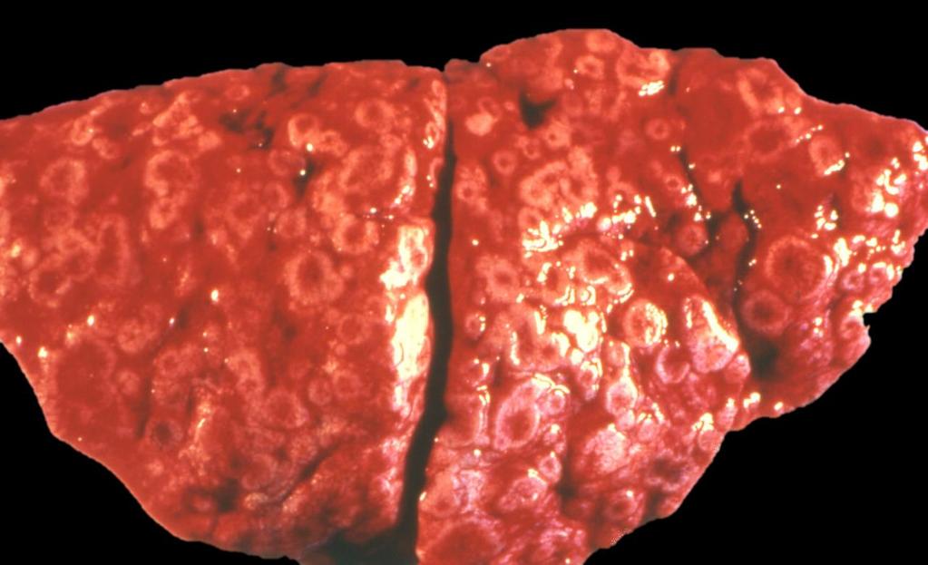
Liver Lab #2. Bacterial Hepatitis
 Liver Lab #2 Bacterial Hepatitis Case: O12561-04. Adult ewe. Describe the lesion: Multifocal large nodules ranging in size from 1-3.5cm in greatest diameter are present within the liver and are filled
Thoracic Surgery; An Overview
 Thoracic Surgery What we see Thoracic Surgery; An Overview James P. Locher, Jr, MD Methodist Cardiovascular and Thoracic Surgery Lung cancer Mets Fungus and TB Lung abcess and empyema Pleural based disease
Malignant Effusions. Anantham Devanand Respiratory and Critical Care Medicine Singapore General Hospital
 Malignant Effusions Anantham Devanand Respiratory and Critical Care Medicine Singapore General Hospital Malignant Effusions Definition: Presence of malignant cells in the pleural space 75% are caused by
Lung tumors & pleural lesions
 Lung tumors & pleural lesions A brief introduction 95% of lung tumors are carcinomas Among the remaining 5%, we will discuss: -Hamartoma the most common benign lung tumor spherical, coin lesion on x-rays
Fungal Diseases of the Respiratory System
 Fungal Diseases of the Respiratory System Histoplasmosis(cave disease) Dr. Hala Al Daghistani Histoplasmosis is a disease caused by the fungus Histoplasma capsulatum. Histoplasma capsulatum, is usually
Pneumothorax lecture no. 3
 Pneumothorax lecture no. 3 Is accumulation of air in a pleural space or accumulation of extra pulmonary air within the chest, Is uncommon during childhood, may result from external trauma, iatrogenic,
Pathology of the Hematopoietic System. Case studies
 Pathology of the Hematopoietic System Case studies Shannon Martinson, September 2015 Signalment: 9 yr-old MC cat Case Study 1 History: Cat had been anorexic and developed bleeding in the eyes Physical
1/13/2014. Proper Radiographs. Proper Radiographs. A Review of Pulmonary Patterns
 Live Webinar A Review of Pulmonary Patterns Sofija R. Liles, DVM, DACVR Proper Radiographs Which views? One lateral plus ventrodorsal (at least) Left lateral is best for thorax Three views for full metastatic
TUBERCULOSIS. By Dr. Najaf Masood Assistant Prof Pediatrics Benazir Bhutto Hospital Rawalpindi
 TUBERCULOSIS By Dr. Najaf Masood Assistant Prof Pediatrics Benazir Bhutto Hospital Rawalpindi Tuberculosis Infectious, Systemic, Chronic granulomatous disease caused by mycobacterium tuberculosis DEFINITION
Examples of Morphologic Types of Pneumonia in Domestic Animals
 Tutorial Module 5 Examples of Morphologic Types of Pneumonia in Domestic Animals Alfonso López Atlantic Veterinary College University of Prince Edward Island Canada 2014 Sept 24, 2014b Examples of Diseases
Manejo Práctico del Derrame Pleural
 Manejo Práctico del Derrame Pleural San José, Costa Rica Junio 29, 2017 Rodrigo Cartín Ceba, MD, MSc Consultant, Pulmonary and Critical Care Medicine Associate Professor of Medicine Mayo Clinic 2010 MFMER
Resident Case Review CHEST. Daria Manos CAR 2016
 Resident Case Review CHEST CAR 2016 Daria Manos Disclosure Speakers bureau, Roche CAR 2016 Daria Manos 1. Recognize common and critical chest radiograph and computed tomography signs and use these clues
Chest XRay interpretation INTERPRETATIONS Identifications: Name & Date Technical evaluation Basic Interpretations
 Chest XRay interpretation INTERPRETATIONS Identifications: Name & Date Technical evaluation Basic Interpretations TECHNICAL EVALUATION 1. Projection: AP/PA view To differentiate between AP & PA films,
5 DISTURBANCES IN CIRCULATION. Congestion / Hyperemia Haemorrhage Thrombosis Embolism Ischemia Infarction Oedema Shock Sludged blood Model Questions
 5 DISTURBANCES IN CIRCULATION Congestion / Hyperemia Haemorrhage Thrombosis Embolism Ischemia Infarction Oedema Shock Sludged blood Model Questions CONGESTION/ HYPEREMIA Hyperemia is increased amount of
Serous fluids. Dr. Mohamed Saad Daoud
 Serous fluids 1 Reference Books: Urinanalysis and body fluids (Susan King Strasinger- Marjorie Schaub De Lorenzo) Fifth edition 2 The closed cavities of the body namely, the pleural, pericardial, and peritoneal
PATHOLOGY OF THE CARDIOVASCULAR SYSTEM
 PATHOLOGY OF THE CARDIOVASCULAR SYSTEM Lecture 3: Pericardium and Endocardium Shannon Martinson, 2018 VPM 2220 Systemic Pathology II http://people.upei.ca/smartinson/ PERICARDIUM AND EPICARDIUM Normal
Auscultation of the lung
 Auscultation of the lung Auscultation of the lung by the stethoscope. *Compositions of the stethoscope: 1-chest piece 2-Ear piece 3-Rubber tubs *Auscultation area of the lung(triangle of auscultation).
Note the large area of necrosis (N) which appears as a pale discolored bone
 which appears as a pale discolored bone") Bone Injury and Inflammatory Bone Diseases Alfonso López Atlantic Veterinary College University i of Pi Prince Edward d Il Island January 7, 2010 Bone Necrosis / Cross Section N The texture of the necrotic
Bone Injury and Inflammatory Bone Diseases Alfonso López Atlantic Veterinary College University i of Pi Prince Edward d Il Island January 7, 2010 Bone Necrosis / Cross Section N The texture of the necrotic
Key Difference - Pleural Effusion vs Pneumonia
 Difference Between Pleural Effusion and Pneumonia www.differencebetween.com Key Difference - Pleural Effusion vs Pneumonia Pleural effusion and pneumonia are two conditions that affect our respiratory
Pyothorax (Pus in the Pleural Space, the Space between the Chest Wall and the Lungs) Basics
 Basics") Pyothorax (Pus in the Pleural Space, the Space between the Chest Wall and the Lungs) Basics OVERVIEW Accumulation of pus within the pleural space (the space between the chest wall and lungs, which is lined
Pyothorax (Pus in the Pleural Space, the Space between the Chest Wall and the Lungs) Basics OVERVIEW Accumulation of pus within the pleural space (the space between the chest wall and lungs, which is lined
Chapter 13. Injuries to the Thorax and Abdomen
 Chapter 13 Injuries to the Thorax and Abdomen Anatomy Review Thoracic cage has 12 pairs of ribs. The first 7 pairs connect directly to sternum. Pairs 8 through 10 connect via common costal cartilage. Pairs
CLINICAL FEATURES IN PULMONARY TUBERCULOSIS
 CLINICAL FEATURES IN PULMONARY TUBERCULOSIS Dr. Amitesh Aggarwal Department of Medicine Tuberculosis Captain of all the Men of Death Great White Plague devastating effect on society 100 years ago one in
Difficulty Breathing and Respiratory Distress Basics
 Difficulty Breathing and Respiratory Distress Basics OVERVIEW Difficulty breathing (known as dyspnea ) a subjective term that in human medicine means an uncomfortable sensation in breathing or a sensation
Proceedings of the 10th International Congress of World Equine Veterinary Association
 www.ivis.org Proceedings of the 10th International Congress of World Equine Veterinary Association Jan. 28 Feb. 1, 2008 - Moscow, Russia Next Congress: Reprinted in IVIS with the permission of the Conference
Pleural syndrome Tuberculous pleurisy
 Pleural syndrome Tuberculous pleurisy Etienne Leroy Terquem Pierre L Her SPI / ISP Soutien Pneumologique International / International Support for Pulmonology Pleural effusion: Findings of fluid between
Impression smear from a nasal mass on a 2 year old cat Presented with: one month duration of epistaxis
 Impression smear from a nasal mass on a 2 year old cat Presented with: one month duration of epistaxis Identify the structures neutrophils macrophages x40 Organisms 8 30 micron in size, with variable capsule
Bronchogenic Carcinoma
 A 55-year-old construction worker has smoked 2 packs of ciggarettes daily for the past 25 years. He notes swelling in his upper extremity & face, along with dilated veins in this region. What is the most
Cardiothoracic and Cardiothoracic Surgery ICD-10-CM 2014: Reference Mapping Card
 2014: Reference Mapping Card 162.3 Malignant neoplasm upper lobe lung 162.5 Malignant neoplasm lower lobe lung 162.9 lung/bronchus 396.2 396.3 Mitral insufficiency, aortic stenosis Mitral aortic valve
Lung Cancer - Suspected
 Lung Cancer - Suspected Shared Decision Making Lung Cancer: http://www.enhertsccg.nhs.uk/ Patient presents with abnormal CXR Lung cancer - clinical presentation History and Examination Incidental finding
Circulatory Disturbances 5: Thrombosis, Embolism, Infarction, Shock
 Circulatory Disturbances 5: Thrombosis, Embolism, Infarction, Shock Shannon Martinson, Feb 2016 http://people.upei.ca/smartinson/ VPM 152 General Pathology Thrombosis, Embolism, Infarction, Shock Learning
Evaluation of the chest Part II.
 Evaluation of the chest Part II. Nagy Endre SZEGEDI TUDOMÁNYEGYETEM ÁOK, RADIOLÓGIAI KLINIKA, SZEGED ANATOMY parenchyma: alveoloacinar system, pulmonary arteries and veins interstitium: connective tissues
Circulatory Disturbances 1: Introduction and Edema
 Circulatory Disturbances 1: Introduction and Edema Shannon Martinson, January 2016 http://people.upei.ca/smartinson/ VPM 152 General Pathology INTRODUCTION NORMAL CIRCULATORY SYSTEM Important concepts
Pneumothorax and Chest Tube Problems
 Pneumothorax and Chest Tube Problems Pneumothorax Definition Air accumulation in the pleural space with secondary lung collapse Sources Visceral pleura Ruptured esophagus Chest wall defect Gas-forming
Pulmonary Problems of the Neonate. Jon Palmer, VMD, DACVIM Chief, Neonatal Intensive Care Service New Bolton Center, University of Pennsylvania, USA
 Pulmonary Problems of the Neonate Jon Palmer, VMD, DACVIM Chief, Neonatal Intensive Care Service New Bolton Center, University of Pennsylvania, USA Lower Respiratory Diseases Ventilation/Perfusion Abnormalities
Esophageal Perforation
 Esophageal Perforation Dr. Carmine Simone Thoracic Surgeon, Division of General Surgery Head, Division of Critical Care May 15, 2006 Overview Case presentation Radiology Pre-operative management Operative
GUIDELINES FOR DIAGNOSIS OF UNILATERAL PLEURAL EFFUSION. Pakistan Chest Society
 GUIDELINES FOR DIAGNOSIS OF UNILATERAL PLEURAL EFFUSION Pakistan Chest Society Message by chairman guideline committee Guidelines for pleural disease working group Expert review committee INTRODUCTION
EDEMA. Learning Objectives
 EDEMA Learning Objectives Define edema Recognize and be able to describe the gross and microscopic appearance of edema Know the four pathophysiological mechanisms by which edema develops Understand the
CHEST INJURIES. Jacek Piątkowski M.D., Ph. D.
 CHEST INJURIES Jacek Piątkowski M.D., Ph. D. CHEST INJURIES 3-4% of all injuries 8% of patients hospitalized due to injuries 65% of patients who died at the accident place CLASSIFICATION OF THE CHEST INJURIES
SPOTLIGHT ON PERICARDIAL DISEASE
 Vet Times The website for the veterinary profession https://www.vettimes.co.uk SPOTLIGHT ON PERICARDIAL DISEASE Author : DAN FORSTER Categories : Vets Date : February 16, 2009 DAN FORSTER discusses the
PATHOLOGY OF LIVER & BILIARY TRACT. Lecture 5. Idiopathic & proliferative conditions; diseases of the biliary tract
 PATHOLOGY OF LIVER & BILIARY TRACT Lecture 5 Idiopathic & proliferative conditions; diseases of the biliary tract Enrique Aburto Winter 2015 IX. Diseases of uncertain origin Equine serum hepatitis Idiopathic
The Thorax The Ever Challenging Pulmonary Patterns
 The Thorax The Ever Challenging Pulmonary Patterns Lisa G. Britt, DVM, MS, Diplomate American College of Veterinary Radiology, Clinical Assistant Professor @ University of Missouri s College of Veterinary
ANATOMY OF THE PLEURA (contd) III. Histology: covered by a single layer of mesothelial cells. Within the pleura are blood vessels, mainly capillaries,
 III. Histology: covered by a single layer of mesothelial cells. Within the pleura are blood vessels, mainly capillaries,") PLEURAL DISEASES By: SETIA PUTRA TARIGAN Pulmonary Department, Faculty of Medicine, Sumatera Utara University/ Adam Malik Hospital Medan 2008 ANATOMY OF THE PLEURA I. Pleura is the serous membrane: 1.
PLEURAL DISEASES By: SETIA PUTRA TARIGAN Pulmonary Department, Faculty of Medicine, Sumatera Utara University/ Adam Malik Hospital Medan 2008 ANATOMY OF THE PLEURA I. Pleura is the serous membrane: 1.
TUMOR,NEOPLASM. Pathology Department, Zhejiang University School of Medicine,
 TUMOR,NEOPLASM Pathology Department, Zhejiang University School of Medicine, 马丽琴,maliqin198@zju.edu.cn The points in this chapter What is a neoplasm (conception) Morphology of neoplasm Macroscopy of Neoplasm
The Respiratory System
 The Respiratory System Respiratory Anatomy Upper respiratory tract Nose Nasal passages Pharynx Larynx Respiratory Anatomy Functions of the upper respiratory tract: Provide entry for inhaled air Respiratory
Pleural fluid. creatinine - urinothorax haematocrit -haemothorax bilirubin gut perforation. Fluid samples 1st Plain Universal ( cell count)
") Examination Purpose of test Sample 17725 Fluid Profile (appearance, culture, WBC differential, ph, total protein, glucose, amylase, triglyceride, albumin, HDL) Peritoneal/ascitic and pleural fluid are
Examination Purpose of test Sample 17725 Fluid Profile (appearance, culture, WBC differential, ph, total protein, glucose, amylase, triglyceride, albumin, HDL) Peritoneal/ascitic and pleural fluid are
CALGARY ZONE PULMONARY REFERRAL QUICK REFERENCE
 CALGARY ZONE PULMONARY REFERRAL QUICK REFERENCE EMERGENCY (Patient needs to be seen immediately) Hemoptysis (Active & 2 TBSP per day) Hypoxemia (if resting O2 SAT 85%) Pulmonary embolism (Acute - known
HEMODYNAMIC DISORDERS
 HEMODYNAMIC DISORDERS Normal fluid homeostasis requires vessel wall integrity as well as maintenance of intravascular pressure and osmolarity within certain physiologic ranges. Increases in vascular volume
Pathophysiology. Tutorial 3 Hemodynamic Disorders
 Pathophysiology Tutorial 3 Hemodynamic Disorders ILOs Recall different causes of thrombosis. Explain different types of embolism and their predisposing factors. Differentiate between hemorrhage types.
Pathology of the Hematopoietic System. Lecture 3: Spleen and thymus
 Pathology of the Hematopoietic System Lecture 3: Spleen and thymus Shannon Martinson, March 2012 Spleen Structure and Function Present in the left cranial part of the abdomen within the greater omentum
Pleural Effusions. Kyle J Henry, MD Pulmonary/ CCM Fellow PGY4 (210) (602)
 (602)") Pleural Effusions Kyle J Henry, MD Pulmonary/ CCM Fellow PGY4 (210) 275 8583 (602) 202 0351 None Disclosures Objectives Understand the presentation of a pleural effusion How to diagnose and treat Differentiate
Pleural Effusions Kyle J Henry, MD Pulmonary/ CCM Fellow PGY4 (210) 275 8583 (602) 202 0351 None Disclosures Objectives Understand the presentation of a pleural effusion How to diagnose and treat Differentiate
General Pathology. Hemorrhage (Web)
") General Pathology Hemorrhage (Web) Paul Hanna Feb 2015 Hemorrhage escape of blood from the cardiovascular system may be external or internal Hemorrhage Causes Trauma Sepsis, viruses or toxins Coagulation
General Pathology Hemorrhage (Web) Paul Hanna Feb 2015 Hemorrhage escape of blood from the cardiovascular system may be external or internal Hemorrhage Causes Trauma Sepsis, viruses or toxins Coagulation
Complicated echinococcal cyst to Biopsy or not to biopsy. V. Rusanov MR Kramer Pulmonary Institute, Rabin medical center
 Complicated echinococcal cyst to Biopsy or not to biopsy V. Rusanov MR Kramer Pulmonary Institute, Rabin medical center Case 1 84 y.o. Male, Iraq descend, past smoker 40 PY Medical History- HTN, Rheumatoid